
Abstract
A variety of 3-dimensional (3D) digital imaging modalities are available for whole-body assessment of genetically engineered mice: magnetic resonance microscopy (MRM), X-ray microcomputed tomography (microCT), optical projection tomography (OPT), episcopic and cryoimaging, and ultrasound biomicroscopy (UBM). Embryo and adult mouse phenotyping can be accomplished at microscopy or near microscopy spatial resolutions using these modalities. MRM and microCT are particularly well-suited for evaluating structural information at the organ level, whereas episcopic and OPT imaging provide structural and functional information from molecular fluorescence imaging at the cellular level. UBM can be used to monitor embryonic development longitudinally in utero. Specimens are not significantly altered during preparation, and structures can be viewed in their native orientations. Technologies for rapid automated data acquisition and high-throughput phenotyping have been developed and continually improve as this exciting field evolves.
Genetically engineered mice (GEM) play a central role in the functional annotation of the human genome. 15,41 They are used extensively to study the role of genetics in normal development as well as the molecular pathogenesis of disease and ultimately in the evaluation of treatment effects for disease models. A number of techniques for creating GEMs are available, such as gene targeting, trapping, and N-ethyl N-nitrosourea (ENU) mutagenesis. 42,63 Their applications in mouse models for studying the human genome are far reaching. However, our ability to create GEMs far exceeds our ability to fully characterize their phenotypes. In many cases, evaluation is limited to a targeted phenotype assessment where the function of a particular gene is known. This may preclude a comprehensive review of the GEM, and off-target pathological changes may never be identified.
Whole-body morphological assessment of a GEM is typically accomplished through pathological review in which gross necropsy and systematic histological review of major tissues and organs are performed. 3,14 This type of review is both labor and time intensive and requires veterinary pathologists with specialized training. 91 It is clear that additional techniques are required for rapid phenotyping as well as for understanding complex morphological processes. One set of tools emerging to address the need for rapid comprehensive phenotyping are high-resolution imaging modalities such as X-ray microcomputed tomography (microCT) and magnetic resonance microscopy (MRM). These modalities provide a number of advantages for the comprehensive evaluation of GEMs. Their 3-dimensional (3D) high-resolution nature allows observations between the scale of gross and microscopic pathological changes. These techniques are noninvasive, which allows review of intact objects in their native configuration, and they are nondestructive, which allows for complete histological review of the specimens following imaging. These techniques can be used to complement the standard pathology review and are becoming more necessary for high-throughput morphological phenotyping. The goal of this review is to present an overview of 3D digital imaging modalities that are available to veterinary pathologists for morphological assessment of genetically engineered embryos and adult mice. Examples of how these modalities have been used to interrogate specific genetic phenotypes are provided. The capabilities and limitations of each of the 3D imaging modalities are discussed.
Imaging Study Design
The experimental design of an imaging phenotyping study is similar to that of a standard pathology phenotyping study. 54 It is important for the imaging scientist to work closely with the primary investigator and the veterinary pathologist to understand the overall study protocol and the genetic makeup of the mutant. This will help in identifying the imaging modality most suitable for highlighting the phenotypical difference of interest and design of the imaging part of the study protocol. It is also important to evaluate age- and gender-matched controls, heterozygotes, and animals obtained from different litters. 45
Microimaging studies are not intended to supplant standard histological review of mutant phenotypes. They can be used as a primary screen to identify potential mutants for further histological review. They can also be used in secondary and tertiary reviews to complement the standard 2D histology data. Additionally, they can guide pathologists on where to best focus their review.
Embryos
Many GEMs exhibit late gestational lethality. 95 The information regarding the cause of the lethality may be lost in traditional 2D histological staining. 3D imaging modalities such as MRM and microCT can be very useful for identifying the structural morphological changes associated with such a phenotype. Other modalities such as optical projection tomography (OPT) and episcopic imaging can be used to obtain 3D fluorescent images for genetic mapping and visualizing signaling pathways. Ultrasound biomicroscopy (UBM) is a unique modality that can be used to interrogate developing embryos in utero.
Magnetic Resonance Microscopy
MRM is typically used to evaluate soft tissue in specimens. The images are created by measuring the precession and relaxation of nuclei in tissue after they have been aligned in a strong magnetic field and selectively excited by the application of a radiofrequency signal. The most abundant nuclei found in vertebrates are the hydrogen nucleus, often referred to as a proton in MRM. The differences in hydrogen distribution (ie, water content of soft tissue) within the body provide the gray-level contrast differences observed within the MRM image. Different types of image weighting (ie, T1, T2, and proton density) are used to manipulate image contrast and allow us to visualize and resolve different structures within the sample.
Requirements for MRM include magnets capable of generating high magnetic field strengths such as 7–22 T, small radiofrequency coils (on the order of 25 mm diameter and less), and fast gradients. The achievable resolution of MRM in embryos is on the order of 20 μm, but the time required to image a specimen at this spatial resolution can be several hours. Recent technical contributions used to speed up image acquisition include 3D gradient echo sequences 81 and gadolinium staining to reduce spin-lattice relaxation time (T1) in the tissues. 70
An immersion fixation–staining approach is used to prepare the embryo for MRM imaging. The fixation–staining solution is made up of a fixative solution such as Bouin’s solution or formalin and gadopentetate dimeglumine (GD) such as Magnevist (Bayer HealthCare Pharmaceuticals Inc, Wayne, New Jersey) or ProHance (Bracco Diagnostics, Princeton, New Jersey) in a 20:1 volume ratio. Each embryo is immersed in 15 mL of the fixation–staining solution at room temperature depending on the size and stage of the specimen (eg, from 10 minutes for E10.5–E12.5 up to 24 hours for E18.5). Later-stage specimens require immersion in 30 mL of fixation–staining solution and intraperitoneal or subcutaneous injections of the solution in neck, abdomen, and lower body prior to immersion. Fixation and staining are then stabilized after the appropriate fixation time by immersing the embryo in 15 mL of 1:200 GD–phosphate-buffered saline solution. The specimens can be stored for several weeks in this solution at 4°C. Additional details on this preparation can be found in Petiet and Johnson. 70
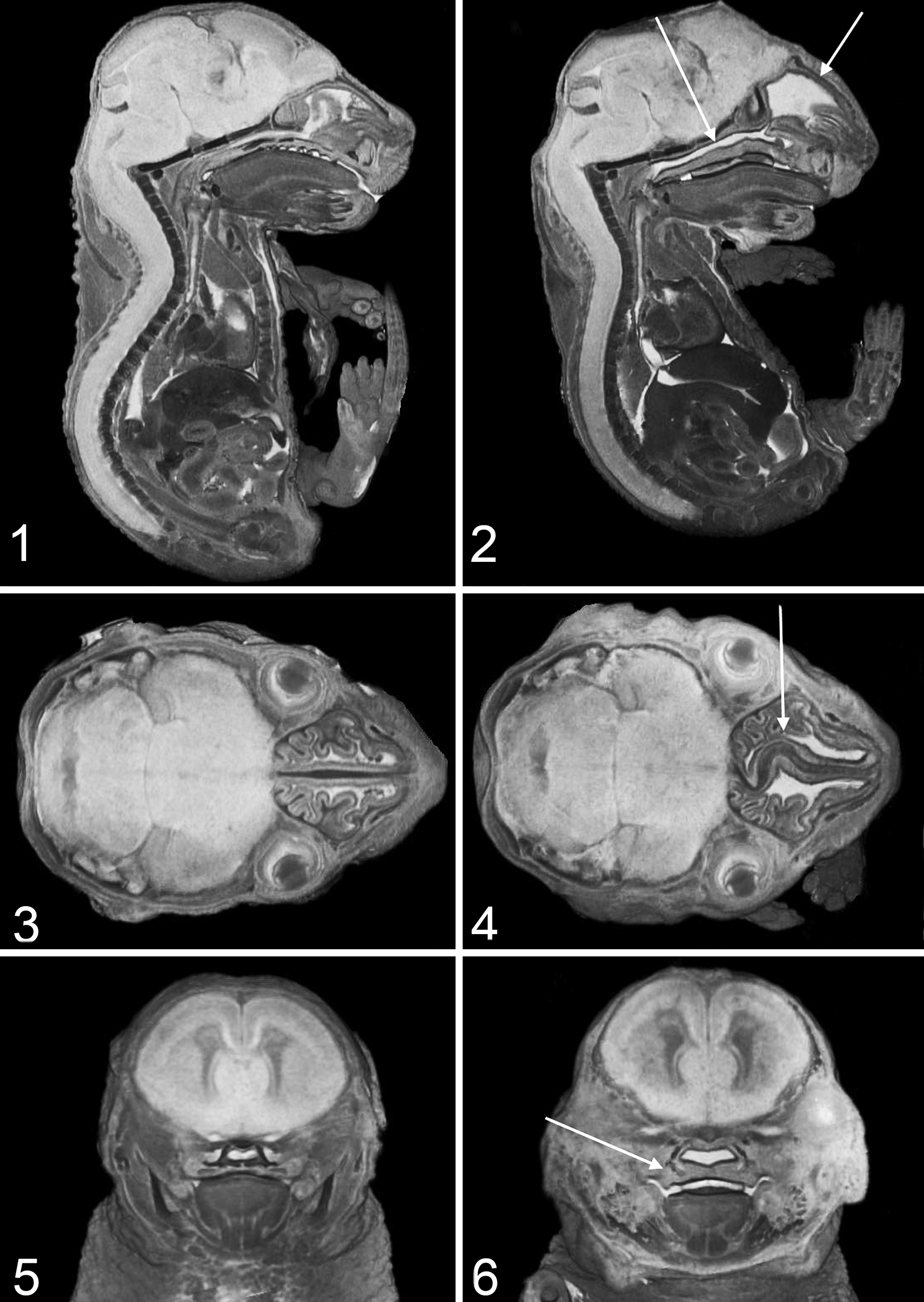
MRM has been shown to be useful for identifying neural and cardiac malformations in developing embryos. For example, Parnell et al 67 used MRM to evaluate ethanol-induced brain abnormalities in 17 GD embryos, whereas Cleary et al 18 established optimum protocols for phenotyping cardiac abnormalities in 15.6 dpc Chd7 mutants. An example of a known morphological phenotype in E17.5 neural-crest–specific Prkar1a (TEC1KO) 39 embryos is illustrated in Figs. 1–6. Berrios-Otero et al 10 developed a unique vascular perfusion staining technique to study cerebral artery development in E10.5 and E17.5 embryos.
Staining techniques coupled with the use of optimized radiofrequency coils designed for multiembryo imaging 82,105 have made MRM a plausible tool for high-throughput phenotypical screens. For MRM to be truly useful for high-throughput screening, automated algorithms for registering specimens, segmenting, 107 and characterizing biological structures in the images are required. 17,104 Digital atlases of normal developmental anatomy 23,71 provide the basis for identifying subtle phenotypic differences in automated high-throughput screens.
X-Ray Microcomputed Tomography
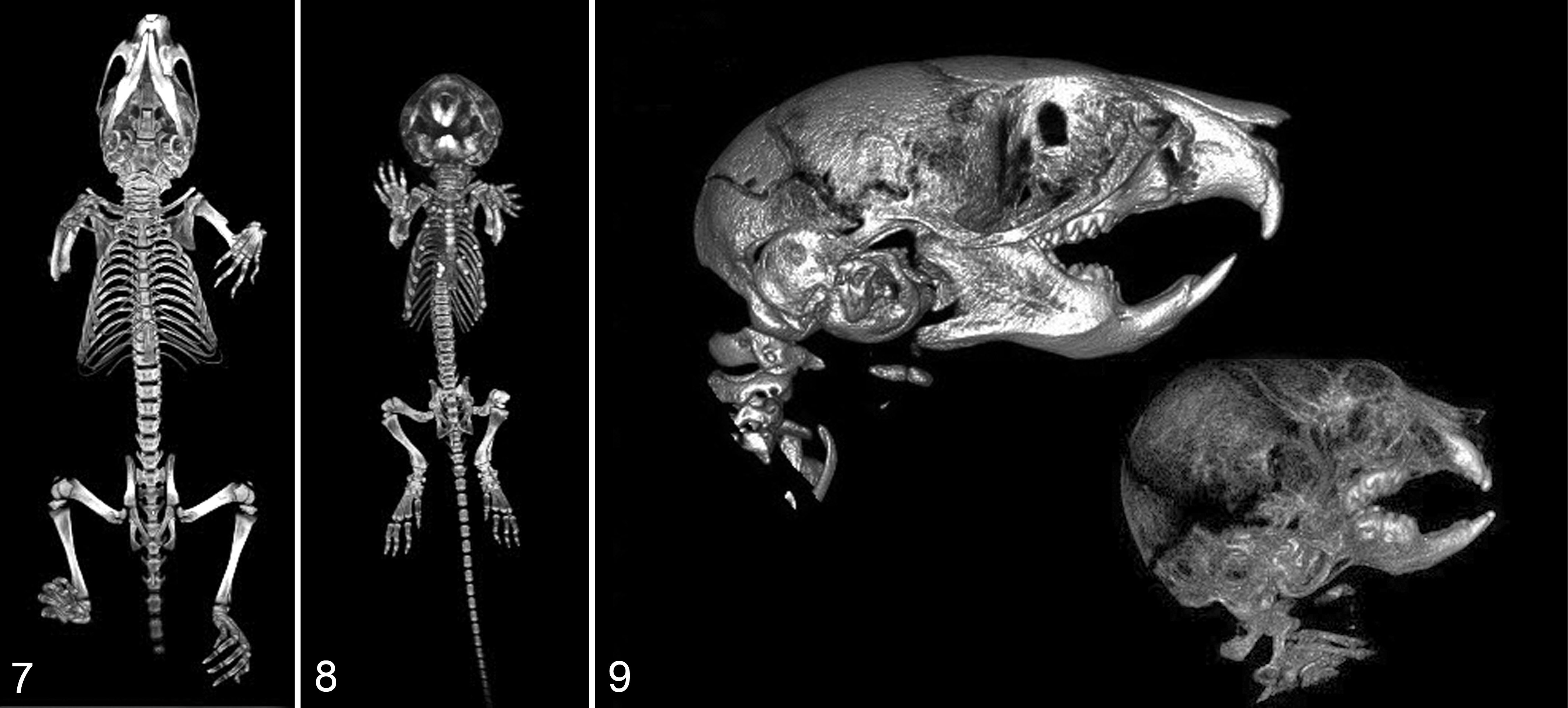
X-ray microCT is a 3D imaging modality based on the reconstruction of multiple X-ray projections taken around a specimen. 8 The differential absorption of X-ray photons as they pass through different tissues in a specimen gives rise to contrast differences within the image. The spatial resolution for whole-body microCT images is typically on the order of 20–40 μm. Higher resolution images are possible with microCT imaging and are used for evaluating isolated anatomical structures such as femurs and tibias. MicroCT is an excellent means of obtaining 3D information regarding skeletal development in embryos 35,96 and has several distinct advantages over the traditional clear staining methods used in histological analysis. 26 These advantages include greatly reduced specimen preparation time, higher morphometric precision available with 3D data, and accessibility to bone volume and mineral density metrics. Sample preparation is nondestructive so that histological analysis can be performed after microCT acquisition. It is particularly useful for examining abnormalities in the developing skull such as those observed in E17.5 neural-crest–specific Prkar1a KO embryos. 39 High-throughput imaging has been demonstrated for rat and rabbit fetuses. 103
A variety of different contrast agents or “stains” have been used to provide soft tissue contrast in microCT images of embryos. Osmium tetroxide was initially proposed for midgestational embryos (E9–E12.5) 38 and was used to highlight the trigeminal ganglion in E13 embryos. 4 Its low tissue penetration combined with its volatile, toxic, and expensive nature led to the investigation of other stains such as phosphotungstic acid (PTA) in water, iodine potassium iodide (IKI, Lugol’s solution), and 1% iodine in ethanol or methanol. 56 These stains are easier to obtain and prepare and can be used on variety of previously fixed specimens. Detailed methods for preparation of these soft tissue stains can be found in references 56 and 57. Degenhard et al 22 illustrated the use of 25% Lugol solution for differential staining in Plxnd1 (Plexin D1) mutant embryos. 2 MicroCT has also been used for evaluating craniofacial abnormalities in mutant embryos without the use of staining. 62,68 Specimen dehydration and tissue shrinkage must be taken into account when making any quantitative structural measurement of fixed tissue. 80
Optical Projection Tomography
Optical projection tomography is a high-resolution 3D imaging modality that is based on optical microscopy projections taken from multiple angles around a specimen. 84 It can be used to image standard histological, fluorescent, and in situ hybridization stained specimens. It is limited to imaging small translucent specimens (ie, embryos <E13.5) and tissues with a homogenous refractive index. The effective spatial resolution over 1-cm 3 field of view is 5–10 μm. It is a good technique for imaging early-stage embryogenesis and has been used to create a 3D atlas of normal vascular development between E8 and E10 embryos 98 as well as identify internal structural abnormalities such as myocardial hypertrophy and pericardial effusion in Men1 null 10.5 to 13.5 dpc embryos 49 and liver abnormalities in E13.5 Wt1 embryos. It has most recently been used to evaluate the 3D distribution of Wnt gene expression 92 and the RET signaling pathway 31 in the early stages of lung development.
Episcopic Imaging
Episcopic imaging techniques are based on serially imaging the block-face of fixed and embedded specimens. 100 The most recent ones include serial surface imaging microscopy (SIM) 34 and high-resolution episcopic microscopy (HREM). 101 SIM uses wax or resin as the embedding medium and histological stains applied to the surface of the specimen before imaging and milling. HREM uses resin as the embedding medium and eosin for unspecific tissue staining along with immunofluorescent staining for gene expression mapping. Episcopic imaging techniques, on average, produce isotropic data sets with 2-μm voxel size, which is sufficient for analyzing cell morphological characteristics, counts, and distribution. HREM has been used to evaluate cardiac malformations in E14.5 and E18.5 in transchromosomic Tc1 embryos. 28 It has also been coupled with MRM as a high-throughput high-resolution imaging screen for a large-scale ENU mutagenesis study. 74
A potential downside to using fixation and wax or resin embedding is that fluorescent protein signal can be lost. An alternative is to snap freeze an embryo and image the block face of tissue and use cryoimaging as practiced by our group. 77,89 An advantage of this approach is that one obtains color bright field anatomy as well as molecular fluorescence imaging. The ability to reliably image fluorescent proteins opens up the possibility of fluorescent imaging of cell lineage using genetic approaches such as knock-out/knock-in and the Cre/loxP system with a fluorescent reporter gene read out. 21,53,61,97
Ultrasound Biomicroscopy
Ultrasound biomicroscopy is a unique modality that can be used to interrogate and phenotype live embryos in utero. 2D and 3D B-mode imaging can be used to evaluate the morphometry of organs and structures of embryos, 60 and Doppler imaging can be used to monitor blood flow and evaluating cardiovascular function 19 as well as for the dynamic assessment of embryo–placental circulation in the developing mouse embryo. 58,72 UBM uses transducers that operate in the 30- to 50-MHz range. The resulting image resolution is on the order of 30–40 μm, and the depth of penetration is limited to 5–15 mm, which means that all of the embryos in a single dam cannot be observed. Other limitations include movement of embryos in utero that limits the ability to longitudinally track the same embryo over time 66 and anesthesia effects on maternal and embryonic hemodynamics. UBM has shown to be most useful in elucidating mechanisms of heart failure in mutant embryos. 59,73,85,93
Adults
Whole-body imaging of adult mice poses a unique challenge because of their large size. A tradeoff between obtaining low-resolution data or higher resolution data with increased acquisition time and significantly larger data size must be considered in the design of the imaging study.
Magnetic Resonance Microscopy
MRM has also been used to image whole-body adult mice for ex vivo phenotyping of GEM. 37 The mice must be prepared with gadolinium staining similar to that described for embryos. Whole-body perfusion fixation–staining methods are used for mice and can be achieved via the jugular vein and left carotid artery 37 or by ultrasound-guided left ventricular catheterization. 106 Whole-body perfusion takes approximately 30 minutes per animal. Data sets with 50-μm isotropic resolution are possible, with higher resolutions obtainable for excised organs. Higher resolution images require longer scan times, so techniques for imaging multiple fixed mice simultaneously have been developed. 12
The primary application of ex vivo MRM has been in the morphological phenotyping of mouse brains in models of neurological disorders. 36,64 The initial step in developing the analytical platform for comparative neuroanatomy has been the development of mouse brain digital atlases with annotated substructures. Several of these have already been created for commonly used inbred mice. 1,5,16,25,43,47,52 The more recent atlases 5,25 were obtained at very high spatial resolutions (21–42 μm) allowing a greater number of anatomical details to be delineated, were imaged in situ (within the skull) so that they do not suffer from the shape and tissue distortion observed in earlier atlases of excised brains, and are based on multiple specimens, thus incorporating strain variations in the atlas. Digital techniques for automatically segmenting structures within MRM images of the brain and nonlinear registration of 3D data to an atlas have also been developed. 2,9,79,83 Many of these atlases are available online (http://www.mouseimaging.ca/technologies/C57Bl6j_mouse_atlas.html, http://www.birncommunity.org/, http://www.loni.ucla.edu/Atlases/Atlas_Detail.jsp?atlas_id=9) and can be used by investigators for comparative phenotyping. Applications that make use of these atlases include the evaluation of anatomical brain anomalies in normal 6,87,88 and mutant 7,40,50,55,86 mice. Atlases based on MRM images have also been used to facilitate the segmentation and alignment of 3D histological images of mouse brain. The spatially aligned and reconstructed histological images provide a high level of anatomical and functional detail not directly available from the MRM images. 46,51
X-Ray Microcomputed Tomography
Micro-CT is an excellent modality for interrogating the 3D skeletal anatomy of adult mouse phenotypes. It has been used to identify skeletal abnormalities in Pthlh 94 (PTHrP) null mice, as illustrated in Figs. 7–9, and osteopotentia (AI848100)-mutant mice, 75 limbs of Ank mutant mice, 30 and Grem1 (ld1LSK) mutant mice, 69 as well as abnormalities in sacral spine of MllPtd/Wt mice. 26 It has also been useful for identifying cranial abnormalities in Zmpste24-deficient mice, 20 Tg(Col2a1-Map2k1) 1 (MEK1) mutant skulls, 11 Fgfr2CLR /+ (C342Y) mutants, 29 and Fgfr2 +/ P253R mutant mice. 99 The temporal bone in normal mice has been investigated using microCT in order to characterize the anatomy of middle and inner ear structures. 48 Automated quantitative morphometry of craniofacial metrics is necessary for screening large numbers of mice and identifying subtle malformations not readily observed by visual inspection. 27,65
MicroCT has also been used extensively for evaluating trabecular and cortical bone microstructure in femur and vertebrae. A review of bone microstructure in 12 inbred mouse strains examined genetic factors that influence bone morphological makeup. 78 One aspect of phenotyping that limits the comparison of results between studies is the lack of standardization on how data are collected, processed, and reported. A set of guidelines for bone microstructure terminology, analysis, and outcomes has recently emerged to help standardize reporting between different studies. 13
Cryoimaging
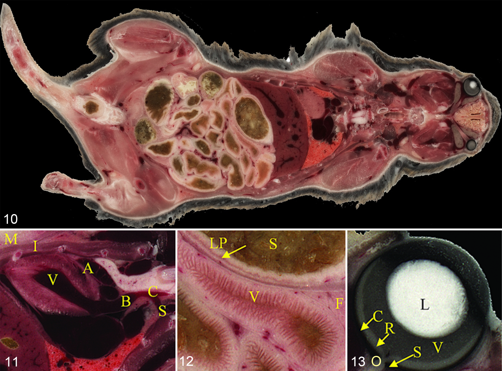
The block face cryoimaging system (http://www.bioinvision.com/) created by our group 77,102 can be used to image a whole adult mouse at microscopic resolution. The system acquires 3D microscopic color brightfield anatomy and molecular fluorescence cryoimage volumes. The system consists of a whole mouse cryomicrotome, microscope, and robotic imaging system positioner for tiled image acquisitions of the block face. Mice are prepared using a cryoembedding compound OCT (Optimal Cutting Temperature, Tissue-Tek, Terrance, California) and fast-frozen in liquid nitrogen. Block face histological and fluorescence images can be obtained with resolutions down to 5 μm pixels and slice thickness of 5–40 μm. Whole mouse 3D data sets on the order of 70–250 GB are acquired. Specialized, multiscale visualization techniques for color volume rendering have been developed. 32,33 Examples of color bright field images taken from 3D cryoimage volumes are shown in Figs. 10–13. With molecular fluorescence imaging, one has the option of imaging fluorescently labeled cancer and stem cells as well as microscopic distributions of imaging agents and nanoparticle theranostics. 89 Postprocessing techniques have also been developed for optimized fluorescence visualization and combined brightfield rendering such as those illustrated in Figs. 14. 44,90 The application of this novel technology has recently been demonstrated in multiscale characterization of Pck1 mutant mouse using semiautomated techniques for organ segmentation and subsequent morphological quantification. 76
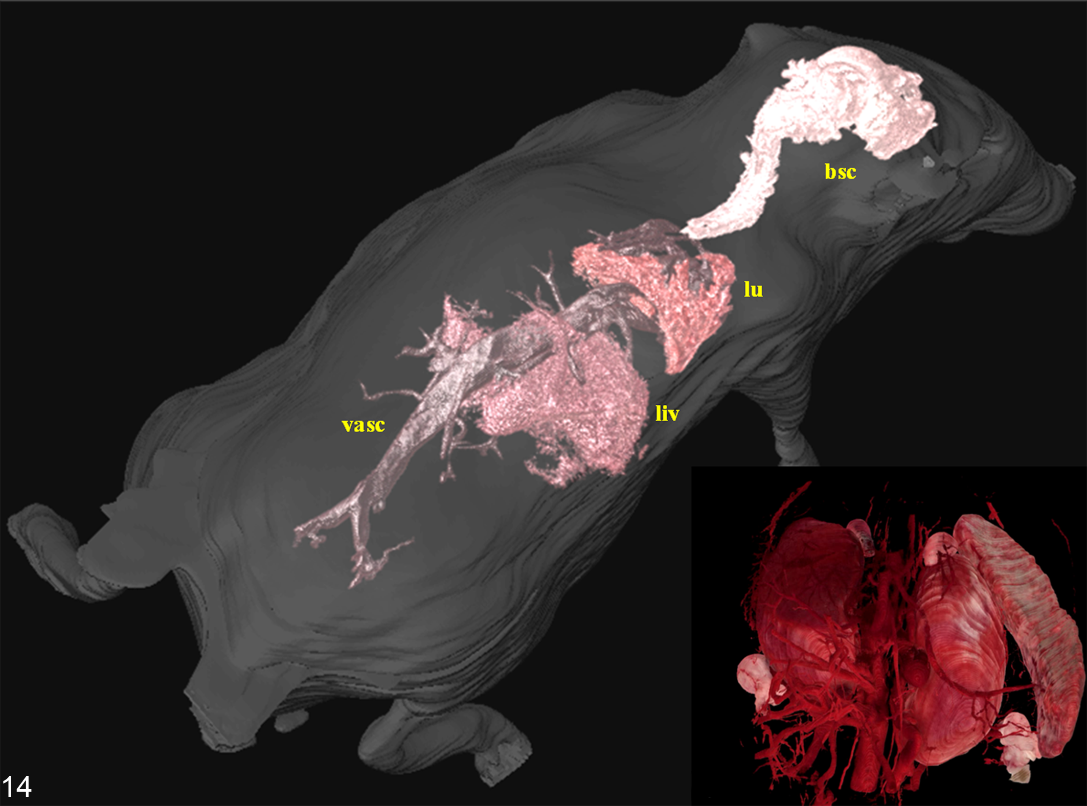
Adult wild-type mouse. Segmented organs from a whole mouse cryoimage volume rendered within a transparent surface of the skin. Organs and organ systems include brain and spinal cord (bsc), lungs (lu), liver (liv), and vasculature (vasc). The inset shows vascular detail in the kidney region. Organ segmentation was performed using an interactive, color-based 3D algorithm. These segmented structures not only are useful for interactive visualization but can be used to calculate morphometric parameters such as organ volume.
Digimouse is a 3D whole-body mouse atlas that is available online (http://neuroimage.usc.edu/Digimouse.html). It was created from microCT and color cryoimages 24 using a 28-g normal male nude mouse. The spatial resolution of the atlas is limited to 100 μm; however, the original higher resolution cryoimages are available for download from the Web site. Major organs are segmented and color-coded for visualization.
Discussion
There are significant advantages to using high-resolution 3D imaging for GEM phenotyping. These include that additional 3D information is available to pathologists for their review, the sample preparation techniques are less laborious than traditional histological methods, the data are in digital format that lends itself to automation and quantitative review, and with many of the modalities presented here, the sample preparation is nondestructive, allowing further 2D histological analysis in targeted regions. Disadvantages include imaging instrumentation that can be costly to operate and maintain and limited access to high-end instrumentation such as the high field-strength magnets used for MRM.
Several aspects of high resolution 3D imaging need to be further addressed in order for these technologies to make it into mainstream use. For example, methods and protocols for decreased acquisition times must continue to be addressed, and standardized imaging protocols and reporting methods must be developed. Automated analysis techniques for quantitative morphometric comparisons need further testing. Development of graphics and visualization techniques for manipulating large color data sets and multimodality data obtained from different scales is essential. Veterinary pathologists, genetic engineers, and imaging scientists will need to work together to develop these tools and determine how best to integrate them into the veterinary pathology workflow.
Footnotes
Acknowledgements
We thank Dr. L. Kirschner and Dr. R. Toribio of The Ohio State University for use of their GEMs in creating the MRM and microCT images presented in Figs. 1–6 and 7–![]() , respectively, as well as Dr. M. Gargesha of BioInVision, Inc (Cleveland, Ohio) for providing the cryoimaging images from CryoViz used in this report.
, respectively, as well as Dr. M. Gargesha of BioInVision, Inc (Cleveland, Ohio) for providing the cryoimaging images from CryoViz used in this report.
The author(s) declared the following potential conflicts of interest with respect to the research, authorship, and/or publication of this article: Dr. Wilson has a financial interest in BioInVision, Inc.
The author(s) disclosed receipt of the following financial support for the research, authorship, and/or publication of this article: Some of the work presented here was partially supported by P30-CA016058 R42-CA124270, R21HL108263, and R41HD063241. Its contents are solely the responsibility of the authors and do not necessarily represent the official views of US National Institutes of Health.