
Abstract
A well-demarcated mass was found by computed tomography in the left cerebellar hemisphere of a 4-year-old male Boxer with acute onset of progressive central vestibular syndrome. At necropsy, the pink, gelatinous mass was in the flocculonodular lobe. Histologically, neoplastic tissue arose from the granular layer of the cerebellar cortex and consisted of sheets of oval to round hyperchromatic cells, consistent with the diagnosis of medulloblastoma. Synaptophysin and neuron-specific enolase immunoreactivity supported the neuronal origin of the neoplastic cells; furthermore, a weak to moderate c-kit expression was detected, as reported in pediatric medulloblastoma. Telomerase activity of tumor cells was demonstrated by immunohistochemistry and by the telomere repeat amplification protocol, suggesting involvement of this enzymatic pathway.
Keywords
Case History
A 4-year-old male Boxer was evaluated because of a 2-week history of loss of balance, head tilt, and ataxia of acute onset and progressive course. On neurological examination, the dog had depressed mental status, disorientation, and a gait characterized by moderate left drifting. Ipsilateral proprioceptive deficits and bilateral loss of menace reaction were also recorded. The clinical signs were consistent with a central vestibular syndrome. A computed tomography scan of the skull revealed an irregularly isodense, space-occupying lesion in the area of the left cerebellar hemisphere that, after intravenous (IV) injection of the contrast medium (Iodixanole, 320 mg/ml; 2 ml/kg IV, as a bolus) had haphazard enhancement. Given the rapid worsening of the dog’s condition, it was euthanatized at the owner’s request.
Necropsy Findings
At necropsy, a pink, gelatinous, 1.5 cm × 3.0 cm mass was in the left flocculonodular lobe of the cerebellum (Fig. 1) with compression of the fourth ventricle. No other central nervous system lesions were discovered, and there was no evidence of distant metastasis. Samples of the cerebellar mass were fixed in 10% neutral buffered formalin, processed routinely into paraffin, sectioned at 4 μm, and stained with hematoxylin and eosin (HE).
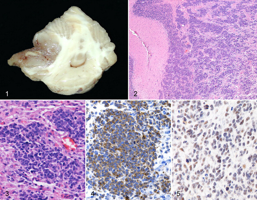
Cerebellum; dog. The medulloblastoma forms a well-demarcated mass in the left cerebellar hemisphere, with compression of the cerebellopontine angle and the fourth ventricle.
Histologic Findings
The neoplastic tissue arose from the granular layer of the cerebellar cortex (Fig. 2) and extended into the leptomeninges and Virchow-Robin spaces. It consisted mostly of solid nests of oval to round cells with hyperchromatic nuclei, indistinct nucleoli, and scanty cytoplasm, in a fine fibrovascular stroma (Fig. 3). There were 2 to 5 mitotic figures per 400× field. A less conspicuous population of elongated or stellate cells with slender cytoplasmic processes, consistent with astrocytes, was interspersed between the nests of neoplastic cells. Concurrent findings included foci of intratumoral mineralization, endothelial cell hypertrophy and hyperplasia, and edema of the adjacent neuropil. Based on the anatomic location, the cytologic features of the neoplastic cells, and the extension of the mass into the leptomeninges, the diagnosis was medulloblastoma.
Immunohistochemistry
Immunohistochemistry was applied to serial tissue sections. After blocking of endogenous peroxidases (0.3% hydrogen peroxide for 30 minutes) and antigen unmasking (microwave oven or enzymatic digestion, depending on the primary antibody), sections were incubated overnight at 4°C with primary antibodies against neuron-specific enolase (NSE; 1:600, Dako, Glostrup, DK), S100 protein (1:1600, Dako), glial fibrillary acidic protein (GFAP; 1:8000, Dako), synaptophysin (SYN; 1:20, Dako), vimentin (1:100, Dako), CD3 (1:75, Dako), CD79 (1:10, Dako); KIT (1:100, Dako), cytokeratin AE1/AE3 (CKAE1/AE3; 1:50, Dako), β-catenin (β-cat; 1:100, Neomarkers, Freemont, CA), Ki67 antigen (1:30, Dako), p53 (1:100, Neomarkers), Bcl-2 protein (1:100, Biogenex, San Ramon, CA), or human telomerase reverse transcriptase (h-TERT) (1:50, Novocastra, Newcastle upon Tyne, UK). A streptavidin–biotin–peroxidase technique (LSAB, Dako) was the detection method. After incubation in chromogenic substrate solution (diaminobenzidine [DAB] 0.04% and H2O2 0.001% in phosphate-buffered saline [PBS]) for 7 minutes, the sections were rinsed in PBS and in running tap water, counterstained with hematoxylin, dehydrated, and mounted with DPX (Fluka, Buchs, Switzerland). As negative control, an isotype-matched irrelevant antibody was used instead of the primary antibody. Positive control tissues were brain for NSE, S100 protein, GFAP, and SYN; skin for vimentin, CKAE1/AE3, and β-cat; lymph node for CD3 and CD79; intestine for KIT, Ki67 antigen, p53 protein, and Bcl-2 protein; and testis for h-TERT.
The neoplastic cells had diffuse intense cytoplasmic expression of SYN (Fig. 4), NSE, and vimentin and mild to moderate, focal, cytoplasmic labeling for KIT and CKAE1/AE3. The spindle/stellate cells strongly expressed vimentin and GFAP. The remaining differentiation markers were not expressed by the neoplastic cells or glial cells. Nuclear expression of Ki67 antigen was evident in 30% of the neoplastic cells. h-TERT immunoreactivity was evident as granular nuclear/nucleolar labeling in about 75% of the neoplastic cells (Fig. 5).
Telomerase Assay
Telomerase activity was investigated in a sample of the tumor, frozen in liquid nitrogen, with the telomere repeat amplification protocol (TRAP) method (Telo TAGGG telomerase PCR ELISA Plus, Roche Diagnostics, Mannheim, Germany). The sample had marked telomerase activity, with the recorded difference of absorbance of 0.512 units. (Baseline difference of absorbance in normal canine testis, used as positive control, was 0.2 units.)
Discussion
Medulloblastoma is a primitive neuroectodermal tumor that arises from the cerebellum, 5 usually in juvenile animals or humans. 11 In dogs, medulloblastoma has been reported mainly in adults, 3 to 10 years of age, with no sex or breed predilection. 10 In this case, immunohistochemical expression of synaptophysin and neuron-specific enolase supported neuronal origin of the neoplastic cells. The neoplastic cells also expressed vimentin and broad-spectrum cytokeratins, typical of immature and multipotential cells. 9 Medulloblastoma cells have been reported to express GFAP, in particularly aggressive cases, in a human and a dog. 12,10 In this case, however, GFAP expression was limited to the population of spindle/stellate cells, thereby supporting their identification as reactive astrocytes.
The immunohistochemical expression of the receptor tyrosine kinase KIT in this canine medulloblastoma is noteworthy because in pediatric medulloblastoma, the overexpression of KIT and the amplification of its gene suggest a possible role for KIT in tumor initiation and progression. 1,2 The fact that the tyrosine kinase inhibitor sunitinib has induced apoptosis and growth arrest in medulloblastoma cell lines makes KIT a potential therapeutic target. 13
Telomerase is a ribonucleoprotein that permits cell immortalization by catalyzing synthesis and elongation of telomeric repeats at the chromosomal ends. Telomerase is normally active in continuously replicating cells, such as germ and hemopoietic cells but is not expressed in most somatic cells. 8 Telomerase activation is detectable in several tumors and is regarded as a fundamental step in the development and progression of human and canine central nervous system tumors, including medulloblastoma. 3,6 –8 Telomerase expression may be evaluated by immunohistochemistry for hTERT; telomerase activity can be measured more accurately in snap-frozen tissue samples with the TRAP, which allows amplification of the telomerase reaction product by polymerase chain reaction and measures enzymatic efficiency. 4 Telomerase has been demonstrated by hTERT immunohistochemistry in a series of canine and feline meningiomas 7 and canine brain tumors, including meningioma, astrocytoma, oligodendroglioma, and 1 medulloblastoma, 6 but TRAP has not been applied to canine brain tumors. In this case, both hTERT immunohistochemistry and TRAP demonstrated a high telomerase activity, suggesting the importance of this enzymatic pathway in canine medulloblastoma.
Because of the difficulty of surgical excision, the prognosis for medulloblastoma is poor. The expression of KIT and telomerase activity in human and canine medulloblastomas warrants further investigation of KIT and telomerase as potential targets for novel chemotherapeutic agents.