
Abstract
Coccidioidomycosis is a systemic fungal infection endemic to the southwestern United States. Although cell-mediated immunity is considered critical in control of the infection, little is known of the cellular population in naturally occurring lesions. To characterize the lymphocytic infiltration, archived formalin-fixed, paraffin-embedded tissues (subcutis, pericardium/heart, lung, bone, and synovium) from 18 dogs with coccidioidomycosis were studied with immunohistochemistry for CD3 and CD79a. In nearly all lesions, T lymphocytes were more numerous than B lymphocytes and were distributed throughout the lesion with concentration in the periphery of granulomas, whereas B lymphocytes were mostly confined to the periphery of granulomas. The predominance of T lymphocytes in lesions of canine coccidioidomycosis was independent of the tissue evaluated, the number of intralesional organisms, and the nature or severity of the inflammatory response.
Coccidioidomycosis is caused by Coccidioides posadasii or Coccidioides immitis, both of which are endemic to the semiarid desert regions of the southwestern United States. The disease begins as a respiratory infection in nearly all cases; 8 when it escapes host control, progressive respiratory disease develops, and infection can disseminate to almost any tissue. However, 60% of human 3 and 70% of canine 9 infections are subclinical and, at least in humans, result in lifelong immunity to reinfection. 3 The durability of immunity in the dog is less well understood, but most dogs are probably protected for life as well. 5
A successful host response to Coccidioides infection is dominated by cell-mediated immunity,1,2,6,7 but few studies have compared the nature of the cellular response in progressive systemic infection with that in subclinical, controlled infection. In an immunohistochemical study of human coccidioidal lesions, pulmonary granulomas had roughly equivalent numbers of T and B lymphocytes, with concentration of T cells in the periphery of granulomas and B cells in perilesional lymphoid aggregates. 4 In experimental murine coccidioidomycosis, 10 mice of a resistant strain had smaller lesions with mild increases in intralesional macrophages and T lymphocytes, as well as few organisms in granulomas, whereas mice of a susceptible strain had precipitous reduction of lymphocyte (especially T lymphocyte) populations, pulmonary consolidation with mainly neutrophilic inflammation, and innumerable organisms as they became moribund. The fungal burden, severity of inflammation, and clinical signs in mice with intermediate resistance to coccidioidomycosis were intermediate between those of susceptible and resistant mice. The purpose of the study reported here was to phenotype the lymphocytes in naturally occurring lesions of canine coccidioidomycosis.
Case Selection
All available histologic slides from canine cases of coccidioidomycosis were reviewed as archived in the Arizona Veterinary Diagnostic Laboratory between 2004 and 2007. Fifteen biopsy cases and 3 necropsy cases, with spherules in histologic sections and sufficient tissue in the corresponding paraffin block, were selected for immunohistochemistry. All but 2 cases had disseminated disease, defined as spread beyond the lung, based on either the tissues evaluated or the clinical information provided with the submission.
Methods
Paraffin blocks were retrieved corresponding to the slides in which fungal organisms were observed (1 to 4 blocks per case). Two 5-μm-thick sections were sequentially cut from each block for immunohistochemistry with an automated slide stainer (Dakocytomation, Carpinteria, CA). A mouse monoclonal anti-human CD79a (Dakocytomation) and rabbit polyclonal anti-human CD3 (Cell Marque Corp, Rocklin, CA) were the primary antibodies. The CD79a antibody was detected with goat anti-mouse conjugated to horseradish peroxidase (LSAB2, Dakocytomation). The anti-CD3 was detected with anti-rabbit horseradish peroxidase polymer (Envision, Dakocytomation). Diaminobenzidine was the chromogen. Slides were counterstained with hematoxylin. Lesions were scored by one observer (L.F.S.), blinded to the clinical history, for density of CD3+ and CD79a+ cells on a scale of 0 to 4 as reported in mice: 10 0, no cells; 1, individual, scattered cells, occasional small clusters visualized at ≥ 20×; 2 and 3, increasing cell density easily visualized at low magnification; 4, dense aggregates in granulomas with diffuse cell infiltration throughout the lesion.
Histologic and Immunohistochemical Findings
Immunohistochemistry was applied in 18 cases comprising 5 tissues: 4 pericardium/heart, 7 subcutis, 4 lung, 1 synovium, and 2 bone. The 3 necropsy cases each had involvement of pericardium/heart; 1 of the 3 had pulmonary granulomas, but no spherules were observed.
Based on the nature of the inflammatory response, the lesions were evenly distributed among suppurative, pyogranulomatous, and granulomatous categories. Spherules were fewer than 25 per slide, regardless of the type or severity of inflammation. Many lesions, especially in sections with only 1 or 2 organisms, had thin-walled, empty spherules that were interpreted as dead or inactive. Inflammation was disproportionately severe for the few organisms observed in most sections. Except for 2 lung sections with discrete granulomas < 1 cm in diameter, the lesions had multifocal necrotic centers with diffuse infiltration by macrophages, fibroblasts, and lymphocytes that generally extended to the borders of the section. Of the 4 pericardial lesions, 3 had granulomatous inflammation with fibrosis of tissue borders.
Immunohistochemically, the lesional density of T lymphocytes was significantly greater than that of B lymphocytes (mean T-cell score, 2.8; mean B-cell score, 1.6; P = .001, Kruskal–Wallis). (Details of the immunohistochemistry results are in Table 1.) In most sections, T lymphocytes were concentrated at the periphery of granulomas but were diffusely and often densely scattered throughout the lesion (Fig. 1), whereas B lymphocytes were mostly confined to the periphery of granulomas (Fig. 2).
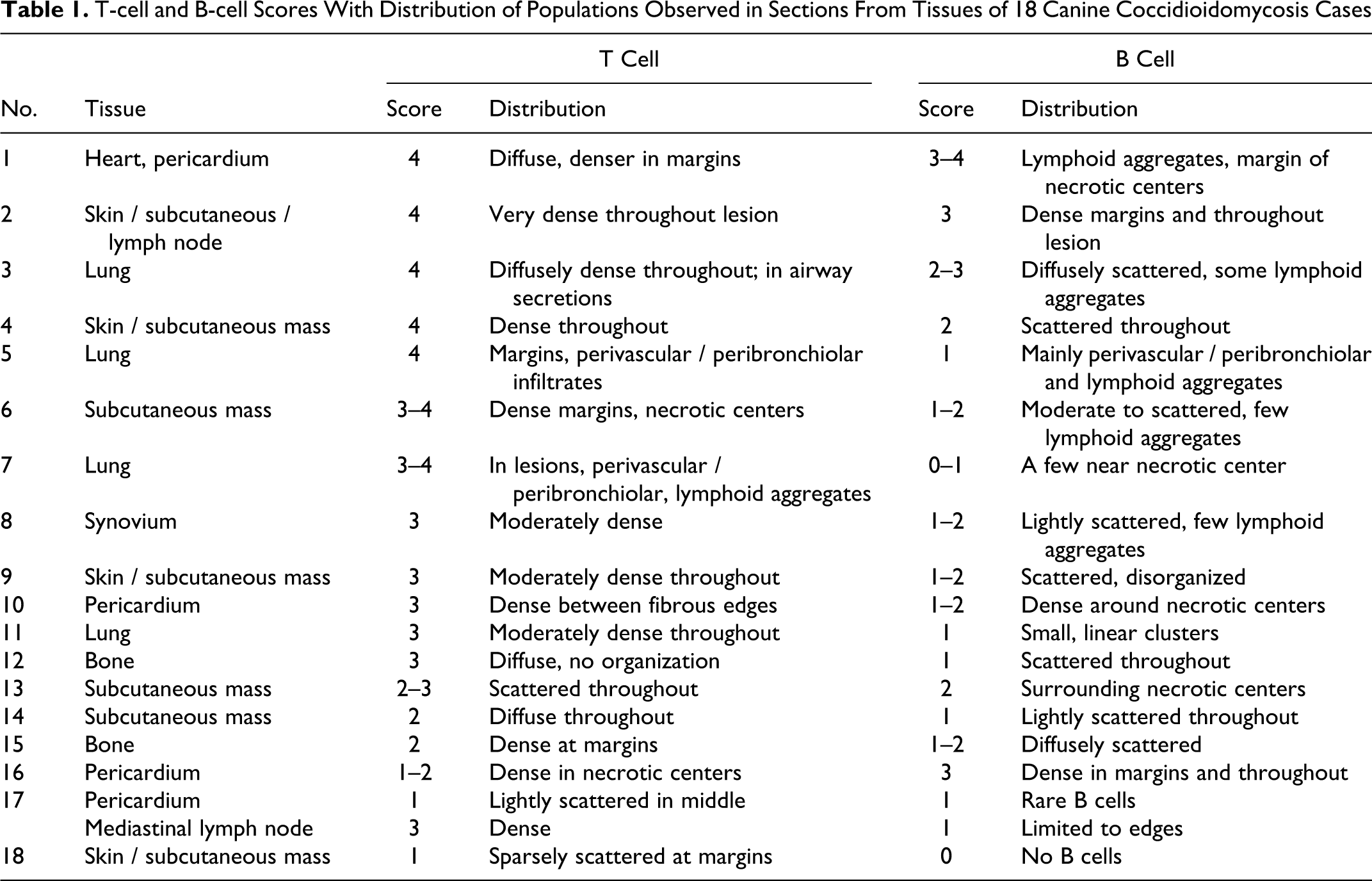
T-cell and B-cell Scores With Distribution of Populations Observed in Sections From Tissues of 18 Canine Coccidioidomycosis Cases
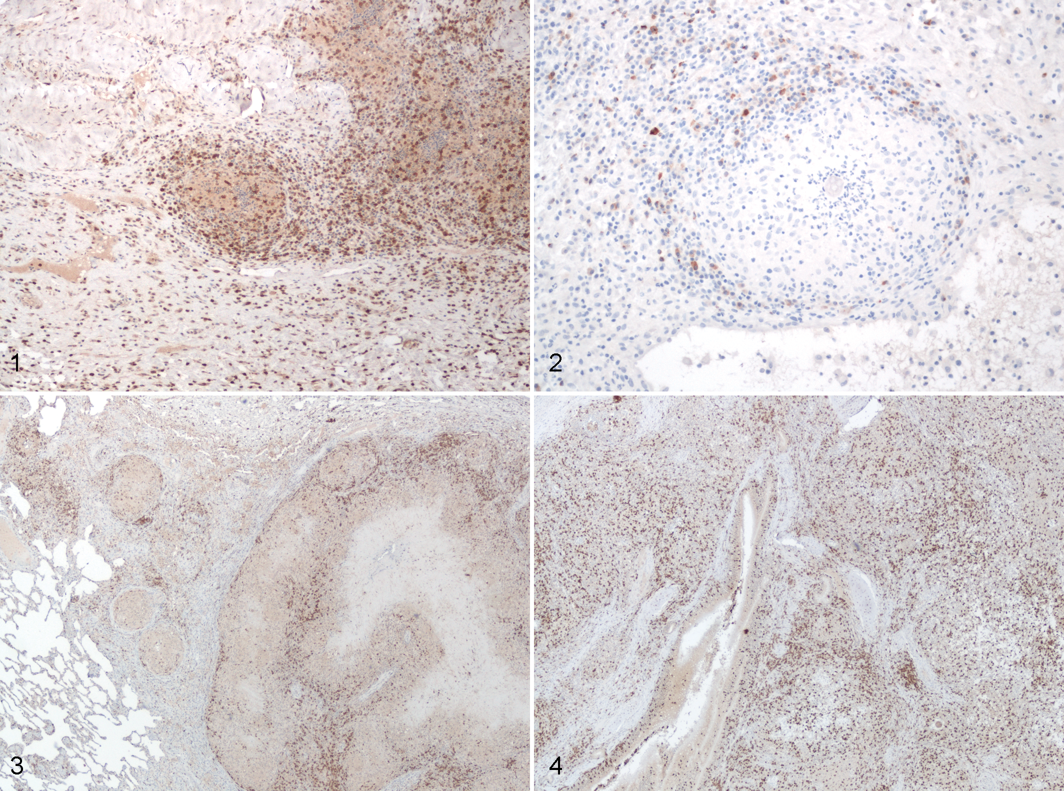
Small, organized pulmonary granulomas (Fig. 3) had numerous T lymphocytes and fewer B lymphocytes at the periphery, few spherules in the necrotic center, and a distinct border of fibroblasts. In contrast, consolidated lung lesions without discrete granulomas (Fig. 4) also had few spherules, but T lymphocytes remained numerous and were distributed throughout the section without obvious areas of concentration.
Discussion
In this study of naturally occurring coccidioidomycosis in dogs, lesions contained few spherules, regardless of the tissue infected, and the number of spherules did not correlate with the nature or severity of the inflammatory response. Whether the infection was in the lung or in extrapulmonary tissues and whether the inflammatory response was suppurative, pyogranulomatous, or granulomatous, T lymphocytes were more numerous than B lymphocytes.
Based on the lesions of experimental murine coccidioidomycosis, 10 dogs were expected to have similar or lower density of T lymphocytes relative to B lymphocytes, especially in uncontrolled lesions—that is, large, suppurative, or extrapulmonary. In mice, T cells tended to be most numerous in the periphery of granulomas, in lymphoid aggregates around well-organized granulomas, and in perivascular/peribronchiolar lymphoid aggregates. The resistant strain of mice had similar T-cell scores at all time points, whereas susceptible mice had a profound reduction in T lymphocytes in the terminal stage of infection. Vaccination of susceptible mice protected them from death and resulted in a statistically significant increase in T lymphocytes (above that of innately resistant mice); T-cell density in pulmonary lesions correlated with survival. 10 However, even vaccinated mice had less dense T-lymphocytic infiltration than that in dogs.
The low number of Coccidioides spherules and the organized granulomas in canine tissues were reminiscent of lesions of experimental coccidioidomycosis in innately resistant or vaccinated mice, 10 even though the infection in most dogs in this study was considered uncontrolled. Susceptible mice developed consolidated lung lesions with numerous spherules and neutrophils but marked paucity of lymphocytes, especially T lymphocytes. 10 Although most dogs in this study were considered susceptible to coccidioidomycosis (systemic disease and consolidated lesions with suppurative inflammation were observed), intralesional spherules were never numerous. Overall, although T-lymphocyte density was greater in dogs than in mice, both the pulmonary and the disseminated canine lesions of coccidioidomycosis most closely resembled those in mice with intermediate resistance, in which severe inflammation—rather than proliferation of the organism, per se—appeared to contribute significantly to morbidity. 10
Vaccination of susceptible mice against Coccidioides results in upregulation of T lymphocytes and increased survival after experimental infection, 10 thereby suggesting a role for cell-mediated immunity in control of the infection. Disseminated coccidioidomycosis is believed to result from a failure in cell-mediated immunity. Thus, the high density of CD3+ lymphocytes in these canine lesions (most of which were deemed “uncontrolled”) and the relatively lower density of B cells were unexpected and distinct from the lymphocytic populations in human 4 or mouse lesions. 10
Unfortunately, because the outcome is unknown for most of these cases, the relationship between the lymphocytic response and survival in canine coccidioidomycosis could not be determined. It was also impossible to correlate the canine lymphocytic response with resistance to coccidioidomycosis because most of the examined tissues were extrapulmonary and thus indicative of failure to contain the infection within the lung. However, these dogs—most of which were alive at sample collection and had organized granulomas even in cases of disseminated disease—are apparently more resistant than susceptible mice, which die within days of pulmonary coccidioidomycosis.
Footnotes
The authors declared no potential conflicts of interests with respect to the authorship and/or publication of this article.
The authors disclosed receipt of the following financial support for the research and/or authorship of this article: This work was funded by the National Institutes of Health, National Institute of Allergy and Infectious Diseases, grant No. PO1-AI061310-03.