
Abstract
Schwannomas are uncommonly recognized in horses. This study describes cutaneous schwannomas in 22 horses aged 8 to 25 years: 12 male, 7 female, and 3 of unknown sex. The horses had solitary cutaneous masses: 9 on the head, 3 on the neck, and the others on the shoulder, hip, thorax, abdomen, rump, extremities, or tail. The location of 1 tumor was unknown. The dermal tumors were well demarcated and expansile. Twelve had a multinodular pattern, whereas 10 formed a single nodule. Antoni A areas were observed in all tumors, and 10 tumors contained Antoni B areas. In Antoni A areas, the densely packed spindle-shaped neoplastic cells were arranged in short fascicles with nuclear palisading. In the hypocellular Antoni B areas, neoplastic cells were separated by abundant myxomatous stroma. Tumors commonly had hyalinization of stroma and vessel walls and ancient change. Cellular vacuolation was observed in 18 tumors. In all 22 cases, neoplastic cells were immunopositive for S100 protein. Expression of laminin and glial fibrillary acidic protein was observed in all 6 tumors evaluated by immunohistochemistry for these markers. One tumor was examined ultrastructurally: Neoplastic cells had branched cytoplasmic processes and were surrounded by an external lamina. Follow-up information was available 8 months to 10 years postexcision for 9 horses, for which surgical excision of the tumor was curative. The equine cutaneous schwannomas in this study had microscopic features like those of human schwannoma and had benign clinical behavior. Correct classification of equine cutaneous schwannoma will facilitate accurate prognosis and appropriate treatment.
Keywords
In human medicine, benign peripheral nerve sheath tumors (PNSTs) are a diverse group of neoplasms that includes 4 main types: schwannoma (synonym: neurilemmoma), neurofibroma, perineurioma, and ganglioneuroma. The 4 types of benign PNST have characteristic diagnostic features, and their distinction is important for prognosis and treatment. In particular, the identification of patients with multiple neurofibromas may lead to the diagnosis of von Recklinghausen disease (neurofibromatosis I), which has implications for family members. 3,41 In the veterinary literature, however, the terms benign PNST, schwannoma, and neurofibroma are often used interchangeably, 7,15,31 whereas ganglioneuroma is classified as a separate entity. 15,16 Although rare cases of perineurioma (originally designated as benign hypertrophic neuropathy) have been reported in domestic animals, 2,9,18 these tumors are not included in most textbooks of veterinary pathology, 15,19 or they are mentioned only briefly. 16 The generic diagnosis of “benign PNST” instead of schwannoma and neurofibroma has been recommended in the veterinary literature because the criteria to distinguish between these two are not well established for animals. 7,15 In contrast, we believe that benign PNSTs can be subclassified in veterinary medicine; indeed, variants of neurofibroma have been recently described in several species. 30
In domestic animals, benign PNSTs may develop in the skin, in peripheral or cranial nerves, and, rarely, in other tissues or organs. Cutaneous benign PNSTs are rarely reported in dogs, cats, horses, and cattle. 5,12,30 Cutaneous schwannomas can be observed as spontaneous tumors in mice; 23 a plexiform cutaneous schwannoma has been reported in a pig. 38 In horses, the eyelids are considered a predilection site for cutaneous PNSTs. 5,22 In addition to forming in the skin, 5,22,36 equine benign PNSTs have been recognized in the intestinal wall. 14,21
The diagnosis of human schwannoma and neurofibroma is usually based on the identification of characteristic histologic features with immunohistochemistry for cellular markers and/or the detection of ultrastructural features. 3,25,28,33,41 Schwannomas are well-demarcated expansile tumors entirely composed of neoplastic Schwann cells. The spindle-shaped to ovoid neoplastic cells form Antoni A and Antoni B areas. Antoni A areas are composed of short fascicles of densely packed neoplastic cells that may have nuclear palisading and Verocay bodies, the formation of which may be incomplete. 3,10,26,28,33,41,43 In the hypocellular Antoni B areas, the neoplastic cells are embedded in abundant myxoid stroma. Hyalinization of stroma and vessel walls is common. As a manifestation of degenerative change, some tumors contain scattered neoplastic cells with enlarged bizarre nuclei (referred to as ancient change) or areas of tumor sclerosis. 28 Immunohistochemistry for S100 protein, a calcium-binding protein expressed by Schwann cells, labels most (if not all) neoplastic cells in benign schwannomas. 33,41 Moreover, neoplastic Schwann cells often produce glial fibrillary acidic protein (GFAP) to a variable extent 20,27 and are surrounded by an external lamina containing laminin and collagen IV. 28,44 On ultrastructural examination, neoplastic Schwann cells are identified by their complete and often-reduplicated external lamina and complex interdigitating cytoplasmic processes. 3,28,41 In contrast, neurofibromas are well to poorly demarcated tumors that contain a mixture of Schwann cells, perineurial cells, and fibroblasts. Their histologic hallmark is the close association of the elongated cells with ropey collagen fibers. Immunohistochemistry for S100 protein labels the Schwann cells but not fibroblasts or perineurial cells. Ultrastructural examination confirms the presence of Schwann cells, perineurial cells, and fibroblasts. 3,28,41
Human neurofibroma and schwannoma are sporadic or develop in association with the inherited diseases neurofibromatosis 1 and 2, respectively. They are subclassified into several variants based on their growth pattern and microscopic features. Recognized growth patterns of schwannoma are uninodular and multinodular (plexiform). Microscopic variants of schwannoma are classical (synonym: conventional), cellular, pigmented (synonym: melanotic), lipoblastic, ancient, collagenous, epithelioid, and glandular. 3,10,24,28,41 To our knowledge, this comprehensive study is the first of the histologic features and clinical behavior of equine cutaneous schwannoma. The purpose of this investigation was to facilitate accurate identification of equine schwannomas and distinguish them from other spindle cell tumors of the skin.
Materials and Methods
Clinical Cases
All cases were excisional biopsy specimens that were submitted in formalin over a 29-year period (1979–2008). Case Nos. 1–21 were submitted to the Department of Biomedical Sciences, College of Veterinary Medicine, Cornell University. Case No. 22 was submitted to the Diagnostic Laboratory at the Royal Veterinary College.
The signalment, tumor location, and duration were obtained from the clinical history. The tumor size was obtained from the history or by measuring the histologic section(s). The latter was used only if the tumor size was not reported and neoplastic tissue was contained within the margins of the examined section(s). In cases with longitudinal and transverse sections of the mass, the length, width, and thickness of the neoplasm were measured. In cases with only 1 section, the width and the thickness of the mass was measured.
In March 2009, we asked the following question of the veterinary surgeons who submitted the biopsy samples: Was removal of the tumor curative or was recurrence and/or metastasis observed? In 9 cases, the requested follow-up information was received (Table 1 ).
Signalment and Neoplastic Features in 22 Horses With Cutaneous Schwannoma a
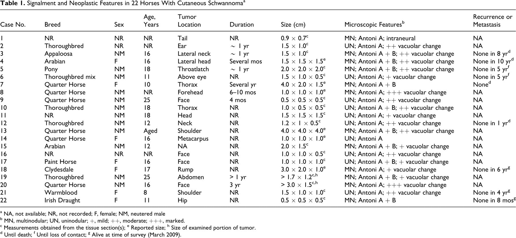
a NA, not available; NR, not recorded; F, female; NM, neutered male
b MN, multinodular; UN, uninodular; +, mild; ++, moderate; +++, marked.
c Measurements obtained from the tissue section(s)
d Until death
e Reported size
f Until loss of contact
g Alive at time of survey (March 2009).
h Size of examined portion of tumor.
Histopathology and Immunohistochemistry
The formalin-fixed biopsies were processed routinely, embedded in paraffin, sectioned, and stained with hematoxylin and eosin (HE). All tumors were evaluated immunohistochemically for S100 protein (Dako, Carpinteria, CA; no pretreatment); 6 tumors (Nos. 5, 7–10, 22) were evaluated for laminin (Dako; steaming in citrate buffer) and 6 (Nos. 5–10) for GFAP (Novocastra, Newcastle Upon Tyne, UK; steaming in citrate buffer). As chromogen, 3,3′-diaminobenzidine tetrahydrochloride was used. In negative controls, the primary antibody was replaced by nonimmune serum. Appropriate positive controls were used.
Electron Microscopy
Electron microscopy was performed on tumor No. 3. Formalin-fixed tissue was further fixed in 2.5% glutaraldehyde in 0.1M sodium cacodylate, washed 3 times in cacodylate buffer, and then postfixed in 2% osmium tetroxide. After further washing, the tissue was dehydrated in graded ethanol solutions, incubated in 100% acetone, and embedded in epon araldite plastic. From 1-μm-thick toluidine blue–stained sections, ultrathin sections of selected areas were contrasted with lead citrate and uranyl acetate and examined in a Philips 300 transmission electron microscope.
Results
Signalment, Tumor Location, Size, and Duration
Table 1 cites the signalment and clinical history for each of the 22 horses. The horses were 8 to 25 years of age, with a mean and median age of 16 years. The age was not provided in 5 cases. Twelve horses were geldings, 7 were female, and in 3 cases, the sex was unknown. There were 6 Quarter Horses, 4 Thoroughbreds, 3 of unspecified breeding, 2 Arabians, and 1 Appaloosa, Clydesdale, Irish Draught, Paint, Thoroughbred mix, Warmblood, and pony. Nine tumors were on the head, 3 on the neck, 2 on the thorax, 2 on the shoulder, and 1 on the abdomen, rump, hip, metacarpus, tail base, and unspecified location. All but 4 tumors were less than 2 cm in maximal dimension. Tumors were described as soft (n, 1) to firm (n, 2) and fleshy (n, 1) and not painful (n, 2) to extremely painful (n, 1). For 9 horses, the duration of the neoplastic growth was reportedly from a few months to 1–3 years. The tumors of case Nos. 3, 5, 9, 14, and 20 had a reported recent increase in size.
Microscopic Features
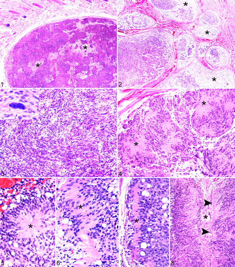
Table 1 lists the growth patterns and other microscopic features of the 22 schwannomas. In 10 cases, the tumor was uninodular (Fig. 1). In the other 12 cases, multiple distinct nodules were surrounded and separated by fibrous tissue (Fig. 2). Growth within a nerve was detected in one (No. 1) of the multinodular tumors. Thirteen tumors were dome shaped; all formed well-demarcated and expansile dermal masses surrounded by a fibrous capsule or compressed fibrous tissue. The neoplastic cells were spindle shaped to ovoid, had indistinct cell borders, single nuclei, and moderate amounts of eosinophilic cytoplasm. The elongate to ovoid nuclei contained stippled chromatin and one to several inconspicuous nucleoli. Cellular pleomorphism, anisocytosis, and anisokaryosis were generally mild, but scattered neoplastic cells had enlarged or lobulated nuclei (Fig. 3, inset) consistent with ancient change, a degenerative change well recognized in human schwannoma 28 and recently described in equine cutaneous melanocytic nevi. 29 The mitotic index (counted in Antoni A areas) ranged from 0 to 6 mitotic figures (average, 2) per 10 high-power fields (400×). The mitotic activity in 2 tumors that had doubled in size (No. 5) and had recent growth (No. 20) was 4 and 3 mitotic figures per high-power field (10×), respectively, just slightly above the average. All tumors contained Antoni A areas; 10 tumors had smaller Antoni B areas. Within the Antoni A areas, the elongated neoplastic cells were arranged in short fascicles separated by small to moderate amounts of stroma (Fig. 3). Nuclear palisading was observed in all tumors to a variable extent (Fig. 3). Tumors contained arrangements of neoplastic Schwann cells in Verocay bodies and rosettes (Figs. 4–7). Novel features included a pattern resembling half of a Verocay body with a single nuclear palisade and eosinophilic Schwann cell processes that formed the margin of a tumor nodule (Fig. 8).
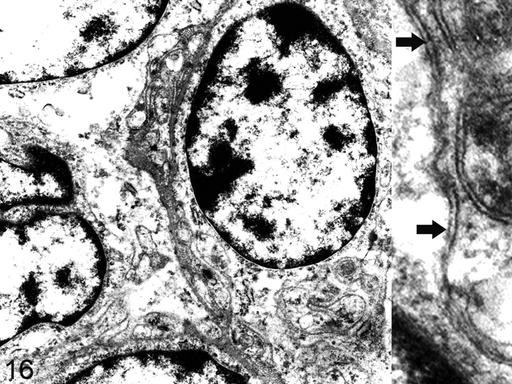
Equine cutaneous schwannoma, No. 3. Neoplastic Schwann cells have an external lamina and branched, interdigitated cytoplasmic processes. Inset: The richly folded cytoplasmic processes are surrounded by a complete external lamina (arrows). Electron microscopy.
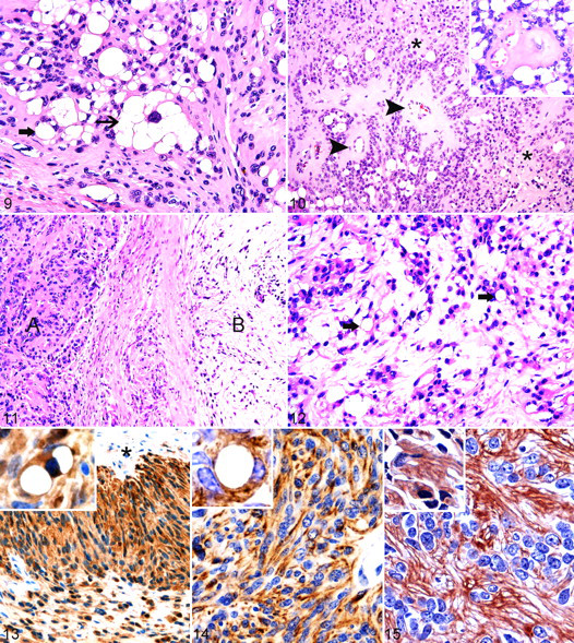
Most tumors had vacuolar change, mainly in Antoni A areas (Figs. 9, 10). Eighteen tumors contained a variable number of cells with a single large clear vacuole surrounded by a narrow rim of cytoplasm and an eccentric nucleus. In 16 of these tumors, a few cells had multiple smaller clear, distinct cytoplasmic vacuoles and a single central or eccentric nucleus (Fig. 9). Vacuolated cells constituted various percentages of cells within the tumors: < 5% (n, 6), 10 to 40% (n, 10), and > 60% (n, 2). A few vacuolated cells contained enlarged nuclei suggestive of ancient change (Fig. 9). Antoni B areas, which occupied approximately 5 to 40% of the tumor mass, were either intermingled with Antoni A areas (Fig. 2) or separated from them by fibrous septa (Fig. 11). Within the hypocellular Antoni B areas, the neoplastic cells were embedded in abundant myxomatous stroma (Fig. 11). In 2 tumors, a transition was observed between highly vacuolated Antoni A and Antoni B areas (Fig. 12), suggesting that Antoni B areas could develop secondary to cellular degeneration in Antoni A areas, as has been proposed. 32
A variable amount of stromal and capillary vessel hyalinization was observed in 21 tumors (Fig. 10) and gave the tumors a characteristic appearance that facilitated their recognition. Some tumors contained small hemorrhages (n, 10) and a low to moderate number of hemosiderin-laden macrophages (n, 11). Tumor necrosis (n, 4), ectatic vessels (n, 3), microcystic change (n, 2), ulceration (n, 1), and lymphocytic infiltration (n, 2) were uncommon findings.
Immunohistochemistry and Electron Microscopic Examination
Neoplastic cells, including those with cytoplasmic vacuoles, uniformly expressed S100 protein in all 22 cases. The immunoreactivity was cytoplasmic and, in some cells, also intranuclear (Fig. 13 ). All 6 tested tumors expressed GFAP; moderate immunoreactivity was observed in the neoplastic cells: 5% (No. 7), 30% (No. 6), and 75 to 90% (Nos. 5, 8–10). Some of the vacuolated cells were GFAP immunopositive (Fig. 14 ). Neoplastic cells were surrounded by laminin-positive material in all 6 tumors examined. Some tumor cells contained intracytoplasmic laminin-positive granular material (Fig. 15 ). The intensity and extent of laminin expression varied. Of the 2 tumors composed of Antoni A tissue only, the entire tumor was strongly immunolabeled in one case (No. 9); in the other case (No. 8), 40% of the tumor had weak laminin expression. The other 4 tumors—composed of Antoni A and B tissue—had weak to moderate laminin expression in Antoni A areas to a variable extent: 100% (No. 22), 50% (No. 5), 35% (No. 7), and 10% (No. 10). In 1 tumor (No. 22), moderate laminin expression was also observed in the Antoni B area.
On ultrastructural examination of 1 tumor, neoplastic cells were identified as Schwann cells by their complete and focally redundant external lamina and cytoplasmic processes that branched and interdigitated (Fig. 16 ).
Biologic Behavior
The surgical excision of the tumor appeared to have been curative for all horses for which follow-up information was available (n, 9; Table 1). No recurrence or metastasis was detected between 8 months and 10 years after surgical excision, although several horses (Nos. 3, 4, 7, 12) died for reasons unrelated to the schwannoma.
Discussion
In these 22 equine cutaneous schwannomas, the tumors were well-demarcated uninodular or multinodular dermal masses with Antoni A areas (and, in some tumors, Antoni B areas), nuclear palisading, and hyalinization of stroma and vessel walls. The neoplastic cells expressed S100 protein, laminin, and GFAP. We have observed in domestic animals variants of schwannoma and neurofibroma 30 similar to those recognized in human beings and therefore propose that the human classification can be cautiously applied to the diagnosis of these tumors in domestic animals.
Because the veterinary literature lacks established criteria for the diagnosis of benign PNSTs, schwannoma, and neurofibroma, their application varies from one pathologist to another. Recent practice in veterinary pathology has been to classify tumors with features of schwannoma or neurofibroma as benign PNSTs. 15 One textbook of equine dermatology uses the term schwannoma for all types of benign PNSTs of horses and describes 3 histopathologic patterns of schwannoma—namely, neurofibroma, neurilemmoma, and plexiform. 31 Thus, the comparison of this series of schwannomas with published cases is problematic.
The expression of S100 protein in schwannomas of dogs and cats is reportedly variable, 16 whereas in our experience, benign schwannomas of horses (this study) and cats (unpublished) are uniformly positive for S100 protein. Certain Schwann cell populations are GFAP positive, 11 and a variable GFAP expression has been observed in most schwannomas. 20,27 GFAP expression by neoplastic Schwann cells can be helpful for the differentiation of benign PNSTs from other spindle cell tumors. 7,27 Schwann cells synthesize external lamina components and are surrounded by a complete external lamina. 25,41 Thus, schwannomas are commonly immunopositive for laminin and collagen IV, which are components of an external lamina. 7,44 The variation in the extent and intensity of the laminin immunoreactivity in the 6 tumors examined in the current study was likely caused by differences of antigen preservation in the formalin-fixed paraffin-embedded tissues because the internal positive controls (the external lamina of vessels and Schwann cells in peripheral nerves) also had similar variations in immunoreactivity.
Benign and malignant PNSTs are said to be rare in horses. 31,39 In a survey of equine cutaneous neoplasia (in total, 536 skin tumors), only 1.1% were schwannomas. 39 In agreement with the literature, we rarely encounter equine PNSTs among our diagnostic cases, although it is our experience that equine schwannoma is more common than equine neurofibroma. 30
The equine cutaneous schwannomas identified in this study occurred as small solitary benign masses in 22 mature horses (mean and median age, 16 years) of a variety of breeds—especially, Quarter Horses and Thoroughbreds. A breed predilection for equine PNSTs has not been reported, and the observed predominance of Quarter Horses and Thoroughbreds might reflect an increased prevalence of schwannoma in these breeds or a higher submission from these 2 breeds compared to other breeds. PNSTs are more frequently observed in male horses than in female horses. 37 In this study, males (all gelded) were slightly more common than females (12 versus 7), but 3 tumors occurred in horses of unknown sex, and the overall case numbers are small. PNSTs are more commonly diagnosed in middle-aged to older horses, 5,35 and all equine schwannomas of this study were observed in this age group.
The 22 schwannomas had been present for weeks to months to years (3 years) and had remained quiescent or, in some cases, had local growth. Microscopic examination revealed uninodular or multinodular proliferation of spindle cells in the dermis, arranged in Antoni A (and, in some cases, Antoni B) patterns. In some schwannomas, a transition could be observed between Antoni A areas containing numerous vacuolated cells and Antoni B areas. This supports the theory that Antoni B areas develop secondary to degenerative changes. 32 Many of the equine tumors contained arrangements of neoplastic Schwann cells similar to conventional Verocay bodies and to others that, in humans, have been designated incomplete or abortive. 10,26,43 Some schwannomas contained rosettes resembling those identified in human schwannoma. 28
Stromal and vascular hyalinization and ancient change, which are common features of human schwannoma, 28,41 were commonly observed in equine schwannomas. Some of the equine tumors had a multinodular growth pattern, which can be observed in some variants of human schwannoma. 10,28,41,43
In human medicine, the modifier plexiform is used for neurofibromas with tumor growth in adjacent nerves, which results in marked multinodular enlargement. 41 The term plexiform is also applied to schwannomas and a variety of other tumors composed of multiple adjacent but separate nodules. In humans, the differentiation of plexiform schwannoma from plexiform neurofibroma has prognostic implication because plexiform neurofibromas are not solitary tumors but occur in patients with neurofibromatosis 1 (von Recklinghausen disease). 41 A solitary cutaneous plexiform schwannoma has been reported in a pig, 38 and a plexiform growth pattern of equine cutaneous schwannoma has been recognized. 31 Only 1 of the equine tumors in the current study grew within preexisting nerves, but in 12 tumors, the growth pattern was multinodular and in a human classification would probably be deemed plexiform.
Many of these 22 schwannomas contained variable numbers of vacuolated cells, some of which had enlarged and occasionally lobulated nuclei resembling the ancient change of the more conventionally spindle-shaped neoplastic Schwann cells. Some vacuolated cells expressed S100, GFAP, and laminin, corroborating their identity as neoplastic cells. The cause for the cellular vacuolation is unknown, but based on the histologic appearance, lipid accumulation has to be considered. Cellular vacuolation due to adipocyte differentiation is observed in lipoblastic schwannoma, a rare human variant. 24 Adipocyte differentiation is also a rare finding in human neurofibroma 24 and has been reported in human meningioma. 1
Differentiation of equine cutaneous schwannoma from sarcoid, the most common equine dermal tumor, 5 should not be difficult, because sarcoids lack Antoni A and B patterns, Verocay bodies, hyalinization, and cellular vacuolation. Perhaps some variants of intradermal melanocytic tumors with spindle cell morphology and limited pigmentation (which would also be S100 protein positive) would present a greater diagnostic challenge, although (1) melanophages will often be present and neither Verocay bodies nor hyalinization has been reported as a salient feature of these tumors 34 and (2) neoplastic melanocytes are immunonegative for GFAP. 17 Equine cutaneous schwannomas with vacuolar change would need to be distinguished from spindle cell lipomas should these tumors be recognized in horses. So far, spindle cell lipomas have been described only in human beings 4,40,45 and dogs. 6 Due to the presence of multivacuolated cells that resemble lipoblasts, as well as the scattered enlarged nuclei, the equine cutaneous schwannomas would need to be distinguished from spindle cell liposarcoma, 42 although this tumor has not been reported in domestic animals to date. Another differential is that of well-differentiated lipoma-like liposarcoma, which contains lipoblasts and has been described in dogs. 6 Mature adipocytes and well-differentiated neoplastic adipocytes are S100 immunopositive and are surrounded by a laminin-positive pericellular membrane. 8,45 In lipomas and liposarcomas with spindle cells, the spindle cell population is immunonegative for S100 and laminin, or it shows only a mild multifocal labeling for one or both antigens. 4,8,42,45 Lipomas and liposarcomas are GFAP immunonegative 6 and lack Antoni A and B areas, Verocay bodies, and prominent vascular hyalinization.
Case outcome was known for 9 horses, none of which had any evidence of recurrence or metastasis 8 months to 10 years after surgical excision. Therefore, complete surgical excision is likely to be curative for equine cutaneous schwannomas. The recognition of schwannoma and neurofibroma as separate entities of benign PNSTs in veterinary medicine will allow more accurate diagnosis. It is likely that the diagnosis of schwannoma and neurofibroma and their variants in the horse and other domestic animals has implications for prognosis and therapy similar to those for the sporadic forms of these tumors in humans. For example, diffuse neurofibromas are infiltrative 41 and therefore often difficult to excise; in fact, removing plexiform neurofibromas may involve significant nerve sacrifice. 13 The complete excision of schwannoma and solitary neurofibroma, however, is usually curative. 13 Finally, although malignant transformation occurs in some neurofibromas in human patients with neurofibromatosis type I, malignant transformation of sporadic schwannoma (or neurofibroma) is rare, and we are not aware of documented progression of schwannoma to malignancy in horses or other animals.
Footnotes
Acknowledgements
We thank Professor K. C. Smith (Royal Veterinary College) for helpful comments on the article, Ms J. Cramer (Cornell University), Ms M. E. Beissenherz (University of Missouri), and the histopathology laboratory at the Royal Veterinary College for immunohistochemistry. We are grateful to Drs D. T. Lamb, J. Kolb, R. Austin, L. F. Karcher, D. Pantano, A. Dwyer, K. T. Kay, C. E. Juul-Nielsen, and their associates and the Liphook Equine Clinic for providing the follow-up information.
The authors declared that they had no conflicts of interest with respect to their authorship or the publication of this article.
The authors declared that they received no financial support for their research and/or authorship of this article.