
Abstract
An adult cat was presented with the history of 3 months' weight loss and more recent loss of balance and ataxia. An abdominal mass was palpable; results of neurologic examination suggested a brainstem disorder. The owners elected euthanasia. Postmortem findings included suppurative jejunal lymphadenitis and bilateral demyelination in the ventral pons with sparing of axons and neuronal soma. The location and character of the lesion mimicked those of human central pontine myelinolysis, an iatrogenic condition that may follow rapid correction of hyponatremia or develop spontaneously in patients with malnutrition or energy deprivation. In this cat, the poor nutritional state may have contributed to the development of this novel pontine lesion.
Central pontine myelinolysis (CPM) in humans, also known as the osmotic demyelination syndrome, has been associated with the rapid intravenous correction of hyponatremia. Spontaneous cases of CPM have been associated with chronic diseases such as alcoholism, malignant tumors, and other conditions with the common feature of altered nutrient supply. Histologically, CPM is characterized by a symmetrical, bat wing–shaped, sharply delineated area of demyelination in the center of the pons. Areas of demyelination may develop in other brain locations. Oligodendrocytes are lost, but axons and neuronal soma, as well as vessels, are spared. Inflammation is typically absent, although activated microglia and lipid-laden macrophages are found. In humans, the major symptoms are behavioral and attributed to disruption of the ascending reticular activating system. 2 Localized myelinolysis following correction of hyponatremia has been reported in the dog, but no case resembling CPM has been reported in the cat. 6
History
A 7-year-old, neutered male, mixed-breed cat was presented with a history of progressive weight loss of 3 months' duration. Loss of equilibrium and ataxia had progressed over the last 2 weeks to include occasional falls and depression. Nystagmus, with no preferred direction, and slight placing reaction delay were observed on neurological examination. An intra-abdominal mass was palpated. With suspected brainstem involvement and a possible intra-abdominal neoplasm, the owners requested euthanasia and allowed necropsy.
Histologic Methods
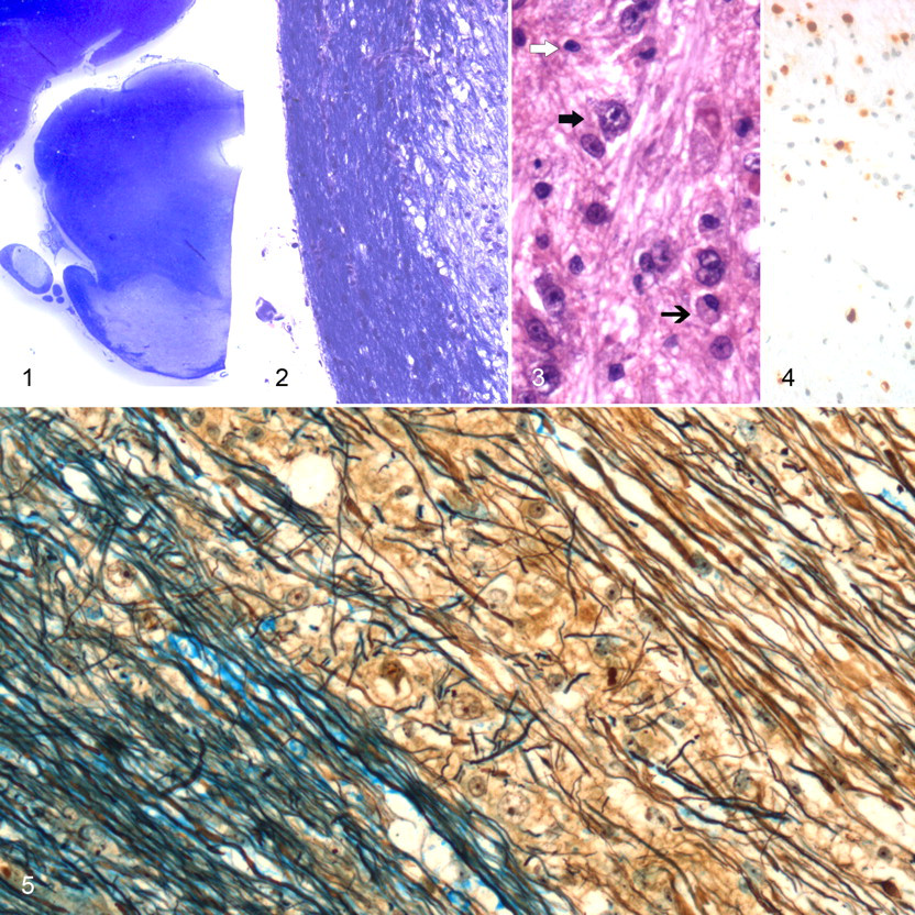
The brain and the abdominal mass, identified as an enlarged jejunal lymph node, were fixed in formalin. Following fixation, the brain was bisected sagittally and each half was further cut transversely into 10 slices. Following processing and embedding in paraffin, 6-μm sections of brain were stained with hematoxylin and eosin (HE), luxol fast blue/cresyl echt violet, and, for selected sections, a combined luxol fast blue–Bielschowsky technique. Immunohistochemistry (peroxidase–antiperoxidase method with diaminobenzidine chromogen) was conducted using antibodies against glial fibrillary acidic protein and the antibody MAC387 to label astrocytes and macrophages, respectively. 7 Because canine distemper virus (CDV) causes demyelination in dogs and has been reported in large felids, 5 DVD12 (Custom Monoclonals International, W. Sacramento, CA), a monoclonal antibody against CDV, was also used; the positive control was cerebellum from an experimentally CDV-infected dog (courtesy of Professor M. Vandevelde). Sections without primary antibody were included as negative controls. Sections of lymph node were stained with HE, Gram’s stain, Ziehl-Nielsen, Grocott’s methenamine silver, and Giemsa methods.
Pathologic Findings
The cat had little remaining adipose tissue. No gross lesions were observed in the brain. On histologic examination of the brain, lesions were restricted to the pons. An irregular, sharply delineated pale area of demyelination was in the pontine base (Figs. 1 and 2). Although not symmetrical, the lesion was bilateral. Demyelination mainly involved the corticospinal tracts with a substantial involvement of the transverse fibers of the pons, extending into the brachium pontis, and a minor involvement of the medial lemniscus. Lipid-laden macrophages were present throughout the lesion (Figs. 3 and 4); astrocytes were essentially unchanged. Neuronal perikarya were preserved within the lesion (Figs. 2 and 3), and Bielschowsky impregnation demonstrated preservation of most axons within the demyelinated area (Fig. 5 ). Discrete perivascular accumulations of mononuclear leukocytes, no more than 1 cell thick, were within and at the border of the demyelinated area. No giant or multinucleated astrocytes, which might have suggested CDV infection, were noticed, and immunohistochemistry for CDV was negative. The enlarged jejunal lymph node contained several micro-abscesses, but neither the histologic changes nor the various types of bacteria present pointed to a specific entity.
Discussion
The nature and topography of the brain lesions in this cat were reminiscent of human CPM. Some asymmetry of the demyelinated focus, as in this cat, is not exceptional in human cases. 4 The wasted nutritional status of this cat brings to mind spontaneous human cases of CPM in which wasting may be part of the pathogenesis. Reduced brain osmolyte concentrations might be the link between spontaneous cases of CPM and iatrogenic cases that follow rapid correction of hyponatremia. In hyponatremia, it is proposed that the brain cannot cope with the rapid increase in interstitial concentrations of sodium and chloride ions that develop with intravenous treatment. 3 Taurine is one of the important organic osmolytes that contribute to the protection of brain cells from dehydration during rapid correction of hyponatremia. Given the dietary dependence of cats on this amino acid, 1 the development of CPM in a wasted cat is not surprising. However, taurine tissue concentration was not documented in this case. The source and specific cause of the bacterial jejunal lymphadenitis remain unknown but account for the cat’s wasted condition and could have contributed to the development of pontine myelinolysis.
Canine distemper virus infection was excluded in the face of only a solitary pontine lesion, the absence of viral inclusions, and the negative immunohistochemistry. Furthermore, although CDV infection is well recognized in lions and tigers, it is virtually unknown in domestic cats. Other, usually multifocal, demyelinating entities, such as multiple sclerosis and progressive multifocal leukoencephalopathy, 4 have not been described in cats and were considered extremely unlikely in this case.
Footnotes
Acknowledgement
The authors thank Ms. Marlene Nardi, Cornell University, for the excellent histochemistry.
The authors declared that they had no conflicts of interest with respect to their authorship or the publication of this article.
The authors declared that they received no financial support for their research and/or authorship of this article.