
Abstract
In this retrospective study, spontaneous osteosarcomas were found in 85 of 1,202 (7.1%) nonobese diabetic (NOD) and NOD-derived mice. Gross tumors were evident at an average age of 155.8 days in male mice and 151.4 days in female mice. Compared with male mice, female mice had a statistically insignificant higher incidence: 56 cases (8.3% of 672) versus 28 cases (6.1% of 458). NOD/ShiLtJ mice had the highest incidence, with 39 cases among all the strains and substrains represented (3.2% of 1,202 necropsies), whereas NOD.SCID substrains had the highest incidence, with 16 cases among the various NOD-derived substrains (1.3% of 1,202 necropsies). There was a statistically significant difference in tumor incidence between NOD/ShiLtJ and NOD.SCID mice. Tumors were more frequent in the appendicular skeleton (55.7%) than in the axial skeleton (44.3%) and most often arose from the femurs. Histologically, osteoblastic osteosarcoma was the most common tumor type, with 79 cases (94%), followed by mixed osteosarcoma, with 5 cases (6%). Metastases were rare, with only 2 cases (2.3%).
Osteosarcomas are malignant mesenchymal tumors in which tumor cells typically produce osteoid or bony matrix. 6,13,14,27 –30 They most commonly arise within the medullary cavities of bones, particularly in the metaphyseal regions of long bones, and invade the adjacent cortex. These are referred to as central osteosarcomas. 27 –29 Osteosarcomas less commonly arise from the periosteum; these are called peripheral osteosarcomas and are of two types—namely, periosteal and parosteal (juxtacortical) osteosarcomas. 27 –29 Osteosarcomas rarely arise in extraskeletal soft tissues independent of adjacent bones. 8,27 –29 Extraskeletal osteosarcomas compose about 5% of all human osteosarcomas. 8
The nonobese diabetic (NOD) inbred mouse strain was developed in 1974 after a female mouse of the cataract Shionogi strain was found to spontaneously develop hyperglycemia. 2,15,16,18 Selective breeding of the progeny of this diabetic female gave rise to the NOD inbred strain that was found to develop insulin-dependent diabetes mellitus (IDDM). 2,16,18 NOD mice harbor a unique major histocompatibility complex (MHC) haplotype, termed H-2g7, which is essential for, and is the highest genetic contributor to, diabetes susceptibility. 2,26 This MHC haplotype does not express an I-E molecule, because of a defective Eα locus. 2,26 The immune-mediated insulitis in these mice is characterized by a mononuclear infiltrate of the pancreatic islets, leading to destruction of insulin-producing β cells. This results in a marked decrease in pancreatic insulin content, leading to the development of IDDM with typical nonfasting plasma glucose levels higher than 250 mg/dL. 2,15,16 Most NOD male mice develop IDDM by 12 to 16 weeks of age, whereas most females typically develop IDDM by 26 to 30 weeks of age. 2,15,16 Early onset of the diabetic phenotype has contributed to the popularity of the NOD strain and subsequent development of numerous genetically manipulated substrains—many of which have not been completely backcrossed to the parent line and therefore have a genetically mixed background. Presently, there are more than 200 NOD substrains, all derived from the original NOD mouse, with variable genetic contributions from other inbred strains, leading to a variety of phenotypes. The development of insulitis has been shown to be polygenic and dependent on diet, health status, and housing conditions, all contributing to the variation in age of disease onset. 2,15,16 Whereas the NOD strain and NOD-derived substrains of mice are best known for their traits pertinent to diabetes and autoimmunity, the occurrence of spontaneous osteosarcomas in these mice has only been marginally mentioned. 15
Whereas several murine models of genetically engineered 5,11,13,14,21,30,33 and induced 19,24,31 bone tumors are extant, spontaneous osteosarcomas are relatively rare in different strains of laboratory mice. Previous incidence reports of spontaneous osteosarcomas in inbred strains of mice include 0.066% in 18-month-old female BALB/c mice, 9 0.04 to 0.50% in male B6C3F1 mice, and 0.15% in female B6C3F1 mice. 1,3,31 There have also been single-case reports of osteosarcoma in a C57BL/6J mouse and a SCID/Sed mouse. 4,12 In contrast to these reports, pathologists at the Jackson Laboratory have, with some regularity, observed spontaneous osteosarcomas in NOD and NOD-derived mice. This retrospective study purports to provide a description and preliminary characterization of spontaneous osteosarcomas in the NOD and NOD-derived substrains of mice.
Materials and Methods
Included in the present study were 1,202 mice, 30 days or older, representing the NOD strain and 101 NOD-derived strains, substrains, and crosses. All mice were part of colonies maintained at the Jackson Laboratory, Bar Harbor, Maine, and all were submitted to the diagnostic service for necropsy between January 1987 and May 2009. Criteria for submission were clinical assessment of a “sick mouse” or the presence of a grossly visible tumor. Complete necropsy was performed on all mice, and collected tissues were fixed in Tellyesniczky/Fekete fixative (100 mL, 70% ethanol; 5 mL, 37 to 40% formalin; 5 mL, glacial acetic acid). Appropriate tissues were decalcified using 10% formic acid (Formical-2000, Decal Chemical Corporation, Tallman, New York) or 3% hydrochloric acid (Cal-Ex, Fisher Scientific, Fairlawn, New Jersey) for 24 hours. Tissues were paraffin embedded and stained with hematoxylin and eosin. One or more bones were available for microscopic examination from 261 cases (156 females, 102 males, and 3 for which sex was not available from the archives). For all cases that had a diagnosis of osteosarcoma, other bone tumor, or any mesenchymal neoplasm, archived glass slides were retrieved and reviewed. Eighty-four cases of osteosarcoma were used for histologic typing, and one was excluded. Osteosarcomas were subclassified according to criteria described in the International Classification of Rodent Tumors: The Mouse. 6 Chi-square analyses were performed on the data using JMP-7 statistical analysis software (JMP, Cary, North Carolina).
Results
Of 1,202 mice with the NOD genetic background that were examined, 85 (7.1%) had a verifiable diagnosis of osteosarcoma. Female mice had a higher incidence, with 56 cases versus 28 for males (sex was not available for 1 case of osteosarcoma). Chi-square analysis revealed this difference to be statistically insignificant, χ2 = 1.99, SE = .12, P = .1581. The average age at necropsy was 155.8 days in males and 151.4 days in females (Table 1 ).
Incidence of Osteosarcoma in NOD and NOD-Derived Mice
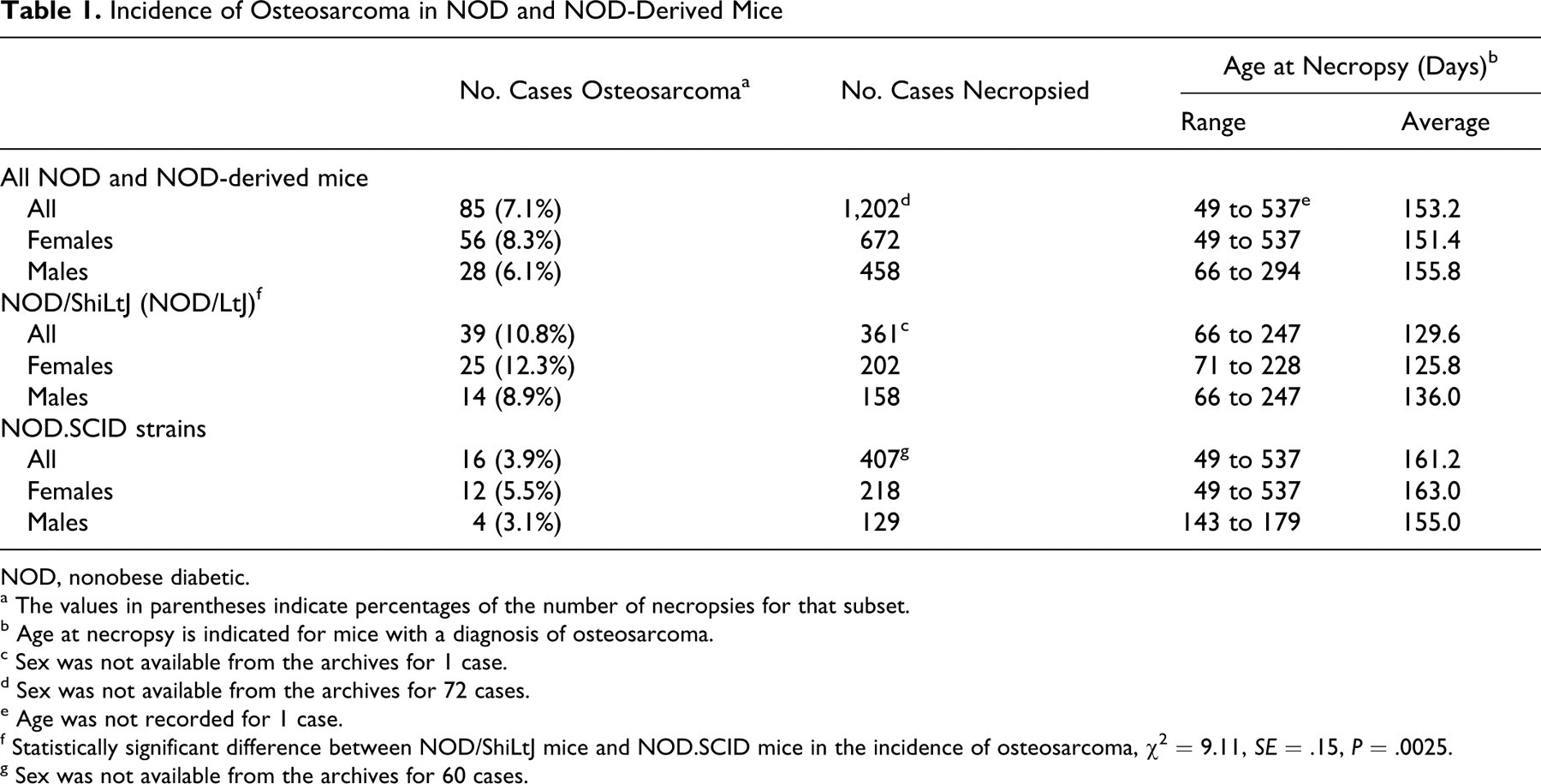
NOD, nonobese diabetic.
a The values in parentheses indicate percentages of the number of necropsies for that subset.
b Age at necropsy is indicated for mice with a diagnosis of osteosarcoma.
c Sex was not available from the archives for 1 case.
d Sex was not available from the archives for 72 cases.
e Age was not recorded for 1 case.
f Statistically significant difference between NOD/ShiLtJ mice and NOD.SCID mice in the incidence of osteosarcoma, χ2 = 9.11, SE = .15, P = .0025.
g Sex was not available from the archives for 60 cases.
Among all the mice represented, NOD/ShiLtJ mice had the highest incidence, with 39 cases (3.2% of 1,202 necropsies), and the youngest average age of incidence, at 129.6 days. NOD.SCID substrains, with 16 cases (1.3% of 1,202 necropsies), were most represented among all NOD-derived substrains, with a slightly higher average age of incidence, at 161.2 days. A statistically significant difference was evident between NOD/ShiLtJ (10.8% of 361 necropsies) and NOD.SCID (3.9% of 407 necropsies) mice (Tables 1 and 2 ), χ2 = 9.11, SE = .15, P = .0025.
Distribution of Osteosarcoma Cases Among the NOD Strain and NOD-Derived Substrains of Mice
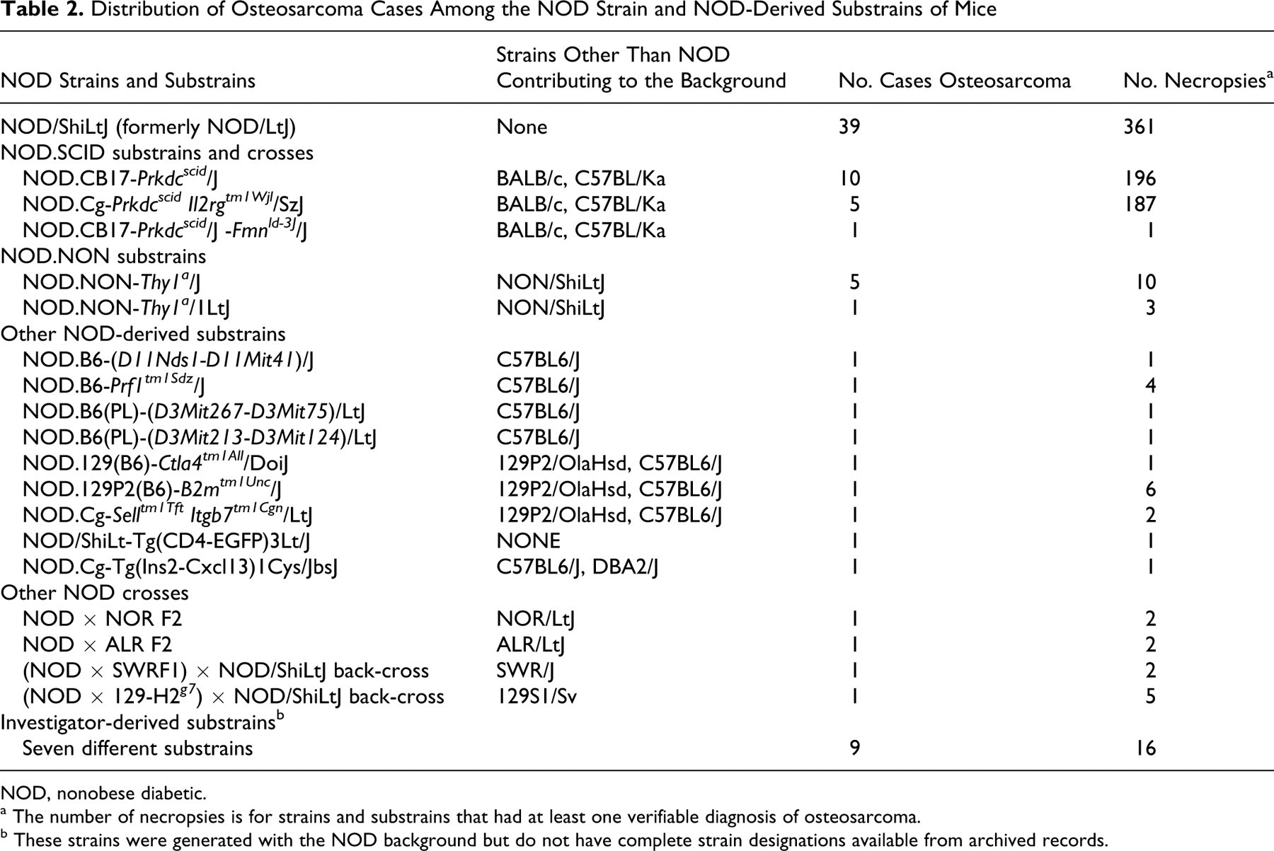
NOD, nonobese diabetic.
a The number of necropsies is for strains and substrains that had at least one verifiable diagnosis of osteosarcoma.
b These strains were generated with the NOD background but do not have complete strain designations available from archived records.
Tumors were more frequent in the appendicular skeleton than in the axial skeleton and were most often observed in the hind limbs, typically arising at the femurs (Figure 1). Other sites on the appendicular skeleton included the pelvic bones, metatarsals, and bones of the forelimb. Tumors were also observed on sites of the axial skeleton, such as cranial bones, mandible, vertebrae, ribs, and sternum. Of 79 cases that had records of the anatomical site of incidence available from the archives, 44 cases (55.7%) were noted on the appendicular skeleton, 29 (36.7%) on vertebrae, and 6 (7.6%) on cranial bones.
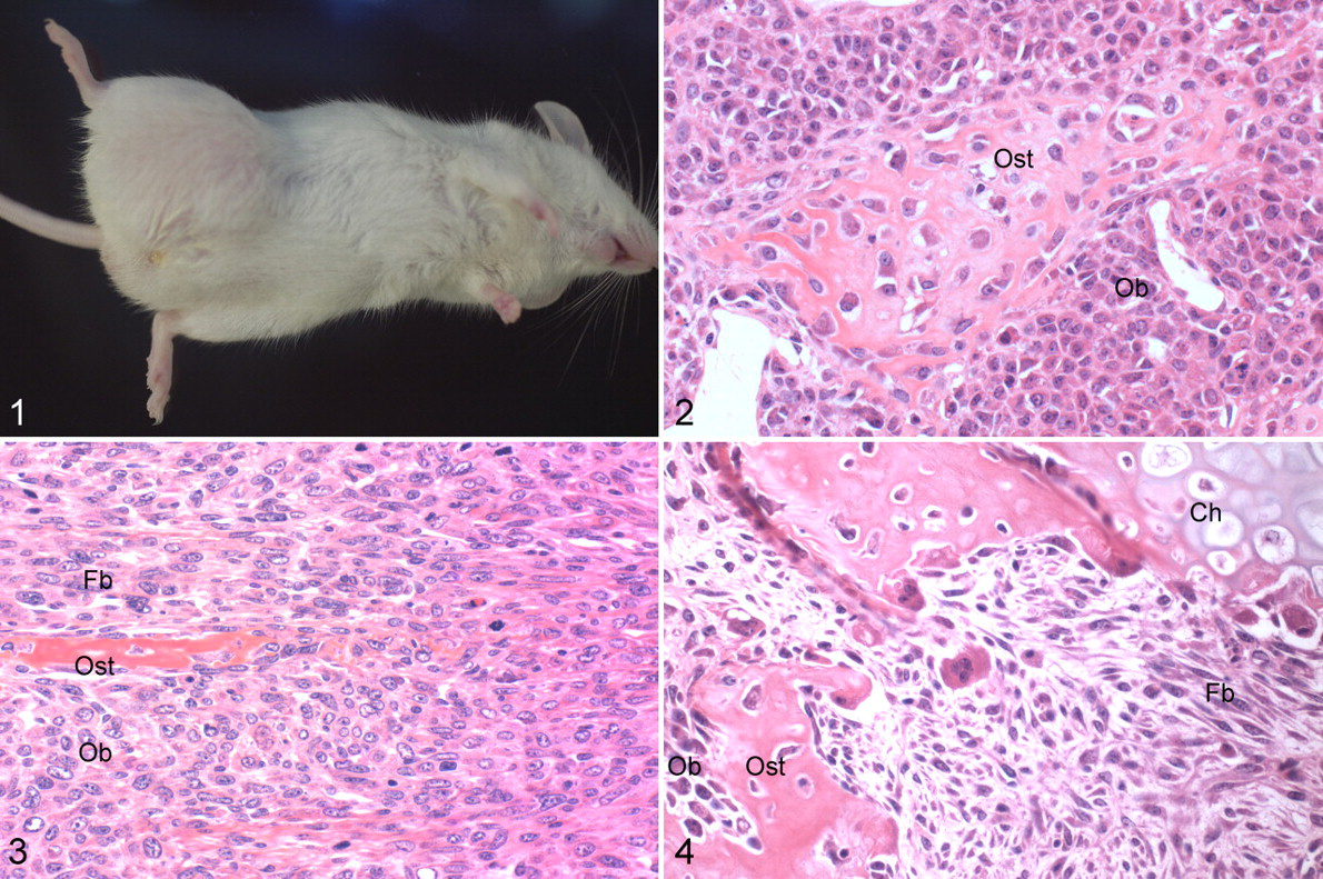
The most common microscopic tumor type, with 79 cases (94%), was osteoblastic osteosarcoma (Figure 2), as characterized by sheets and bundles of ovoid to polygonal neoplastic cells with occasional multinucleate forms. Foci of osteoid formation were evident in most cases. These tumors were often locally invasive into the surrounding soft tissue. Five cases (6%) of mixed osteosarcoma were also observed, and they were characterized by a mixture of osteoblastic polygonal cells and fibroblast-like spindle cells (Figure 3). A low-grade mixed osteosarcoma that resembled a fibro-osseous pseudo-tumor also had cartilaginous foci (Figure 4). There was no documentation of gross metastases. Only two microscopic metastases were noted, in the wall of the stomach and duodenum in two separate cases (2.3%). An embolus of osteoblastic osteosarcoma was also noted within the right ventricle of the heart.
In addition to osteosarcomas, several other types of tumors were noted in these mice (Table 3 ). Lymphoma was the most prevalent tumor type, and as published previously, 10,25 NOD.CB17-Prkdcscid /J mice were most represented, with 58 cases (29.6% of 196 necropsies).
Tumor Incidence in the NOD Strain and NOD-Derived Substrains of Mice
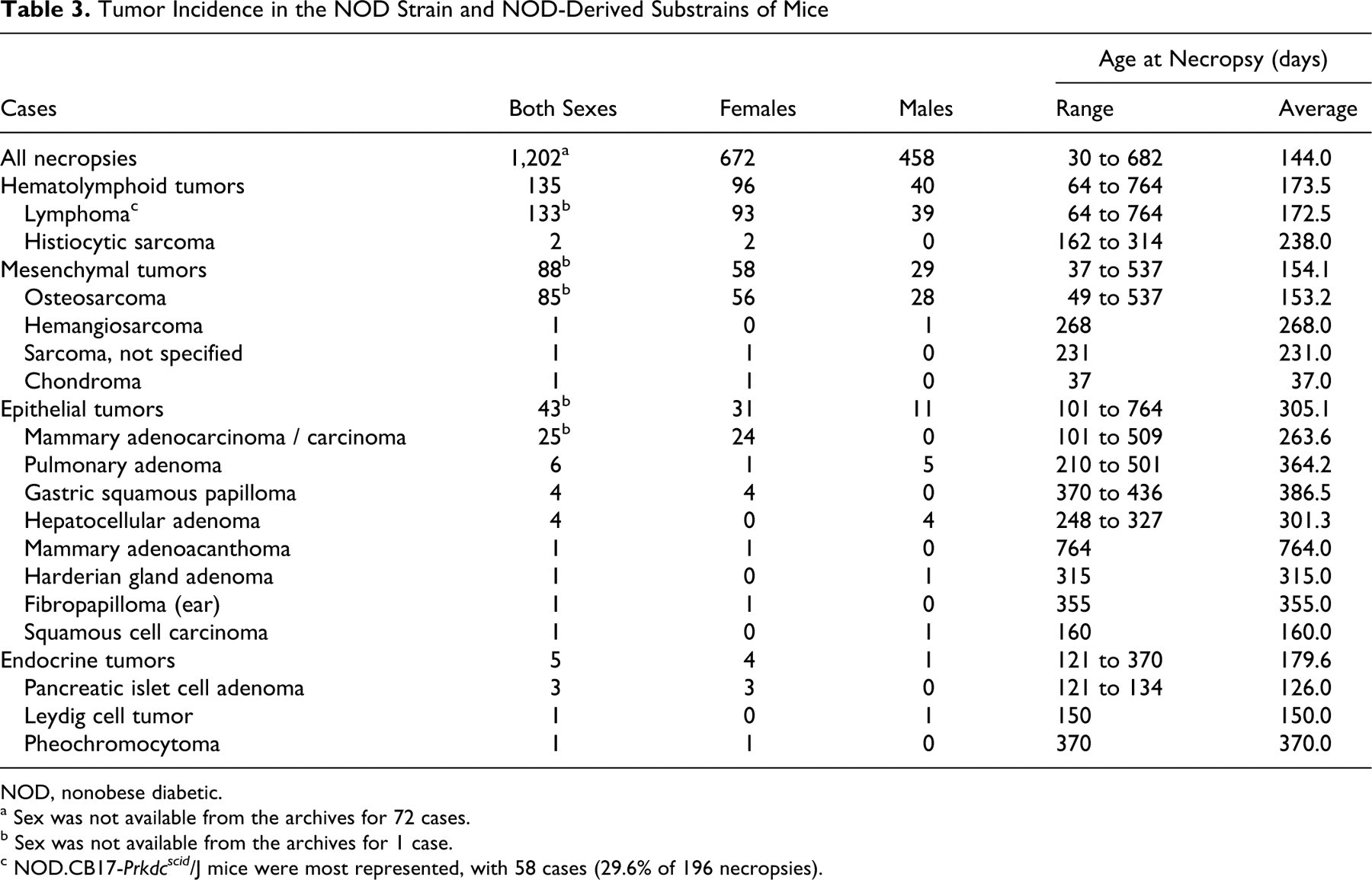
NOD, nonobese diabetic.
a Sex was not available from the archives for 72 cases.
b Sex was not available from the archives for 1 case.
c NOD.CB17-Prkdcscid /J mice were most represented, with 58 cases (29.6% of 196 necropsies).
Discussion
This study describes the incidence and features of spontaneous osteosarcomas in the NOD strain and NOD-derived substrains of mice. Compared with previous reports of spontaneous osteosarcomas in other inbred strains of mice, the incidence of spontaneous osteosarcomas in these mice was 10-fold greater. 1,3,9,23,31 In addition, the average age of gross detection of osteosarcomas in this study was 153.2 days. With the exception of an unusual case of spontaneous osteosarcoma in a 35-day-old C57BL6/J mouse, 4 the age of onset in this study was much earlier than that of other reports—namely, 530 days and 300 days in a SCID/Sed mouse 12 and an AKR/Ms mouse, 22 respectively.
NOD mice exhibit multiple aberrant immunophenotypes, including defects in immunoregulatory functions of antigen-presenting cells, regulation of the T lymphocyte repertoire, NK cell function, cytokine production from macrophages, and wound healing. 7 Although the genetic basis of increased incidence and earlier onset of spontaneous osteosarcomas in NOD and NOD-derived mice remains unknown, the unique MHC haplotype, H-2g7, shared between NOD substrains is likely to play a role in reduced tumor inhibition. In addition, MHC Class IA (MICA) is one of the major ligands that activate the immune receptor NKG2D, which is expressed on NK cells and cytotoxic T lymphocytes. MICA was found to be upregulated in osteosarcomas compared with benign bone tumors and normal bone, and it is suspected to have a role in MICA-NKG2D-mediated immunosurveillance in osteosarcoma patients. 17,34 With these facts, further investigations can include the possibility of MHC profiles and related immunologic pathways bearing on the propensity of NOD mice to develop spontaneous osteosarcomas at a relatively young age.
There have been reports of retrovirally induced osteosarcomas in NIH Swiss 32 and BALB/c 26 mice and isolation of retroviral particles in spontaneous osteomas in (C3H × 101) F1 19 and CD-1 20 mice. Note that in contrast to the tumors in this study, the spontaneous osteomas from which retroviral particles were identified were morphologically distinct, 19,20 and the previously reported retrovirus-associated osteosarcomas were experimentally induced by administration of FBJ murine osteosarcoma virus 32 and Moloney murine sarcoma virus. 24 A retroviral etiology is thus unlikely for the tumors presented in this study.
Tumors were most common in the appendicular skeleton as described in other mouse strains as well as other species. The most prevalent histologic type was osteoblastic osteosarcoma as described in humans, 29 in dogs, 28 and in some of the previous reports of murine osteosarcomas, with the exclusion of a report on female BALB/c mice in which half the osteosarcomas were diagnosed as fibroblastic osteosarcomas. 9 In concordance with some of the previous reports on murine osteosarcomas, tumor incidence was slightly higher in females, which contrasts with the higher incidence of osteosarcomas in human males and appendicular osteosarcomas in canine males. 28,29 The sexual dimorphism in tumor incidence in this study was found to be statistically insignificant.
Among the various strains and substrains represented in this study, NOD/ShiLtJ mice had the highest tumor incidence. The statistically significant difference in the incidence compared with NOD.SCID strains could be a reflection of the disparate genetic and immunologic profiles of these mice or, alternatively, a consequence of the latter developing more lymphomas. What remains to be explored is whether there is a genetic basis for the higher incidence of osteosarcomas in NOD/ShiLtJ mice and the significance of the scid mutation, if any, in mitigating susceptibility to the development of osteosarcomas.
Unlike in other species and some other strains of mice, metastases were not frequent. It is possible that mice with the NOD background have mutations in some of the genes, or reduced levels of one or more of the proteins, known to play cardinal roles in metastasis. Alternatively, given that many of the tumors were detected early while relatively small, it is possible the mice did not live long enough to develop metastatic tumors.
Many epithelial, mesenchymal, and endocrine tumors were also noted in the NOD and NOD-derived mice. Although the incidence of these tumors is presented for the combined pool of all NOD and NOD-derived mice examined, note that these tumor types and their incidences might vary amid NOD-derived substrains as reflections of genetic backgrounds and modifications.
Footnotes
Acknowledgements
We thank Dawna Boggess for assistance in retrieval of archived case materials, the Department of Histopathology and Microscopy Sciences for preparation of histologic sections from archived paraffin blocks, and Weidong Zhang of the Department of Computational Sciences for assistance with JMP-7 software for statistical analysis.