
Abstract
An 18-month-old cross-bred goat was presented with generalized erythema and thinning of the hair coat, as well as localized moderate scaling. Histopathological evaluation of skin biopsies showed hyperplasia and marked disruption of the infundibular epithelium owing to a predominant infiltrate of macrophages with multinucleated histiocytic giant cells and some lymphocytes, plasma cells, and eosinophils. Examination of peripheral blood and skin by polymerase chain reaction gave positive results for ovine herpesvirus type 2 consistent with a diagnosis of malignant catarrhal fever.
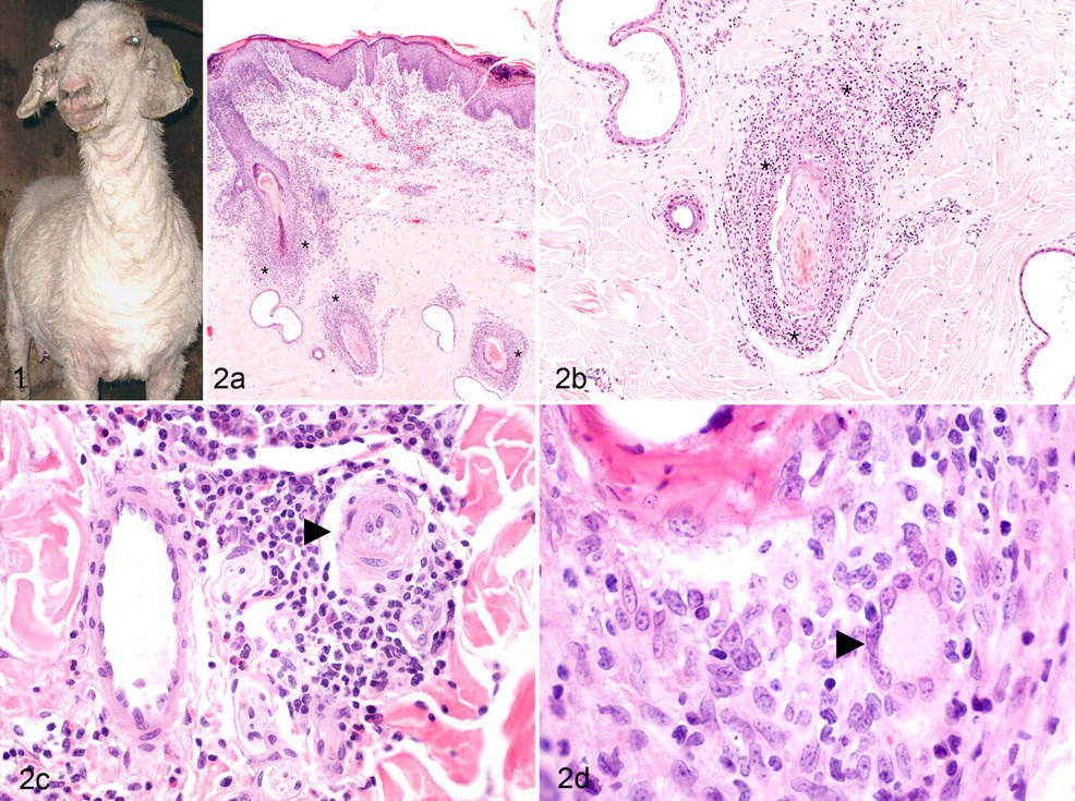
An 18-month-old cross-bred doe presented with a history of 4 weeks' duration that started with multifocal erythematous papules, particularly on the distal limbs, which progressed to generalized erythema with localized scaling and thinning of the hair coat. There was moderate focal scaling and crusting of the pinnae, nares, and peribuccal skin (Fig. 1). No ocular or mucocutaneous lesions were observed, and the skin lesions were not pruritic. The goat shared grazing and shelter with a breeding flock of sheep, and there were beef cattle on the farm. The owners reported that mastitis had developed shortly before the onset of the skin lesions. Three samples of skin were excised and placed into formalin fixative, and a heparinized blood sample was collected. The skin condition deteriorated; the goat became depressed and had a reduced appetite; and it was euthanized. The owners did not give permission for postmortem examination. There was no skin disease reported in the sheep and cattle on the farm and no history of malignant catarrhal fever (MCF) in the cattle on this and neighboring farms.
Differential Diagnoses
Alopecia with scaling in goats may be associated with demodicosis, dermatophytosis, and bacterial pyoderma. The focal crusting could be due to dermatophilosis and pemphigus foliaceus. Similar systemic signs and widespread distribution of skin lesions have been seen in cattle with MCF associated with ovine herpesvirus type 2 (OvHV-2) infection.
Microscopic Findings
Microscopic examination revealed marked irregular epidermal hyperplasia with compacted ortho- and parakeratotic hyperkeratosis and severe layered inflammatory scaling and crusting. The inflammatory scaling and crusts contained gram-positive coccoid bacteria and variably sized subcorneal eosinophilic pustules. There was mild multifocal lymphocytic, eosinophilic, and neutrophilic exocytosis into the hyperplastic epidermis. The infundibular epithelium was also hyperplastic and hyperkeratotic. The internal and external root sheaths of the isthmus and, to a lesser extent, infundibular portion of the follicular wall were heavily infiltrated by macrophages, with fewer lymphocytes, plasma cells, and eosinophils and with occasional multinucleated histiocytic giant cells (Fig. 2). Macrophages, lymphocytes, and plasma cells, with fewer eosinophils, surrounded the hair follicles and infiltrated the sebaceous glands, resulting in their degeneration. Arterioles in the deep dermis were occasionally surrounded by an admixture of macrophages and lymphocytes and plasma cells; some mural hemorrhage was present. However, infiltration of the vascular wall by inflammatory cells or deposition of fibrin was not observed. Multiple periodic acid–Schiff-stained sections were examined and did not reveal fungal organisms.
Laboratory Findings
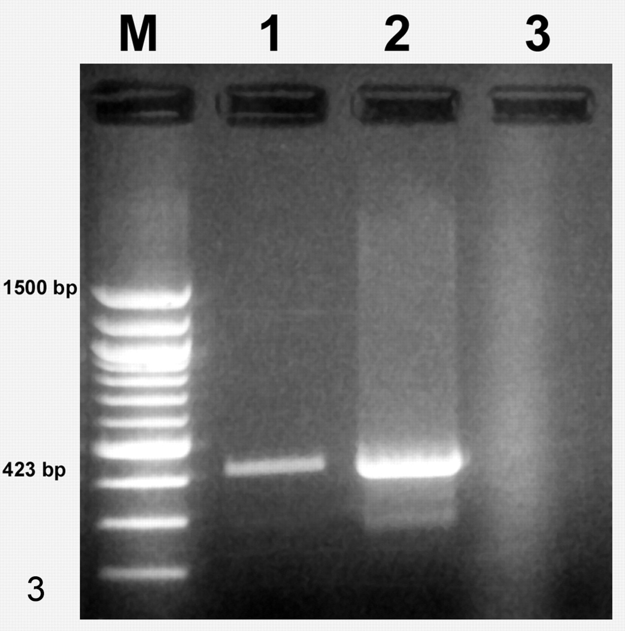
An antemortem heparinized blood sample was submitted for detection of OvHV-2 DNA by real-time polymerase chain reaction (PCR). 10 Total DNA was extracted from the buffy coat, and OvHV-2 DNA was detected, with a cycle threshold (CT) value consistent with a viral DNA load in excess of 1 per cell (OvHV-2 CT = 22.6; β-actin CT = 23.5), supportive of active OvHV-2 replication in the goat. The skin samples were also examined for OvHV-2 by a gel-based PCR method following DNA extraction from wax blocks according to a previously described method—as validated according to American Association of Veterinary Laboratory Diagnosticians guidelines and based on a positive control sample from a known positive case—with a subsequent positive result and strong gel band (Fig. 3). 1
Diagnosis
The diagnosis was probable MCF-like cutaneous lesions owing to OvHV-2 infection.
Discussion
Although fungal culture was not performed, multiple periodic acid–Schiff-stained sections were negative for dermatophytes, so dermatophytosis was considered unlikely. Demodex mites were not evident, and careful examination failed to reveal acanthocytes, so the histopathology did not support a diagnosis of pemphigus foliaceus. The bacterial infection was considered to be secondary surface pyoderma, and the morphology of the organisms was inconsistent with Dermatophilus spp.
MCF is a potentially fatal viral disease characterized by lymphoproliferation, vasculitis, and erosive-ulcerative mucosal and cutaneous lesions. 9 The disease is caused by the members of a group of gammaherpesviruses that are asymptomatic in their reservoir hosts but capable of causing disease in other susceptible species. In the United Kingdom, the form usually seen is sheep-associated MCF owing to infection with OvHV-2. Sheep-associated MCF has been described in goats in the United Kingdom and Germany. 6,11 The authors described clinical disease in 4 goats that had contact with sheep. Diagnosis was confirmed by demonstration of characteristic histopathological lesions and demonstration of OvHV-2 DNA by PCR. The clinical presentation common to each was pyrexia with neurological signs (mainly, ataxia and tremors). Although one goat had bilateral corneal opacity, the typical “head and eye” form described in other susceptible species was not seen. In the case reported here, the goat presented with cutaneous lesions as the predominant clinical sign.
The distinct histopathological findings in this case were similar to those observed in Sika deer with MCF where mural folliculitis with multinucleate giant cells were associated with caprine herpesvirus type 2 infection. 2,4 Extensive skin disease can also be a feature of MCF in cattle, including a dermal infiltrate of multinucleate giant cells, in the absence of other clinical signs. 3,5,8 In the goat reported here, infection with OvHV-2 most likely resulted from exposure to sheep on the same farm. Although the OvHV-2 status of the sheep was unknown, essentially all sheep in the United Kingdom are considered to be OvHV-2 carriers. The prevalence of OvHV-2 infection in goats is unknown, and it is unclear if goats can act as a reservoir for the virus. Although asymptomatic infection of goats with OvHV-2 has been reported, 7 this case illustrates the potential susceptibility, albeit rare, of goats to clinical disease with OvHV-2 infection.
Although a more definitive diagnosis could have been reached on the basis of a full postmortem examination with the observation of characteristic histopathologic lesions in various organs typical of OvHV-2 infection, we consider the evidence sufficient to conclude that the MCF-like cutaneous lesions in this goat probably were associated with OvHV-2 infection, which has not been reported. Furthermore, fungal culture of hair and scale material, as well as microscopic examination of samples collected by deep skin scraping, would have helped to confirm the histopathological findings and rule out dermatophytosis and demodicosis.
Footnotes
Acknowledgements
Part of this work was carried out by the Veterinary Laboratories Agency under the scanning surveillance contract funded by Defra Food and Farming Group to support the detection and investigation of new or emerging diseases. The real-time polymerase chain reaction was performed by the Virus Surveillance Unit at the Moredun Institute, which is funded by the Scottish government. The histopathology was carried out at Abbey Veterinary Services and the polymerase chain reaction assay at Michigan State University.