
Abstract
Spontaneous basal cell carcinoma (BCC) is very rare in rats, with an incidence rate of only 0.14% reported in aged animals. A spontaneous BCC occurred in a 7-week-old Sprague-Dawley rat housed in a specific-pathogen-free animal facility. The tumor was a single, well-delineated reddish-brown subcutaneous mass measuring 2 × 2 cm and located in the left inguinal region. Microscopically, the tumor consisted of basaloid cells in lobular and cribriform growth patterns and with a high mitotic rate. Immunohistochemically, cytokeratin 14 (an indicator for basal keratinocytes of the epidermis) showed strong reactions throughout the whole tumor, and cytokeratin 18 showed weak but positive reaction in the majority of nested tumor cells. To the authors' knowledge, this is the first report of spontaneous BCC occurrence in young Sprague-Dawley rats.
Basal cell carcinomas (BCCs) are keratinocyte tumors that are so named because of their histological resemblance to the epidermal cells along the basement membrane—the “basal” layer. 4 In laboratory rats, only 0.14% of 1,433 aged rats developed BCC according to Zwicker et al. 12 Keratoacanthoma and squamous cell carcinoma were the most frequent epithelial neoplasms reported in aged Sprague-Dawley (SD) rats. Because BCC is considered a low-incidence tumor and has slow-growing characteristics in rodents, 2,5,9 a spontaneous BCC is a rare case in the young SD rat.
A subcutaneous mass was found in the left inguinal region of a 7-week-old SD male rat housed in a specific-pathogen-free animal facility and was referred to our laboratory for a diagnosis from a commercial animal breeder (case No. 1). The tumor mass was easily palpable, although the rat was of a relatively young age and in good physical condition. Excised tissues were fixed in 10% buffered neutral formalin and embedded in paraffin wax. Thin-sliced sections were analyzed with light microscopy following hematoxylin and eosin (HE) staining. Selected sections were labeled with antibodies for cytokeratin 14 (CK14; PH503, Birmingham, UK) and CK18 (PH504, Birmingham, UK) using an avidin–biotin complex kit (Biogenex, San Ramon, CA). Slides were analyzed by light microscopy following counterstaining with hematoxylin.
Grossly, the subcutaneous tumor was encapsulated by solid, scirrhous reddish-brown fibrous tissue. On cross section, tumor tissue was firm and milky grey. Microscopically, neoplastic tissue appeared well demarcated; it extended down into the subcutis and was encircled by dense collagenous fibrous tissue (Fig. 1). Tumor tissue was composed of lobules and islands of neoplastic cells with variable infiltration of polymorphs and macrophages. Solid sheets of tumor cells were interspersed with cribriform areas composed of spaces lined by a single layer of cuboidal cells morphologically distinct from surrounding basaloid cells (Fig. 2). Tumor cell nests consisted mainly of basaloid cells with hyperchromatic nuclei and little cytoplasm. Cells showed predominantly atypical morphologies with a high mitotic index (Fig. 3). Palisaded cells at the periphery of the lobules were not prominent except in the superficial aspects of the neoplastic tissue. The stroma was infiltrated by lymphocytes and plasma cells. Metastases were not detected in other organs.
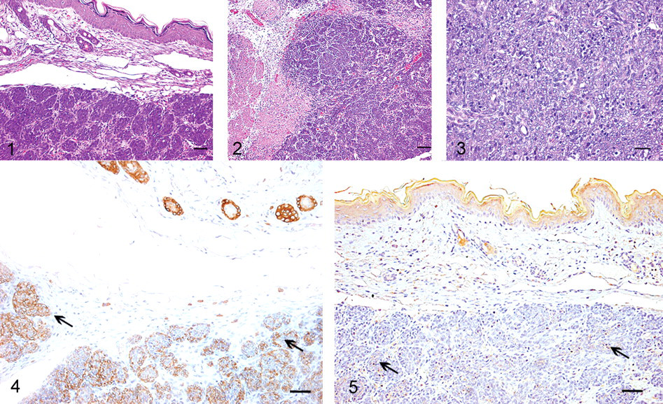
Results of CK immunostaining are presented in Figs. 4 and 5. In the adjoining normal skin, strong positivity for CK14 was detected in the upper aspects of the epidermis and in the internal layer of the infundibular and isthmic outer root sheath. Other normal follicular cells showed moderate, weak, or negative reactivity. Nested tumor cells showed a strong CK14 positive signal in the cytoplasm. Comparatively, the signal for CK18 in tumor cells was relatively weak, similar to that observed in the suprabasal layers and the sebaceous ductlike structure of adjacent normal tissue (Figure 5).
CKs are highly useful markers for the recognition of differentiation and morphological features of BCC due to the specificity in various epithelial cells. 10 They are classified into two subtypes—type I, or acidic CKs (9–20); type II, or basic CKs (1–8)—and they are coexpressed in pairs. 6 The expression patterns of CKs change during keratinocyte differentiation. The expression of CKs in BCC has been demonstrated by immunohistochemical staining in various degrees and combinations. 1,8,9,11 BCC is characterized by the presence of a specific set of acidic CKs (14–17). CK18 is present in a majority of adenocarcinomas and BCC but not in squamous cell carcinomas. 7 Our results showed positive signals for both CK18 and CK14 within tumor cells, indicating a type of BCC.
CK14 is also used as squamous cell carcinoma marker. 3 However, as shown in Fig. 2, this tumor contains lobular and insular lesions composed entirely of basaloid cells resembling the basal layer of the epidermis. Based on the cell morphology, the absence of keratinization or keratin pearl formation, and the immunohistochemical profile, this tumor is most consistent with BCC. To our knowledge, this is the first report of spontaneous BCC occurrence in young SD rats.
Footnotes
Acknowledgements
We thankfully acknowledge financial support from a Korea Research Foundation grant (KRF-005-E00077) and additional financial support from the BK21 Program for Veterinary Science.
The authors declared that they had no conflicts of interests with respect to their authorship or the publication of this article.
The authors declared that they received no financial support for their research and/or authorship of this article.