
Abstract
Objective
Although ultrasound can be considered an assistant method, successful placement of a central venous catheter (CVC) in infants is still challenging. The incidence of CVC placement-related complications is still high. Therefore, this systematic review aimed to synthesize evidence to assess the effects of ultrasound-guided CVC placement on adverse outcomes in infants and neonates aged <12 months.
Methods
PubMed, Ovid, EMBASE, and the Cochrane Library were searched to identify potentially relevant studies. The main outcome was the incidence of adverse events, which included inadvertent arterial puncture, hematoma, pneumothorax and hemothorax, catheter kinking, threading, and malpositioning problems, venous thrombosis, catheter-related infection, phlebitis, and cardiac tamponade.
Results
Eleven studies involving 2097 patients were included in the final analysis. The odds of inadvertent arterial puncture, and catheter kinking, threading, and malpositioning problems were lower in the ultrasound group than in the control group. No significant difference was detected in the incidence of hematoma or venous thrombosis between the control and ultrasound groups. Other complications, such as pneumothorax, hemothorax, phlebitis, and cardiac tamponade, rarely occurred.
Conclusion
Ultrasound-guided CVC placement can improve the safety of punctures in neonates and infants. CVC punctures should be guided in real-time by ultrasound.
Keywords
Background
Central venous catheter (CVC) placement is commonly used for continuous hemodynamic monitoring, prolonged parenteral nutrition, and administration of drugs, especially for critically ill patients with difficulty in establishing peripheral veins or those having major surgery. The most commonly used sites include the internal jugular vein (IJV), subclavian vein, brachiocephalic vein, and femoral vein. 1 Traditionally, successful CVC placement mainly depends on physicians’ experience and palpation of anatomical landmarks. However, CVC placement-related mechanical complications are high. In pediatric patients, the overall incidence of catheter-related mechanical complications is 19%, including failure to place a catheter (9.3%), hematoma (4.9%), arterial puncture (2%), and pneumothorax (1.5%). 2 This safety issue is particularly important for neonates and infants because their veins have a limited diameter and can greatly vary depending on body weight. The incidence of local hematoma related to CVC placement is approximately 5% to 10% in neonates and infants.3,4 Life-threatening complications such as cardiac tamponade have also been reported.5,6
Real-time ultrasound guidance for the insertion of a CVC has benefits in adults and children.7,8 A meta-analysis involving 4185 procedures showed that adults with ultrasound-guided CVC placement achieved an 82% reduction in cannulation failure, a 75% decrease in hematomas, and a 79% decline in pneumothorax. 9 Although ultrasound is widely used, successful placement of CVC in newborns and infants remains challenging, even in experienced personnel. 8 A study reported that in low birth weight newborns, up to eight attempts were required to achieve the success of right IJV vein catheterization, and only 50% of patients had successful catheterization within one attempt. 3 Infants had not been included in previous analyses, but with the increasing amount of reports, this is now possible. Ultrasound guidance is useful not only for detecting immediate insertion-related complications (i.e., hematomas, pneumothorax and hemothorax, and catheter kinking, threading, and malpositioning problems), but also for late complications (i.e., thrombosis, phlebitis, and infections).
Whether ultrasound can avoid complications in infants aged <12 months is unclear. Therefore, this study aimed to synthesize evidence to assess the effects of ultrasound-guided CVC placement on adverse outcomes in neonates and infants (aged <12 months).
Methods
Search strategy and article selection
This systematic review was conducted according to the Preferred Reporting Items for Systematic Reviews and Meta-Analysis (PRISMA) framework. The protocol for this review has been registered at https://www.crd.york.ac.uk/prospero (registration number CRD42023410234).
All available literature on ultrasound-guided CVC placement published before 13 February 2023 was searched without language restriction. The search strategies were created by the corresponding author (YC). The following medical terms were searched: “neonates,” “infants,” “ultrasound-guided,” “central venous catheters” “complications,” and “adverse events.” Search strategies are detailed in Supplemental File 1. Two co-authors (YW and QZ) separately searched PubMed, Ovid, EMBASE, and the Cochrane Library to identify potentially relevant studies. They also independently perused each remaining full-text article to determine candidates. Discrepancies were resolved by discussion and finally determined by the corresponding author (YC). The search strategies were in line with the following criteria.
Inclusion and exclusion criteria
The inclusion criteria were as follows. Neonates and infants aged <12 months were included. Regarding types of interventions, we included previous publications that supported the concept that ultrasound-guided puncture/cannulation is not required for umbilical venous catheters and epicutaneo-caval catheters, but it is mandatory for CVCs in neonates and children, as well as for peripherally inserted central catheters (PICCs) in children.10,11 Therefore, only pediatric patients who received centrally inserted central catheters (CICCs), femorally inserted central catheters, and PICCs were enrolled in the current analysis. Types of comparison included non-ultrasound-guided CVC placement, such as the traditional/conventional landmarks technique, X-ray examinations, or other non-ultrasound-guided techniques. The included studies needed to report complications or adverse events, such as inadvertent arterial puncture, hematoma, catheter-related infections, pneumothorax and hemothorax, and others. Randomized, non-randomized, and observational studies were included. The exclusion criteria were as follows: duplicated publications, conference abstracts, or letters without full text; study protocols without results; non-original studies; and case reports.
Two co-authors (QH and QZ) independently extracted the following data from each study (if relevant): authors, the year of publication, study design, type of cases included, sample size, practitioner, intervention, and CVC-related outcomes. The third co-author double-checked for data accuracy. Randomized, controlled trials (RCTs), non-RCRs, and observational studies with a control group (patients who did not receive ultrasound-guided CVC placement) were included.
Quality assessment
Two reviewers (YC and YW) independently evaluated the quality of each RCT using Review Manager Version 5.3 for Windows (RevMan, The Cochrane Collaboration, Oxford, UK). However, the Newcastle–Ottawa Scale (NOS) was used for non-RCTs. 12 This quality assessment tool for RCTs included the following seven parts: random sequence generation, allocation concealment, performance bias, detection bias, attribution bias, reporting bias, and others. The risk of bias was evaluated at three levels (low risk, unclear risk, and high risk). The NOS contains a nine-point scale for the quality of selection, comparability, and outcome of the study participants. A high-quality study was defined as an NOS score ≥6.
In addition, the Grading of Recommendations, Assessment, Development, and Evaluations (GRADE) approach methodology was conducted to evaluate the quality of evidence for each outcome, regarding the risk of bias, inconsistency, indirectness, imprecision, and publication bias. Two authors (TG and YW) independently assessed the certainty of evidence, and resolved any discrepancies through consultation with the corresponding author.
Statistical analysis
Dichotomous outcomes are expressed as n (%), and the odds ratio (OR) with the 95% confidence interval (95% CI) was calculated. Statistical heterogeneity was estimated by the I2 statistic. According to previous publications,13,14 if a value of I2 > 50%, which may represent evidence of significant heterogeneity, the random-effect model was used; otherwise, a fixed-effect model was used. A sensitivity analysis was performed to investigate possible explanations for the high heterogeneity. We performed random effects models for all outcomes as a sensitivity test for the fixed effects. Data analysis was performed using Review Manager software (RevMan, version 5.3). A p value <0.05 was considered statistically significant.
Results
Search results
This systematic review identified 1471 potentially relevant articles (581 in PubMed, 58 in Ovid, 554 in Embase, and 278 in the Cochrane Library). Of these, 180 were duplicates and were removed. We then excluded 1251 studies because they involved animals (n = 6), non-neonates or non-infants (n = 244), irrelevant studies (n = 625), reviews or cases (n = 171), and others (n = 205). Forty-one studies were assessed by full-text review. Finally, 11 studies involving 2097 patients were included in the final analyses.15–25 A PRISMA flow diagram is shown in Figure 1.

PRISMA flow diagram. PRISMA, Preferred Reporting Items for Systematic Reviews and Meta-Analysis.
Trial characteristics
Of the 11 eligible studies, 6 were RCTs,15–20 One study used a non-randomized, prospective design to compare ultrasound-guided line insertion in the IJV with standard PICC line placement, 21 and the remaining four articles were retrospective studies.22–25 The puncture sites included the femoral vein, IJV, supraclavicular vein, and a PICC. The characteristics of the enrolled studies, including authors, year, study design, type of cases included, sample size, intervention, relevant reported outcomes, and results, are shown in Table 1.
Study characteristics.

RCT, randomized, controlled trial; PICC, peripherally inserted central catheter; IJV, internal jugular vein; RT-US, real-time ultrasound; CL, conventional landmark; USG-CVC, ultrasound-guided central venous catheter.
Outcomes: ① Inadvertent arterial puncture; ② hematoma; ③ pneumothorax and hemothorax; ④ Catheter kinking, threading, and malpositioning problems; ⑤ venous thrombosis; ⑥ catheter-related infection; ⑦ phlebitis; and ⑧ cardiac tamponade.
During CVC placement in neonates and infants, inadvertent arterial puncture was the most commonly reported adverse event, followed by hematoma, catheter-related infections, pneumothorax and hemothorax, catheter kinking, threading, or malpositioning problems, venous thrombosis issues, cardiac tamponade, and phlebitis.
Quality assessment
The Cochrane risk of bias assessment tool was used to assess the quality of the six RCTs,15–20 and the NOS was used to assess the quality of the five non-RCTs.21–25 The average NOS score of cohort studies was 6 (range: 4–8). The results of the quality assessment for the RCTs and non-RCTs are shown in Supplemental files 2 and 3, respectively.
Inadvertent arterial puncture
The incidence of accidental arterial puncture was reported in eight studies.15–18,20,22–24 In the ultrasound group, the incidence of arterial puncture ranged from 0.0% to 6.5%, while in the control group, it ranged from 0.0% to 25%. Ultrasound-guided CVC placement significantly reduced the incidence of inadvertent arterial puncture compared with the control group (OR: 0.24; 95% CI 0.12–0.50; p < 0.001; heterogeneity: I2 = 17%) (Figure 2). The GRADE of evidence was considered to be moderate owing to the enrollment of three non-randomized studies and a lack of blinding in eight studies (Table 2).

Forest plot of the incidence of inadvertent arterial puncture. M-H, Mantel–Haenszel; CI, confidence interval.
GRADE of evidence of ultrasound-guided CVC placement compared with other methods of CVC placement.

GRADE, Grading of Recommendations, Assessment, Development, and Evaluations; CVC, central venous catheter; OR, odds ratio; CI, confidence interval.
Inconsistency: the results are consistent across studies, and any inconsistency may be serious enough to downgrade the quality of evidence for this outcome.
Indirectness: the evidence directly answers the health care question asked, and any indirectness of available evidence may be serious enough to downgrade the quality of evidence for this outcome.
Imprecision: The results are precise enough, and any imprecision of the results may be serious enough to downgrade the quality of evidence for this outcome. Imprecision is defined differently by authors of systematic reviews and by guideline panels.
Hematoma
Hematoma was reported in five studies of CVC placement.15,17,18,20,21 The pooled incidence of hematoma was 5.3% (10/187) in the ultrasound group. There was no significant difference in the incidence of hematoma between ultrasound guidance and other methods (OR: 0.73; 95% CI 0.33–1.64; p = 0.45; heterogeneity: I2 = 3%) (Figure 3). The GRADE of evidence was considered to be moderate owing to enrollment of one non-randomized study and a lack of blinding in five studies (Table 2).

Forest plot of the incidence of hematoma. M-H, Mantel–Haenszel; CI, confidence interval.
Pneumothorax and hemothorax
Four studies reported the incidence of pneumothorax and hemothorax,15,20,21,24 and only one case occurred in the control group. 15 The GRADE of evidence was considered to be moderate owing to enrollment of two non-randomized studies and a lack of blinding in four studies (Table 2).
Catheter kinking, threading, and malpositioning problems
Four studies mentioned the incidence rates of catheter kinking, threading, or malpositioning problems, which ranged from 0.0% to32.5% in the ultrasound group and 3.8% to 67.5% in the control group,15,19–21 with a significant difference between the two groups (OR: 0.31; 95% CI: 0.15–0.62; p < 0.01; heterogeneity: I2 = 0%) (Figure 4). The GRADE of evidence was considered to be moderate owing to enrollment of one non-randomized study enrollment and a lack of blinding in three studies (Table 2).

Forest plot of the incidence of catheter kinking, threading, and malpositioning problems. M-H, Mantel–Haenszel; CI, confidence interval.
Venous thrombosis
Only one study reported the incidence of venous thrombosis. 22 There was no significant difference in the incidence of venous thrombosis between the ultrasound and control groups, but there was a tendency towards significance (OR: 0.45; 95% CI: 0.19–1.02; p = 0.06) (Figure 5).
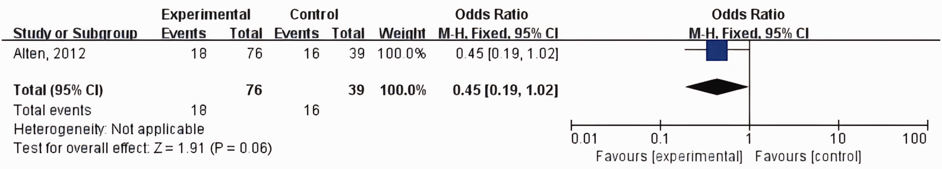
Forest plot of the incidence of venous thrombosis. M-H, Mantel–Haenszel; CI, confidence interval.
Catheter-related infection
Five trials reported the outcome of catheter-related infection.19,20,22–24 The incidence rate of catheter-related infection was significantly higher in the ultrasound group than in the control group (OR: 2.73; 95% CI: 1.37–5.45; p < 0.01; heterogeneity: I2 = 0%) (Figure 6). The GRADE of evidence was considered to be low owing to enrollment of three non-randomized studies, a lack of blinding in four studies, wide CIs, and plausible confounding concerns (Table 2).

Forest plot of the incidence of catheter-related infection. M-H, Mantel–Haenszel; CI, confidence interval.
Phlebitis and cardiac tamponade
One trial examined the effect of ultrasound-guided technology in preventing phlebitis during peripheral deep venous catheterization. However, no significant difference was detected in the incidence of phlebitis between the ultrasound and control groups. 25 CVC-related cardiac tamponade was not observed in the 1333 newborns who were included. 24
Sensitivity analyses
Although all of the I2 values were lower than 20%, we performed random-effect models for all outcomes as a sensitivity test for the fixed effects (Supplementary File 4).
Discussion
To the best of our knowledge, this is the first systematic review to evaluate ultrasound-guided CVC placement-related complications in neonates and infants younger than 12 months. We found that the three most commonly reported complications during CVC placement were inadvertent arterial punctures that were reported by eight studies, followed by hematoma (5 studies) and catheter-related infections (5 studies). Ultrasound-guided technology achieved a lower incidence rate of short-term complications (e.g., inadvertent arterial puncture, and catheter kinking, threading, and malpositioning) than other methods. However, ultrasound-guided technology was associated with a higher incidence rate of catheter-related infections.
Many health professionals believe that ultrasound guidance can facilitate CVC placement. However, some providers are still reluctant to use ultrasound guidance. According to surveys, ultrasound guidance is still underused during central venous catheterization, with a usage rate of approximately 36% to 68%.26–28 The main reasons for not using ultrasound guidance are as follows: unnecessary (36%), limited availability of an ultrasound machine (28%–33%), and a prolonged perceived procedure time (19%).27–28 In the current study, neonates and infants (aged < 12 months) were chosen as the study population because CVC placement is one of the most common procedures performed in these patients to measure pressure or secure intravenous access. Additionally, the diameters of the vessels in these young patients are small and their immune systems are premature, which makes puncture challenging and concomitant with a high incidence rate of cannulation-related complications. Theoretically, the incidence of CVC-related complications in infants is higher than in older children and adults, as reported by Chuan et al. 29
In our study, we summarized published studies that reported CVC-related complications in neonates and infants aged < 12 months. The puncture sites included the IJV, subclavian vein, femoral vein, and PICCs. Among them, the IJV and PICCs were the first choices during neonatal and infant CVC cannulation because of their easy identification/use and because they were unlikely to lead to lethal complications, 30 which is in line with previous studies in pediatrics.31,32 The choice of the puncture site still depends upon local conditions and providers’ experiences.
In the ultrasound group, the incidence rates of hematoma, accidental arterial puncture, and catheter kinking, threading, and malpositioning problems were 5.3%, 2.2%, and 11.7%, respectively. These were the three most common CVC-related complications. The incidence rates of those adverse events in the control group were 7.4%, 2.0%, and 23.3%, respectively. These results suggested that ultrasound-guided CVC placement was beneficial in reducing inadvertent arterial puncture and catheter kinking, threading, and malpositioning problems. In most clinical practices, the ultrasound probe is removed once the vein is punctured. A guide wire is inserted and a dilator dilates the skin. A CVC is then placed. However, the incidence rate of CVC malposition can be up to 11.7% even with ultrasound guidance and 23.3% without ultrasound guidance, which is higher than that reported in adults (5.01%). 33 Therefore, we recommend using ultrasound not only to puncture the central vein but also to ensure the desired destination.
In the current systematic review, only one retrospective study including 115 patients reported the incidence of venous thrombosis. Therefore, we did not find that ultrasound-guided CVC placement reduced long-term complications such as venous thrombosis, but there was a tendency for significance (p = 0.06). In the future, a large RCT is required to confirm the effect of ultrasound-guided CVC placement on a reduction in venous thrombosis.
Surprisingly, real-time ultrasound guidance appears to have disadvantages in catheter-related infections. Bayoumi et al. believed that a CVC may be more susceptible to infection because of its insertion site. 24 A retrospective study showed that CVCs had a higher incidence of catheter-related infections than PICCs, supporting this possibility of susceptibility to infection. 34 The authors believed that their result could be explained by minor microbial density and a lower temperature at the PICC placement site compared with those of other CVCs, including at the neck and groin.34,35 Evecit et al. showed that the ultrasound-guided group had a longer duration of a PICC in place than the control group (21.61 days vs. 8.36 days, p < 0.001). 23 The total number of catheter days during hospitalization was reported to be an independent risk factor for central line-associated bloodstream infection. 36 Therefore, we believe that the only conclusion that can be made from these studies is that catheters were associated with events such as infections, and did not cause infection because the actual cause was unknown. CVC procedures must always be performed under strict aseptic conditions (e.g., cap, mask, sterile gown, sterile gloves, and large sterile fields), with maximum hand hygiene, regardless of the use of real-time ultrasound-guided insertion. 37 Additionally, real-time ultrasound-guided cannulation is recommended because it can be used to evaluate all possible options, recognize potential diseases, estimate the diameter of vessels and choose the optimal size of kit, verify the correct position of the catheter tip, and immediately detect mechanical complications. 11
Moreover, Jarraya et al. concluded that a high incidence of complications was associated with difficult catheter placement and in patients with cancer and severe thrombocytopenia.38,39 To reduce catheter-related morbidity, an ultrasound-guided approach, a multidisciplinary teaching program to improve nursing skills, and the use of less invasive devices for patients with cancer and comorbidity are important.
To date, no special guideline for CVC placement is available for infants. However, the use of real-time ultrasound guidance for CVC placement is recommended as the first-line procedure because it considerably reduces early mechanical complications (inadvertent arterial puncture, and catheter kinking, threading and malpositioning).
There are some limitations to the current systematic review. First, high-quality evidence such as RCTs was not sufficient. Only six RCTs were included in the final analysis and their sample size was small. Second, the site of CVC puncture or the types of CVCs greatly varied among the included studies. The decision of a puncture site depends on a surgical wound, the patient’s physique, or infection, and providers may choose the optimal kit and site after fully considering the patient’s condition and the risks and benefits. Additionally, the providers’ experience is one of the most important factors affecting outcomes. Among the enrolled studies, Hosokawa et al. included practitioners who were fellows without special training in pediatric central venous catheterization, 17 whereas three other studies involved experienced providers16,22,24 or fellows.15,18 Five of the studies did not include the characteristics of the practitioners.19–21,23,25 Therefore, selection bias is possible. Finally, although the study population consisted of newborns and infants, there were differences in their weight and height, ranging from <1.5 kg to 10 kg, which directly led to varying degrees of difficulty in puncture.
Conclusions
Ultrasound-guided CVC placement may be performed in all settings, such as emergency departments, operating rooms, intensive care units, and the bedside. The use of the ultrasound image technique not only reduces the occurrence of inadvertent arterial puncture and catheter kinking, threading, and malpositioning during CVC placement, but also improves the safety of puncture. We recommend that all CVC punctures should be guided in real-time by ultrasound in neonates and infants. However, sufficient simulation training is essential. A practice guide for safe central venous catheterization released in 2019 suggested educating novice operators at the start of the clinical training period. 30
Supplemental Material
sj-pdf-1-imr-10.1177_03000605241287168 - Supplemental material for Systematic review of ultrasound-guided central venous catheter placement-related complications in neonates and infants aged <12 months
Supplemental material, sj-pdf-1-imr-10.1177_03000605241287168 for Systematic review of ultrasound-guided central venous catheter placement-related complications in neonates and infants aged <12 months by Yu Cui, Yu Wang, Tianqing Gong, Qinghua Huang and Qian-Qian Zhang in Journal of International Medical Research
Supplemental Material
sj-pdf-2-imr-10.1177_03000605241287168 - Supplemental material for Systematic review of ultrasound-guided central venous catheter placement-related complications in neonates and infants aged <12 months
Supplemental material, sj-pdf-2-imr-10.1177_03000605241287168 for Systematic review of ultrasound-guided central venous catheter placement-related complications in neonates and infants aged <12 months by Yu Cui, Yu Wang, Tianqing Gong, Qinghua Huang and Qian-Qian Zhang in Journal of International Medical Research
Supplemental Material
sj-pdf-3-imr-10.1177_03000605241287168 - Supplemental material for Systematic review of ultrasound-guided central venous catheter placement-related complications in neonates and infants aged <12 months
Supplemental material, sj-pdf-3-imr-10.1177_03000605241287168 for Systematic review of ultrasound-guided central venous catheter placement-related complications in neonates and infants aged <12 months by Yu Cui, Yu Wang, Tianqing Gong, Qinghua Huang and Qian-Qian Zhang in Journal of International Medical Research
Supplemental Material
sj-pdf-4-imr-10.1177_03000605241287168 - Supplemental material for Systematic review of ultrasound-guided central venous catheter placement-related complications in neonates and infants aged <12 months
Supplemental material, sj-pdf-4-imr-10.1177_03000605241287168 for Systematic review of ultrasound-guided central venous catheter placement-related complications in neonates and infants aged <12 months by Yu Cui, Yu Wang, Tianqing Gong, Qinghua Huang and Qian-Qian Zhang in Journal of International Medical Research
Footnotes
Author contributions
Y.C. contributed to the study conception and design, analysis, and interpretation of data. Y.W. and T.G. drafted the initial manuscript. Y.C. reviewed and revised the manuscript. Y.W., Q.Z., and Q.H. collected the data. All authors have read and agreed to the published version of the manuscript.
Declaration of conflicting interest
The authors declare that there is no conflict of interest.
Data availability statement
Not applicable because this is a systematic review.
Funding
This study was supported by the Science & Technology Department of Sichuan Province (2023NSFSC1626), Health Commission of Sichuan Province (No. 21PJ131), and Yingcai Scheme of Chengdu Women’s and Children’s Central Hospital (YC2021006).
Supplemental material
Supplemental material for this article is available online.
References
Supplementary Material
Please find the following supplemental material available below.
For Open Access articles published under a Creative Commons License, all supplemental material carries the same license as the article it is associated with.
For non-Open Access articles published, all supplemental material carries a non-exclusive license, and permission requests for re-use of supplemental material or any part of supplemental material shall be sent directly to the copyright owner as specified in the copyright notice associated with the article.