
Abstract
Angiosarcoma is the most invasive and malignant cardiac tumor and most commonly originates from the right atrium. Early diagnosis is essential, and echocardiography has an important role in diagnosis. This tumor grows aggressively, and metastases to other sites makes it difficult to control. Surgical treatment remains the best option for patients who do not respond to chemoradiotherapy. We herein report a case of a 17-year-old patient with cardiac angiosarcoma who presented with dyspnea, chest pain, dry cough, and fever. Although we considered the most probable diagnosis to be constrictive pericarditis, pathologic examination revealed a primary angiosarcoma originating from the pericardium. The patient underwent total pericardiectomy. However, despite receiving chemotherapy for 2 weeks postoperatively, she developed complications including leukopenia and eventually died of respiratory failure. Late diagnosis of angiosarcoma often occurs, resulting in progression to end-stage disease and a very poor prognosis. Therefore, a thorough understanding of this entity, knowledge of its pitfalls in management, and establishment of an accurate treatment guideline would help to develop a reliable and life-saving treatment approach for these patients.
Keywords
Introduction
Metastasis to the heart, especially from lung carcinoma, is 100 to 1000 times more common than a primary cardiac tumor. The incidence of primary cardiac tumors ranges from only 0.001% to 0.030%, and 75% are benign. Among all malignant primary cardiac tumors, 95% are sarcomas and the rest are lymphomas. The most frequent malignant primary tumors of the heart are those with undifferentiated characteristics, followed by sarcomas (i.e., angiosarcoma, leiomyosarcoma, and rhabdomyosarcoma). 1
The most common sarcomas are tumors of vascular origin, particularly angiosarcoma. Primary cardiac angiosarcoma is the most invasive and malignant primary cardiac tumor.2,3 Primary cardiac sarcoma is an extremely rare clinical entity with an incidence of only 0.0001%. 4 Angiosarcomas are mostly found in patients aged 30 to 40 years, and they are twice as likely to develop in men than in women. 1 Angiosarcoma is associated with nonspecific symptoms such as dyspnea, cough, heart failure, and arrhythmia. 5 In the present report, we describe a 17-year-old female patient who was diagnosed with cardiac angiosarcoma. She underwent radical resection and chemotherapy but eventually died. Echocardiography is one of the most useful diagnostic tools for cardiac masses, exhibiting a sensitivity of 93% for detection of such lesions; however, computed tomography (CT) and magnetic resonance imaging (MRI) are often used to detect metastatic disease sites.6,7 Echocardiography findings include heart block, arrhythmia, and nonspecific T-wave and ST changes. 1
The poor prognosis of primary cardiac angiosarcoma is related to its aggressive behavior, early metastasis to distant organs, and late diagnosis, often making it a fatal condition. Surgery is challenging because of the infiltrative characteristics of the tumor and the high probability of recurrence after treatment. 8
We herein report a case of cardiac angiosarcoma, present a review of the literature regarding this tumor, and discuss its management to prevent probable complications. We believe that a thorough understanding of the disease etiology and pathogenesis can help improve therapeutic approaches in the clinical setting.
The reporting of this study conforms to the CARE guidelines. 9
Case presentation
A 17-year-old female patient was admitted to our hospital because of an 8-month history of dyspnea, a 10-day history of dry cough, and a 2-day history of periodic fever with no obvious cause or specific pattern. The patient had a rheumatic disease of unknown origin for which she had begun treatment with colchicine and prednisolone 2 years previously. She had no remarkable family history, and her vital signs on admission were stable with a blood pressure of 120/90 mmHg, pulse rate of 81 beats/minute, respiratory rate of 17 breaths/minute, and temperature of 37°C.
The patient’s cardiovascular examination was notable for a tumor plop sound at the apex and mild edema in the lower extremities. Other physical examination findings were normal.
Laboratory findings were as follows: white blood cell count, 11,300/µL; hemoglobin concentration, 11.9 g/dL; hematocrit, 35.8%; and platelet count, 350,000/µL. Electrolyte panel findings were as follows: sodium, 141 mmol/L and potassium, 4.0 mmol/L. Echocardiography findings were as follows: pulmonary artery pressure 45/25 mmHg (mean, 26 mmHg) and right ventricular pressure, 45/26 mmHg with a normal coronary artery on angiographic examination.
Transesophageal echocardiography (TEE) revealed a moderate amount of pericardial fluid and constrictive pericarditis, but this was not considered the definitive diagnosis. CT and brain MRI showed no metastasis. An electrocardiogram showed sinus rhythm (Figure 1), and a chest radiograph showed edema on both sides of the pleura and left-sided pleural effusion (Figure 2). An operation was performed by a standard median sternotomy after consultation with a multidisciplinary team. We obtained the patient’s consent to treatment.
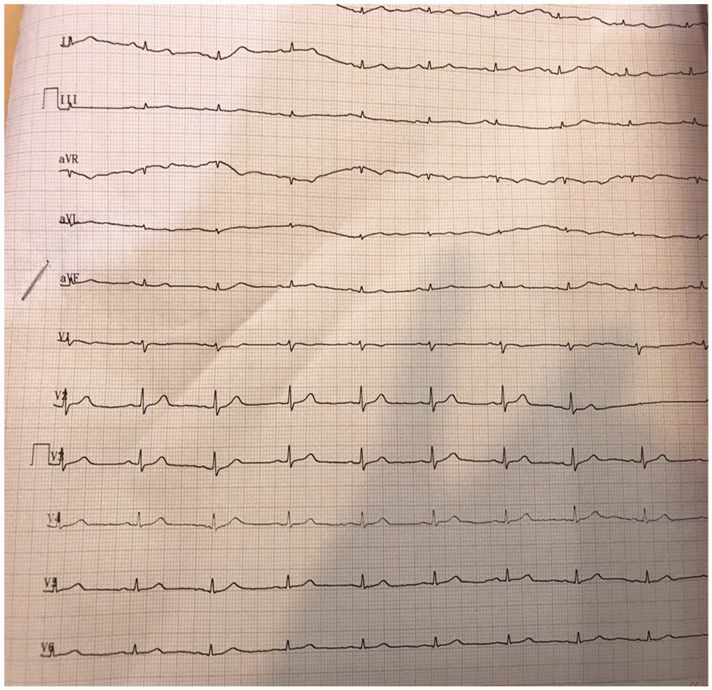
Electrocardiogram showing sinus rhythm.

Chest radiograph showing edema on both sides of the pleura and left-sided pleural effusion.
Upon opening the pericardium, extensive adhesions were observed between the pericardium and the heart such that we separated the right atrium from the pericardium with difficulty (Figure 3). A 4-cm section of pericardial tissue was removed and sent to the pathology department.

Adhesions of the pericardium.
The pathological diagnosis was angiosarcoma, and total pericardiectomy was performed. After the operation, the patient was sent to the intensive care unit, and 5 days later, she was discharged in stable condition. Ten days later, the patient began chemotherapy with docetaxel for two cycles, but the tumor was still progressing. Chemotherapy-induced leukopenia then developed. Within 2 weeks of discharge, she returned to the hospital because of dyspnea and unfortunately died of respiratory failure during that admission.
Discussion
Malignant primary cardiac neoplasms are rare tumors with an incidence of approximately 25%. 2 They can markedly compromise a patient’s life expectancy, mainly because they are often diagnosed after metastasis has occurred. Metastasis commonly involves the lung (20.0%–55.6%), liver (10.0%–22.2%), and bone (10.0%–20.0%). Additionally, affected patients often have nonspecific symptoms such as dyspnea, pericardial effusion, and chest pain (occurring in 50%–80%, 29%–56%, and 10%–39% of patients, respectively), resulting in the initial consideration of more common diseases. 10 Primary cardiac angiosarcoma is the most common (33%) and aggressive type of malignant primary cardiac tumor. 11
A systemic review of primary cardiac angiosarcoma revealed a median age of 45 years and a 1.5-times higher susceptibility in men than in women for this type of tumor. One study involving 162 patients showed that multimodality management could increase the overall survival duration from 6 months after surgery alone to 13 to 27 months with the addition of adjuvant chemoradiotherapy. 12 Additionally, a review of 18 patients with confirmed primary cardiac angiosarcoma in a single institution over a 20-year period showed that 89% of patients had tumors originating from the right atrium. Furthermore, 44% of patients presented with locally advanced disease and 56% with metastasis at the time of diagnosis. The overall survival duration was 13 months, indicating a poor prognosis. 13 The reported mortality rates range from 64.7% to 100%. 10
Various imaging modalities are employed to help with the diagnosis. The initial and most widely available modality used is echocardiography. TEE, a first-line diagnostic tool, has 97% sensitivity for identifying heart masses. 8 However, a study comparing ultrasonography and MRI for intracardiac masses showed that MRI provided an accurate diagnosis in 100% of cases whereas transthoracic echocardiography did so for only 82% of cases. Additionally, MRI could detect masses in locations missed by transthoracic echocardiography or TEE, such as intramural tumors or tumors that extended to the inflow or outflow of the heart. However, no ultrasonographic findings were missed by MRI in that study. 14 Combined with the diagnostic features obtained by these imaging modalities, a biopsy can be performed under TEE guidance. 15
Although the definitive pathologic diagnosis in our case was angiosarcoma, clinicians should also be aware of mimickers of angiosarcoma. For instance, constrictive pericarditis should be considered as a differential diagnosis. In another study similar to ours, a patient with angiosarcoma was misdiagnosed with constrictive pericarditis because of her cardiac MRI findings, particularly pericardial enhancement. In addition, cardiac catheterization and pericardiectomy demonstrated a thick, dense, and adherent pericardium, and dissection was difficult; thus, the diagnosis of pericarditis was reasonable. However, surgical pathology unexpectedly revealed angiosarcoma. 16 Zhang et al. 4 reported that the lack of a stalk was a standard finding that may help distinguish angiosarcoma from benign myxoma and papillary fibroelastoma.
Surgery is considered the treatment of choice for all types of primary cardiac tumors whenever it can be performed. Nevertheless, surgery has not yet been proven to be the safest option with the best outcome in patients with malignant tumors. In a study of surgical resection of primary cardiac tumors in 91 patients, the follow-up results demonstrated that all 5 patients with malignant tumors who survived the surgery died within 3 years of the first operation (either within 1–2 months after the surgery or because of recurrence and reoperation). 17 Adjuvant chemotherapy is administered postoperatively in some patients. The most routinely used chemotherapy agents are anthracyclines (e.g., doxorubicin), ifosfamide, and taxanes (e.g., paclitaxel). Combination regimens such as AIM (doxorubicin, ifosfamide, and mesna) have also been proven efficient. 8 Besides chemotherapy, radiotherapy is also recommended for locally advanced disease. Concurrent chemotherapy and radiotherapy have also been used, leading to prolonged survival rates and lower morbidity. For example, one patient underwent a novel therapy regimen involving concurrent proton beam therapy and paclitaxel at a dose of 80 mg/m2 each week for seven rounds; however, the therapy was terminated in the third cycle because of dermatitis. The patient then continued adjuvant chemotherapy with docetaxel at 100 mg/m2 on day 8 and gemcitabine at 900 mg/m2 on days 1 and 8. Compared with the patients undergoing surgery alone (mean survival of 6 months postoperatively), this patient was alive 18 months after completing the therapy and experienced no recurrence based on CT scans. 18
Immunotherapy trials for the treatment of angiosarcoma are currently being performed, specifically by targeting the site involved (i.e., the endothelium). However, based on the most recent update on 17 July 2023, none of these studies has shown any beneficial outcomes, and more studies therefore need to be conducted. Among the immunotherapeutic agents studied, bevacizumab (an anti-vascular endothelial growth factor drug) and tyrosine kinase inhibitors (sorafenib, axitinib, pazopanib, and regorafenib), all of which target either platelet-derived growth factor receptor alpha or vascular endothelial growth factor, have gained popularity. 19
Moreover, the use of minimally invasive methods is rapidly increasing. In one study, for example, cardiopulmonary bypass via percutaneous femoral cannulation was performed for resection of intracardiac lesions from 2018 to 2020. Notably, a shorter hospital stay and lower mortality rate were achieved. 20
Studies have shown that the prognosis of cardiac angiosarcoma is generally based on the stage of the disease. The microscopic surgical margins are associated with overall survival; in one study, microscopically negative margins (R0 resection) were associated with a survival time of 17 months, whereas microscopically positive margins (R1 resection) were associated with a survival time of only 6 months. 21
In conclusion, primary cardiac angiosarcoma is a rare malignant mesenchymal tumor originating from the endothelium of cardiac vessels. Early detection and management are associated with challenges because of the rarity, early metastasis, and poor prognosis of this tumor. Various imaging techniques and histopathological examinations are required to rule out other differential diagnoses, and a multidisciplinary approach and involvement of specialties are necessary for successful treatment. Further research is needed to enhance our understanding of this challenging tumor and to discover better diagnostic techniques, treatment regimens, and follow-up protocols.
Ethics
In accordance with standard guidelines, the present study did not require ethical approval because of its retrospective design and its focus on a single case report. We have stringently anonymized all patient-related data and ensured its utmost confidentiality. Furthermore, the presentation of the case does not allow for the identification of the patient and thereby adheres to the principles of patient privacy and autonomy. The study did not involve any interventions, alterations in patient care, or additional procedures beyond standard clinical care. The study aimed solely at educational purposes, intending to share knowledge and insights without any commercial interests. The report aligns with the institution’s policies regarding the publication of case reports.
Footnotes
Informed consent
Informed consent for publication was not applicable because we have de-identified all of the patient’s details.
Acknowledgement
The authors are grateful to the staff in the medical records section of the hospital for providing the patient’s medical records.
Author contributions
Conceptualization, A.O. and S.S.; investigation, A.O. and S.S.; resources, A.O.; data curation, A.O.; writing—original draft preparation, A.O.; writing—review and editing, S.S.; supervision, A.O. All authors have read and agreed to the published version of the manuscript.
Data availability statement
The data that support the findings of this study are available from the corresponding author upon reasonable request.
Declaration of conflicting interests
The authors declare that there is no conflict of interest.
Funding
This research received no specific grant from any funding agency in the public, commercial, or not-for-profit sectors.