
Abstract
The maxillary first premolar commonly has one or two root canals; the presence of three canals is rare. These root canals are easily missed during treatment, resulting in failure of root canal therapy. The present report describes a case in which three root canals in the maxillary first premolar were diagnosed by cone-beam computed tomography. The herein-described patient was successfully treated using three-dimensional reconstruction technology to determine the form and direction of curvature of the root canals.
Keywords
Introduction
The root canal system of the maxillary first premolar is subject to great variation because of its complex internal anatomical structure. The presence of one or two root canals is common for the maxillary first premolar, but the presence of three is rare. 1 Li et al. 2 observed the maxillary premolars in Chinese patients by cone-beam computed tomography (CBCT) and found that the incidence of a maxillary first premolar with three root canals was only 0.5%. A complex root canal usually has a low rate of therapeutic success, and a missed root canal usually results in failure of therapy. CBCT three-dimensional (3D) imaging overcomes the limitations of the conventional preoperative X-ray two-dimensional imaging using periapical films and provides comprehensive information regarding the number, form, and curvature of root canal aberrations. 3 Preoperative scanning by CBCT improves the clinician’s anatomical understanding of the root canals and avoids iatrogenic accidents, such as lateral penetration, bottom penetration, and step formation, thus greatly improving the success rate of root canal therapy for complex structures. The present report describes a patient with three root canals in the premolar diagnosed at the Affiliated Stomatological Hospital of Nanchang University and provides a reference for the diagnosis and treatment of patients with a similar condition.
Case report
A 22-year-old male student was transferred to the Department of Endodontics, Affiliated Stomatological Hospital of Nanchang University for diagnosis and treatment on 19 September 2019. The main complaint was root canal obstruction during “nerve-killing” treatment for a toothache at another hospital. The patient provided written consent for publication of his data. The study protocol was approved by the ethics review committee of the Affiliated Stomatological Hospital of Nanchang University. The reporting of this study conforms to the CARE guidelines. 4
Clinical and imaging examinations (Figure 1) revealed white filling in the distal part of tooth 14, with caries under the filling but with no probing pain, percussion pain, or looseness. An X-ray scan of the periapical area was normal, but the canal image was not clear. CBCT conducted in our hospital (Figure 2) showed three root canals in tooth 14: two on the buccal side and one on the palatal side with a curved

Preoperative examinations. (a) Preoperative X-ray film and (b, c) Preoperative images of the mouth.
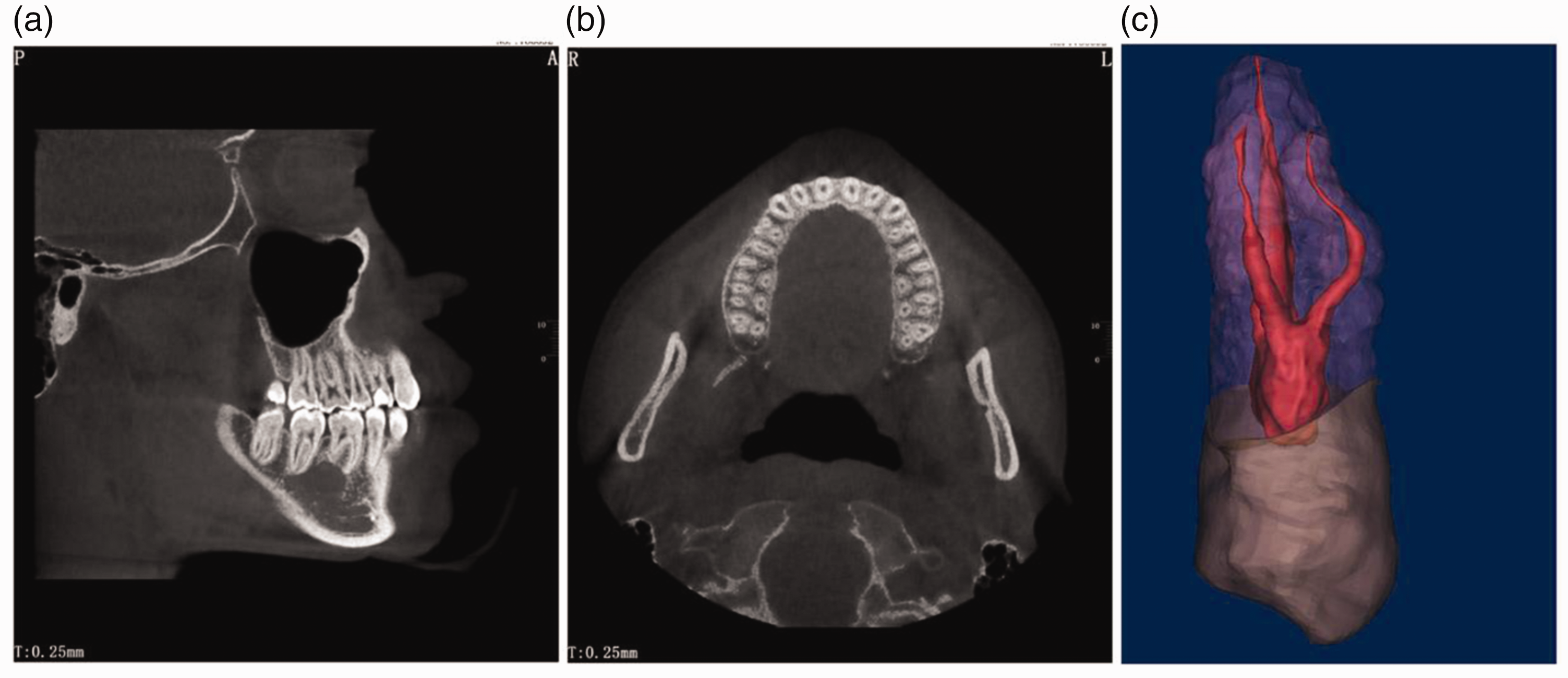
Cone-beam computed tomography (CBCT) and three-dimensional reconstruction. (a) CBCT showed two buccal roots of tooth 14, and the proximal middle root was curved. (b) CBCT showed that tooth 14 had three independent root canals and (c) The measured declination of the
Images obtained during the treatment process are shown in Figure 3. All existing fillings and decay in tooth 14 were removed, the caries were repaired with resin (Filtek Z350; 3M, Saint Paul, MN, USA), and a rubber dam was applied. The root canals were examined under a microscope (Zumax Medical, Suzhou New District, China), and three were found. A pre-bent stainless steel K-file (Dentsply Sirona, Charlotte, NC, USA) was used to dredge the root canals to 10#, and a root apex locator (VDW, Munich, Germany) was used to measure the length. The root canals were prepared with a nickel-titanium (NiTi) file (Orodeka, Jining, China) with a working length of 15.0 mm for the
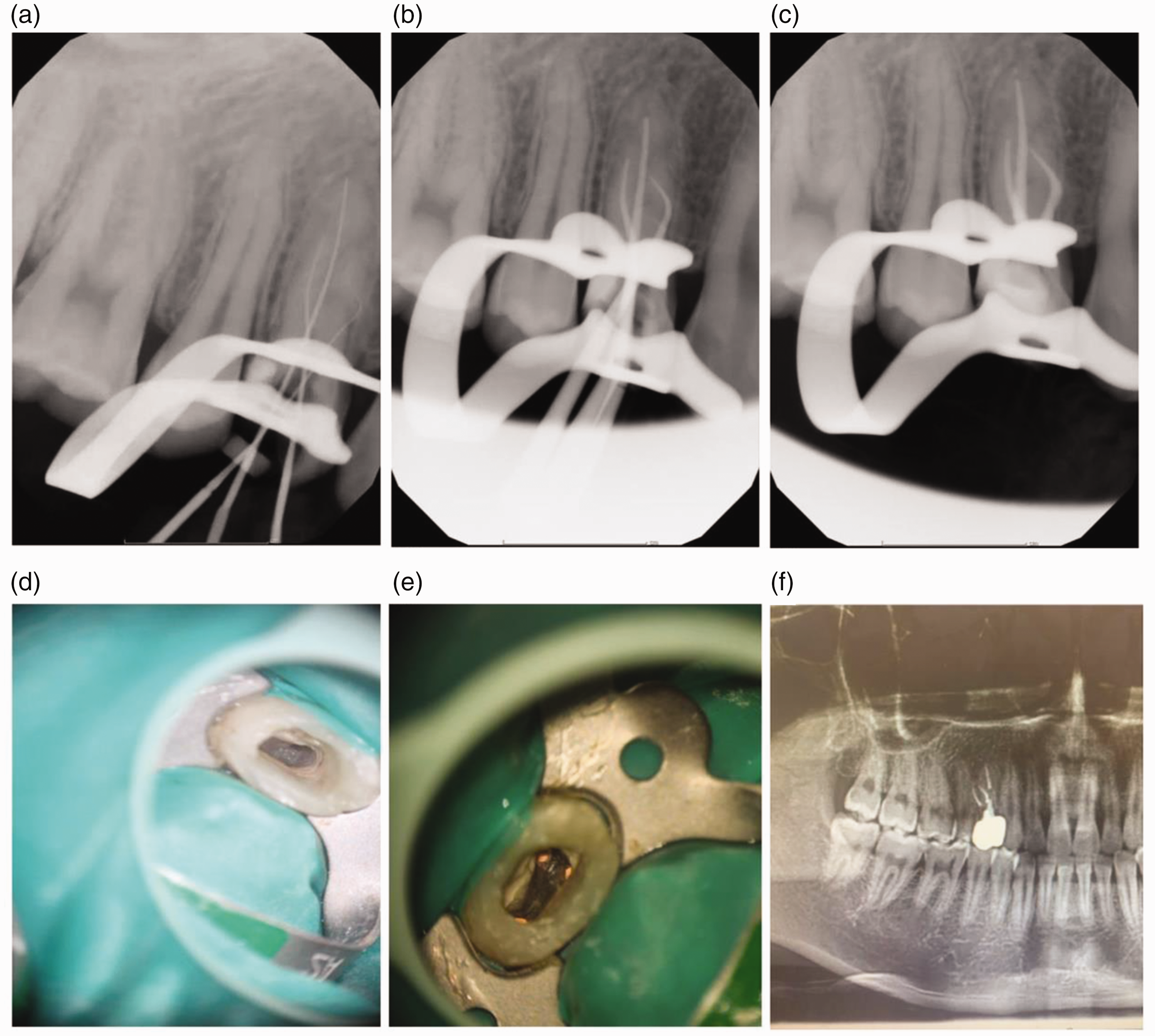
Treatment process and follow-up visit. (a–c) X-ray films during root canal therapy. (d, e) Root canal opening under a microscope and (f) X-ray film of the patient during a follow-up visit.
Discussion
The root canal system may be subject to multiple anatomical variations; hence, a missed root canal usually leads to failure in root canal therapy, especially for a nonvital tooth. Anatomical differences in the maxillary first premolar have been reported in many studies. Vertucci 6 classified the root canals of the maxillary premolars into eight forms and pointed out that the maxillary first premolar is the only tooth that exhibits all eight root canal forms. However, the incidence of a maxillary first premolar having three root canals is low, ranging from only 0.4% to 2.6%. 1
Microscopy and X-ray scanning from different angles can detect the anatomical changes during root canal therapy. However, such plain films show only two-dimensional images and may not reflect the complete anatomy of the root canals. Michetti et al.
7
found that CBCT improves the detection rate of root canals by 40% compared with conventional X-ray films. Furthermore, compared with CBCT, 3D reconstruction enables an intuitive view and provides accurate measurement data.
3
In the present case, we collected 3D data from CBCT scans and reconstructed the model to understand the anatomical structure of the root canals in the sagittal, coronal, and fracture planes
8
and accurately locate the direction and angle of the
In this case, we accurately located the root canal orifices using 3D reconstruction technology and set the declination of the
A good opening of the pulp cavity and careful exploration of the root canal at the bottom of the pulp cavity also helped to avoid missing the third root canal. In a study by Arisu and Alacam, 10 the buccolingual opening was extended distally to form a T shape for a tooth with three root canals to facilitate exploration of the root canal opening and straight access. If the mesiodistal distance of the middle one-third of the root is equal to or greater than the mesiodistal distance of one-third of the root neck on a preoperative plain film, the tooth might have three roots. 11
In conclusion, although it is rare for a maxillary premolar to have three root canals, an endodontist should always be cautious about the complexity of the root canal system in clinical diagnosis and treatment and should carefully read the imaging information and perform preoperative CBCT when necessary. Additionally, an understanding of the root canal forms and variations is essential for clinicians, and they should therefore have the necessary anatomical knowledge of normal root canals and variants. A microscope should be used during surgery for careful exploration of the root canals to improve the success rate of root canal therapy and preservation of the diseased tooth.
Footnotes
Acknowledgement
The authors would like to thank the patient described in this case report.
Declaration of conflicting interests
The authors declare that there is no conflict of interest.
Funding
The authors disclosed receipt of the following financial support for the research, authorship, and/or publication of this article: This research was supported by a grant from the Science and technology project of Jiangxi Administration of traditional Chinese Medicine (2020B0172).