
Abstract
We herein present a rare case of breast fibromatosis, the contrast-enhanced ultrasonography (CEUS) findings of which we believe have never been described. The high similarity between the clinical and imaging manifestations of breast cancer makes its differential diagnosis difficult. In this report, we describe the CEUS findings of a less common type of fibromatosis, discuss the potential value of CEUS to differentiate it from malignant breast lesions, and briefly review the literature.
Keywords
Introduction
Breast fibromatosis, also known as aggressive fibromatosis, desmoid tumor, or low-grade fibrosarcoma, 1 is a rare benign stromal tumor that represents only 0.2% of all breast tumors. 2 Despite its benign nature and lack of metastatic potential, the tumor can be locally aggressive with high rates of recurrence, especially when surgical excision is ineffective.3–5 However, there are still major difficulties in the preoperative diagnosis of breast fibromatosis. Although B-mode ultrasonography (US) and mammography (MG) are widely used to characterize breast lesions, they may demonstrate low specificity in differentiating breast fibromatosis and malignant breast tumors according to previous case reports.3,5,6 Breast magnetic resonance imaging is also not usually helpful in the diagnosis of fibromatosis. 5 Are there other imaging methods for preoperative identification?
The American College of Radiology (ACR) published the fourth edition of the Breast Imaging-Reporting and Data System (BI-RADS) lexicon for US in 2003, and this was updated in 2013. 7 According to the ACR BI-RADS US lexicon, BI-RADS category 3 lesions are defined as “probably benign” with a low chance of malignancy (<2%), whereas category 4 lesions have a broad range of probability (2%–95%) and are subcategorized into 4a, 4b, and 4c. The probability of malignancy for category 4a lesions is 3% to 10%, that for category 4b lesions is 11% to 50%, and that for category 4c lesions is 51% to 94%. These extremely wide ranges lead to a very high rate of unnecessary biopsies, although the positive rate is only 17% for category 4 lesions. 8 Studies have shown that contrast-enhanced US (CEUS) may be a promising tool for improving the diagnostic accuracy of breast lesions compared with conventional US, particularly for BI-RADS category 4a and 4b lesions.9–16 However, CEUS of breast fibromatosis has not been described in the literature to date. This report presents a rare case of breast fibromatosis evaluated by high-frequency US and CEUS. The purpose of this report is to show that CEUS might be helpful in the diagnosis of breast fibromatosis and exclusion of malignant disease.
Case report
A 31-year-old woman presented to the outpatient department with dimpling on the left breast. The patient had no history of malignant tumors, no relevant family or genetic history, and no previous breast procedures. On clinical examination, a 2.0 × 1.5 cm2 firm, ill-defined, mobile, smooth mass was noted on the upper outer quadrant of the left breast. There was no tenderness on palpation and no axillary lymph node enlargement. MG revealed no distinct findings (Figure 1(a), (b)). High-frequency US showed a hypoechoic irregular nodule, approximately 21 × 18 mm2 in size, with a partially indistinct and lobulated margin (Figure 1(c)). On color Doppler examination, only a vascular spot was seen in the lesion (Figure 1(d)). Strain elastography revealed an extremely low strain value in the lesion (Figure 1(e)). All findings suggested malignancy. This lesion was categorized as BI-RADS 4b, indicating a malignant lesion. Contrast agent began to show synchronous wash-in with iso-enhancement compared with the surrounding breast tissue 12 s after injection (Figure 1(f)). The micro-bubbles almost completely filled the lesion within 25 s, and the margin and shape could not be distinguished after enhancement (Figure 1(g)). Based on the enhancement pattern revealed by CEUS imaging, the possibility of a benign lesion was considered.
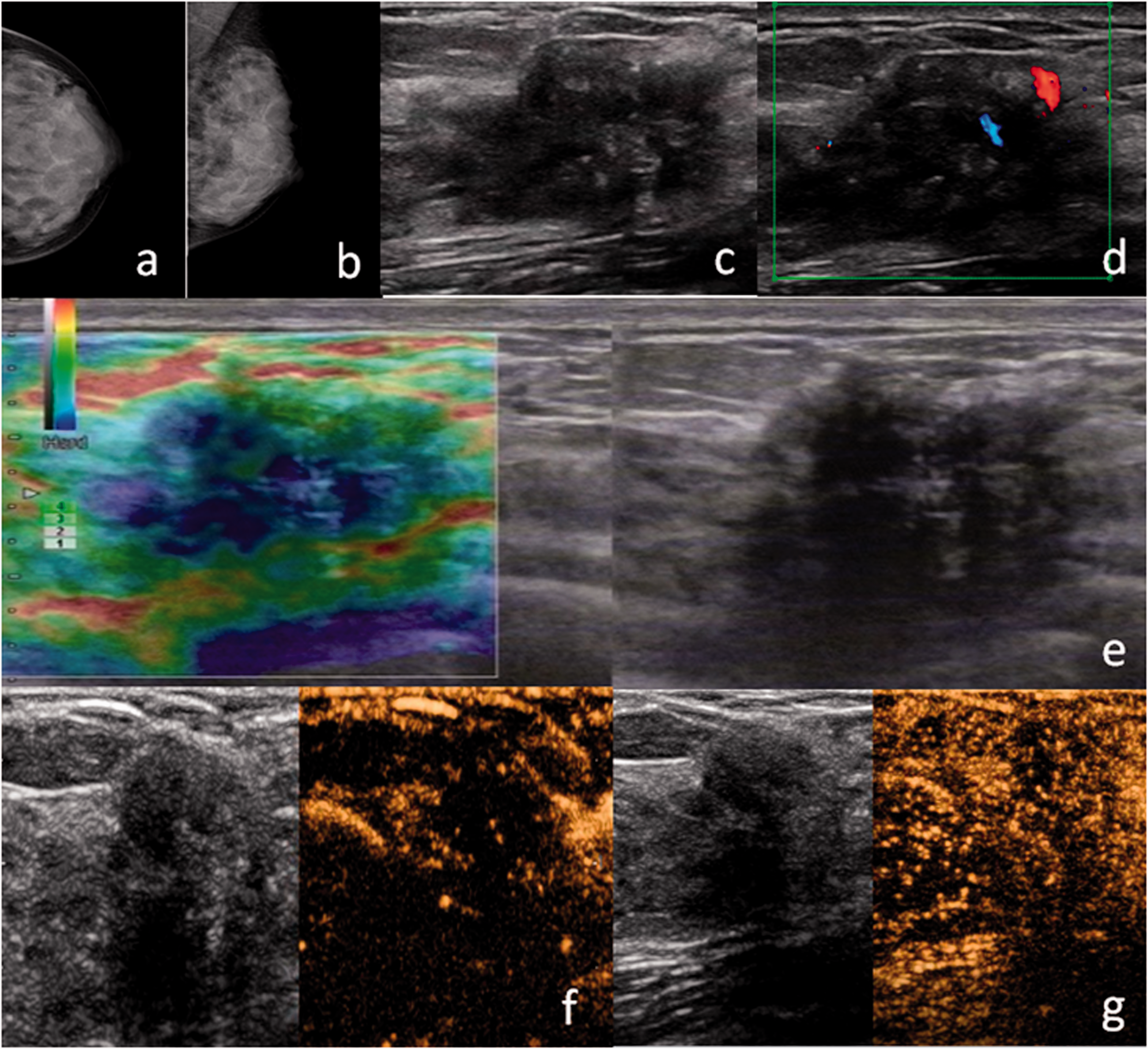
Mammography and conventional ultrasonography findings. Routine (a) craniocaudal and (b) mediolateral oblique mammography showed a relatively dense breast with focal asymmetry, classified as Breast Imaging-Reporting and Data System (BI-RADS) 0. (c) Ultrasonography demonstrated an ill-defined irregular hypoechoic breast lesion categorized as BI-RADS 4b. (d) Only a vascular spot was seen in the lesions. (e) The lesion was hard and given a strain elastography score of 5. (f) Contrast agent began to exhibit synchronous wash-in with iso-enhancement compared with the surrounding breast tissue 12 s after injection. (g) The micro-bubbles almost completely filled the lesion within 25 s, and the margin and shape could not be distinguished after enhancement.
Thus, the lesion was downgraded to BI-RADS category 3. The images were analyzed by two senior sonographers with >5 years of experience in breast CEUS. The final histological confirmation (200×) revealed fibromatosis (Figure 2(a)–(f)). The immunohistochemistry results were as follows: smooth muscle actin (+), desmin (+), S-100 (−), β-catenin (+), progesterone receptor (−), estrogen receptor (−), CD34 (+), and Bcl-2 (+). Nuclear accumulation of β-catenin was present in stromal cells but not in endothelial cells or vascular smooth muscle cells, which showed only cytomembrane staining (Figure 2(d), 400×). Combined with these results, fibromatosis was considered. Six months later, a breast mass reappeared in the area of the previous surgery and a second lumpectomy was performed. The pathology result was the same as that after the first surgery.

(a) Histological analysis revealed an infiltrative stromal process with interlacing bundles of fibroblasts and myofibroblasts. No nuclear atypia or mitosis was observed. (b) Immunohistochemically, the tumor cells were positive for (b) smooth muscle actin, (c) desmin, (d) β-catenin, (e) CD34, and (f) Bcl-2. Based on these findings, the tumor was diagnosed as desmoid-type fibromatosis.
Discussion
Breast fibromatosis is a rare benign stromal tumor that is usually described only in case reports or small case series. It has a high recurrence rate (ranging from 21% to 27%)2,17 after incomplete surgical resection because of its locally infiltrative nature. However, on MG, conventional US, and strain elastography, fibromatosis exhibits atypical morphological characteristics, making it difficult to differentiate from malignant breast lesions.
Although MG and conventional US remain the standard imaging methods for breast lesions, both MG and US have a sensitivity <75%. 18 Strain elastography provides information on the degree of stiffness of the lesion. However, none of these techniques can provide information on the microcirculation of breast masses. CEUS is a novel technique for the detection of microvascular structures. Several previous studies have shown the usefulness of CEUS in distinguishing benign and malignant breast masses.9–16 Some studies have revealed that benign masses tend to be hypovascular or homogeneously enhancing on CEUS.10,16 According to a prospective study, CEUS can be used as an additional tool to evaluate uncertain BI-RADS category 4 breast masses with MG and conventional US. Potentially, these masses could be downgraded to BI-RADS category 3 with 6-month follow-up instead of biopsy if they are either nonenhancing with a circumscribed margin or enhancing with an oval shape and homogeneous enhancement. 13 Xiao et al. 15 established a five-point system to analyze CEUS images. The authors determined that lesions displaying iso-enhancement and synchronous enhancement with surrounding tissue without a clear outline in CEUS images could be considered benign and thus downgraded. 15 In the present study, CEUS findings of the lesion showed iso-enhancement and synchronous enhancement without a clear outline. This is similar to the description of the enhanced pattern of CEUS in benign breast lesions in the above-mentioned study.
The application of CEUS in breast disease remains controversial because the diagnostic criteria vary across regions and the morphological characteristics overlap between benign and malignant breast masses. Moreover, the pathological subtypes in previous studies were relatively simple. Whether breast CEUS is suitable for the diagnosis of rare breast tumors, such as breast fibromatosis, should be further studied, and more cases should be accumulated.
Conclusion
Breast fibromatosis is a mimicker of malignancy and may exhibit suspicious morphological findings on all imaging modalities because of its local infiltrative behavior. Features on CEUS may suggest a benign entity, but histopathological examination remains the gold standard in the diagnosis of this disease.
Supplemental Material
sj-pdf-1-imr-10.1177_03000605211010619 - Supplemental material for Contrast-enhanced ultrasound of breast fibromatosis: a case report
Supplemental material, sj-pdf-1-imr-10.1177_03000605211010619 for Contrast-enhanced ultrasound of breast fibromatosis: a case report by Shanhong Lin, Yong Cao, Libin Chen, Mei Chen, Shengmin Zhang and Xiupeng Jia in Journal of International Medical Research
Footnotes
Ethics statement
Written informed consent for the publication of all clinical details and images was obtained from the patient. The local Ethics Committee of Ningbo First Hospital waived the requirement for approval of this case study. The reporting of this study conforms to the CARE guidelines. 19
Declaration of conflicting interest
The authors declare that there is no conflict of interest.
Funding
This study was supported by the Natural Science Foundation of Ningbo (2019A610308).
References
Supplementary Material
Please find the following supplemental material available below.
For Open Access articles published under a Creative Commons License, all supplemental material carries the same license as the article it is associated with.
For non-Open Access articles published, all supplemental material carries a non-exclusive license, and permission requests for re-use of supplemental material or any part of supplemental material shall be sent directly to the copyright owner as specified in the copyright notice associated with the article.