
Abstract
Objective
Regulator of calcineurin 1 (RCAN1) controls plasticity of the nervous system and depressive conditions by regulating brain-derived neurotropic factor (BDNF) and plays a crucial role in neural and cardiac pathways. The apolipoprotein E gene (ApoE) is a robust risk factor for progression of Alzheimer’s disease. A fatty diet is considered detrimental for metabolic disorders, such as obesity and cardiovascular diseases.
Methods
We examined the neuronal and cardiac protective roles of RCAN1 in ApoE−/− mice that were fed a high- or low-fat diet with and without voluntary movement for 3 months. Organ weights, laboratory data, histology, RNA expression, and behavior were examined.
Results
A high-fat diet with exercise improved depressive function, as examined by the forced swimming test, and RCAN1 mRNA expression was induced in the hippocampus. A low-fat diet with exercise resulted in a reduced body weight, higher heart weight/body weight ratio, and lower circulating triglyceride levels compared with a low-fat diet without exercise. RCAN1 mRNA expression was increased in cardiomyocytes in ApoE−/− mice.
Conclusions
The combination of a high-fat diet and exercise might reduce depressive function, whereas a low-fat diet with exercise leads to cardioprotection. Induction of RCAN1 expression might affect neuroplasticity and cardiac function.
Keywords
Introduction
Regulator of calcineurin 1 (RCAN1) regulates plasticity of the nervous system and depressive conditions by regulating brain-derived neurotropic factor (BDNF).1,2 Gene expression of RCAN1 is enriched in central neurons 3 and cardiomyocytes. 4 Further, RCAN1 knockout exacerbates ischemic injury in cerebral and cardiac cells. 4 Synaptosomal dysregulation has been reported in apolipoprotein E (ApoE)−/− mice compared with ApoE+/+ mice. 5 Moreover, ApoE gene polymorphisms are one of the strongest genetic risk factors for development of Alzheimer’s disease (AD). RCAN1 in the central nervous is overexpressed in patients with AD and might be related to the pathogenesis of neuronal degeneration. 6 Recently, a study showed that the processes of learning and remembering are closely related to body mass and energy homeostasis. 7 RCAN1 might also play a crucial role in neuronal and cardiac pathways, 8 and its gene expression is highly correlated with metabolic syndrome. 9 Specifically, with high caloric intake, RCAN1 mediates the suppressive effect of energy consumption and regulates control of obesity. 9
RCAN1 is a new RNA-binding factor that regulates neuronal apoptosis, and its RNA structure has a neuroprotective role. Induction of RCAN1 inhibits T cell activation and nuclear factor-κB signal transduction, and suppresses neuronal cell apoptosis. 3 However, the role of RCAN1 in the brain is controversial. 10 Overexpression of RCAN1 induces neurodegeneration in diseases, such as AD and Down syndrome. Short-term induction of RCAN1 expression shows a protective effect in cell survival. 10 However, the role of RCAN1 in the brain and its association with food intake and/or physical exercise are not clearly understood.
This study aimed to determine the effects of fat intake and exercise on cognition and behavioral tasks of ApoE−/− mice, as well as changes in RCAN1 and BDNF gene expression. Because RCAN1 and BDNF help regulate neuronal plasticity, we hypothesized that induction of RCAN1 and/or BDNF changes cognition and behavior through fat intake and exercise. Our results could be helpful in improving food intake and exercise for protection of neurodegenerative and depressive disorders in humans.
Materials and methods
Mice
Forty-five 2-month-old male BALB/c.KOR/Stm Slc-Apoeshl mice (Japan SLC, Inc., Shizuoka, Japan) were kept in a room at a temperature of 22°C with a 12-hour light–dark cycle. The mice were administered either a high-fat diet (45% energy from fat, 24% fat content by weight, 4.73 kcal/g; D12451, Research Diet; Sankyo Labo Service Corporation, Inc., Japan) or a control diet (10% energy from fat, 4.3% fat content by weight, 3.85 kcal/g; D12450H, Research Diet), and drinking water was provided ad libitum.
All animal experiments followed the Institutional Guidelines of the Kanazawa Medical University (KMU) and the guiding principles of the Physiological Society of Japan. These experiments were approved by the Animal Care Committee of KMU and were performed at the Animal Care Center of KMU.
Experimental design
Twenty mice were divided into four groups (n = 5 in each group). The high-fat diet with exercise (HFEX) group consisted of mice fed with a high-fat diet (D12451; Research Diet) whereas the low-fat diet with exercise (LFEX) group consisted of mice with a low-fat diet (D12450H, Research Diet). Both groups were allowed voluntary exercise and were kept in individual cages with running wheels. The high-fat diet with no exercise (HFNE) and low-fat diet with no exercise (LFNE) groups consisted of mice fed with high-fat and low fat diets, respectively, without exercise. They were housed separately in individual cages. All mice were fed for 12 weeks.
We collected lipid data of wild-type mice, but the data are not shown because the behavior of 5-week-old ApoE−/− mice in a previous study was reported to be the same as that of wild-type mice. 11 Therefore, we used ApoE−/− mice without a high-fat diet or exercise as the control group.
During this time, 25 other mice were separated into high-fat (n = 15) and low-fat diet groups (n = 10), and neither group was allowed to exercise. At termination, the following behavioral tests were performed. The Morris water maze test was used for spatial learning, the novel object recognition test was used for ability of recognition, and the forced swimming test was used to determine the depressive condition (see details below). After these behavioral tests, the mice were fasted overnight and blood samples were collected from the heart under anesthesia. Their brain and heart tissues were used for analysis.
Measurement of organ weights
All mice were euthanatized using pentobarbital and cervical dislocation was performed. These procedures were approved by the KMU Animal Care Committee Guidelines. The organ weights of brain and heart were measured as previously described. 12
Laboratory analysis
After the behavioral tests and euthanasia, blood was collected from the heart and was centrifuged at 302 × g at 4°C for 10 minutes. Each plasma sample was stored in a refrigerator at 5°C. Total cholesterol (T-CHO), low-density lipoprotein cholesterol (LDL-C), high-density lipoprotein cholesterol (HDL-C), and triglyceride (TG) levels were evaluated by the KMU Hospital Laboratory.
Histological analysis
After taking blood, the brain and heart were gently excised and fixed in 10% neutral buffered formalin. The hippocampi were precisely removed and processed as described previously. 10 Histological analysis was performed in all groups. Hematoxylin and eosin staining was performed for morphological analyses.
Real-time PCR
The hippocampus and heart were immediately harvested for RNA for gene expression analysis, which was performed as described previously. 11 RNA extraction was performed using an RNeasy Kit (Qiagen, Valencia, CA, USA) according to the manufacturer’s protocol. PCR was performed using primers that were specific for RCAN1 isoform 1 and glyceraldehyde-3-phosphate dehydrogenase (GAPDH) (for normalization) using a CFX96 Real Time PCR machine according to the manufacturer’s instructions (Bio-Rad, Hercules, CA, USA). The primer sequences for BDNF, RCAN1, and GAPDH were described previously. 12 These analyses were examined using the ABI PRISM7700 Sequence Detection System (Thermo Fisher Scientific, Waltham, MA, USA).
Behavioral tests
Open field test
The open field test is an experimental tool for rodents that evaluates their general locomotor activity, anxiety, and willingness to explore. The mice were placed in an empty round maze (φ = 80 cm, height = 50 cm), which was divided into outer (zone 1) and inner zones (zone 2). The mice were allowed free and uninterrupted movement in the maze for 10-minute periods, and their movement was tracked using SMART v 2.0 (Panlab Harvard Apparatus, Holliston, MA, USA). The amount of time spent in zone 2 (Tinner/Ttotal×100%) was statistically analyzed.
Novel object recognition test
The novel object recognition test is dependent on murine spontaneity of exploring objects, and specifically, that of novel areas rather than familiar areas. Each mouse was individually habituated to an empty box (40 × 25 × 20 cm) for 3 minutes, then taken out for a 5-minute rest. After resting, a 3-minute training session was carried out in which mice searched for two equal objects (objects A and A1) that were placed in two adjoining nooks, 10 cm from the wall. The exploration process was defined as sniffing or touching the substance with their noses or forepaws, and the exploration times for each object were recorded. Twenty-four hours after the training, the object to which mice showed less interest was replaced by a new object (object B), and all mice were allowed to explore the box for 3 minutes with one familiar object (object A or A1) and one novel object (object B). Data were collected by analyzing the recognition index, which was calculated as TB/(TA + TB), where TA is the exploration time with the familiar object A and TB is the exploration time with the new object.
Morris water maze test
The Morris water maze test was used to assess spatial memory. Maze training was held between 17 and 22 hours, and the maze was constructed from a round pool with a clear Plexiglas platform that was immersed 10 mm under the water’s surface. All mice participated in four forced swimming sessions per day for 5 days. The swimming pool was divided into five zones, and each session was performed by setting mice into one of the four quadrants on the edge of the pool. Each session was terminated 5 s after the mouse reached one platform or when the maximum 60 s had elapsed. The mice were then allowed to rest for 30 s between each swimming session. The mice participated in the trial for 60 s with the platform removed after the last experiment. The swimming traces for mice were analyzed using SMART v 2.0 (Panlab Harvard Apparatus). The percentage of time spent in a target zone, in which the platform was set, was analyzed.
Forced swimming test
The forced swimming test is used to identify behaviors, such as depressive-like conditions.10 The mice were forced to swim for 10 minutes daily for 2 consecutive days (days 1 and 2) in a clear cylinder tank (∅ = 24 cm, height = 60 cm, depth = 25 cm) filled with water at 25°C. The total swimming distance and immobility time were measured and analyzed.
Statistical analysis
Statistical analysis was performed using IBM SPSS Statistics for Windows, version 22.0 (IBM Corp., Armonk, NY, USA), and the results are expressed as the mean ± standard deviation. Comparative analyses of the behavior tests, body and organ weights, blood biochemical parameters, and gene expression were performed using the Student’s t-test or nonparametric tests. P < 0.05 was considered statistically significant.
Results
Organ weights
The mean body weight of the HFEX group was similar to that of the HFNE group, whereas that of the LFEX group was significantly lower than that of the LFNE group (P < 0.05, Table 1). Brain and heart weights were not significantly different among the four groups. The mean brain/body weight ratio of the HFEX group was slightly higher than that of HFNE group, but the difference was not significant. The mean brain/body weight ratio of the LFEX group was significantly higher than that of the LFNE group (P < 0.05). The mean heart/body weight ratio of the HFEX group was significantly higher than that of the HFNE group, and that of the LFEX group was significantly higher than that of the LFNE group (both P < 0.05).
Organ weights.
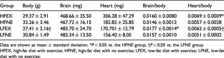
Data are shown as mean ± standard deviation. *P < 0.05 vs. the HFNE group, †P < 0.05 vs. the LFNE group.
HFEX, high-fat diet with exercise; HFNE, high-fat diet with no exercise; LFEX, low-fat diet with exercise; LFNE, low-fat diet with no exercise.
Laboratory analysis
T-CHO levels not significantly different among the groups. Mean TG levels were significantly lower in the HFEX group than in the HFNE group (P < 0.05). Mean TG levels were significantly lower in the LFEX group than in the LFNE group (P < 0.05). Mean HDL-C levels were significantly lower the LFEX group than in the LFNE group (P < 0.05). LDL-C levels were not significantly different among the four groups (Table 2).
Laboratory data.

Data are shown as mean ± standard deviation. *P < 0.05 vs. the HFNE group, †P < 0.05 vs. the LFNE group.
T-CHO, total cholesterol; TG, triglycerides; HDL-C, high-density lipoprotein cholesterol; LDL-C, low-density lipoprotein cholesterol; HFEX, high-fat diet with exercise; HFNE, high-fat diet with no exercise; LFEX, low-fat diet with exercise; LFNE, low-fat diet with no exercise.
Histological analysis
Hippocampal neuronal cells were not different among the four groups.
The left ventricular wall thickness in the HFEX group was significantly lower than that in the HFNE group (P < 0.05, Table 3). The diameter of myocytes was similar among the four groups (Table 3).
Heart wall thickness.

Data are shown as mean ± standard deviation. *P < 0.05 vs. the HFNE group.
LVWT, left ventricular wall thickness; VST: ventricular septal thickness; RVWT: right ventricular wall thickness; HFEX, high-fat diet with exercise; HFNE, high-fat diet with no exercise; LFEX, low-fat diet with exercise; LFNE, low-fat diet with no exercise.
mRNA expression
RCAN1 mRNA expression in the hippocampus of the HFEX group was significantly higher than that in the HFNE group (P < 0.05, Figure 1a). Further, RCAN1 mRNA levels in the hearts of the LFEX group were significantly higher than those in the LFNE group (P < 0.05, Figure 1b). BDNF mRNA expression in the hippocampus was not different among the four groups (Figure 2a). Cardiac BDNF mRNA expression in the LFEX group was significantly higher than that in the HFEX group (P < 0.05, Figure 2b).
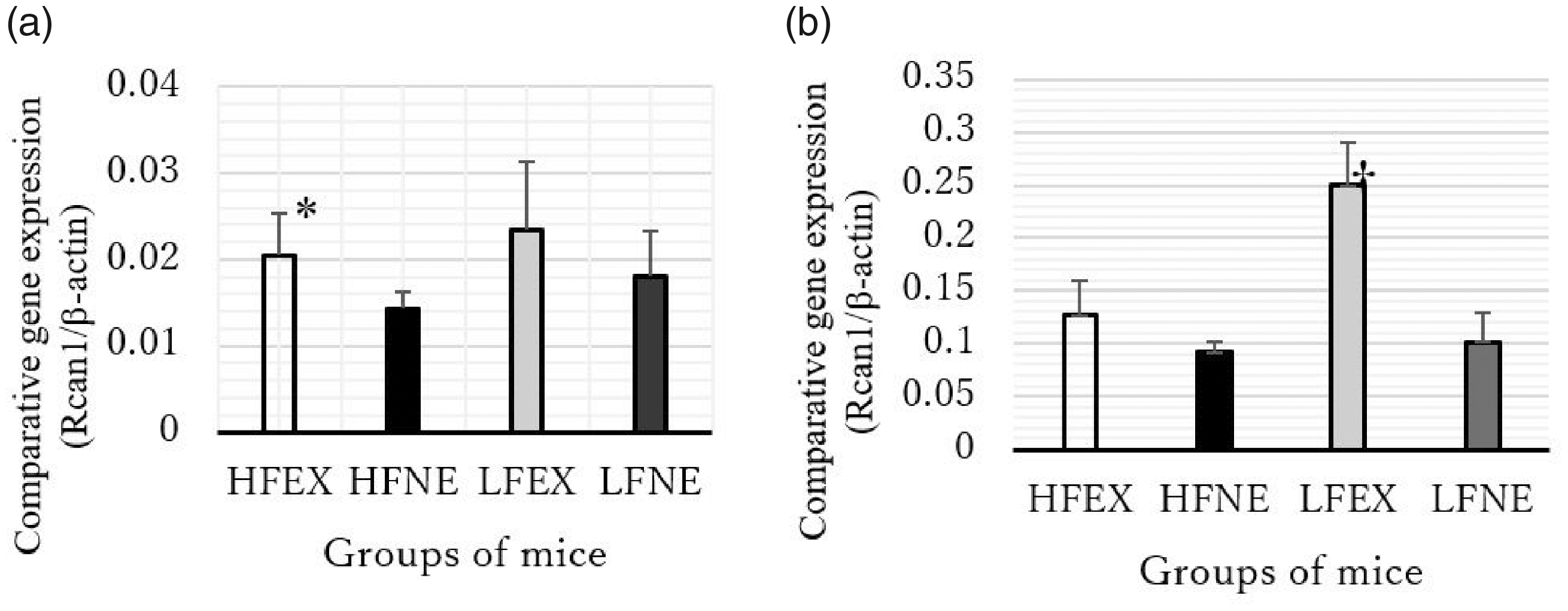
RCAN1 mRNA expression in (a) the hippocampus and (b) heart in each group. *P < 0.05 vs. the HFNE group, †P < 0.05 vs. the LFNE group. Error bars represent the standard deviation.

BDNF mRNA expression in (a) the hippocampus and (b) heart in each group. ΔP<0.05 vs. the HFEX group. Error bars represent the standard deviation.
Behavioral tests
We found no significant difference in the percent time spent in the open field test among the four groups (Figure 3a). In the novel object recognition test, there was also no significant difference in the exploration time among the four groups (Figure 3b). Moreover, in the Morris water maze test, no significant difference in escape latency was observed among the four groups (Figure 4).

Results of the open field test and the novel object recognition test. There were no significant differences in the percent time in the center in the open field test (locomotor activity) (a) and exploration time of recognizing a novel object (recognition memory) (b) among the four groups. Error bars represent the standard deviation.

Results of the Morris water maze test. There was no significant difference in the time of escape latency among the four groups. Error bars represent the standard deviation.
In the forced swimming test, the swim distance (days 1 and 2) in the HFEX group was significantly longer than that in the HFNE group (P < 0.05). Furthermore, the immobility time on day 2 in the HFEX group was significantly shorter than that in HFNE group (P < 0.05, Figure 5).

Results of the forced swimming test. *The swim distance on days 1 and 2 in the HFEX group was significantly longer than that in the HFNE group (P<0.05) (a). △The time to immobility on day 2 in the HFEX group was significantly shorter than that in the HFNE group (P<0.05) (b). Error bars represent the standard deviation.
Discussion
Our findings suggested that a high-fat diet with exercise improved the depressive state and induced RCAN1 mRNA expression in the hippocampus. However, a low-fat diet with exercise showed cardioprotective effects (e.g., lower body weight and TG levels), increased RCAN1 mRNA expression in cardiomyocytes, and decreased circulating TG levels in ApoE−/− mice. Hippocampal neuronal cells of ApoE−/− mice are susceptible to age-related and cytotoxic synaptic loss. 4 Synaptosomal dysregulation mediated by Aβ5 and cognitive decline 11 are enhanced in ApoE−/− mice compared with normal mice. Further, polymorphisms in ApoE have been studied as strong genetic risk factors for AD. 13 Neural overexpression of RCAN1 has been recognized in patients with AD and might be related to the pathogenesis of neuronal degeneration. 14
ApoE is the main protein involved in lipoprotein transport in neural systems. Peripheral ApoE is associated with progression of coronary heart disease, whereas central ApoE is associated with progression of dementia and AD. Recent studies have shown an association between ApoE and morbid obesity in experimental animals. 15 Further, apolipoproteins play a key role in HDL metabolism. 16 Therefore, the presence of lipoproteins is one of the main criteria for classification of HDL. A lack of ApoE might also induce lower circulating HDL-C levels in ApoE−/− mice. Accordingly, human longevity can be inherited, and ApoE is related to a lower odds ratio with respect to survival to the 90th and 99th percentiles of age7,16 ApoE-ε4 carrier status has important implications for associations between the brain and cardiovascular health in aging adults. 17 A lack of ApoE and lipid overload are closely related to dysfunction in the brain and heart. 18
Elevated RCAN1 expression after several hours in the brain can be neuroprotective under acute stress. However, long-term elevation of RCAN1 expression is critical in neurodegeneration in diseases, such as Down syndrome and AD. RCAN1 is a physiological modulator of oxidative stress. 10 Induction of RCAN1 might result in expression of important molecules for physiological adjustments to exercise-induced stress. 19 In this study, we found induced RCAN1 mRNA expression in the hippocampus after chronic exercise and higher lipid intake, but there was no change in hippocampal BDNF expression. The beneficial effect of induction of RCAN1 by chronic exercise in the hippocampus has not been fully studied yet. However, chronic exercise with higher lipid intake could play a key role in depressive function.
Calcineurin exerts pleiotropic effects, including heart valve formation and cardiac muscle hypertrophy. 20 Our study showed that a low-fat diet with exercise resulted in higher RCAN1 expression in myocytes and a higher heart/body weight ratio without ventricular dilatation or myocyte hypertrophy compared with a low-fat diet without exercise. Induced expression of cardiomyocyte-specific RCAN1 also inhibits myocardial hypertrophy induced by pathological or physiological stimuli in mice. Further, deletion of the RCAN1 gene exacerbates myocardial infarction induced by ischemia/reperfusion injury in mice.4,20 overexpression of RCAN1 in cardiomyocytes results in hyperfunction of mitochondria with augmented rates of oxygen consumption and reactive oxygen levels. Similar findings have been observed with milder overexpression of cardiac RCAN1 derived from trisomy 21 human-induced pluripotent stem cells. 4 This induced expression of RCAN1 might be an adaptational mechanism associated with hyperoxidative stress in patients with Down syndrome. This mechanism also operates in the nervous system against ischemia or reperfusion and merits further investigation. Further investigation is also required to test whether induction of the RCAN1 gene regulates myocardial protection through mitochondrial Ca2+ uptake. With caloric abundance, suppression of energy expenditure by RCAN1 overexpression might contribute to the growing epidemic of obesity. 9 Although a high-fat diet can induce functional and structural neuronal plasticity and activate orexin-producing neurons, which prevent depressive behavior, this beneficial function is limited within 4 weeks of high-fat diet intake.21,22 Our study showed that a 12-week high-fat diet with exercise improved the depressive condition of ApoE−/− mice. Therefore, the effect of a high-fat diet might continue longer than 4 weeks in combination with chronic voluntary movement. A low-fat diet in women is beneficial to cardiovascular health as shown by the Women’s Health Initiative Dietary Modification Trial.23 However, a high-fat diet is associated with impaired memory. 24 We found that a high-fat diet with exercise suppressed the depressive state compared with a high-fat diet without exercise and also induced hippocampal RCAN1 mRNA expression.
Moderate exercise can contribute to long-lasting protection against cardiovascular diseases, even with a high-fat diet. 25 However, no study has reported the beneficial effects of voluntary exercise with a high-fat diet on the depressive state. Exercise can reverse the harmful effects of a high-fat diet on behavior. 26 Further, a high-fat diet induces continuous low-grade inflammation. Therefore, the benefits of exercise might contribute to this finding via its anti-inflammatory effects. Indeed, a previous study showed that aerobic exercise reversed cardiac remodeling by reducing general inflammation, fibrotic changes, and apoptosis in high-fat diet-fed rodents. 27 Physical exercise also reduced insulin resistance and apoptosis in hippocampal neurons through improved function of mitochondria. These results suggest that physiological exercise improves neural plasticity in the hippocampus and reduces neural apoptosis, potentially reducing memory disturbances associated with high-fat diet-induced obesity. 28 Exercise also improves the capacity of the serotonergic system in the dorsal raphe, which prevents the depressive state associated with high-fat diet-related obesity. 29
One limitation of our study is the short (3 months) period of chronic exercise. A longer duration of physical exercise might alter RCAN1 gene expression and affect recognition tests more remarkably. Cardiac function and parameters should also be analyzed more precisely in further experiments. Protein analysis, such as that of calcineurin, RCAN1, and BDNF, is required for a more precise conclusion. Moreover, improved mobility may have affected the mice in our study. Therefore, the changes induced by exercise could be because of better mobility and not better cognition alone. We did not find any significant difference in neuronal changes among the four groups. We observed no pathological changes in the brain in 5-month-old ApoE-deficient mice, similar to a previous report. 30 Older mice should be examined in a future study on neuronal alternation in the brain.
This is the first report to demonstrate that a combination of 3 months of physical exercise and a high-fat diet reduce depressive dysfunction by inducing RCAN1 mRNA expression in the hippocampus in ApoE−/− mice. Further investigation of the RCAN1 gene in the brain and heart, as well as the relationship between nutritional alterations and RCAN1 expression, will lead to greater understanding of calcineurin function in human organs. A high-fat diet and voluntary exercise may be beneficial for the depressive condition, whereas a low-fat diet and voluntary movement are cardioprotective. Our findings could be beneficial for physicians.
Footnotes
Acknowledgements
Declaration of conflicting interest
The authors declare that there is no conflict of interest.
Funding
This study was supported by a Grant-in-Aid for Scientific Research of the Ministry of Education, Culture, Sports, Science, and Technology (grant number: 17K09330).