
Abstract
The Papez circuit is an important brain structure that is closely associated with learning and memory. In this report, we present four patients with anterograde amnesia as the main manifestation induced by Papez circuit infarction. In addition, we review the distribution of the responsible arteries in key and rare regions to investigate the pathogenesis of these infarctions.
Keywords
Introduction
The Papez circuit was first proposed by J.W. Papez in 1937. It is a closed circuit that is formed by a neural pathway originating from the hippocampus, which returns to the hippocampus through the mammillary body, the anterior thalamic nucleus, and the cingulate gyrus. 1 The Papez circuit is closely associated with learning and memory. At present, there are only a few published reports of infarction in the key regions of the Papez circuit.2,3 The majority of these have been single case reports. The current report comprehensively analyzes the clinical features, imaging features, and responsible arteries that are involved in memory impairment caused by infarction of the Papez circuit (the hippocampus, mammillary body, anterior thalamic nucleus, and fornix column). The aim of this report is to improve the understanding of specific cognitive impairments that are induced by infarction of the Papez circuit.
Case reports

(a) Diffusion-weighted imaging indicating acute cerebral infarction in the left hippocampus. (b) Magnetic resonance angiography indicating localized stenosis in the left posterior cerebral artery

(a) Diffusion-weighted imaging indicating bilateral fornix column infarction. (b) Magnetic resonance angiography indicating changes in intracranial arteriosclerosis.
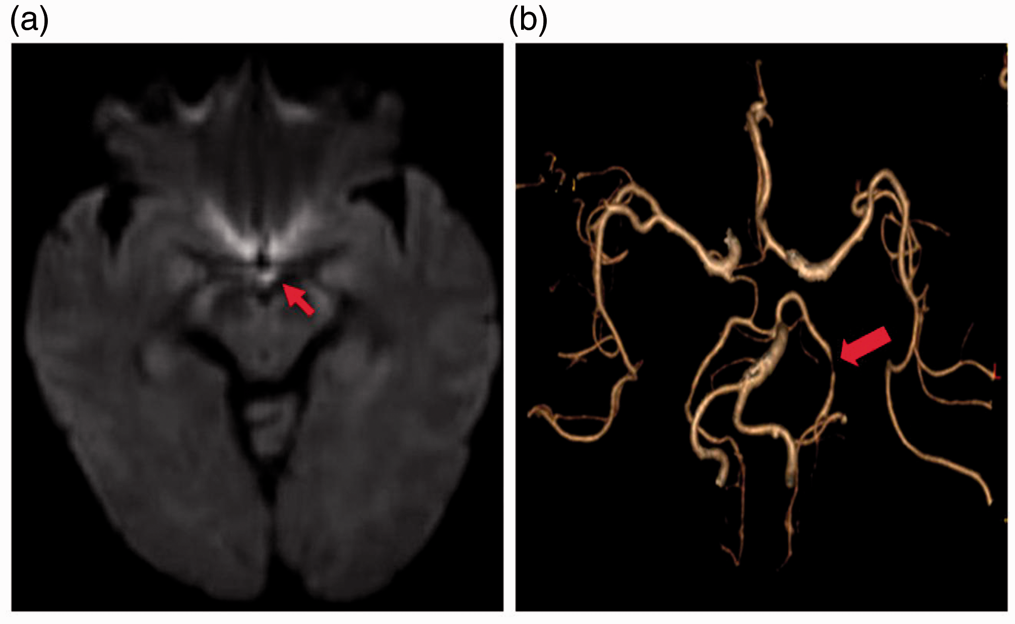
(a) Diffusion-weighted imaging indicating a left mammillary body infarction. (b) Computed tomography angiography indicating moderate to severe stenosis in the P2 segment of the left posterior cerebral artery.
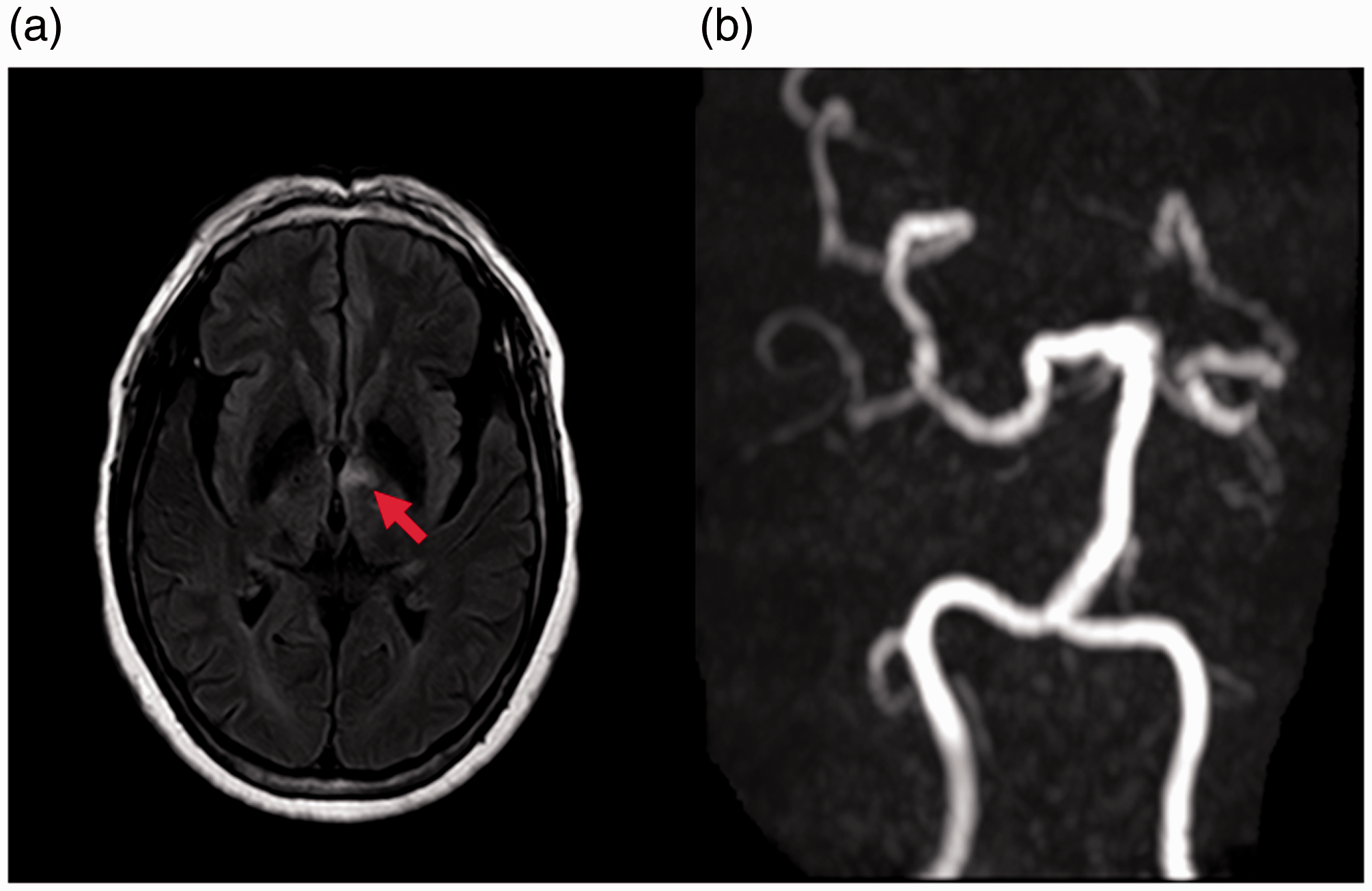
(a) Fluid-attenuated inversion recovery imaging indicating a focus in the left thalamus. (b) Magnetic resonance angiography indicating an opening in the left posterior communicating artery.
Discussion
Amnesia is frequently observed in the clinic. Based on the affected content and time sequence, amnesia can be classified as anterograde, retrograde, borderline, or progressive. 4 Damage to the bilateral marginal system circuit (hippocampal structure, mammillary body, anterior nucleus of the thalamus, cingulate gyrus, parahippocampal cortex, and their connections through the fornix and mammillary body–thalamic conduction tract) can cause obvious and persistent anterograde memory deficits. 5
The hippocampus, located between the thalamus and the medial temporal lobe, is part of the limbic system. It is responsible for the storage, transformation, and orientation of long-term memory. Damage to the hippocampus leads to recent memory storage disorder. The hippocampus requires a large amount of energy and is susceptible to ischemia–hypoxia. 6 Patient 1 was characterized as having simple anterograde amnesia. Combined with his MRI results, a diagnosis of hippocampal infarction was clear. The short-term and rapid recovery of memory in this patient was considered to be related to the compensation of contralateral hippocampal function. The hippocampus is supplied by the terminal artery, including the anterior, middle, and posterior arteries. The anterior hippocampal artery originates from the anterior choroidal artery, while the middle and posterior hippocampal arteries originate from the posterior cerebral artery. 7 Brain MRA demonstrated extensive intracranial arteriosclerosis in Patient 1. Combined with the distribution of hippocampal infarcts, the left posterior cerebral artery was considered as the vessel responsible for the infarct. The fornix is the nerve bundle that connects the hippocampus and the mammillary body. It is mainly composed of efferent bundles of the hippocampus. In addition, it contains several afferent and connecting bundles to the hippocampus. Any damage to the fornix leads to memory loss. The most prominent manifestation of fornix column infarction is apparent memory disorder 8 and Korsakoff syndrome. Patient 2 was diagnosed with anterograde amnesia, with decreased short-term memory and mild impairment in mathematical calculation ability. This finding was consistent with those of previously published studies. The blood supply to the fornix column is through the subcallosal artery, which is a single branch from the anterior communicating artery. 9 Because the bilateral fornix column is supplied by a single artery, fornix column lesions induced by vascular disease have bilateral involvement. 10 The patient had bilateral fornix column infarction and this was consistent with the aforementioned vascular distribution.
The mammillary bodies are part of the hypothalamus and are located behind the tuber cinereum. They are a pair of protuberances in the fovea between the midbrain feet. The deep surface of the mammillary body is the nucleus. Damage to the mammillary body can lead to poor memory, mental retardation, decreased ability for calculation and understanding. 11 Patient 3 was diagnosed with simple memory impairment, which was consistent with previous reports of mammillary body infarction. Head MRI showed a left mammillary body infarction. The vascular supply to mammillary bodies has been documented in only a small number of publications. 12 Based on these studies, the main blood supply to mammillary bodies are the side branches of the posterior communicating artery and the posterior cerebral artery. 12 From the aforementioned distribution of mammillary body vessels, the left posterior cerebral artery was considered to be the vessel responsible for the mammillary body infarction.
The thalamus is the largest structure of the mammalian diencephalon and plays an important role in maintaining alertness, emotion, and other cognitive functions. The thalamus structure is complex, with abundant blood supplies. When different arteries are occluded, different clinical symptoms are induced. One of the most common types is top of the basilar syndrome,13,14 while others include simple thalamus syndrome and thalamic dementia. Thalamic dementia is a type of vascular dementia that is relatively rare in the clinic. The majority of cognitive, language, and mental behavior disorders caused by infarction involve the anterior thalamic nucleus and the mammillary–thalamic tract. Most of the responsible vessels are branches of either the posterior communicating artery or the tubercular artery of the thalamus. 15 Patient 4 had clinical and imaging features that were consistent with anterior thalamic infarction. The infarct involved the mammillary–thalamic tract and the anterior thalamic nucleus. The responsible vessel was the thalamic nodal artery.
The majority of vascular cognitive dysfunction is caused by infarctions in key regions of the Papez circuit, which induce simple memory defects and slow responses. These infarctions usually lack other signs and symptoms of nerve defects. Because the infarct focus is often small and the location is unusual, head computed tomography scans often show no abnormal findings. This usually results in misdiagnosis.
The present case studies suggest that, for patients with simple acute-onset memory impairment (and especially short-term memory impairment) and risk factors for vascular disease, special attention should be paid to regions such as the fornix column and mammillary bodies, to avoid misdiagnosis.
Footnotes
Declaration of conflicting interest
The authors declare that there is no conflict of interest.
Ethics and consent
This study was approved by the institutional research ethics committee of Harrison International Peace Hospital (reference number 2018-1-004). This study received verbal consent from all patients or patients’ parents or caregivers.
Funding
This study was funded by the Medical Science Research Project of Hebei province in 2019 (no. 20191782).