
Abstract

303
BRAIN-0035
Poster Session
DYSFUNCTION OF MOUSE CEREBRAL ARTERIES DURING EARLY AGING
Abstracts
1Institute for stroke and dementia (ISD), LMU, Munich, Germany
2Department of Pharmacology, University of Vermont, Burlington, USA
3Graduate School of Systemic Neuroscience (GSN), LMU, Munich, Germany
Abstract
304
BRAIN-0452
Poster Session
DYNAMIC MAGNETISATION TRANSFER MRI: CARDIAC PULSATION IN AGING BRAIN TISSUE
Aging
1Lloyds Institute of Neuroscience, Trinity College Dublin, Dublin, Ireland
2School of Medicine & Medical Science, University College Dublin, Dublin, Ireland
3School of Psychology, Bangor University, Wales, United Kingdom
Abstract
The aging process profoundly impacts the brain and the heart at multiple levels, ranging from sub-cellular to macro-structural [1, 2]. In the brain, aging causes deterioration of neuronal and mitochondrial membranes, which leads to the loss of cellular integrity and impaired neuronal function [3]. At the same time, the cardiac dynamics are altered, which influences the brain metabolism. Here, we want to study the interplay between heart and brain in a new fashion. Our hypothesis is that using our method of ultrafast magnetisation transfer MR [4] we can measure water dynamics changes in brain tissue which depend on cell integrity and on cardiac pulsation.
The MRI sequence consists of a magnetisation transfer (MT) preparation phase followed by single-slice echo-planar imaging sequence. MT effect was introduced by a negative (-5236.8 ± 894.18 Hz) and a positive off frequency (6982.5 ± 894.18 Hz) RF pulse. Imaging parameters were voxel size of 3.5 x 3.5 x 3 mm, TR = 60 ms, TE = 18 ms, FA = 35 degrees and 3000 repetitions.
48 subjects (25 between 18 and 29 years old, and 23 over 65 years old) were scanned with the protocols approved by the local ethics committee.
A comparison between the time series for both age groups is shown in figure 1a-b. In both cases, the time series of all the voxels across the slice were averaged to showcase the difference between populations. Strong cardiac constant peaks are resolved only for the young subjects while for the older subjects the strong cardiac peaks are diminished. The Fourier Transform of the time series was calculated in each case (Figure 1c-d). The frequency spectra for the young group present strong cardiac frequencies. However, the spectra for the older group show stronger harmonics or envelope waves or both, in addition to the weaker cardiac frequencies. The envelope waves have a beat frequency of the (cardiac frequency)/n, where n takes values of (2,3,4,6,9 …). These results are consistent over all subjects.
Abstract
Time series and frequency spectra for a young (a and c) and an old (b and d) subjects.
To our knowledge, the observation of a cardiac related MT change and its variation with aging has not yet been reported. Most likely, as the brain ages the deterioration of the cellular membranes lowers the magnitude of the exchange between the pool of free water and the macromolecules. Since the pressure wave is thought to interrupt this exchange [4], the effect of the pressure wave in the old age population diminishes. Taken together with the additional variation of the cardiac dynamics, the shape and number of frequencies in the spectra change between age groups. It is our understanding that using this method we can increase our knowledge of the interplay between heart and brain in aging.
[Figure] Time series and frequency spectra for a young (a and c) and an old (b and d) subjects.
References
305
BRAIN-0153
Poster Session
REVERSAL OF BETA-AMYLOID-INDUCED NEUROTOXICITY IN PC12 CELLS BY CURCUMIN, THE IMPORTANT ROLE OF ROS-MEDIATED SIGNALING AND ERK PATHWAY
Aging
1Key Lab of Cerebral Microcirculation in Universities of Shandong, Taishan Medical University, Taian, China
2Nursing Department, Taishan Vocational College of Nursing, Taian, China
3School of Basic Medicine, Taishan Medical University, Taian, China
Abstract
Abstract
The study was supported by the National Natural Science Foundation of China No.81471212, 81271275, 81070947, 30770759 to B.-L. Sun; Natural Science Foundation of Shandong No. ZR2012HZ006 to B.-L. Sun.
*Corresponding author: Cun-dong Fan, Xiao-yan Fu, Bao-liang Sun, Tel: +86-538-6230027, E-mail: 
References:
306
BRAIN-0436
Poster Session
ELICITATION THRESHOLD OF CORTICAL SPREADING DEPOLARIZATION INCREASES WITH BRAIN MATURATION AND ISCHEMIA
Aging
1Department of Medical Physics and Informatics Faculty of Medicine & Faculty of Science and Informatics, University of Szeged, Szeged, Hungary
2Donald W. Reynolds Department of Geriatric Medicine Reynolds Oklahoma Center on Aging, University of Oklahoma Health Sciences Center, Oklahoma City, USA
Abstract
Objectives
Spreading depolarization (SD) is a wave of synchronized depolarization of neurons and glia cells, which propagates across the cerebral gray matter at a rate of 2–5 mm/min. SD is rapidly followed by a local hemodynamic response, the kinetics of which is characteristic of the metabolic state of the tissue. SD is associated with migraine aura, and also occurs in the vicinity of ischemic brain lesions, where it is considered to aggravate the initial damage. Even though age is a highly significant predictor of both migraine and stroke, very few studies investigated whether aging has an impact on SD evolution.
The major goal of this study was to determine the electric threshold of SD elicitation before and during incomplete forebrain ischemia in rats of various age groups of early adulthood.
Methods
Animal procedures were approved by the Ethical Committee for Animal Care of the University of Szeged adhering to national regulation. Male Sprague-Dawley rats (n=37, age groups: 7, 8, 9, 10, 12, 16 and 30 week-old) were anesthetized with isoflurane in N2O:O2. Both common carotid arteries were dissected for the latter induction of incomplete forebrain ischemia by occlusion of the vessels (2VO). Two, separate craniotomies were created on the right parietal bone for SD elicitation and data acquisition. SDs were triggered by cathodal direct current stimulation of the dura, allowing the calculation of the exact amount of current delivered. To determine the threshold of an SD, the current was elevated stepwise, until SD was detected. SDs were identified using DC-and local-field potentials. SD related hemodynamic changes were recorded by Laser-Doppler flowmetry. In order to discriminate between the non-ischemic and ischemic threshold of SD elicitation, 3 SDs were elicited prior ischemia induction. Three additional SDs were elicited following 2VO.
Results
Abstract
The threshold of SD elicitation was higher during ischemia as compared with the non-ischemic condition in all age groups studied; statistical significance was found in the 10-30 week-groups. The threshold proved to be the lowest in the 9-week-old group, both for non-ischemic and ischemic SDs, being significantly lower than in the 12-30-week-old groups.
Conclusions
Our data demonstrate that the threshold to trigger SDs increases with progressing life time, as described previously in brain slices.[1] The susceptibility of the brain to SD may be determined by the gray matter’s biochemistry and cytoarchitecture (e.g. density of dendritic spines, volume of extracellular space) that undergo adaptational changes with maturation. The threshold of SD elicitation also increased during ischemia, coinciding with earlier findings. [2] Since decreasing pH has been known to hinder SD evolution, acidosis created by ischemia is suggested to inhibit SD elicitation.
Grant support: János Bolyai Research Scholarship of the Hungarian Academy of Sciences and The Hungarian Scientific Research Fund: OTKA K111923

References
307
BRAIN-0182
Poster Session
AGE-ASSOCIATED ALTERATIONS OF ANTIOXIDANT STATUS, CALCIUM HOMEOSTASIS AND GLUCOSE TRANSPORTER IN FEMALE RAT BRAIN: NEUROPROTECTIVE ROLE OF ESTRADIOL
Aging
1School of Life Sciences, Jawaharlal Nehru University, New Delhi, India
Abstract
During normal aging, brain experiences structural, molecular, and functional alterations. Aging in females and males is considered as the end of natural protection against age related diseases like osteoporosis, coronary heart disease, diabetes, Alzheimer’s disease and Parkinson’s disease. Protection from age-related disorders is provided by several factors, including estrogens. These changes increase during menopausal condition in females when the level of estradiol is decreased. The objective of this study was to observe the changes in activities of superoxide dismutase (SOD), glutathione S-transferase (GST), Ca2+ATPase, intracellular calcium levels, DNA degradation and glucose transporter 4 (GLUT4) occurring in brains of female albino Wistar rats of 3 months (young), 12 months (adult) and 24 months (old) age groups, and to see whether these changes are restored to normal levels after exogenous administration of estradiol (0.1 µg/gm body weight for one month). The results obtained in the present work revealed that normal aging was associated with significant decrease in the activities of SOD, GST, Ca2+ATPase and GLUT4 levels in the brains of aging female rats, and an increase in DNA degradation and intracellular calcium levels. Administration of E2 brought these changes to near normalcy. It can therefore be concluded that E2’s beneficial effects seemed to arise from its antioxidant and antilipidperoxidative effects, implying an overall neuroprotective and anti-aging action. The results of this study will be useful for pharmacological modification of the aging process and applying new strategies for control of age related disorders.
308
BRAIN-0245
Poster Session
DELAYED INTRACRANIAL PRESSURE ELEVATION FOLLOWING ISCHEMIC STROKE IS PREVENTED BY EARLY AND SHORT HYPOTHERMIA TREATMENT IN AGED RATS
Aging
1School of Biomedical Sciences and Pharmacy, University of Newcastle, Newcastle, Australia
Abstract
309
BRAIN-0160
Poster Session
AGING-ASSOCIATED INFLAMMATION IN THE VISCERAL ADIPOSE TISSUE AND THE BRAIN ARE REDUCED BY RESVERATROL
Aging
1Pharmacology, Ewha Womans University College of Medicine, Seoul, Korea
Abstract
References:
310
BRAIN-0515
Poster Session
HUMAN ADIPOSE-DERIVED MESENCHYMAL STROMAL CELL ADMINISTRATION IMPROVES OUTCOME IN AGED STROKE MICE
Aging
1Neuroscience, IRCCS - Istituto di Ricerche Farmacologiche Mario Negri, Milan, Italy
2Cerebrovascular Diseases, Fondazione IRCCS Istituto Neurologico Carlo Besta, Milan, Italy
Abstract
References:
311
BRAIN-0657
Poster Session
LONG-TERM RELATIONSHIP OF SERUM BRAIN-DERIVED NEUROTROPHIC FACTOR WITH CEREBRAL BLOOD FLOW AND COGNITION IN COGNITIVELY NORMAL ELDERS
Aging
1Epidemiology, University of Pittsburgh Graduate School of Public Health, Pittsburgh, USA
2Psychiatry, University of Pittsburgh, Pittsburgh, USA
3Medical Gerontology, Trinity College Dublin, Dublin, Ireland
4Neuroepidemiology Section, National Institute on Aging, Bethesda, USA
5Sticht Center on Aging, Wake Forest School of Medicine, Winston-Salem, USA
6Psychiatry, University of California San Francisco, San Francisco, USA
Abstract
Abstract
Brain regions with significant correlations of brain-derived neurotrophic factor and resting cerebral blood flow.

312
BRAIN-0711
Poster Session
CHARACTERIZATION OF AGE-RELATED CHANGES IN MICROGLIA/MACROPHAGE POLARIZATION AND FUNCTIONAL OUTCOMES IN A MOUSE MODEL OF ISCHEMIC STROKE
Aging
1Neurology, Department of Neurology University of Pittsburgh, Pittsburgh, USA
Abstract
One reason for thefailure of translating the successes in stroke models to the clinical applications is that themajority of experimental stroke studies have used young adult animals, whilestroke in humans mainly afflicts the elderly. It is therefore necessary toinvestigate the different pathological changes after stroke in young and oldanimals, and elucidate how these differences contribute to stroke outcomes. Inthe present study, we investigated the difference between aged and young micein their responses to ischemic challenge, focusing on infarct volume, long termfunctional outcomes, and M1-M2 polarization of microglia/macrophage.
Young (2 monthold) or aged (18 month old) male mice were subjected to permanent tandemocclusion of left distal middle cerebral artery (dMCAO) and ipsilateral commoncarotid artery (CCA). Sensorimotor and cognitive behavioral tests wereperformed up to 35d after stroke. Infarct volumes were quantified at 2d afterstroke. The expression of M1 (CD16 and CD32) and M2 (CD206 and Arg1) markers wasexamined by immunohistochemical stainings and reverse-transcriptase polymerasechain reaction (RT-PCR) at 1, 3,7, 14 and 35d after stroke.
Brain infarct at2d after stroke was significantly larger in aged mice as compared to youngadults (young 15.6±2.3 vs aged 20.7±1.8%, p<0.01). Remarkably, aged miceexhibited more severe long-term sensorimotor deficits, as manifested bydeteriorated performance in rotarod and hang wire tests up to 35d stroke. The agedmice also showed significantly worse long-term cognitive deficits as measured byMorris water maze test at 21d after stroke. RT-PCRand immunohistochemistry staining of M2 or M1 markers revealed a similar trendof change in microglia/macrophage polarity after stroke between young and agedmice. The expression of M2 markers peaked around 7d after stroke and theexpression of M1 markers peaked later around 14-21d after stroke, suggesting aM2-to-M1 phenotype shift with the progress of stroke. Interestingly, the agedmice exhibited a trend of reduced extent of M2 polarization after stroke ascompared to young adults.
This distal MCAOmodel of stroke consistently result in ischemic brain injury with very lowmortality and long-term behavioral deficits, and therefore is suitable for theevaluation of long term stroke outcome. The aged mouse exhibits deterioratedfunctional outcome after stroke, which might associated with reduced M2microglia/macrophage polarization.
313
BRAIN-0684
Poster Session
CARRIER MEDIATED DELIVERY SYSTEM BEARING DOPAMINE FOR EFFECTIVE MANAGEMENT OF PARKINSONISM
Neurodegeneration
1Department of Pharmacy, Manav Bharti University, Kanpur, India
2R&D, KRV Hospitals Pvt. Ltd., Kanpur, India
Abstract
Delivery of drug and sustaining it in effective concentration in brain is challenging due to blood brain barrier. In the present investigation, amino acid coupled liposomes bearing dopamine-HCl were prepared to deliver drug to the brain utilizing receptor-mediated transcytosis for effective management of parkinsonism.
L-lysine stearylamine conjugate (LSC) was synthesized & LSC coupled liposomes bearing dopamine HCl was prepared by lipid cast film method. Formulations were analyzed for average vesicle size, drug entrapment, in-vitro drug release and in-vivo efficacy of the formulations was assessed by measuring the reduction in the degree of drug induced catatonia in albino rats.
Average particle size was found in the range of 1.92-0.80 mm. There was increase in the size for coupled liposomes due to the inclusion of LSC in liposomal bilayers. The percent encapsulation efficiency decreased from 46.82±2.17% in uncoupled to 38.13±1.18% in coupled liposomes. The in-vitro drug release after 24hrs was 58.9±2.94% with uncoupled while the coupled liposomes showed 43.7±2.18% drug release. The lower value for coupled formulation could be due to the retardation of drug release caused due to the incorporation of LSC in the liposomal bilayers, which enhanced the structural integrity of the bilayer. In-vivo study reveals that the animals receiving uncoupled liposomes showed partial reduction and animals that received coupled liposomes showed almost complete reduction in catatonia.
Fluoresence study clearly indicates the uptake of 6-CF in blood vessels and accumulated in brain. This could be due to enhanced uptake of Lysine coupled liposomes through amino acid transporters present at BBB surface.
314
BRAIN-0142
Poster Session
ALTERATIONS OF BRAIN MORPHOLOGY AND ENERGY METABOLISM UNDERLYING MEMORY DETERIORATION IN INSULIN-RESISTANT GOTO-KAKIZAKI RATS
Neurodegeneration
1Laboratoire d'imagerie fonctionnelle et métabolique, Ecole polytechnique fédérale de Lausanne, Lausanne, Switzerland
Abstract
This work was supported the Swiss National Science Foundation (grant 148250), and the Centre d’Imagerie BioMédicale (CIBM) of the UNIL, UNIGE, HUG, CHUV, EPFL and the Leenaards and Jeantet Foundations.
References:
315
BRAIN-0285
Poster Session
ALTERED THALAMIC GLUCOSE METABOLISM IN PARKINSON’S DISEASE
Neurodegeneration
1Neurology, University Hopsital, Köln, Germany
Abstract
The thalamus is the main relay station in the basal ganglia circuitry and the key output pathway for basalganglia-cortex loops. Pathophysiological alterations of thalamic nuclei play a critical role in the development of parkinsonian symptoms like akinesia or rigidity.
Historically, these changes have been conceptualized as a consequence of nigral degeneration; nowadays there is also histopathological evidence that suggests a direct pathology of thalamic nuclei in PD disease. Recent imaging studies could show an increased glucose metabolism in the total thalamus. However, these latest imaging findings did not consider the organization into distinct nuclear regions. Due to the different involvement of thalamic subnuclei we wanted to precise the involved regions by analysis of the glucose metabolism in parkinsonian patients.
In total 28 akinetic-rigid PD patients and 11 healthy controls were examined with high resolution 18-Fluoro- deoxyglucose PET-imaging. An in-house created VOI-atlas was used to define the local metabolism in the total thalamus and thalamic subnuclei. The VOIs of the atlas were used to calculate the rCMRGlc in the regarding regions. Results for the overall thalamic uptake and the uptake in the thalamic sub nuclei were compared between parkinsonian patients and healthy controls.
Analysis of glucose metabolism revealed a bilaterally increased metabolism in the PD group for the total thalamus in all nuclei except the left dorsal medial nucleus, the left and right nucleus anterior. Statistical analysis showed significantly higher glucose metabolism for the following subnuclei: bilateral ventral lateral nuclei, ventral posterior lateral nuclei and the left nucleus anterior.
The ventrolateral nuclei and the ventral posterior lateral nuclei showed an increased glucose metabolism in PD patients compared to healthy controls. These findings argue for a critical (maybe compensatory) role of the thalamus as a major relay station in the striatothalamocortical network and against a direct disease pathology.
316
BRAIN-0362
Poster Session
REPRODUCIBILITY OF ABNORMAL BRAIN METABOLISM ASSOCIATED WITH PROGRESSIVE SUPRANUCLEAR PALSY: NETWORK AND REGIONAL COMPARISONS BETWEEN A US AND A CHINESE COHORT
Neurodegeneration
1PET center, Huashan Hospital, Shanghai, China
2Department of Neurology, Huashan Hospital, Shanghai, China
3Center for Neurosciences, Feinstein Institute for Medical Research, Manhasset New York, USA
Abstract
Objectives
PET with FDG has been used for identification of disease-specific metabolic brain networks associated with parkinsonian disorders (1-3). We have previously shown that brain network and its expression in idiopathic Parkinson’s disease were highly reproducible across the patient populations and tomographs (4). In this study, we aimed to evaluate the reproducibility of disease-specific brain network and regional cerebral metabolism associated with progressive supranuclear palsy (PSP) by comparing clinically-confirmed patients with PSP in a US and a Chinese cohort.
Methods
The US cohort consisted of 10 patients with PSP and 10 age-matched healthy controls scanned on a GE Advance PET camera. The sample size was the same in the Chinese cohort scanned on a Siemens Biogragh 64 PET/CT system in China. The study at each site was approved by the respective IRB for which the subjects had signed written informed consent. We first used the network analysis to identify a PSP-related metabolic pattern (PSPRP) in each cohort and computed the corresponding network scores prospectively in the other cohort. We also localized the regions with abnormal metabolic differences in the same sets of FDG PET images by using conjunction and interaction analyses with SPM. The reproducibility of PSP-specific network and metabolic distribution was examined across the study populations, PET instruments and analytical approaches.
Results
Both cohorts revealed similar PSPRPs characterized by metabolic decreases in the medial prefrontal cortex/cingulate, ventrolateral prefrontal cortex, striatum, medial thalamus, and midbrain, along with covarying metabolic increases in the hippocampus and parieto-temporal regions. PSPRP scores were similarly elevated (P<0.0001) in the patients relative to the controls in the derivation cohort in the USA and in the validation cohort in China or vice versa. PSPRP scores correlated strongly (R≥0.96; P<0.001) in the two corresponding cohorts of patients and healthy controls from the USA and Chinese sites, respectively.
We observed that PSP patients from two cohorts shared a great number of overlapping areas with regional metabolic abnormalities in both cortical and subcortical areas (FWE P<0.05). Relative metabolism decreased in the medial prefrontal cortex/cingulate, ventrolateral prefrontal cortex, striatum, medial thalamus, and midbrain but increased in the hippocampus and parieto-temporal regions. Volume of interest analyses confirmed the significant group differences (P<0.001) in these brain regions.
Conclusion
This study demonstrated the high comparability and reproducibility of PSP-related brain network and regional metabolism across patient populations, tomographs and imaging techniques. Activity of this brain network may serve as a reliable and objective marker of PSP for clinical applications.

References:
317
BRAIN-0495
Poster Session
DIFFERENT RATES OF DOPAMINE TRANSPORTER LOSS IN PARKINSON’S DISEASE AS MEASURED WITH [123I]β-CIT AND [123I]FP-CIT SPECT
Neurodegeneration
1Global Exploratory Medicine, UCB Pharma, Braine l'alleud, Belgium
2Molecular NeuroImaging, Institute for Neurodegenerative Disorders, New Haven, USA
3Neuroscience, Intracellular Therapies Inc, New York, USA
4Imanova Ltd, Imperial College London, London, United Kingdom
Abstract
OBJECTIVES: Progressive loss of dopamine transporter (DAT) density in Parkinson’s disease (PD) is well established and considered a biomarker of the underlying dopamine neuron loss. In large, longitudinal studies with [123I]β-CIT SPECT in early PD (CALM-PD1; ELLDOPA2; PRECEPT3), the annualized rate of loss in the striatum was approx. 5%, while in studies using [123I]FP-CIT SPECT in similar patients (PROUD4, PPMI5) the annualized rate of loss was approx. 12%. The explanation for this apparent discrepancy is not known.
METHODS: In the absence of input function data and the possibility to explore kinetics of the two tracers directly, data from Seibyl6 were used. In that study, control and PD subjects were scanned with both [123Iβ-CITand [123I]FP-CIT to obtain the SUR ratio with the occipital lobe as the reference region. Under the assumption that the SUR is directly proportional to the binding potential (BPND), one would expect a linear relationship between the two tracers across subjects and regions intercepting at the origin according to,

where K D is the tracer affinity and f ND is the tracer tissue free fraction. The BPND measures were plotted against each other and their relationship investigated.
RESULTS: Plotting the BPND measures against each other yielded a linear relationship, but with a positive y-intercept (Figure 1a). Transformation of the derived linear regression enabled the measured reduction in [123I]β-CIT to be expressed in terms of the measured reduction in [123I]FP-CIT (Figure 1b). Linear regression showed that one should expect [123I]β-CIT to underestimate [123I]FP-CIT in the control population by 22.5% and in the PD population by 44.2%. Applying this correction factor to the rates of loss observed in the longitudinal studies in PD shows that the rates of decline of the actual specific binding are equivalent. What is not known is which tracer is biased. Any kinetic effect is unlikely to explain the difference, as the regression line is straight. A difference in selectivity (e.g. vs. the serotonin transporter) or a metabolite that is taken up heterogeneously across regions are more likely explanations for the observed bias.
CONCLUSIONS: In order for DAT imaging to be a useful biomarker of disease progression in disease-modifying trials in PD, ideally the change in DAT imaging over time would closely parallel the underlying change in pathology. At this time, it is unclear which of [123I]β-CIT or [123I]FP-CIT provides the best estimate of the underlying pathology. Longitudinal studies with other DAT tracers (e.g. [11C]PE2I) may be useful to clarify this question.

REFERENCES
318
BRAIN-0558
Poster Session
LOCALIZATION OF THE SIGMA-2 RECEPTOR/PGRMC1 IN NEURONS AND GLIA
Neurodegeneration
1Radiology, University of Pennsylvania, Philadelphia, USA
Abstract

References.
319
BRAIN-0456
Poster Session
EARLY ALTERATIONS OF THE CORTICAL PYRAMIDAL CELLS IN THE 3XTG-AD MOUSE MODEL OF ALZHEIMER’S DISEASE
Neurodegeneration
1CNRS UMR 8246, Neuroscience Paris Seine, Paris, France
2CNRS UMR 9199, Commissariat à l’Energie Atomique et aux Energies Alternatives (CEA) Département des Sciences du Vivant (DSV) Institut d’Imagerie Biomédicale (I2BM) MIRCen Université Paris-Sud Université Paris-Saclay, Fontenay-aux-Roses, France
3Centro de Estudios Científicos, Arturo Prat 514, Valdivia, Chile
Abstract
Supported by the Agence Nationale pour la Recherche (ANR 2011, MALZ 003 01, BC); the “IHU Institut de Neurosciences Translationelles de Paris”' (ANR-10-IAIHU-06) and the Association France Alzheimer (ASTROinAD).
References:
320
BRAIN-0575
Poster Session
CAN AMIDE PROTON TRANSFER MRI GIVE ADDITIONAL INFORMATION ABOUT HUNTINGTON’S DISEASE?
Neurodegeneration
1INSTITUTE OF NEUROLOGY, UCL, London, United Kingdom
2Institute of Neurology, University College London, London, United Kingdom
3Department of Medical Genetics, University of British Columbia, Vancouver, Canada
4Department of Neurology, University Medical Center, Leiden, Netherlands
5Department of Neurology, Ulm University, Ulm, Germany
6CHDI, HighQ Foundation Inc, New York, USA
Abstract
Huntington’s disease (HD) is a polyglutamine disease for which no disease-altering treatment currently exists. It is caused by a genetic error on the HTT gene responsible for the formation of polyglutamine tracts (chain of glutamine amino acid) and eventually of the production of Huntingtin protein in the cytoplasm. In healthy individuals the polyglutamine chain consists of no more than 36 repeats. However, in individuals with more than 36 repeats the production of Huntingtin protein takes an altered form, the mutant Htt (mHtt) which is found to be associated with increased neuronal decay1,2.
While Magnetisation Transfer Imaging (MTI) has been linked to myelin loss3, Amide Proton transfer (APT) MRI is known to be sensitive to protein size and concentration4. In this study we investigate both MTI and APT MRI as potential biomarkers in Huntington’s disease patients.
Premanifest/early Huntington’s patients (n=21) and healthy controls (sex matched, n=21) were recruited as part of the third visit of the TRACKON-HD study and scanned on a 3T Philips MR scanner in Leiden. The protocol consisted of 3 seconds saturation (50% duty cycle, 50ms duration, FA=540° for the APT and FA= 1620° for MTI) followed by 2 seconds delay and a GRASE (GRadient And Spin Echo) readout (resolution 4mm3). Frequency offsets included MT (at 10ppm), APT (-4 to -3 & 3 to 4ppm) and a reference scan without saturation. B0 maps were also acquired and used for correction. The APT was calculated as the asymmetry at 3.5ppm while MTI was calculated as a ratio to the reference.
Region of Interest analysis performed over six brain areas (as shown in figure 1) revealed no significant differences in MTI between healthy controls and patients, however APT signal showed significant alterations in both the putamen and globus pallidus regions. Such changes could be indicative of alterations in the total protein structure or concentration in the corresponding regions for HD patients, independently from any measureable atrophy. The lack of changes in MTI would indicate a relative maintenance of the myelin architecture in the same regions.
To our knowledge this work demonstrates for the first time that APT might be used to provide additional information for HD patients which however needs to be included in longitudinal studies as a potential biomarker of disease progression or response to treatment.


References:
321
BRAIN-0178
Poster Session
QUANTITATIVE T2, T2* AND T2‘- MR IMAGING IN PATIENTS WITH ISCHEMIC LEUKOARAIOSIS DETECT MICROSTRUCTURAL CHANGES AND CORTICAL HYPOXIA
Neurodegeneration
1Hospital of Goethe-University, Institute of Neuroradiology, Frankfurt am Main, Germany
Abstract
Objectivs: High-resolution, motion-corrected T2, T2* and T2’-mapping has been shown to depict microstructural changes and oxygenation status in men. It should therefore assess the grade of structural damage (T2) and hypoxia (T2’) in patients with ischemic leukoaraiosis in white matter (WM) lesions and the normal appearing WM and grey matter (GM) particularly if combined with cortical CBF-mapping and quantification of cortical GM and WM atrophy.
Methods. 15 patients with ischemic leukoaraiosis and 15 age-matched healthy controls were included. High-resolution, motion-corrected T2, T2* and T2’-imaging, CBF-mapping (PASL), and segmentation of GM and WM was used to depict specific changes in both groups. All parameters were compared between patients and healthy controls, using t-testing (p ≤ 0.05).
Results. Compared with controls, patients showed significantly increased T2 in lesions (p<0.01) and unaffected WM (p=0.05) as well as increased T2* in lesions (p=0.01). A strong trend towards a decrease of T2’ could be shown in the patients in unaffected WM (p=0.09) and GM (p=0.06). Both lesions and unaffected WM and GM showed significantly decreased volume in the patient-group (p<0.01). No differences of PASL-based CBF could be shown.
Conclusion. Non-invasive quantitative T2, T2* and T2’-mapping can detect subtle structural und metabolic changes in ischemic leukoaraiosis. While conventional MR imaging only visualizes changes that reflect a broad spectrum of pathologies, quantitative T2, T2* and T2’ imaging assess more specifically the pathophysiology of GM and WM damages in patients with IL and might therefore be used as a monitoring and prognostic tool.
322
BRAIN-0290
Poster Session
ISCHEMIC INSULT INDUCES COFILIN ROD FORMATION AND CAUSES SYNAPSE LOSS IN RATS
Neurodegeneration
1Institutes of Brain Science, Fudan University, Shanghai, China
Abstract
323
BRAIN-0266
Poster Session
INHIBITION OF HISTONE DEACETYLASES 3 ALLEVIATES MEMORY DEFICITS IN ALZHEIMER'S DISEASE BY MODULATION OF SYNAPTIC PLASTICITY
Neurodegeneration
1Department of Neurology, the Affiliated Drum Tower Hospital of Nanjing University Medical School, Nanjing, China
2Department of Neurology, Nanjing Drum Tower Hospital Clinical College of Traditional Chinese and Western Medicine, Nanjing, China
3Department of Neurology, the Affiliated Drum Tower Hospital of Nanjing University Medical School Nanjing Drum Tower Hospital Clinical College of Traditional Chinese and Western Medicine, Nanjing, China
Abstract
324
BRAIN-0271
Poster Session
ORIDONIN ATTENUATES Aβ1–42-INDUCED SYNAPSE LOSS VIA THE BDNF/TRB/CREB SIGNALING PATHWAY
Neurodegeneration
1Department of Neurology, Nanjing Drum Tower Hospital Clinical College of Traditional Chinese and Western Medicine, Nanjing, China
2Department of Neurology, the Affiliated Drum Tower Hospital of Nanjing University Medical School, Nanjing, China
3Department of Neurology, the Affiliated Drum Tower Hospital of Nanjing University Medical School Nanjing Drum Tower Hospital Clinical College of Traditional Chinese and Western Medicine, Nanjing, China
Abstract
325
BRAIN-0761
Poster Session
ALTERATION OF ICTAL AND INTERICTAL PERFUSION IN PATIENTS WITH PAROXYSMAL KINESIGENIC DYSKINESIA
Neurological Diseases
1Neurology, The Catholic University of Korea Incheon St. Mary's Hospital, Incheon, Korea
Abstract
Although previous cerebral blood fl ow studies have suggested that the basal ganglia or thalamus are involved in the pathogenesis of paroxysmal kinesigenic dyskinesia (PKD), the precise anatomic substrate or pathophysiological networks associated with PKD remain unclear. Here, ictal and interictal single photon emission computed tomography (SPECT) in 2 patients with idiopathic PKD compared to 6 age-matched normal controls and the perfusion fi ndings of subtraction ictal SPECT co-registered to MRI (SISCOM) in 1 patient are reported. The interictal and ictal perfusion changes were diff erent in each of the patients and there were no consistent anatomic substrates observed. 2 patients had signifi cant perfusion changes in the left frontal/temporal cortices compared to controls, whereas the others showed an increased uptake of 99m Tc-ethyl cysteinate dimer (ECD) in the left occipital area on subtraction SPECT imaging. The results of this study suggest that the pathophysiology of PKD cannot be simply explained by lesions of the basal ganglia or thalamus, and that other associated areas of the cortex are likely involved in these movement disorders.

326
BRAIN-0066
Poster Session
STRIATAL DOPAMINE TRANSPORTER INTEGRITY VS. WHOLE-BRAIN DISEASE-RELATED GLUCOSE METABOLIC PATTERNS IN PARKINSONISMS
Neurological Diseases
1Human Anatomy and Cell Science, University of Manitoba, Winnipeg, Canada
2Neurology, Asan Medical Center, Seoul, Korea
3Center for Neurosciences, Feinstein Institute for Medical Research, Manhasset, USA
Abstract
References
327
BRAIN-0061
Poster Session
FREQUENCY-DEPENDENT NEURAL ACTIVITY IN PATIENTS WITH UNILATERAL VASCULAR PULSATILE TINNITUS
Neurological Diseases
1Department of Radiology, Beijing Friendship Hospital, Beijing, China
2Department of Radiology, Beijing Tongren Hospital, Beijing, China
Objectives: Previous resting-state functional magnetic resonance imaging (RS-fMRI) studies have shown that neurological changes are important findings in vascular pulsatile tinnitus (PT) patients1
Abstract
Methods: Here we utilized R-fMRI to measure the amplitude of low-frequency fluctuations (ALFF) in forty patients with unilateral PT and forty age-, gender-, education-matched normal control subjects. Two different frequency bands (slow-4, 0.027-0.073 Hz; slow-5, 0.010–0.027 Hz) were analyzed to examine the intrinsic brain activity in details2-3.
Results: Widespread ALFF differences between the two bands were observed, predominantly including the aMPFC (anterior medial prefrontal cortex)/ACC (anterior cingulate cortex), PCu (precuneus), part of the lateral regions of bilateral superior temporal gyrus etc (Figure 1). Compared to controls, PT patients had increased ALFF values mainly in the PCu, bilateral IPL (inferior parietal lobule), left IFG (inferior frontal gyrus), right IFG/anterior insula, and decreased ALFF values in the multiple occipital areas including bilateral middle-inferior occipital lobe and part of bilateral cerebellum posterior lobe (Figure 2). Intriguingly, the ALFF abnormalities in aMPFC/ACC, PCu, right IPL and some regions of occipital and parietal cortices were greater in the slow-5 band compared to the slow-4 band (Figure 3, A and B). Additionally, the THI score of PT patients was positively correlated with changes in slow-5 (r=0.368, p=0.019) and slow-4 (r=0.342, p=0.031) band in PCu. PT patients enrolled in this study did not show any gray matter volume changes.
Conclusions: This study demonstrated widespread alternation of baseline brain activities in PT patients. The pathophysiological mechanism of these results should be carefully determined to be helpful in the neurological studies of PT patients.
The main effect for frequency band on ALFF. The hot color represents a higher ALFF in the slow-5 band than in the slow-4 band, whereas the cool color represents a lower ALFF. The main effect for group on ALFF. The hot color represents a higher ALFF in pulsatile tinnitus (PT) patients than in the healthy controls. The interaction between frequency band and group on ALFF. Greater group differences in the aMPFC/ACC, PCu, right IPL and some regions of occipital and parietal cortices and cerebellar showed greater group differences in slow-5 band compared to the slow-4 band.


References:
328
BRAIN-0469
Poster Session
AMINO ACID TISSUE LEVELS AND NEURONAL DAMAGE IN CEREBELLUM AFTER STATUS EPILEPTICUS IN THE IMMATURE RAT
Neurological Diseases
1Neurociencias, Neuroetologia, México, Mexico
2Epilepsia, Centro de Investigaciones Cerebrales, México, Mexico
Its known that status epileptics (SE) induces neuronal cell damage in the developing rat brain (1,2). However, the consequences of SE on the cerebellum has been less explored
Abstract
Experiment 1: Animals were sacrificed by decapitation 24 hours after induction of SE. Cerebellum was removed and vermis and hemispheres was dissected out on ice. Tissue was homogenized in 0.1 M perchloric acid containing 4 mM sodium bisulfate. Homogenates were centrifugate and supernatant was used to quantify gamma-aminobutyric acid (GABA), glutamate, aspartate,alanine, glycine, glutamine and taurine concentrations by HPLC; pellet was used to determine protein leves by bradford’s methods.
Experiment 2: Animals were transcardially perfused with 4% paraformaldehyde and 0.9% sodium chloride, 24 hours after induction of SE. Cerebellum was removed, postfixed and embedded in paraffin; 10 um-thickness sagittal section from medial vermis were stained with Fluoro-Jade B (F-JB) or hematoxylin-eosin satining.
This studied was supported by CONACyT Doctoral fellowship 161511(JOR)
References:
329
BRAIN-0388
Poster Session
INFLAMMATION COMBINED WITH ISCHEMIA PRODUCES MYELIN INJURY AND PLAQUE-LIKE AGGREGATES OF MYELIN, Aβ AND APP IN ADULT RAT BRAIN
Neurological Diseases
1Neurology, MIND University of California at Davis, Sacramento, USA
Abstract
Background: Ischemia, white matter injury, and Alzheimer’s disease (AD) pathologies often co-exist in aging brain. How one condition predisposes to, interacts with or perhaps causes the others remains unclear.
Objectives: To better understand the link between ischemia, white matter injury and AD, adult rats were administered lipopolysaccharide (LPS) to serve as an inflammatory stimulus, and 24h later subjected to 20-minute focal cerebral ischemia (IS) followed by 30-minute hypoxia (H).
Methods: Myelin and axonal damage, as well as amyloid beta (Aβ) and amyloid precursor protein (APP) deposition were examined by Western blot and immunocytochemistry following LPS/IS/H. Findings were compared to the 5XFAD mouse AD brain.
Results: Myelin/axonal injury was observed bilaterally in cortex following LPS/IS/H, along with an increase in IL-1, granzyme B and LPS. APP deposition was present in ischemic striatum in regions of myelin loss. Aβ1-42 and APP were deposited in small foci in ischemic cortex that co-localized with myelin aggregates. In the 5XFAD mouse AD model cortical amyloid plaques also co-localized with myelin aggregates.
Conclusions: Lipopolysaccharide/ischemia/hypoxia produce myelin injury and plaque-like aggregates of myelin. APP and Aβ deposition co-localize with these myelin aggregates.


330
BRAIN-0499
Poster Session
THE EFFECT OF EXERCISE ON THE MICROVASCULATURE OF THE SENSORIMOTOR CORTEX IN A MOUSE MODEL OF ALZHEIMER'S DISEASE.
Neurological Diseases
1Physical Sciences Platform, Sunnybrook Research Institute, Toronto, Canada
2Laboratory Medicine and Pathobiology, University of Toronto, Toronto, Canada
3Medical Biophysics, University of Toronto, Toronto, Canada
4Medical Biophysics, Mouse Imaging Centre, Toronto, Canada
Abstract


References:
331
BRAIN-0564
Poster Session
ABNORMAL METABOLIC NETWORK ACTIVITY IN IDIOPATHIC RAPID EYE MOVEMENT SLEEP BEHAVIOR DISORDER BASED ON METABOLIC PET AND PERFUSION MRI
Neurological Diseases
1PET Center Department of Nuclear Medicine, Huashan Hospital Fudan University, Shanghai, China
2Center for Neurosciences, The Feinstein Institute for Medical Research North Shore-Long Island Jewish Health System, Manhasset, USA
3Department of Neurology, Huashan Hospital Fudan University, Shanghai, China
Abstract
References:
332
BRAIN-0384
Poster Session
ELLAGIC AICD PREVENTS KAINIC ACID-INDUCED EPILEPTOGENESIS IN MICE
Neurological Diseases
1College of Veterinary Medicine and Research Institute of Veterinary Medicine, ChungBuk National University, Cheongju-si, Korea
Abstract
References
333
BRAIN-0363
Poster Session
EFFECT OF HYPERFIBRINOGENEMIA-INDUCED CAVEOLAR TRANSCYTOSIS ON SHORT-TERM MEMORY
Vascular Cognitive Impairment
1Physiology & Biophysics, University of Louisville, Louisville, USA
Abstract
Objectives:
Many inflammatory and cognitive disorders are accompanied by elevated blood level of fibrinogen (Fg), called hyperfibrinogenemia (HFg). We showed that acute increase of Fg content to its pathological level (4 mg/ml) enhanced pial venular permeability in mice1. Plasma proteins may pass through endothelial barrier via two major, paracellular and/or transcellular, pathways. We hypothesized that HFg increases cerebrovascular permeability through mainly the transcellular transport leading to accumulation of Fg in subendothelial matrix and forming a complex with other proteins such as cellular prion protein (PrPC)2. The latter is involved in memory loss2,3.
Methods:
Dual-tracer probing method4 was used to define prevailing role of paracellular or transcellular pathway and assess the changes in pial venular permeability in wild type (WT, C57BL/6J) and transgenic, HFg mice. Fluorescein isothiocyanate (FITC) and bovine serum albumin conjugated with Alexa fluor-647 (BSA-647) were infused to the animals, and leakage of each dye was assessed at 10th, 20th, 40th, 60th and 90th min after infusion. Role of caveolar transcytosis in cerebrovascular protein leakage was studied in HFg mice in the presence or absence of in vivo siRNA against Caveolin-1. Formation of Fg-PrPC complex was assessed in mouse brain cryo-sections by immunohistochemistry. Short-term memory of mice was evaluated by a novel object recognition test3.
Results:
Overall, BSA-647 leakage (202±8,% of baseline) was more in HFg than that (136±8,%) in WT mice. Leakage of FITC was greater in HFg animals compared to WT group at 20th and 40th minutes of observation, while BSA-647 leakage was greater than in WT group starting from 20th minutes. Thus, HFg caused a transient opening of gaps between endothelial cells. After 40th minutes effect of HFg subsided and the difference between FITC leakages in these two animal groups vanished. BSA-647 continued to cross vascular wall even after paracellular pathway was no longer overstimulated by HFg. Thus, BSA-647 moved first through both pathways and later (when gaps were closed) by transcytosis. In HFg mice, overall BSA-647 traversing of vascular wall (193±15,%) was lowered (112±4,%) in the presence of siRNA against Caveolin-1 without affecting the FITC leakage. Fg-PrPC complex formation in HFg mice was enhanced compared to that in WT mice and this was directly correlated with greater loss in short-term memory.
Conclusion:
HFg increases cerebrovascular permeability via mainly caveolar transcytosis and enhances Fg-PrPC complex formation, which amplifies short-term memory loss and suggests a functional role of Fg in vasculo-neuronal pathology.
Supported by NIH grant NS-084823
References:
334
BRAIN-0292
Poster Session
BRAIN CONNECTIVITY CHANGES IN A MOUSE MODEL OF VASCULAR COGNITIVE IMPAIRMENT.
Vascular Cognitive Impairment
1Department of Experimental Neurology Center for Stroke Research Berlin (CSB), Charité Universitätsmedizin, Berlin, Germany
2German Center for Neurodegenerative Diseases (DZNE), German Center for Neurodegenerative Diseases (DZNE), Berlin, Germany
3School of Life Sciences Medical School Queen’s Medical Centre, University of Nottingham, Nottingham, United Kingdom
4Expert Ymaging SL, Expert Ymaging SL, Barcelona, Spain
Abstract
Abstract

References:
335
BRAIN-0723
Poster Session
COGNITIVE FUNCTION AND CEREBROVASCULAR RESERVE IN PATIENTS WITH SEVERE STENO-OCCLUSIVE DISEASE OF AN INTERNAL CAROTID ARTERY OR A MIDDLE CEREBRAL ARTERY
Vascular Cognitive Impairment
1Neurosurgery, Saitama Medical Center Jichi Medical University, Saitama, Japan
Abstract
336
BRAIN-0842
Poster Session
CONNECTOME MODELING TO PREDICT FUNCTIONAL INACTIVATION AFTER ISCHEMIC STROKE
Vascular Cognitive Impairment
1Neurological Surgery, UCSF, San Francisco, USA
2Anatomy, Uni Rostock, Rostock, Germany
3Neurosurgery, UCSF, San Francisco, USA
Abstract
337
BRAIN-0514
Poster Session
SIMVASTATIN RESCUES COGNITIVE AND CEREBROVASCULAR DEFICITS INDUCED BY HIGH CHOLESTEROL DIET IN A MOUSE MODEL OF CEREBROVASCULAR DISEASE.
Vascular Cognitive Impairment
1Laboratory of Cerebrovascular Research Montreal Neurological Institute, Montreal Neurological Institute McGill University, Montreal, Canada
Abstract
Acknowledgements: Supported by grants from the Canadian Institute of Health research (CIHR, MOP-126001) and the Heart and Stroke Foundation of Québec.
References:
338
BRAIN-0116
Poster Session
AMELIORATING EFFECT OF MINOCYCLINE AGAINST 3-NITROPROPIONIC ACID-INDUCED COGNITIVE DYSFUNCTION AND BRAIN OXIDATIVE STRESS IN MALE RATS
Animal models
1Department of Medical Surgical Nursing, College of Nursing, Riyadh, Saudi Arabia
2Department of Zoology, College of Science, Riyadh, Saudi Arabia
Abstract
Objective:
3-Nitropropionicacid (3-NP) is reported to cause decreased motor performance in animals withlesions primarily in brain regions like hippocampus and striatum. It is aneurotoxin which evokes an experimental model of Huntington's disease.Oxidative stress has also been suggested to play a role in 3-NP toxicity;however, the process behind the oxidative damage is not fully understood.Minocycline, a semi synthetic second-generation tetracycline, has been shown tohave robust neuroprotective effects in rodent models of variousneurodegenerative diseases. Recent studies have clearly demonstrated thatincreased oxidative stress is one of the major deleterious events in3-NPA-induced neurodegenerative process.
Method:
Inthe present study we investigated the effects of minocycline on cognitivebehavioral dysfunction and brain oxidative stress induced by the administrationof 3-NP to adult male rats. 3-NP (20 mg/kg) was given daily i.p. toanimals for 7 days. Minocycline (50 and 100 mg/kg) was administered orally, 30min before 3-NP administration for seven days. 24 h after the last 3-NP dose,the animals were subjected to cognitive behavioural assessments(including shuttle-box and water-maze tests). The animals were sacrificedto remove their hippocampus and striatum for biochemical assessments ofoxidative stress indices in these brain regions. Ethical approval was obtainedfrom the Institutional Animal Care and UseCommittee and all care and handling of the animals were humane and inaccordance with the guidelines of EthicsCommittee Review Board of the College of Pharmacy of King Saud University,Riyadh, Saudi Arabia.
Results:
Minocyclinedose-dependently ameliorated 3-NP-induced dysfunction in cognitive behavior. Inaddition, 3-NP produced a marked increase in lipid peroxidation levels measuredas thiobarbituric acid reactive substance (TBARS), and decreased the activitiesof reduced glutathione (GSH), catalase (CAT) and superoxide dismutase (SOD)activities in a dose-dependent manner. Pre-treatment of 3-NP injected rats withminocycline resulted into ameliorating these alterations in all studiedparameters.
Conclusion:
Thepresent finding suggest for the neuroprotective effect of minocyclineagainst 3-NP – induced cognitive dysfunction probably mediated by virtue of itsantioxidant activity. Further studies on these lines may help in foridentifying Minocycline as a possible pharmacological treatment forcognitive impairment and dementia problems in Huntington's patients.
339
BRAIN-0359
Poster Session
CREATION OF A NOVEL PRECLINICAL MODEL OF PRIMARY BLAST-INDUCED TRAUMATIC BRAIN INJURY BY USING LITHOTRIPSY SHOCK WAVE
Animal models
1Neurology, University of Minnesota, Minneapolis, USA
2Department of Public Health Sciences, Karolinska Institute, Stockholm, Sweden
3Urology, Cleveland Clinic, Cleveland, USA
Abstract
Objective: We present a novel method to induce blast traumatic brain injury (bTBI) using shockwave (SW) lithotripsy in rats with histological, angiographic, and behavioral outcomes over the course of injury and recovery similar to those observed in clinical settings.
Background: bTBI is the “signature” closed-head injury of the recent Iraq and Afghanistan wars. There are a variety of methods used to study the effects of bTBI including utilizing explosives that can replicate characteristic blast waves; however, such methods are impractical requiring, for example, large open-field space and handling explosives. SW lithotripsy utilizes an electrohydraulic generator that can cause reproducible neurotrauma injury clinically relevant to blast exposure, providing small focused zones of pressure which affords the greatest opportunity for inducing focal brain injury.
Methods: To induce bTBI, anesthetized rats were placed on a lithotripsy machine (shown in Figure 1) to deliver 5 SW pulses of 24kV with 60 Hz frequency to the right frontal cortex of each rat’s brain. Animals were assigned to three sacrifice endpoints: 24hrs, 72hrs, and 168hrs. Neurological and behavioral assessments (Garcia's test, beam-walking, Rotarod, and elevated-plus-maze) were performed at 3, 6, 24, 72, and 168hrs post-injury, if applicable. We performed digital subtraction angiography (DSA) to assess presence of cerebral vasospasm. Damage to brain tissue was assessed by an overall histological severity (OHS) score based on injury depth, area of hemorrhage, and extent of axonal injury.
Results: Except for beam-walking, OHS significantly correlated with the other three behavioral outcomes and with at least one measurement during the first 6hrs. OHS correlated most strongly with anxiety at the baseline and 6hrs post-injury (rbaseline=-0.75, r6 hrs=0.85; P<0.05). Median hemispheric differences for contrast peak values (CPV), obtained from DSA studies, for 24, 72, and 168hrs endpoints were 3.45%, 3.05% and 0.2%, respectively, with significant differences at 24 vs. 168 hours (p<0.05) and 72 vs. 168 hours (P<0.01). According to the nonparametric test results, the differences in CPV were associated with the study endpoints (P<0.01).
Conclusion: We successfully established a preclinical rat model of bTBI with characteristics similar to those observed in clinical cases. This new method may be useful for future investigations aimed at understanding bTBI pathophysiology.

340
BRAIN-0165
Poster Session
MECHANISM OF POST-STROKE DEMENTIA: INTERACTION BETWEEN TERRITORIAL INFARCTION AND CHRONIC CEREBRAL HYPOPERFUSION
Animal models
1Neurology, School of Medicine Konkuk University, Seoul, Korea
Abstract
341
BRAIN-0166
Poster Session
COGNITIVE IMPAIRMENTS IN A RAT MODEL OF STREPTOZOTOCIN-INTRAVENTRICULAR INJECTION: INTERACTION BETWEEN DIABETES AND ALZHEIMER’S DISEASE
Animal models
1Neurology, School of Medicine Konkuk University, Seoul, Korea
Abstract
Background: Diabetes is well known as one of the major risk factors in Alzheimer’s disease. However, the mechanism has not been clearly elucidated how diabetes exacerbated cognition in terms of the vascular or Alzheimer pathology. Rat model of streptozotocin-intraventricular injection (STZ-icv) has been recently proposed as an animal model for diabetic dementia or sporadic Alzheimer’s disease. We investigated cognitive impairments in STZ-icv rats and vascular and Alzheimer pathology.
Methods: STZ (3mg/kg) was intraventricularly injected bilaterally in 3-month-aged Wistar rats. Morris water maze task and noble object test were performed for the cognitive evaluation. Vascular pathology including cerebral amyloid angiopathy and Alzheimer pathology including amyloid beta and tau were investigated.
Results: Cognitive impairments were prominent in STZ-icv rats. Pathology of cerebral amyloid angiopathy and Alzheimer disease were increased in a time-dependent manner.
Conclusion: STZ-icv rats may be a useful tool to investigate pathomechanism of diabetic dementia. Cerebral amyloid angiopathy and Alzheimer pathology may be one of main culprits for the diabetic dementia.
342
BRAIN-0252
Poster Session
DEVELOPMENT OF THE RAT VASCULAR DEPRESSION MODEL
Animal models
1Department of Neurosurgery, The University of Tokyo, Tokyo, Japan
2Department of Psychiatry and Neuroscience, Gunma University, Gunma, Japan
Abstract
[Objectives]
Vascular depression of the elderly gets familiar with growing aged society in developed countries. The occurrence of white matter hyperintensities on T2-weighted magnetic resonance images is more frequent in patients with vascular depression patients, compared with intrinsic depression patients. This fact indicates that deep white matter injuries (WMIs) may provoke some kind of vulnerability leading to depression with daily stress. In this study, we have developed a selective WMI rat model with restraint stress (RS) to evaluate the correlation between the WMI and depression.
[Methods]
Sprague-Dawley rats (302-380g, n=108) were used in this study. Selective WMI was induced with bilateral endothelin-1 injection under general anesthesia. Animals were randomly assigned to 4 groups: WMI with RS (group 1); sham operation with RS (group 2); WMI no RS (group 3); sham operation, no RS (group 4). Two weeks after surgery, group 1 and 2 animals received 2 hours of RS a day, for 13 days. Some animals in group 1 and 4 received escitalopram along the protocol. Body weight (BW) was recorded daily and blood samples were collected at three time points for the serum corticosterone level measurment along the protocol. Animals underwent a forced swimming test (FST) on the day following the 13th RS day. Animals were euthanized after the FST, and brain sections analyzed.
[Results]
Conventional histopathology of the operated rat brain revealed the selective damage of the internal capsule. RS significantly suppressed weight gain in groups 1 and 2 compared with non RS groups. Moreover the change in BW over time in group 1 was significantly different from group 2. The body weight reduction in group 1 reversed with the administration of escitalopram. The corticosterone levels were elevated at the seventh stress day and returned to basal levels at the thirteenth day in group 1 and 2. The immobility time on the FST for group 1 was longer than that of other groups.
[Conclusions]
We have investigated whether animals with selective WMI showed evidence of increased stress-induced depressive behavior. Accompanied with WMI, repeated RS induced a reduction in weight gain and prolongation of the immobility time in the FST. These results provide some preliminary evidence that WMI could influence stress vulnerability. Additionally, selective serotonin reuptake inhibitor reversed the weight gain reduction. In order to use this model as rat vascular depression model, further behavioral tests need to be added, but it is considered that this model represents some aspects of the depression related to the WMI, and may have a potential to contribute to the near future aging society.
343
BRAIN-0638
Poster Session
THE ALTERATIONS IN BRAIN FUNCTION DUE TO BRAIN TUMOR GROWTH
Animal models
1Radiology, Washington University, St. Louis, USA
2Pediatrics, Washington University, St. Louis, USA
Abstract

References:
344
BRAIN-0341
Poster Session
ASSESSMENT OF BRAIN DELIVERY AND METABOLISM OF [18F]FDG IN AN EXPERIMENTAL PARABIOSIS MODEL, FOLLOWING SINGLE PARTNER ADMINISTRATION
Animal models
1Molecular Imaging Program at Stanford, Stanford University School of Medicine, Stanford, USA
2Department of Neurological Sciences, Stanford University School of Medicine, Stanford, USA
Abstract
Objectives
Methods
Eight mice were combined in 4 age-matched isochronic parabiotic pairs by connecting the peritoneal cavity2. At day 3, 5, 7,14 and 39 a catheter was placed in the tail vein of one mouse in a pair. The mice pairs were placed in an Inveon microPET/CT (Siemens). And received a 60-min dynamic [18F]FDG PET scan. Tracer activity over time was calculated as a percentage of the injected dose per gram tissue (%ID/g) in each pair and as %uptake in the brain of the non-injected mouse compared with the injected mouse. [18F]FDG brain metabolism was not corrected to blood glucose levels or body weight due to the difficult interpretation of such a result in the parabiotic mice.
Brain [18F]FDG uptake at 60 minutes in the injected mouse of each parabiotic pair was comparable but slightly lower than in control mice, with an average of 7.1 ± 1.1%ID/g at Day 3 to 5.5 ± 1.5%ID/g at Day 39 versus 9.8 ± 2.4%ID/g in control mice. Simultaneously, the brain [18F]FDG uptake at 60 minutes in the non-injected mouse was 0.09 ± 0.06%ID/g at day 3, significantly higher than background levels and was increasing up 0.39 ± 0.11%ID/g at day 14. The rate of [18F]FDG delivery to the brain of the injected mouse was at maximum within 5 minutes, comparable with control mice. However, the delivery to the brain of non-injected mouse was much slower, and still increasing at the 60-minute time point, similarly to what one observes with a slow infusion of a radiotracer.
Our results show that [18F]FDG was delivered to and metabolized in the brain of the injected mouse as early as day 3 in this parabiosis model. However, complex pharmacokinetics of [18F]FDG after microvascular anastomosis formation leads to a slower delivery of [18F]FDG to non-injected mouse. Further PET studies of blood flow (e.g., [15O]water) or angiogenesis (e.g., [18F]FPPRGD2) could offer more insight to the development of this parabiosis model.

References
345
BRAIN-0451
Poster Session
HUMAN NEURAL STEM CELLS ENCODING CHOLINE ACETYLTRANSFERASE GENE RESTORE COGNITIVE FUNCTION AND PHYSICAL ACTIVITY IN ALZHEIMER DISEASE MOUSE MODEL
Animal models
1College of Veterinary Medicine, Chungbuk National University, Cheongju, Korea
2Department of Physiology, Ajou University School of Medicine, Suwon, Korea
3Stem Cell R&D Center, iCellBank, Seoul, Korea
4Division of Neurology, University of British Columbia Hospital, Vancouver, Canada
Abstract
Objectives: Alzheimer disease (AD), one of the most devastating neurological diseases, is characterized by specific memory deficits due to acetylcholine depletion following degeneration of cholinergic system. For AD therapy, administration of acetylcholinesterase (AChE) inhibitors partially recovers cognitive deficits. Since they are only palliative without slowing or reversing disease progress, there is a need for effective therapies for patients with AD, and stem cell-based therapeutic approaches targeting AD should fulfill this requirement.
Methods: We established a human neural stem cell (NSC) line encoding choline acetyltransferase (ChAT) gene, an acetylcholine-synthesizing enzyme. APPswe/PS1dE9 AD model mice transplanted with the F3.ChAT NSCs exhibited improved cognitive function and physical activity. All the animal experiments were conducted according to the Standard Operation Procedures, and approved by the Institutional Animal Care and Use Committee to Chungbuk National University, Korea.
Results: Transplanted F3.ChAT NSCs in the AD mice differentiated into neurons and astrocytes, produced ChAT protein, increased ACh level, and improved the learning and memory function. F3.ChAT cell transplantation reduced Aβ deposits by recovering microglial function; i.e., down-regulation of β-secretase and inflammatory cytokines and up-regulation of Aβ-degrading enzyme neprilysin. F3.ChAT cells restored neurotrophic factors, and induced proliferation of NSCs in the host brain.
Conclusions: These findings indicate that NSCs overexpressing ChAT can ameliorate complex cognitive and physical deficits of AD animals by releasing ACh, reducing Aβ deposit, and promoting neuroregeneration by production of neurotrophic factors. It is suggested that NSCs over-expressing ChAT could be a candidate for cell therapy in advanced AD therapy.
References:
346
BRAIN-0119
Poster Session
MICROSPHERE EMBOLUS FROM THE COMMON CAROTID ARTERY CAN PRODUCE INFARCTION IN THE WATERSHED AREA IN MICE
Animal models
1Department of Neurology, Keio University School of Medicine, Tokyo, Japan
2Department of Neurology, Osaka City University, Osaka, Japan
Objectives
Abstract
A watershed infarction often occurs in patients with severe internal carotid artery stenosis. It has long been assumed that a hemodynamic mechanism plays an important role in this event. In recent years, however, clinical evidence indicates that an embolic mechanism is involved in the watershed infarction.1 In other words, impaired clearance of micro-emboli due to low perfusion pressure is supposed to play a role in cerebral infarction in the watershed area.2 In the present study, we injected fluorescent microspheres with various diameters into the common carotid artery of mice, characterized their distribution, and evaluated a possible embolic mechanism producing the watershed infarction.
C57BL/6 mice (6-8 weeks old) were used. After inhalation anesthesia, we injected fluorescent microspheres made of polystyrene divinylbenzene into the left common carotid artery of the mice. Since a previous study indicated that the microsphere size affected the distribution pattern,3 we used microspheres with four different diameters, i.e., 13, 24, 40, and 69 μm. Microspheres were suspended in saline at a concentration of 1 × 10⁵/ml and a volume of 0.05 ml was injected.4 After 24 hours, the brains were removed and the distribution pattern of the microsphere located in the brain surface or parenchyma was evaluated using a fluorescence microscope. We specified the watershed areas between the anterior cerebral artery and middle cerebral artery or middle cerebral artery and posterior cerebral artery by staining the brain blood vessels with India ink.
The distribution rates of microspheres in the watershed area were 29.5 ± 14.2% with 13 μm microsphere, 58.7 ± 7.4% with 24 μm, 40.4 ± 12.3% with 40 μm, and 14.2 ± 12.8% with 69 μm (mean ± SD). The distribution rate of 24 μm microsphere in the watershed area was significantly higher than those of other microspheres (p<0.05; ANOVA followed by Tukey’s test). In addition, the distribution rates in brain parenchyma were 48.6 ± 7.1% with 13 μm, 31.1 ± 5.4% with 24 μm, and 0.7 ± 1.2% with 40 μm. Microspheres with the diameter of 69 μm were not found in the brain parenchyma. The distribution rate of 13 μm microsphere in the brain parenchyma was significantly higher than those of other microspheres (p<0.05; ANOVA followed by Tukey’s test).
In this study, the mean diameter of the vessels in the watershed area was 26.0 μm (20.1 - 33.1 μm). The vessel diameter in the watershed area was close to that of the 24 μm microspheres, which were distributed in the watershed area. It suggested that microsphere distribution was affected by both microsphere diameter and vessel diameter. Smaller diameter microspheres were not trapped in the watershed area, but in the brain parenchyma. This study suggested an embolic mechanism can produce the typical distribution pattern that resembles a watershed infarction.
References
347
BRAIN-0269
Poster Session
MOUSE MODEL OF LACUNAR INFARCTS WITH LONG-LASTING FUNCTIONAL DISABILITIES
Animal models
1Department of Neurosurgery, Tohoku University Graduate School of Medicine, Sendai, Japan
2Department of Stem cell Biology and Histology, Tohoku University Graduate School of Medicine, Sendai, Japan
Abstract

References:
348
BRAIN-0379
Poster Session
CHARACTERIZATION OF TWO NON-HUMAN PRIMATE MODELS OF SPORADIC AND INHERITED TAUOPATHIES USING [18F]-FDG AND [18F]-DPA714 PET IMAGING
Animal models
1Commissariat à l’Energie Atomique et aux Energies Alternatives (CEA) Département des Sciences du Vivant (DSV), Institut d’Imagerie Biomédicale (I2BM) Molecular Imaging Research Center (MIRCen) Centre National de la Recherche Scientifique (CNRS) Université Paris-Sud Université Paris-Saclay UMR 9199 Neurodegenerative Diseases Laboratory, Fontenay aux Roses, France
2Inserm UMR-S 1172 Université Lille 2 Faculté de Médecine, IMPRT JPARC CMRR CHR, Lille, France
3Department of Clinical Neurosciences (DNC) Laboratory of Cellular and Molecular Neurotherapies (LNCM), Lausanne University Hospital (CHUV), Lausanne, Switzerland
Abstract
Histological analysis demonstrated the presence of tau hyperphosphorylation (AT8) and changes in tau conformation (MC1) in the hippocampus projection areas in both WT h1N4R-Tau and h1N4R-Tau-P301L injected NHPs.
349
BRAIN-0820
Poster Session
A MULTIPLE MICROINFARCTION BASED ANIMAL MODEL FOR VASCULAR DEMENTIA.
Animal models
1neurology research, Henry Ford Hospital, Detroit, USA
Abstract
350
BRAIN-0264
Poster Session
A REPRODUCIBLE MODEL OF STRESS-INDUCED NEONATAL HEMORRHAGIC STROKE IN THE RAT
Animal models
1Biology, Saratov State University, Saratov, Russia
Abstract
The evolution of brain lesions in Groups 2-6 allowed us to classify them in two stages of pre-stroke. Phase one (early) changes (during 8h after stress, Groups 2-3, n=30 in each group) characterized by: accumulation of blood in cerebral veins of pia mater and cortex, the fall of velocity of blood flow, decrease blood outflow from the brain.
The second (transient) phase (12h-20h after stress, Groups 4-6, n=30 in each group) characterized by progression above indicated neurological injuries with appearance of swelling of Betz cells and moderate perivascular edema.
Pre-stroke was associated with upregulation of Sur1, increase B2AR expression and synthesis of beta-arrestin-1.
Stroke is accompanied by decrease permeability of BBB due to higher expression of clauding-5, occluding, ZO-1, collagen IV, laminin and progressively increase in Sur1/B2AR expression and synthesis of beta-arrestin-1 compared with normal stage and pre-stroke.
Using testes adequate age of rats: grid walking, ledged tapered beam, pellet retrieval task, forelimb flexion, forelimb placing, accelerated rotator, adhesive removal test, Morris Water Maze we identify motor, sensory and cognitive deficit in Group 7.
The research supported by grant No 14-15-00128.
351
BRAIN-0256
Poster Session
POST-ISCHEMIC EXPRESSION OF AN ANTI-ANGIOGENIC FACTOR VEGF165B AND ITS INHIBITORY EFFECT ON POST-ISCHEMIC ANGIOGENESIS IN RATS.
Angiogenesis
1Department of Neurology, Brain Research Institute Niigata University, Niigata, Japan
Abstract
Objectives
To determine the relationship between VEGF165b expression and angiogenesis by assessing their timing and localization after acute focal cerebral ischemia.
Methods
Male Sprague–Dawley rats were subjected to acute transient focal cerebral ischemia with an intraluminal suture. The suture was removed 90 minutes after ischemia to allow reperfusion. The animals were sacrificed at 1, 3, 7, or 14 days after the ischemia, and the cortex of the ischemic side was examined. Naïve rats were used as controls. VEGF165b expression was evaluated by Western blotting using antibodies against VEGF165b. Localization of VEGF165b was evaluated with immunostaining using antibodies against VEGF165b, von Willebrand factor (endothelial marker), glial fibrillary acidic protein (astrocyte marker), and microtubule-associated protein 2 (neuronal marker). Proliferating endothelial cells were immunostained with an antibody against Ki-67 (proliferation marker), and endothelial barrier antigen (endothelial marker). Endothelial cells having Ki-67 positive nuclei were considered to be proliferating. The timing and localization of VEGF-associated angiogenesis was evaluated by Western blotting and immunostaining using antibodies against the angiogenesis marker endocan, which is a dermatan sulfate proteoglycan whose expression is upregulated by VEGF signaling.
Results
Western blotting analysis indicated that VEGF165b expression was significantly increased 3 days after ischemia, and immunostaining revealed that it was localized in the endothelial cells of the ischemic core. Proliferating endothelial cells were mainly observed in the ischemic core 3 days after ischemia by immunofluorescence. Endocan expression was observed in peri-ischemic lesions 7 days after ischemia. It was also observed in the ischemic core 3 days after ischemia, but the significant increase in the number of endocan-positive vessels was not observed in ischemic core 3 days after ischemia, compared with control.
Conclusions
We demonstrated that VEGF165b expression was upregulated in the ischemic core 3 days after ischemia. We also demonstrated that endothelial cells proliferated in the ischemic core 3 days after the ischemia, suggesting that angiogenesis can occur in the ischemic core. On the other hand, the significant increase in endocan was not observed in the ischemic core. These findings suggest that angiogenesis in the ischemic core was suppressed by the expression of VEGF165b, which inhibits VEGF signaling. VEGF165b may therefore be a novel target for the treatment of stroke, wherein it enhances angiogenesis after ischemia.
References
352
BRAIN-0857
Poster Session
INCREASED CEREBRAL CAPILLARY DENSITY FOLLOWING ENVIRONMENTAL ENRICHMENT IN MICE
Angiogenesis
1Physiology & Biophysics, Case Western Reserve University, Cleveland, USA
Abstract
References:
353
BRAIN-0409
Poster Session
UNDERSTANDING HOW DIFFERENT STROKE RISK FACTORS AFFECT ANGIOGENESIS IN EXPERIMENTAL CEREBRAL ISCHEMIA IN CO-MORBID RATS ANALYZED BY DCE-MRI
Angiogenesis
1Pharmacology, University Complutense of Madrid, Madrid, Spain
2RMN AND RSE CAI, University Complutense of Madrid, Madrid, Spain
3Biomedical Imaging Technology Unit, Technical University of Madrid, Madrid, Spain
Abstract
During the last three decades, although there has been a large effort to understand the pathophysiology of cerebral ischemia, none of the drugs and neuroprotective strategies useful in experimental studies have succeeded clinically. To increase the translation to humans, the Stroke Therapy Academic Industry Roundtable (STAIR) has recommended to consider different stroke risk factors and imaging techniques in experimental studies(1). As well as acute treatment, the study of long term post-stroke neurorepair mechanisms, such as angiogenesis, may offer new opportunities of treatment with a broader therapeutic window. Angiogenesis, a process increased after cerebral ischemia around the affected brain area, is reduced by risk factors such as age and obesity(2,3). Dynamic enhanced-contrast imaging (DCE-MRI) is an imaging technique widely used in cancer disease to study angiogenesis, but poorly explored in the stroke field(4). Our purpose was to study the influence of different stroke risk factors on the angiogenic process and its evolution after stroke in an experimental model of cerebral ischemia using DCE-MRI.
Twenty month-old corpulent (JCR:LA Cp/Cp, a model of atherosclerosis and obesity) and lean rats were used. Experimental stroke was induced by transient MCAO (90 min) by ligature. Post-stroke angiogenesis was analyzed by DCE-MRI made at 3, 7 and 28 days after tMCAO using a Bruker Biospec BMT 47/40 system (Bruker, Ettligen, Germany) operating at 4.7 T and using a 5-cm anatomically shaped homemade surface coil. Using a T1-weighted imaging sequence, 80 serial MR images were acquired before, during, and after intravenous administration of Gd-DTPA (0.2mmol/kg). Then angiogenesis, determined by brain vessel perfusion, permeability and tissue volume fractions, was analyzed by the Kety-Tofts mathematical model. To confirm MRI findings, immunofluorescence techniques were performed on brain sections. Finally, endothelial progenitor cells (EPCs) properties were evaluated using cultures of spleen EPCs from those animals.
At 7 and 28 days after tMCAO, aged lean animals showed higher brain perfusion in the infarcted area than that observed in corpulent rats and, in both, a reduction in the angiogenic parameters was observed at 28d compared with the 7d time point (140% of MRI signal relative to the basal at 7d versus 118% at 28d). Histological examination confirmed that lean rats had a higher number of blood vessels in the affected area than corpulent rats at 28d. Finally, EPCs cultured from aged lean rats showed more adhesion and migration when compared with EPCs from corpulent rats, demonstrating that risk factors affect the angiogenic properties of these cells.
Our results show that co-morbidities impair the angiogenesis process after cerebral ischemia, and confirm that DCE-MRI is a useful technique to evaluate this process in a non-invasive way.
Bibliography
354
BRAIN-0483
Poster Session
EGF AND B-FGF COMBINED STRONGLY ENHANCED NEUROGENESIS IN THE ISCHEMIC SUBVENTRICULAR ZONE OF THE NEONATAL HYPOXIC ISCHEMIC BRAIN INJURY IN RAT
1Neonatology, Kumamoto University Hospital, Kumamoto, Japan
Abstract
Objectives:We have reported endogenous neural stem cells existed in the ischemic lesion andthe peak of its dividing activity was three days after hypoxia ischemia (HI) inthe neonatal rat brain on XXVI International Symposium on Cerebral Blood Flow,Metabolism and Function. This study was conducted to investigate whether administrationof exogenous EGF and b-FGF combined in the neonatal brain at 3 days after HIcould increase neural stem/progenitor cells as a resource of neuronal repair.
Methods: HI brain injury was induced in 7-days-oldrat pups by the left common carotid artery occlusion followed by 120 minutesexposure to 8% oxygen. Na (nonischemic) animals served as controls. Threedays after HI, exogenous EGF (10 mg/kg)and b-FGF (10 mg/kg) wereinjected into the striatum and the cerebral cortex of the ischemic hemisphereusing stereotaxic technique. Bromodeoxyuridine (BrdU, 50 mg/kg) was injectedintraperitoneally twice a day between 4 and 6 days after HI. Seven days afterHI, all brains were removed and coronal sections were cut using a microtome. TheBrain volume loss was determined by calculating the amount of surviving tissueusing cresyl violet staining. Sections for BrdU staining were pretreated with1N HCl followed by 0.1mol/L boric acid (pH 8.5), then incubated with anti-BrdUantibody. Neural progenitor cells were visualized with anti-DCX immunostaining.All sections were observed using an Olympus microscope or Olympus confocalmicroscope. Immunopositive cell numbers in dorsolateral, striatal, and ventralsubventricular zone (SVZ) was calculated by the MCID image analysis system. All values areexpressed as mean ± SD. Statistical comparisons among groups were determinedusing analysis of variance followed by post hoc analysis using Fisher'sprobable least-squares difference tests.
Results: Administration of EGF and b-FGF combined didnot reduce brain volume loss. In the ischemic hemisphere, administration of EGFand b-FGF combined significantly increased the number of BrdU positive cells inthe dorsal SVZ (774 ± 108), striatalSVZ (283 ± 119) compared to that in vehicle (dorsal; 558 ± 81, striatal; 133 ± 55) and naïve control (dorsal; 449 ± 53, striatal;56 ± 25), respectively, but not in the ventral SVZ.These effects were not detected in the SVZ of non-ischemic hemisphere. Inthe ischemic hemisphere, with administration of EGF and b-FGF, most BrdUpositive cells were oublepositive for DCX.
Conclusions: In vitro neurosphere method, neuralstem cell proliferation is additive in the presence of EGF and b-FGF combined1).Our results suggest that administration of EGF and b-FGF combined in theischemic hemisphere stimulate neural stem/progenitor cell proliferation and theymay be useful as resources for neuronal repair.
Sources of Funding: This work was supported by JSPS KAKENHI Grant Number 25461649.
Bibliography
355
BRAIN-0770
Poster Session
EFFECTS OF CHEMOTHEARAPY AND AN ANTI DEPRESSANT ON THE ASSOCIATION BETWEEN NEURAL STEM CELLS AND ENDOTHELIAL CELLS IN THE NEUROGENIC NICHE
1SCHOOL OF LIFE SCIENCES, University of Nottingham, Nottingham, United Kingdom
Abstract
The chemotherapy drug 5-FU has been used for over 40 years to treat various cancers but can have a deleterious effect on cognition. Antidepressant drugs, of the selective serotonin reuptake inhibitor class (e.g., Fluoxetine), increase the generation of new neurons in the dentate gyrus of the adult mammalian brain and increase cognition. SSRIs can also be neuroprotective against damage to the brain (Lyons et al 2012). The present study was designed to investigate the location of dividing cells which are affected by 5-FU and the impact of chronic Fluoxetine on these cells and the microvasculature.
4 groups of 8 adult male Lister Hooded rats were used. 1. Saline injection; 2. Saline injection and Fluoxetine (10mg/kg/day) in drinking water for 3 weeks; 3. An injection of 5-FU (30mg/kg), no Fluoxetine; 4. Injection of 5-FU and Fluoxetine in their drinking water. Cell proliferation in the SGZ was quantified by Ki67 immunostaining. Microvasculature density was quantified by immuno staining with an antibody against RECA-1 and stereology (ImageJ).
A single dose of 5-FU significantly decreased the number of proliferating cells in the sub granular zone of the dentate gyrus one day after treatment. Proliferating cell number returned to normal after one week. The proportion of dividing cells not associated with the micro vasculature was more reduced than those on the surface of blood vessels. Chronic Fluoxetine treatment prior to chemotherapy prevented the decrease in cell proliferation. Neither chemotherapy nor Fluoxetine affected vascular density in SGZ.
Results of the present study showed that Fluoxetine co treatment with chemotherapy is a viable treatment to prevent the decrease in hippocampal neurogenesis after chemotherapy.
Bibliography
356
BRAIN-0571
Poster Session
PROLONGED HYPOXIA DEPLETES SVZ NEURAL STEM/PROGENITOR CELL POOLS CRITICAL FOR CORTICAL DEVELOPMENT IN PIGLETS
1Center for Neuroscience Research, Children's National Medical Center, Washington DC, USA
2Radiology and Imaging Sciences, NIH, Bethesda, USA
Abstract
Objectives: Many patients suffering from congenital heart disease (CHD) display significant neurological deficits, primarily due to a restricted oxygen supply to the brain during fetal life. The subventricular zone (SVZ) generates neural stem/progenitor cells (NSPCs) that replenish damaged neurons and glia in the brain throughout the human lifespan. The structural and cellular properties of the well-studied rodent SVZ are dissimilar from its human counterpart. The piglet brain is a powerful tool to study human brain development as it displays a highly evolved, gyrencephalic neocortex absent in many other mammals. The aim of this study is to determine the contribution of SVZ NSPCs to cortical development in a human-like, gyrencephalic brain under normal physiological and pathological conditions.
Methods: Female Yorkshire piglets were used in this study. Normal porcine SVZ development was evaluated at 1, 7, and 15 weeks of age with immunohistochemical and anatomical approaches. In vivo cell labeling was performed at 1 week of age to analyze migration and differentiation of SVZ-derived cells in the developing brain. We also performed neurosphere assays at postnatal day 2 and 14. Neonatal piglets were housed in a hypoxic environment between postnatal day 3 and 14. To analyze the effects of prolonged hypoxia, immunohistochemical analysis was performed immediately after hypoxia. Cell-tracker green or super-paramagnetic iron oxide nanoparticles were injected into the SVZ 1 day prior to hypoxia and subsequently analyzed by MRI and histology at 14 days of age.
Results: We found that the porcine SVZ shares significant anatomical/structural similarities to the human SVZ; including nearly identical laminar organization with an astrocyte ribbon. The dorsolateral-SVZ contained the largest number of NSPCs and was the predominant proliferative region in early postnatal development. A majority of NSPCs in the SVZ region generated immature neurons that migrated to the frontal cortices and olfactory bulb, indicating that the SVZ contributes to cortical development. Neurospheres isolated from this cell population also displayed multipotency and a tendency for neuronal differentiation. Following hypoxia, our MRI studies demonstrated a reduction in cortical folding of the frontal cortex; a phenomenon commonly seen in CHD patients. A reduction in cell proliferation and neurogenesis was also seen in the SVZ. Additionally, results from in vivo cell labeling techniques demonstrated that hypoxia limits the contribution of SVZ-derived neurons to postnatal cortical development. Finally, a decrease in the number of immature neurons was displayed within the frontal cortices with no changes in apoptosis.
Conclusions: Our data suggest that hypoxia reduces the generation of neuronal producing NSPCs in the SVZ, which results in delayed/impaired corticogenesis and gyrencephaly. Future studies aimed at determining the mechanisms coordinating the endogenous response of regenerating SVZ NSPCs will be invaluable in developing novel therapeutic targets and approaches to improve the neurological deficits exhibited in CHD patients
357
BRAIN-0411
Poster Session
EFFECT OF DELAYED ADMINISTRATION OF INTERLEUKIN-1 RECEPTOR ANTAGONIST ON NEUROGENESIS AFTER EXPERIMENTAL STROKE IN YOUNG/AGED RATS
Neurogenesis, Angiogenesis, and Gliogenesis
1Pharmacology, University Complutense of Madrid, Madrid, Spain
2Faculty of Life Sciences, The University of Manchester, Manchester, United Kingdom
Abstract
Stroke is a leading cause of death and disability worldwide with current treatment limited to thrombolysis with tissue plasminogen activator (tPA). Although many drugs have been successful in the acute phase of experimental stroke, there has been a lack of translation to the clinic(1), due possibly to the absence of co-morbidities in experimental studies. Interleukin-1 (IL-1) is a key inflammatory mediator of ischemic brain damage(2), and we have shown that administration of IL-1 receptor antagonist (IL-1Ra) is neuroprotective in aged and co-morbid animals(3). As well as acute treatment study of long term post-stroke neurorepair mechanisms, such as neurogenesis, offers new opportunities of treatment with a broader therapeutic window. In this context, negative actions of IL-1 in neurogenesis have been reported in a broad range of conditions, including stress, depression and Alzheimer’s disease(4). Conversely, administration of IL-1Ra has been shown to increase neurogenesis(5). Here we report on effects of delayed IL-1Ra treatment on neurogenesis after stroke in young and aged animals.
Young (2 month-old; n=10) and aged (13 month-old; n=10) rats were exposed to transient middle cerebral artery occlusion (tMCAO). IL-1Ra or placebo was administered subcutaneously (25mg/kg) at 3 and 6h of reperfusion. Infarct volume was assessed at 24h and 7 days after tMCAO by MRI (T2W images) and blood brain barrier (BBB) damage by histology at 7d. At 7d effects of IL-1Ra on the proliferation of stem cells in the subventricular zone (SVZ) and the number of migrating neuroblasts to the ischemic area was analysed by immunofluorescence (BrdU and ki67 for proliferation of stem cells and Doublecortin staining for differentiation and migration of neurobrasts).
Administration of IL-1Ra reduced infarct volume at 24h and 7d in both young and aged rats (around 40% of reduction versus placebo), and also reduced BBB damage at 7d (50% of reduction in both young and aged animals). The post-stroke IL-1Ra administration increases in young and aged rats the proliferation of stem cells and the number of neuroblasts in the SVZ. In addition neuroblast migration was enhanced by IL-1Ra treatment. We observed reduced neurogenesis (proliferation of stem cells and neuroblast number) in aged versus young rats, but effects of IL-1Ra in increasing neurogenesis were comparable in each.
Our results demonstrate for the first time that delayed administration of IL-1Ra during reperfusion is neuroprotective and increases the neurogenesis process after experimental stroke in both young and aged animals, lending further support for development of IL-1Ra as a new treatment for stroke.
References
358
BRAIN-0505
Poster Session
SIMVASTATIN IMPROVES ADULT HIPPOCAMPAL NEURONAL MATURATION BY UP-REGULATING THE WNT/ß-CATENIN PATHWAY IN A MOUSE MODEL OF ALZHEIMER’S DISEASE.
Neurogenesis, Angiogenesis, and Gliogenesis
1Laboratory of Cerebrovascular Research, Montreal Neurological Institute McGill University, Montreal, Canada
2Laboratory of Cerebrovascular Research, Montreal Neurological Institute McGill University, Montreal, Canada
Abstract
359
BRAIN-0312
Poster Session
ENHANCING OF NEUROGENIC AND ANTI-ADIPOGENIC EFFECTS BY ORAL ADMINISTRATION OF ARTEMISIA ANNUA IN DIET-INDUCED ANIMAL MODEL
Neurogenesis, Angiogenesis, and Gliogenesis
1Department of Biomedical Laboratory Science, Soonchunhyang University, Asan, Korea
2Department of Medical Biotechnology, Soonchunhyang University, Asan, Korea
3Department of Life Science and Biotechnology, Soonchunhyang University, Asan, Korea
4Genomic Informatics Center, Hankyong University, Anseong, Korea
5Department of Biotechnology, Hoseo University, Asan, Korea
Abstract
Recently, people become desperate for a solution of reducing body weight at a group of obesity in the developed countries. A lot of natural compounds and chemical drugs have been introduced for decades, however, many pharmacologic disadvantages, from the agents have found and still academia and industries have been searching for more promising agents.
Interestingly, Artemisia annua L extract (AA) has been well known for using anti-malarial agent, however, it has been recently proven to reduce adipocyte differentiation in vitro. Therefore, the positive effects have been a center of attention for overcoming obesity, currently. Unfortunately, the inhibitory effects of adipocyte differentiation have not validated with animal models and the secondary effects in the other organ and tissues also have not been proved yet. Therefore, we daily injected AA to C57BL/6 mice via oral and provided high fat diet to making diet-induced obesity(DIO) and every factors were compared to its control (normal diet group).
The accumulating body weight, related mRNA expression levels of obtained adipose tissues from the animals showed significance at the AA treated groups, and inhibition of neurogenesis in the hippocampus of obese animals was rescued at the AA treated animals. Therefore, these results showed AA has significant positive effects on inhibition of body weight gain and adipocyte differentiation, and increase brain function related with cognitive, learning and memory by hippocampal neurogenesis at the same time.
This work was carried out with the support of “Cooperative Research Program for Agriculture Science & Technology Development (Project No. PJ01104602)” Rural Development Administration, Republic of Korea.
360
BRAIN-0410
Poster Session
NOVEL EFFECTS ON INCREASING NEURONAL DIFFERENTIATION BY MINERALOCORTICOID RECEPTOR ANTAGONIST PF3882845 ADMINISTRATION IN TYPE 2 DIABETIC MICE MODEL
Neurogenesis, Angiogenesis, and Gliogenesis
1Department of Biomedical Laboratory Science, Soonchunhyang University, Asan, Korea
2Department of Internal Medicine, Soonchunhyang University Cheonan Hospital, Cheonan, Korea
3Department of Internal Medicine, Yonsei University Wonju College of Medicine, Wonju, Korea
Abstract
Aldosterone, a primary steroidal hormone released from adrenal glands, regulates the sodium and potassium level, binding to mineralocorticoid receptor (MR). However, it was reported that aldosterone has harmful effect to cause inflammatory reaction, accelerating secretion of various cytokines like MCP-1, IL-1 and IL-6, therefore it is directly associated with damaging tissues. In particular, interleukin-6 has a harmful effect on the hippocampus, impairing learning, cognition and memory function and leading to dementia.
Meanwhile, recent clinical testing has demonstrated that MR antagonist reduces accumulation of collagen, TCF-b, IL-6,Icam-1 which impairs glomeruli, and decreases albuminuria, inhibiting aldosterone reaction. However, there are few researches that MR antagonist including PF3882845 has anti-inflammation effect on hippocampus. In this study, we performed oral administration of PF3882845 (PF3882845 was kindly gifted from Pfizer) to db/db mouse model and investigated if this agent reduces the onset of diabetic nephropathies and improves the hippocampus functions.
Food intakes and body weights were measured weekly, GTT, ITT and albuminuria test were performed at week 12 and IPITT was surveyed at week 16. There were no differences in food intakes, body weights, GTT, ITT and IPITT results between ethanol- and PF3882845-administered groups. However, albuminuria examination and related mRNA expressions were different between db/db + Veh and db/db + PF3882845. Immunohistochemistry result shows that neurogenesis was significantly increased in db/db + PF3882845 group than db/db + Veh group. Western blotting resultof hippocampus protein shows same effect. MR expression was reduced in db/db + PF3882845 than db/db + Veh. Cell proliferation was also different between Veh-and PF3882845-administered group.
In conclusion, these results implicate that PF3882845, MR antagonist, has no noticeable effect on inhibiting diabetic nephropathies. However, in hippocampus, there was positive effect that neurogenesis was increased by PF3882845. This study may be applied to research on relation between MR and hippocampus.
361
BRAIN-0732
Poster Session
LIMB REMOTE ISCHAEMIC POST-CONDITIONING PROTECTS CEREBRAL WHITE MATTER IN A PIGLET MODEL OF PERINATAL ASPHYXIA
Developing Brain
1Neonatology, Institute of Women's Health UCL, London, United Kingdom
2Physics and Bioengineering, UCL NHS Trust, London, United Kingdom
3Medical Physics and Biomedical Engineering, UCL, London, United Kingdom
4Perinatal Imaging and Health, Division of Imaging Sciences and Biomedical Engineering KCL King’s Health Partners St. Thomas’ Hospital, London, United Kingdom
5Inserm, U1141, Paris, France
6University College London, The Hatter Cardiovascular Institute, London, United Kingdom
7Brain Repair and Rehabilitation, Institute of Neurology, London, United Kingdom
Abstract
References:
362
BRAIN-0825
Poster Session
NEUROPROTECTIVE EFFECT OF ARGON COMBINED WITH THERAPEUTIC HYPOTHERMIA IN A PIGLET MODEL OF PERINATAL ASPHYXIA
Developing Brain
1Neonatology, Institute of Women's Health, London, United Kingdom
2Perinatal Imaging and Health, Division of Imaging Sciences and Biomedical Engineering KCL King’s Health Partners St Thomas Hospital, London, United Kingdom
3Physics and Bioengineering, UCLH NHS Trust, London, United Kingdom
4Anesthesiology, University of Wisconsin, Madison, USA
5Brain Repair and Rehabilitation, Institute of Neurology, London, United Kingdom
Abstract
References:
363
BRAIN-0844
Poster Session
DEXMEDETOMIDINE DOES NOT AUGMENT THE NEUROPROTECTIVE EFFECT OF COOLING FOLLOWING HYPOXIA-ISCHEMIA IN NEONATAL PIGLETS
Developing Brain
1Neonatology, Institute of Women's Health, London, United Kingdom
2Perinatal Imaging and Health, Division of Imaging Sciences and Biomedical Engineering KCL King’s Health Partners St Thomas Hospital, London, United Kingdom
3Physics and Bioengineering, UCLH NHS Trust, London, United Kingdom
4Brain Repair and Rehabilitation, Institute of Neurology, London, United Kingdom
5Anesthesiology, University of Wisconsin, Madison, USA
Abstract
References:
364
BRAIN-0273
Poster Session
PET/MR HYBRID SCANNER IMAGING OF CEREBRAL BLOOD FLOW USING 15O-WATER POSITRON EMISSION TOMOGRAPHY AND ARTERIAL SPIN LABELING MAGNETIC RESONANCE IMAGING IN NEWBORN PIGLETS
Developing Brain
1Department of Clinical Physiology Nuclear Medicine and PET, Rigshospitalet, Copenhagen Ø, Denmark
2The Functional Imaging Unit, Glostrup Hospital, Glostrup, Denmark
3Department of Neonatology, Rigshospitalet, Copenhagen Ø, Denmark
Abstract
Objectives:
Abnormality of cerebral blood flow (CBF) distribution can lead to hypoxic-ischemic white matter damage in newborn infants. In this study we measured CBF in seven newborn piglets by comparing simultaneous 15O-water positron emission tomography (PET) and pulsed arterial spin labeling (ASL) MR on a hybrid PET/MR system. The final aim was to develop a clinically useful method for comparison of CBF measurements in white matter in newborn infants.
The experimental protocol for the study was approved by the Danish Animal Experiments Inspectorate and conducted in accordance with the Animal Ethics Policy at the University of Copenhagen. Positron emission tomography was performed with repeated IV injections of 20 and 100 MBq 15O-water to test CBF reliability at low tracer dose. On each piglet, four scans at baseline and four scans after acetazolamide stimulus were performed. Cerebral blood flow was quantified using a 1-tissue-compartment model(1) employing two different input functions: either an arterial input function (AIF) or a non-invasive image derived input function (IDIF) generated from the dynamic PET scan using a VOI in the left ventricle of the heart.
The mean global CBF (95%CL) PET-AIF, PET-IDIF and ASL at baseline were 27 (23;32), 34 (31;37) and 27 (22;32) mL/100g/min, respectively. At acetazolamide stimulus PET-AIF, PET-IDIF and ASL were 64 (55;74), 76 (70;83) and 79 (67;92) mL/100g/min, respectively. At baseline the differences between PET-AIF, PET-IDIF and ASL were 22% (p<0.0001) and -0.7% (p=0.9). At acetazolamide stimulus the differences between PET-AIF, PET-IDIF and ASL were 19% (p=0.001) and 24% (p=0.0003). ASL showed the largest variance when accounting for dose and acetazolamide effect (p=0.01). There was no significant difference in variation between the 20 MBq CBF PET scans compared to the 100 MBq CBF PET scans (p=0.06). Meaningful analysis of white matter CBF was not possible in this piglet model due to insufficient spatial resolution in the 50 g newborn piglet brain. On visual presentation the distribution of regional CBF showed that the variation across voxels was greater in ASL CBF images than in PET CBF images.
Acceptable concordance between global ASL CBF and PET CBF was found during baseline but not during hyperperfusion. A systematic overestimation of global PET CBF was found when using an IDIF, but the difference was acceptable for a clinical research method. Further evaluation of white matter ASL CBF and PET CBF is needed in newborn infants with low absolute rates of flow and brain weights of 150 to 400 g.
Abstract
CBF images of a newborn piglet at baseline showing 15O-water PET, ASL and T1-weighted MPRAGE. Low values voxels in ASL images (blue arrows). CBF quantified in mL/100g/min.

References:
365
BRAIN-0823
Poster Session
DECREASED CELL DEATH IN FEMALE PRIMARY HIPPOCAMPAL NEURONS AFTER IN-VITRO ISCHEMIA: ROLE OF THE ESTROGEN ALPHA AND TYROSINE KINASE B RECEPTOR INTERACTIONS
Developing Brain
1PEDIATRICS, University of Wisconsin-Waisman Center, Madison, USA
2NEUROLOGICAL SURGERY, University of Wisconsin-Madison, Madison, USA
3NEUROSCIENCE, University of Wisconsin-Madison, Madison, USA
Abstract
366
BRAIN-0828
Poster Session
ROLE OF HYPOTHERMIA IN HIPPOCAMPAL TYROSINE KINASE B RECEPTOR PHOSPHORYLATION AFTER NEONATAL HYPOXIC ISCHEMIC ENCEPHALOPATHY
Developing Brain
1Pediatrics, University of Wisconsin-Waisman Center, Madison, USA
Abstract
367
BRAIN-0085
Poster Session
PHENOBARBITAL DELAYS SEIZURES BUT DOES NOT IMPROVE DAMAGE IN A NEONATAL STROKE MODEL
Developing Brain
1UMR1141, INSERM, PARIS, France
Abstract
This work is supported by a FFRE-FRC grant.
REFERENCES
368
BRAIN-0109
Poster Session
PRETREATMENT WITH DEXMEDETOMIDINE OR ERYTHROPOIETIN HAD NO EFFECT ON THE LONG-TERM COGNITIVE FUNCTION AFTER SEVOFLURANE EXPOSURE IN NEONATAL RATS.
Developing Brain
1Anesthesiology and Intensive Care Medicine, Akita University Graduate School of Medicine, Akita, Japan
Abstract
Introduction
It is well known that anesthetic exposure induces neural apoptosis and degeneration in neonatal immature brain1,2). Previous reports suggested that dexmedetomidine3-5) and erythropoietin6) provided neuroprotection against anesthetic-induced neurodegeneration or cognitive impairment in neonatal animal4,5). We examined whether dexmedetomidine and erythropoietin attenuate the long-term cognitive dysfunction after 3% sevoflurane exposure in neonatal rats.
Methods
After approval by the animal research committee, 7-day-old rats received intraperitoneal saline (control), dexmedetomidine (6.6, 12.5, 25 µg/kg) and erythropoietin (60, 120, 600U) respectively, 30 min before 3% sevoflurane exposure for 4 hours with 21% oxygen (n=5, each group). Acquisition trials were carried out using Morris water maze 3 weeks after anesthesia exposure, and rats were evaluated for spatial memory 6 weeks after anesthesia exposure. Fear conditioning test was conducted 5 and 6 weeks after anesthesia exposure. Escape latency, swimming path lengths and freezing time were measured. Data (mean ± SD) were analyzed using one-way ANOVA. P<0.05 was considered statistically significant.
Results

Abstract
Conclusion
Pretreatment with dexmedetomidine or erythropoietin did not affect the long-term cognitive function after 3% sevoflurane exposure for 4 hours in 7-day-old rats
References
369
BRAIN-0864
Poster Session
DEVELOPMENT OF REGIONAL CEREBRAL HEMODYNAMICS IN NEONATES WITH CRITICAL CONGENITAL HEART DISEASE
Developing Brain
1Department of Bioengineering, University of Pennsylvania, Philadelphia, USA
2Department of Physics, University of Pennsylvania, Philadelphia, USA
3Division of Neurology, Children's Hospital of Philadelphia, Philadelphia, USA
4Division of Cardiothoracic Anesthesia, Children's Hospital of Philadelphia, Philadelphia, USA
5Division of Cardiothoracic Surgery, Children's Hospital of Philadelphia, Philadelphia, USA
6Division of Critical Care Medicine, Children's Hospital of Philadelphia, Philadelphia, USA
Abstract
References
370
BRAIN-0579
Poster Session
NEUROPROTECTION OF IMMATURE BRAIN FROM CONTROLLED CORTICAL IMPACT BY AN INHIBITOR OF 20-HETE SYNTHESIS
Developing Brain
1Department of Anesthesiology and Critical Care Medicine, Johns Hopkins University, Baltimore, USA
Abstract
371
BRAIN-0831
Poster Session
INTRAOPERATIVE MEASURES OF CEREBRAL PERFUSION AND OXYGEN CONSUMPTION IN ANAESTHETIZED INFANTS USING DIFFUSE CORRELATION SPECTROSCOPY AND NEAR-INFRARED SPECTROSCOPY
Developing Brain
1Athinoula A. Martinos Center for Biomedical Imaging, Massachusetts General Hospital/Harvard Medical School, Boston, USA
2Department of Anesthesiology Perioperative and Pain Medicine, Boston Children Hospital/Harvard Medical School, Boston, USA
3Fetal-Neonatal Neuroimaging and Developmental Science Center, Boston Children Hospital/Harvard Medical School, Boston, USA
Abstract
We have developed unique, advanced diffuse correlation spectroscopy (DCS) combined with near-infrared spectroscopy (NIRS), enabling, for the first time, continuous quantification of cerebral blood flow (CBF), oxygen saturation (SO2) and metabolic rate of oxygen (CMRO2) in the operating room3. With this method we continuously measured CBF and CMRO2 in infants under general anesthesia during perioperative periods including induction, maintenance, and emergence phases of anesthesia. These measurements allow us detect changes in CMRO2 due to anesthetic depth and abnormal hypotension, and differentiate them from changes in cerebral SO2, CBF and CMRO2 due to hypo/hypercapnia and hypoxia.
372
BRAIN-0833
Poster Session
BEDSIDE MEASURES OF CEREBRAL BLOOD FLOW AND OXYGEN CONSUMPTION CHANGES IN NEWBORNS WITH HYDROCEPHALUS TREATED BY ENDOSCOPIC THIRD VENTRICULOSTOMY COMBINED WITH CHOROID PLEXUS CAUTERIZATION
Developing Brain
1Athinoula A. Martinos Center for Biomedical Imaging, Massachusetts General Hospital Harvard Medical School, Boston, USA
2Fetal-Neonatal Neuroimaging and Developmental Science Center, Boston Children Hospital/Harvard Medical School, Boston, USA
3Departments of Neurosurgery and Global Health and Social Medicine, Boston Children Hospital/Harvard Medical School, Boston, USA
Abstract
Objectives: Prediction and management of hydrocephalus in newborns are particularly challenging. Existing hydrocephalus treatment assessments rely on radiological estimation of ventricular size, but have low correlation with the incidence of neurodevelopmental delay later in life1. Impaired perfusion and oxidative metabolism likely contribute to the development of brain injuries in hydrocephalus. Previous studies using conventional near-infrared spectroscopy (NIRS) found cerebrospinal fluid (CSF) removal is associated with significant improvements of cerebral hemoglobin oxygenation (SO2) and cerebral blood volume (CBV)2. However, these changes are not always reliably correlated with either the volume of CSF removed or the severity of hydrocephalus. Since SO2 changes only indirectly reflect changes of cerebral blood flow (CBF), measurement of CBF may have stronger correlation with disease trajectory. In this study, we use diffuse correlation spectroscopy (DCS) and frequency-domain NIRS (FDNIRS) to make direct quantitative measurements of CBF and metabolic rate of oxygen (CMRO2) at the patient’s bedside and monitor hydrocephalus progression and treatment response. We hypothesized successful hydrocephalus treatment will result in significant increases in both CBF and CMRO2.
Methods: Six newborns (GA: 25.3 -39.1 wks) with progressive hydrocephalus were enrolled in the study. All patients underwent a surgical procedure, endoscopic third ventriculostomy combined with choroid plexus cauterization (ETV/CPC), to treat progressive hydrocephalus. CBF, SO2, CBV and CMRO2 were measured in the frontal, temporal, and parietal regions by FDNIRS-DCS3 before and after surgical procedure. Evaluations of treatment response determined by clinical assessments including CUS, MRI and daily head circumference measurements.
Results: After the ETV/CPC procedure we observed large increases in CBF and CMRO2 (22% and 19%, respectively), while SO2 and CBV increased only 7% and 5% respectively. In all infants, cranial ultrasound also showed reductions of ventricle size after ETV/CPC. The increases in CBF we observed are consistent with results from previous studies after CSF removal4. We also found that by estimating CBF from CBV using the Grubb's relationship5, the approach of prior NIRS studies, significantly underestimates the CBF changes and does not properly reflect intervention-induced changes in cerebral perfusion.
Conclusions: Our preliminary results suggest FDNIRS-DCS bedside measures of CBF and CMRO2 detects hydrocephalus evolution better than CBV and SO2, measures alone, and thus may have potential to be a better patient-specific management tool.
373
BRAIN-0863
Poster Session
PRE-OPERATIVE CEREBRAL HEMODYNAMICS FROM BIRTH UNTIL SURGERY IN INFANTS WITH CRITICAL CONGENITAL HEART DISEASE
Developing Brain
1School of Medicine, New York University School of Medicine, New York City, USA
2Neurology, The Children's Hospital of Philadelphia, Philadelphia, USA
3Bioengineering, University of Pennsylvania, Philadelphia, USA
4Biostatistics and Epidemiology, University of Pennsylvania, Philadelphia, USA
5Cardiothoracic Anesthesia, The Children's Hospital of Philadelphia, Philadelphia, USA
6Cardiac Surgery, The Children's Hospital of Philadelphia, Philadelphia, USA
7Department of Physics and Astronomy, University of Pennsylvania, Philadelphia, USA
8Critical Care Medicine, The Children's Hospital of Philadelphia, Philadelphia, USA
Abstract
Introduction—Infants with critical congenital heart disease (CHD) exhibit a high prevalence of hypoxic-ischemic white matter injury, termed periventricular leukomalacia (PVL). Recent work has shown that the risk for PVL in infants with hypoplastic left heart syndrome and transposition of the great arteries is dependent on the waiting time from birth to surgery. Understanding the changing cerebral physiology during this vulnerable preoperative period may lead to new therapeutic algorithms aimed at prevention.
Methods—Term neonates with critical CHD were recruited for this study. Frequency domain diffuse optical spectroscopy was employed to noninvasively quantify cerebral tissue oxygen saturation (StO2). Daily StO2 measurements were made from day of recruitment until the day of surgery.
Results—We studied 35 neonates with critical CHD. The subjects were placed in 3 groups depending on their cardiac diagnosis: two ventricle with normal arch (N=17), two ventricle with arch obstruction (N=8), or one ventricle with arch obstruction (N=10). In a linear mixed-effects model, time after birth was significantly predictive of StO2 (p<0.01), with StO2 decreasing as a function of time from birth.
Conclusions—We observed decreasing StO2 from birth until surgery in all groups. These results suggest that reported increases in risk for PVL with time-to-surgery could be due to increasing cerebral oxygen extraction. Additionally, these findings suggest that therapeutically increasing cerebral oxygen delivery or decreasing cerebral oxygen demand could mitigate the risk for PVL in cases when earlier surgery is not possible.
374
BRAIN-0827
Poster Session
USING A HYBRID OPTICAL SYSTEM TO DETECT THE UNCOUPLING OF CEREBRAL BLOOD FLOW AND CEREBRAL OXIDATIVE METABOLISM IN PRETERM INFANTS UNDERGOING TREATMENT FOR PATENT DUCTUS ARTERIOSUS
Developing Brain
1Imaging Program, Lawson Health Research Institute, London, Canada
2Physics, Ryerson University, Toronto, Canada
3Neonatology, London Health Sciences Centre, London, Canada
Abstract
OBJECTIVE
Preterm neonates are at risk of ischemic brain injury due to a combination of poor cerebral autoregulation and unstable blood pressure. These factors can lead to transient reductions in cerebral blood flow (CBF), resulting in injury if adequate oxygen delivery is not maintained. Optical methods can provide bedside monitoring of CBF (diffuse correlation spectroscopy, DCS), cerebral oxygenation (StO2 by near-infrared spectroscopy, NIRS) and the cerebral metabolic rate of oxygen (CMRO2 by combining the two methods)1,2. In addition, quantitative measurements can be determined using dynamic contrast-enhanced (DCE) NIRS to measure absolute CBF3,4. The objective of this study was to demonstrate that broadband NIRS combined with DSC could measure the expected uncoupling of CBF and CMRO2 in preterm infants undergoing treatment with the vasoconstrictor, indomethacin5. Broadband NIRS was used as the spectral measurements provides the ability to separate absorption changes caused by blood oxygenation from those caused by the contrast agent (indocyanine green, ICG)6.
METHODS
Experiments involved preterm infants diagnosed with patent ductus arteriosus. NIRS measurements of CBF and StO2 were acquired before and after a 45-min infusion of indomethacin, and DCS data were acquired every 30 s during the infusion to measure relative CBF. DCE NIRS required an intravenous injection of ICG (0.1 mg/kg) and measuring the arterial ICG concentration by dye densitometry5. The pre-infusion CBF measurements were used to convert DCS data into units of CBF3. StO2 was determined using a spectral derivative method to extract estimates of the concentrations of the main absorbers (water, oxy- and deoxy-hemoglobin)6. CMRO2 was determined by combining CBF and StO24.
RESULTS
Experiments were performed on 11 preterm infants (birth weight: 950 ± 159, standard error of mean), and complete DCE NIRS and DSC data sets were obtained from a subset of 8. Mean baseline CBF and CMRO2 measured by NIRS were 18.5 ± 3.3 ml/100g/min and 1.32 ml O2/100g/min, respectively. The CBF reduction caused by indomethacin was 26.4 ± 4.4% measured by DCE NIRS and 27.8 ± 2.2% measured by DCS. Figure 1 shows the CBF change during treatment, as obtained by combining baseline CBF measured by NIRS with flow changes measured by DCS. Despite the significant change in CBF with treatment, the mean change in CMRO2 (-4.0 ± 6.7%) was not significant.

CONCLUSION
The similarity between CBF changes measured by DCS and DCE NIRS is in good agreement with a previous study conducted using a neonatal animal model3. As anticipated5, CMRO2 remained stable despite the drop in CBF due to a compensatory increase in oxygenation extraction. These results demonstrate that broadband NIRS combined with DCS can detect independent changes in perfusion and metabolism. Considering these technologies are safe and portable, they are well suited for neuromonitoring with this fragile patient population.
REFERENCES
375
BRAIN-0230
Poster Session
WHITE MATTER APOPTOSIS IS INCREASED BY DELAYED HYPOTHERMIA AND REWARMING IN A NEONATAL PIGLET MODEL OF HYPOXIC ISCHEMIC ENCEPHALOPATHY
Developing Brain
1Anesthesiology and Critical Care Medicine, Johns Hopkins University School of Medicine, Baltimore, USA
Abstract
References
376
BRAIN-0675
Poster Session
COULD VASOMOTION BE AN EMERGENT EARLY BIOMARKER OF CEREBROVASCULAR DISEASE?
Biomarkers
1Psychology, University of Sheffield, Sheffield, United Kingdom
2School of Systems engineering, University of Reading, Reading, United Kingdom
Abstract
Vasomotion is a low frequency (0.1Hz) oscillation in blood flow that emerges when vascular system or delivery of oxygen to any region of the body is perturbed (1). Two recent articles have shown that cerebral vasomotion can occur in patients with brain tumours (2) and those undergoing cardiac bypass surgery (3). Vasomotion may therefore be a compensatory mechanism to ensure adequate oxygen supply in a compromised circulation and may be a valuable biomarker of cerebrovascular disease. It may also raise a confound in recent fMRI studies investigating changes in spontaneous connectivity networks as possible biomarkers for dementia (4-6). These studies are underpinned by the tacit assumption that the BOLD signal equates to neural activity and is unchanging. However, if neurovascular coupling is debilitated in disease this could suggest such connectivity changes to be the product of an emerging vasomotion and not related to neuronal activity. In this study we manipulated the magnitude of cerebral vasomotion in anaesthetised healthy rats by altering blood pressure and respiratory gases.
Experiments were performed on urethane anaesthetised female Hooded Listar rats. 2D-dimensional imaging spectroscopy (2D-OIS) and multi-channel electrophysiology were used to measure cortical hemodynamics and neural activity respectively (n=10). 2D-OIS allowed localisation of the whisker sensory and motor cortices following presentation of whisker stimuli. The resultant ‘maps’ were used to guide electrode placement in both cortical regions. We then performed two experiments in which spontaneous neural and hemodynamic data were collected in the absence of stimuli for a period of 2100s. A known property of urethane anaesthesia is a reduction of mean arterial blood pressure (MABP). Thus in the first period of experiment 1 animals had low blood pressure (‘vasomotion’ condition) then after 290s a continuous infusion of intravenous phenylephrine began to elevate MABP to physiologically normal levels. In the second experiment the phenylephrine infusion ceased after 290s had elapsed. After 1410s of the data collection period the inspired gases were changed from room air to 100% oxygen. 2D-OIS data was subject to seed based correlation connectivity analysis to assess whether the resultant maps were altered in the different conditions (lowered MABP, normal MABP, lowered MABP with elevated Oxygen).
Low MABP was associated with reliable large cortical vasomotion oscillations in all aspects of hemodynamics. These oscillations qualitatively altered functional connectivity maps across the cerebral cortex. At low MABP increasing oxygen to 100% significantly reduced the magnitude of the vasomotion oscillations.
Data suggest that during perturbations of systemic rodent physiology vasomotion is enhanced and as such may be present at the early stages of cerebrovascular disease as an early defence mechanism to guarantee sufficient transport of oxygen to brain tissue and thus has the potential to be a powerful early biomarker.
References
377
BRAIN-0135
Poster Session
DECREASED CEREBRAL BLOOD FLOW IS ASSOCIATED WITH WORSE COGNITIVE OUTCOME AFTER REPETITIVE CONCUSSIONS IN MICE
Biomarkers
1Biomedical Engineering, Georgia Institute of Technology, Atlanta, USA
2Radiology, Massachusetts General Hospital, Boston, USA
3Pediatrics, Massachusetts General Hospital, Boston, USA
4Sports Medicine, Boston Children's Hospital, Boston, USA
Abstract
For the single CHI model, CBF was measured at baseline, as well as at 4 and 24 h post-CHI or sham injury. For the repetitive CHI model, CBF was measured at the following time points: baseline prior to injury, four hours after the third injury, 4h after the final injury, 72h after the final injury, and 2.5 weeks after the final injury.
Cognitive function was assessed after the final CHI using a version of the Morris water maze (MWM)7.
A single CHI did not lead to MWM cognitive deficits as compared to sham). However, repetitive CHI led to significant cognitive deficits in the injured group as compared to sham in both hidden and visible platform trials of MWM tests (p < 0.001).
In the repetitive concussion model, lower CBF measured both pre-injury and 4 hours after the third concussion was associated with worse performance in the MWM, as quantified by average performance on hidden trial latencies (
378
BRAIN-0011
Poster Session
MATRIX METALLOPROTEINASE-9 AS A MARKER FOR ACUTE ISCHEMIC STROKE AND ITS RELATION TO STROKE SEVERITY.
Biomarkers
1Neurology, Cairo University, Cairo, Egypt
2Clinical Pathology, Cairo University, Cairo, Egypt
Abstract
379
BRAIN-0480
Poster Session
CONSISTENT METABOLISM ACROSS RESTING STATE FMRI NETWORKS BUT DIFFERING METABOLISM ACROSS STATES
Biomarkers
1Diagnostic Radiology, Yale University, New Haven, USA
2Neuroradiology and Nuclear Medicine, Universität München, München, Germany
3Psychiatry, Universität München, München, Germany
4Nuclear Medicine, Uniklinikum, Koeln, Germany
5Diagnostic Radiology and Biomedical Engineering, Yale University, New Haven, USA
Abstract
Resting state functional magnetic resonance imaging (R-fMRI) is a popular way to measure gray matter networks in the human brain. However, the parameters defined as “resting” for R-fMRI scans remain quite variable. For example, subjects may lie with eyes closed or open. Thus far, no study has examined the rationale for choosing the “rest” condition based on metabolic demand. Here, we compare glucose metabolism (CMRglc) between eyes open and eyes closed and also across R-fMRI networks within each state.
***
Simultaneous R-fMRI (TR=2s, 300 images) and fluorodeoxyglucose PET data were recorded in 11 subjects with eyes open and 11 different subjects with eyes closed. Quantitative calibration of the PET data was used to reflect absolute CMRglc in units of μmol/g/min. R-fMRI data were processed with standard R-fMRI processing techniques, both with and without regression of nuisance signals. Three types of network definition were used.
***
***
Globally higher CMRglc was observed in the eyes open state. Although there was a significant CMRglc difference between the two states in both gray and white matter, there was no significant CMRglc difference across gray matter networks in any given state. Although R-fMRI network analysis does not capture the metabolic state difference, R-fMRI networks were reliably detectable regardless of the state. The results presented here thus suggest that the brain easily transitions between these networks and thus, in terms of metabolic demand, either “rest” condition can be chosen for R-fMRI studies.
***
CMRglc across 37 networks created using definition 2. 
380
BRAIN-0673
Poster Session
METABOLIC AND TRANSCRIPTIONAL PROFILING OF ACUTE CEREBRAL ISCHEMIA IN RATS: PART 1. CHANGES OF BRAIN TISSUE
Biomarkers
1Department of Neurosurgery, Kobe university graduate school of medicine, Kobe, Japan
2Division of Evidenced-based Laboratory Medicine, Kobe university graduate school of medicine, Kobe, Japan
Abstract
Objectives:
Little is known about biological changes of brain tissue or blood in acute cerebral ischemia. Known as a method for detecting and analyzing endogenously-synthesized metabolites caused by biological activities comprehensively, metabolomics has attracted a lot of attention recently because metabolites are directly linked to the phenotype compared to genes or proteins. We investigated metabolic and transcriptional profiles of brain tissue in middle cerebral artery occlusion model of rats to explore the specific changes associated with acute ischemia.
Forty adult male Wister rats weighing between 220 and 260 g were used in this study. Focal cerebral ischemia was induced in rats (n=20) using filament occlusion of the middle cerebral artery for 2 hours under inhalation anesthesia. After 2 hours of occlusion, brains were removed quickly, and tissue samples from cortical areas of the ischemic hemisphere were collected for metabolic profiling (n=15) and for transcriptional profiling (n=5). Sham operated rats (n=20) were treated like ischemic rats except that no suture was inserted into the artery and the samples were also collected as controls for each analysis. For metabolic profiling, water-soluble metabolites were extracted and then analyzed using gas-chromatography/mass-spectrometry (GC/MS). For transcriptional profiling, microarray analysis of the global gene expression was performed using Affymetrix GeneChip® Rat Gene 2.0 ST Array. The obtained data were analyzed by multivariate statistical method.
1) Metabolomics: Ninety metabolites were detected by GC/MS. As training set, 10 rats from ischemia group and 10 rats from control group were selected at random. Principal component analysis (PCA) demonstrated clear separation between ischemia and control group on the first component. Heatmap also showed clear differences between the two groups (Figure). Fourteen metabolites including Alanine, GABA, and Citric acid/Isocitric acid had significantly high correlation with the first component of PCA. Using the remaining 10 rats (5 rats from each group) as validation set, PCA with the 14 metabolites showed distinct separation between the two groups. Pathway analyses from these differences in metabolites between the two groups indicated that several pathways including Glutamate metabolism and Glutathione metabolism, with a significant increase of GABA and decrease of Glutamate, were specific to acute cerebral ischemia.
2) Transcriptomics: Of 23,586 genes present on the array chip, ischemia group significantly upregulated 71 genes and downregulated 47 genes compared with control group. Heatmap of genes that had highly significant changes showed clear differences between the two groups (Figure). The significantly changed genes were involved in several pathways including MAPK signaling pathway, TNF signaling pathway, and Oxidative phosphorylation.
Our study revealed that metabolic and transcriptional profiling in brain tissue of cerebral ischemic rats was associated with acute cerebral ischemia. Although the characteristic changes of metabolites were not fully consistent with the alternation in gene expression, it is considered that the discrepancy may reflect the different time course and pattern between the metabolic changes and transcriptional changes. This combination approach may provide new insights into the mechanism of acute cerebral ischemia and be useful for exploring novel biomarkers in the acute stage.

381
BRAIN-0344
Poster Session
SEARCHING FOR A MOLECULAR SIGNATURE OF ALZHEIMER’S DISEASE IN DOWN SYNDROME PLASMA
Biomarkers
1Pharmacology and Therapeutics, McGill University, Montreal, Canada
2Department of Drug Sciences, University of Catania, Catania, Italy
Abstract
References:
382
BRAIN-0800
Poster Session
PRECISION OF PHYSIOLOGICAL PARAMETERS FROM DYNAMIC CONTRAST-ENHANCED MRI IN PATIENTS WITH GLIOMA
Biomarkers
1The University of Manchester Wolfson Molecular Imaging Centre, Institute of Population Health, Manchester, United Kingdom
2The University of Manchester Imaging Sciences, Institute of Population Health, Manchester, United Kingdom
Abstract
The difficulties in precisely estimating physiological parameters is recognized for complex pharmacokinetic models but often overlooked with the simpler commonly used models[1]. Correlations derived from the minimum variance bound can be used as a surrogate measurement of the precision of the physiological parameters when no ground truth is available like in clinical images.

References:
383
BRAIN-0221
Poster Session
EARLY DIAGNOSIS OF BRAIN TUMOURS THROUGH BIOFLUID METABOLOMICS
Biomarkers
1CRUK and MRC Oxford Institute for Radiation Oncology Deparment of Oncology, University of Oxford, Oxford, United Kingdom
2Department of Chemistry, University of Oxford, Oxford, United Kingdom
3Department of Pharmacology, University of Oxford, Oxford, United Kingdom
Abstract

Significant separations were also found between control and day 10 animals for all 4T1-GFP injected mice irrespective of route (q2 = 0.70, 0.63 and 0.78 for intracerebral, intracardiac and intravenous routes, respectively). The metabolites underpinning separations in each case differed indicating differentiation between systemic and CNS metastatic burden, but with common patterns that suggest a “fingerprint” for brain metastasis.
References:
384
BRAIN-0220
Poster Session
THE DIAGNOSTIC USEFULNESS OF DETERMINATION OF MATRIX METALLOPROTEINASE 3 (MMP-3) IN PATIENTS WITH MILD COGNITIVE IMPAIRMENT (MCI)
Biomarkers
1Department of Neurodegeneration Diagnostics, Medical University of Bialystok, Bialystok, Poland
2Department of Biochemical Diagnostics, University Hospital in Bialystok, Bialystok, Poland
3Research-Scientific-Didactic Centre of Dementia-Related Diseases, Medical University of Wroclaw, Wroclaw, Poland
4Department of Biochemical Diagnostics, Medical University of Bialystok, Bialystok, Poland
5Department of Psychiatry and Psychotherapy, Universitätsklinikum Erlangen and Friedrich-Alexander Universität Erlangen-Nürnberg, Erlangen, Poland
Abstract
References:
385
BRAIN-0458
Poster Session
METABOLIC PROFILING TO IDENTIFY DISTINCT CHANGES ASSOCIATED WITH ISCHEMIC CEREBROVASCULAR EVENTS IN CAROTID STENOSIS
Biomarkers
1Department of Neurosurgery, Kobe University Graduate School of Medicine, Kobe, Japan
2Integrated Center for Mass Spectrometry, Kobe University Graduate School of Medicine, Kobe, Japan
3Integrated Center for Mass Spectrometry& Division of Evidenced-based Laboratory Medicine, Kobe University Graduate School of Medicine, Kobe, Japan
4Division of Metabolomics Research & Division of Gastroenterology, Kobe University Graduate School of Medicine, Kobe, Japan
Abstract
Objectives
The underlying metabolic pathophysiology of ischemic stroke remains poorly understood. To explore the candidate biomarkers associated with cerebral ischemia, we investigated metabolic changes in blood of carotid stenosis patients with or without ischemic cerebrovascular events (ICVE).
Methods
Patients considered for carotid endarterectomy (CEA) were prospectively recruited in our hospital. The inclusion criteria of the current study were patients with carotid stenosis and success of sampling of internal jugular vein (IJV) and/or peripheral blood during CEA. Exclusion criteria were emergency CEA for stroke in evolution, crescendo TIA, or major disabling stroke. The patients were classified into 2 groups: ICVE-positive group was defined as those who had experienced any amaurosis fugax, any TIA, or any stroke within one-year before entry; ICVE-negative group was defined as those who had no history of ICVE and no obvious infarction on MRI.
During CEA, plasma from IJV and peripheral vein was collected before carotid artery clamping. All metabolites data measured using gas-chromatography/mass-spectrometry (GC/MS) were analyzed with multivariate statistical method, such as powered partial least squares-discriminant analysis (PPLS-DA) to discriminate ICVE-negative and -positive group.
Results
During the time of the study, 34 patients fulfilled the criteria described above. Nine patients were women. The mean age was 70.4±7.4 years ranging from 53 to 81. The overall average of the degree of ICA stenosis was 70.0±19%, ranging from 50 to 95% according to the North American Symptomatic Carotid Endarterectomy Trial criteria.
Blood samples from the IJVs of 27 patients were used for the IJV analysis. Thirteen patients were classified into the ICVE-positive group, and 14 were classified into the ICVE-negative group. The clinical characteristics did not differ significantly between two groups with the exception of age. We detected 108 kinds of water-soluble metabolites using GC/MS. PPLS-DA of IJV plasma data demonstrated a significant separation between ICVE-negative and -positive group (Figure left) and led to a construction of prediction model to discriminate the two groups with eight metabolites (ascorbic acid, gluconic acid, glycerol, histidine, lactic acid, methionine, p-hydroxybenzoic acid, and valine). Lactic acid and p-hydroxybenzoic acid were significantly elevated and other six metabolites were reduced in ICVE-positive group (Figure right). The prediction accuracy of the linear discriminant analysis model using the scores of the first and second components from the PPLS-DA model with eight metabolites was 0.96 (p=0.00000052).
Multilevel simultaneous component analysis showed significant metabolic changes between IJV and peripheral plasma. Then, we also applied the prediction model with the eight metabolites described above to the classification of the peripheral plasma samples from 32 patients including 26 of the same 27 patients used in the IJV analysis and six additional patients. The clinical characteristics did not differ significantly between two groups. Consequently, the model applied to the peripheral plasma demonstrated the prediction accuracy of 0.84 (p=0.000057).
Conclusions
The current data demonstrated metabolic changes in the plasma were associated with cerebral ischemia. The eight specific metabolites were useful biomarkers for detecting ICVE. Finally, the constructed prediction model may be a first-step of a new strategy in screening of stroke.

386
BRAIN-0470
Poster Session
METABOLOMICS PROFILING OF ACUTE ISCHEMIA IN RATS: PARTII. METABOLIC CHANGES OF BLOOD PLASMA
Biomarkers
1Neurosurgery, Kobe University Graduate School of Medicine, Kobe, Japan
2Evidenced-based Laboratory Medicine, Kobe University Graduate School of Medicine, Kobe, Japan
Abstract
Inpart I study, we reported that metabolicprofiling in brain tissues of acute cerebral ischemic rats revealed thespecific changes.
Inthis Part 2 study, we investigated the metabolic changes of blood plasma associated with acute cerebral ischemia.
Bloodplasma (100 µl) was collected 2 hours after middle cerebral artery occlusion(MCAO) by a nylon monofilament insertion method in Wisterrats (n=14). Plasma samples from sham operated control rats (n=13) were alsocollected. Water-soluble metabolites were extracted and then analyzed usinggas-chromatography/mass-spectrometry (GC/MS). As a training set, 9 rats fromMCAO group and 8 rats from control group were selected at random. As validationset, the remaining 10 rats (5 rats from each group) were used. The obtained data of blood plasma were analyzed bymultivariate statistical method and compared with that of brain tissue.
Eighty-sixmetabolites were detected by GC/MS. Although principal component analysis (PCA)and Heatmap showed clear separation between the MCAO group and control group in the brain tissue, the same unsupervisedanalysis demonstrated only obscure separation between the two groups in thetraining set of blood samples. However,partial least square discriminant analysis (PLSDA)of the blood sample demonstrated clearer separation between the two groups inthe blood samples. Eight metabolites (alanine, glycerol, lactic acid, lysine,proline, pyroglutamic acid, serine, tyrosine) was selected as biomarkercandidates because they had loading >0.1 on the first component of the PLSDA.In the validation set, this PLSDA model could discriminate the control and MCAOgroups with 90% of accuracy (p = 0.046).
Only4 (alanine, glycerol, lactic acid, pyroglutamic acid) of the 8 metaboliteswhich were selected as biomarker candidates in blood samples corresponded tothe biomarker candidates (14 metabolites) in the brain tissue.

In the acute cerebralischemia, metabolic changes of blood plasma do not coincide with that of the brain tissues. These discrepancy may be due to theinfluence of metabolic changes of the whole body by anesthesia and surgicalinvasion, or dilution with systemic blood perfusion. Nevertheless, the current results suggested the blood metabolite could be novel biomarkers for acute ischemic stroke.
387
BRAIN-0496
Poster Session
CD49D EXPRESSION ON T LYMPHOCYTES AND CD8 EFFECTOR PERCENTAGE AS PREDICTORS OF JC VIRUS REACTIVATION IN MULTIPLE SCLEROSIS PATIENTS ON NATALIZUMAB TREATMENT
Biomarkers
1Infection Immunité Inflammation, INSERM U1016, Paris, France
2Public Health and Infectious Diseases Policlinico Umberto I, Sapienza, Roma, Italy
3Neurology, Sapienza, Roma, Italy
Abstract
Objectives
The treatment of relapsing-remitting multiple sclerosis (RRMS) with natalizumab, anti-CD49d monoclonal antibody1, raises the risk of JC virus (JCV) reactivation and the development of progressive multifocal leukoencephalopathy (PML)2. Aim of the study was to identify immunophenotypic markers of viral activation in RRMS patients under natalizumab treatment.
Methods
Blood and urine samples were collected prior to the first administration of natalizumab and after 12 and 24 months (T0, T1, T2, respectively) in 16 RRMS patients and only at T1 and T2 in 10 additional patients. JCV-DNA and peripheral blood T lymphocyte phenotype were detected by real-time PCR and flow cytometry, respectively.
Results
We detected JCV-DNA in both blood and urine (T0: 25% and 19%, T1: 25% and 31%, T2: 31% and 25%, respectively). We observed a reduction of CD49d median fluorescence intensity (MFI) on CD4+ and CD8+ T lymphocytes and their subpopulations, with the exception of naïve cells (CD4 and CD8 central memory [CM] p≤0.0001 and p=0.0087; effector memory [EM] p=0.0388 and p=0.0002; effector [E] p=0.0003 and p=0.0001, respectively) associated with an increased levels of CD8 CM (p=0.0264), EM (p=0.007) and E (p=0.045). CD4 and CD8 immune activation was higher at T2 than T0 (p=0.014 and p=0.016, respectively). Compared with JCV negative subjects, patients with JCV-DNA positive blood and/or urine showed higher levels of CD4 immune activation (p=0.0286, T0 vs T2) and higher percentages of CD4 and CD8 EM and E (p=0.0552 and p=0.0225 at T0 vs T1; p=0.0424 and p=0.0006 T0 vs T2, respectively). Moreover, there was a positive correlation between JCV positivity and CD8 E percentage at T1 and T2 (Spearman r=0.642, p<0.001 and r=0.696, p<0.001, respectively). In multiple linear regression analysis, CD8 E was independently associated with JCV-DNA positivity in blood and/or urine (T1 p<0.001 and T2 p=0.016). The ROC analysis showed that a cut-off of 13.3% for CD8 E at T1 (specificity 83.3%, sensitivity 80%, AUC 0.82, p<0.021) and 11.9% at T2 (specificity 94%, sensitivity 80%, AUC 0.9 p<0.001) was able to predict JCV reactivation.
Conclusions
Natalizumab treatment reduces CD49d expression on T lymphocytes and impairs the migration of T cells from peripheral blood towards the central nervous system (CNS). This could reduce the immune surveillance in CNS with the possibility of JCV reactivation. The percentage of CD8 E could be an useful tool in order to identify patients with an increased risk of JCV reactivation.
References
388
BRAIN-0377
Poster Session
CEREBRAL VASCULATURE AND COGNITIVE IMPAIRMENT IN ACUTE TRYPANOSOMA CRUZI INFECTION
Blood Brain Barrier
1Pathology, Albert Einstein College of Medicine, Bronx, USA
Abstract
The brain is one of the most protected organs in the body, with an intricate blood-brain barrier (BBB) system comprised of endothelial cells, astrocyte end-feet, and pericytes, which tightly regulate the passage of substances. Certain organisms, however, including Trypanosoma cruzi (T. cruzi), are able breach this selectively permeable unit, especially in children and immunocompromised individuals. Damage to the cerebral vasculature is an important feature associated with the neurological impairments in Chagas disease (CD). The vasoactive peptide, endothelin-1 (ET-1), has been shown to mediate BBB permeability, inflammation, and vascular tone, and may be important in the pathogenesis of T. cruzi. ET-1 has been shown to reduce the expression of angiopoietin-1 (Ang-1), an angiogenic growth factor that promotes endothelial quiescence and regulates the expression of tight junction proteins responsible for maintaining BBB stability. We postulate that ET-1 contributes to the pathogenesis of impaired neurological function during T. cruzi infection by disrupting BBB integrity and potentiating endothelial activation and inflammation. To test this hypothesis, C57BL/6 mice were infected with trypomastigotes of the Tulahuen strain of T. cruzi to induce experimental CD. Cognitive function, degree of illness, and levels of inflammatory mediators were assessed in the brains of T. cruzi infected mice and compared to uninfected controls. Acute CD caused an increase in parasitemia 12 days post infection, which correlated with the level of illness as determined by the rapid murine coma and behavior scale (RMCBS). Although T. cruzi infection resulted in decreased RMCBS scores, infection had no adverse effects on body weight or temperature. Object recognition and placement tests revealed significant impairment in the visual and spatial memory of infected mice. These cognitive deficits were associated with increased levels of the pro-inflammatory cytokine IL-1β and pro-inflammatory cell adhesion molecules, E-selectin, VCAM-1, and ICAM-1. Ang-1 has been shown to suppress the expression of E-selectin, ICAM-1 and VCAM-1 on endothelial cells, whereas Ang-2 promotes the expression of these adhesion molecules. qRT-PCR performed on whole brain homogenates revealed elevated levels of ET-1 in the brains of T. cruzi infected mice as compared to uninfected controls. Increased ET-1 expression correlated with decreased Ang-1 protein, resulting in an increased ratio of Ang-2 to Ang-1. An imbalance in the angiopoietin system illustrates endothelial dysfunction, which may contribute to the neurological impairments and activated endothelium observed in experimental CD. Our data demonstrates that acute T. cruzi infection results in an inflamed cerebral vasculature and disruption in the angiopoietin system, which may in part be attributable to the increased levels of ET-1. Further examination of the mechanistic basis of these observations, including the roles of endothelin and angiopoietin are warranted and should provide insight into adjunctive therapy to prevent neurological complications associated with neuro-CD.
389
BRAIN-0397
Poster Session
ULTRASTRUCTURAL ANALYSIS OF BLOOD-BRAIN BARRIER PERMEABILITY IN THE PERI-INFARCT CORTEX OF YOUNG ADULT AND AGED MICE
Blood Brain Barrier
1Medical Science, University of Victoria, Victoria, Canada
Abstract
Following ischemia and for days afterwards, the blood-brain barrier (BBB) is compromised in the peri-infarct zone, leading to secondary injury and dysfunction that can limit stroke recovery. At present there is considerable uncertainty regarding what structural changes could account for BBB breakdown, particularly in aged animals. Here we employed electron microscopy to analyze early and late changes (3 vs. 72 hours) in the BBB in young and aged mice (3-5 vs. 18 month old) subjected to photothrombotic stroke. At both time points and ages, BBB permeability was reliably accompanied by swollen endothelial cells packed with small trans/endocytotic vesicles (∼60 nm in diameter) and vacuoles (>100 nm diameter). In a very small fraction of microvessel tight junctions, we observed a fluid filled space at the junction suggesting that partial, but not complete disruption can occur. Of note, ischemia in young adult mice led to an increase in pericyte area and coverage of endothelial cells, whereas in older mice, pericyte area and coverage was significantly reduced. In both age groups, the basement membrane was expanded, especially at the 3 hour mark. Peri-vascular astrocytes and their mitochondria were severely swollen (∼2-3 fold increase in area) at both times points and ages. At 3 days recovery, astrocytes were replete with glycogen deposits suggesting a change in their energy metabolism and storage. Our results indicate that an upregulation of endothelial transcytosis and vacuolation, rather than a loss of endothelial tight junctions, likely mediates BBB permeability in the peri-infarct cortex. Further, our data suggest that pericyte responses to ischemia are affected by aging.
Abstract


390
BRAIN-0841
Poster Session
MONOCARBOXYLIC ACID TRANSPORTER 1: REGULATION IN RAT BRAIN ENDOTHELIAL CELLS BY THE WNT/β-CATENIN PATHWAY AND CROSSTALK WITH NOTCH SIGNALING
Blood Brain Barrier
1Biomedical Sciences, University of Minnesota Medical School Duluth, Duluth, USA
2Internal Medicine, Yale School of Medicine, New Haven, USA
Abstract
References.
391
BRAIN-0630
Poster Session
ASSESSMENT OF CEREBROSPINAL FLUID LEUKOTACTIN-1 (CCL-15) LEVELS AND BLOOD-BRAIN-BARRIER-PERMEABILITY IN THE CLINICAL SETTING.
Blood Brain Barrier
1Department of Diagnostic Radiology - Molecular Imaging Innovations Institute, NewYork-Presbyterian Hospital - Weill Cornell Medical College, New York, USA
2Department of Neurology, NewYork-Presbyterian Hospital - Weill Cornell Medical College, New York, USA
3Department of Diagnostic Radiology, NewYork-Presbyterian Hospital - Weill Cornell Medical College, New York, USA
Abstract
Objectives: Disruption of the Blood-Brain-Barrier (BBB) has been implicated in the pathophysiology of numerous conditions featuring an activated inflammatory milieu such as CNS malignancy, meningitis and autoimmune disorders, however, to date, there is limited clinical data on BBB permeability (BBBP). Recent studies demonstrated correlation of increase in BBBP with poor outcomes in patients with aneurysmal subarachnoid hemorrhage (SAH). CT Perfusion (CTP) allows assessment of quantitative parameters used to describe BBBP, including KEP, PS, Ktrans, and VE. KEP represents the washout rate constant of contrast agent from the EES (extravascular extracellular space) to the IVS (intravascular space), and is inversely related to BBBP. PS, the permeability surface area product, and represents the flow across the blood vessel wall. Ktrans equals plasma flow and VE represents EES volume per unit volume of tissue, respectively. Assessment of these parameters offers a promising technique to evaluate BBBP in the clinical setting.
On the molecular level, increase in BBBP is thought to be mediated by inflammatory chemokines. The matrix metalloproteinase MMP-9 is inducible by chemokines, and plays a key role in the opening of the BBB. Leukotactin-1 (CCL15) is known to modulate MMP-9 release from macrophages in cardiovascular disease, however, the potential role of CCL15 in BBBP modulation has not been examined to date. The objective of this study is to correlate CCL15 protein levels in cerebrospinal fluid (CSF) with BBBP measured by CTP.
Methods: SAH patients underwent CTP in the early phase after aneurysmal rupture. CTP data were post-processed into BBBP quantitative maps of PS, K-trans, KEP, and VE using Olea Sphere software (Olea Medical, La Ciotat, France). Global cortically based ROI mean values were calculated for each patient.
CSF was collected via indwelling ventriculostomy catheter (placed for intracranial pressure management) within 24 hours of CTP.
CCL15 levels were measured in CSF supernatant using multiplex microbead immunoassay technology (Luminex Corp, Austin, TX).
Results: In this preliminary study, BBBP parameters and CSF CCL15 levels were prospectively assessed in 7 patients with SAH admitted to the Neurological Intensive Care Unit at our institution. Spearman correlation analysis was performed to determine correlation between CCL15 and KEP, PS, Ktrans and VE, respectively. In the case of CCL15 and KEP, a statistically highly significant inverse correlation was found (r = - 0.96, p = 0.0028). Correlation results for PS, Ktrans, and VE were not statistically significant, however, there was a trend for positive correlation as demonstrated in Figure 1.
Conclusions: Elevated BBBP, expressed as low KEP, correlated with elevated CSF levels of CCL15 in the clinical setting in patients with SAH, indicating a pathophysiological correlate for the presumed BBBP disruption via chemokine-induced MMP-9 upregulation. This study is limited due to its proof-of-concept character and small sample size, and future studies evaluating additional chemokines, as well as MMPs, are needed to improve understanding of BBBP disruption and to thereby improve clinical outcomes.

References:
392
BRAIN-0672
Poster Session
REGULATION OF NOD-LIKE RECEPTORS AND INFLAMMASOME ACTIVATION IN CEREBRAL ENDOTHELIAL CELLS
Blood Brain Barrier
1Institute of Biophysics, Biological Research Centre Szeged, Szeged, Hungary
Abstract
Objectives. Cerebral endothelial cells forming the BBB (blood-brain barrier) are at the interface of the immune and the central nervous systems and thus may play an important role in the functional integration of the two systems. Here we investigated how CECs recognize and respond to pathogen- and damage-associated molecular patterns in order to regulate the functions of the neurovascular unit.
Methods. In our experiments we used an in vitro BBB model based on the culture of the hCMEC/D3 human cerebral microvascular endothelial cell line. Expression and regulation of NOD-like receptors (NLRs) was studied by endpoint and real-time PCR. Priming (induction of mRNA expression) of inflammasome components was elicited by LPS (lipopolysaccharide), while activation of inflammasomes was induced by muramyl-dipeptide (MDP). Interleukin (IL)-1β secretion was studied by Western-blot and ELISA.
Results. First we detected the expression of several NLRs – including NOD1, NOD2, NLRC4, NLRC5, NLRP1, NLRP3, NLRP5, NLRP9, NLRP10, NLRP12, NLRA and NLRX – in brain endothelial cells. Inflammatory cytokines, such as IFN-γ, TNF-α, and IL-1β had stimulatory effect on the transcription of many of these receptors. Expression of key inflammasome components (NOD2, NLRP3 and caspase-1) along with inflammasome-activated IL-1β could be induced by priming with lipopolysaccharide (LPS) and activation with muramyl dipeptide (MDP). In addition, combined treatment with LPS and MDP resulted in IL-1β secretion in a caspase- and ERK1/2 kinase-dependent manner.
Conclusions. Our findings demonstrate that NLRs and inflammasomes can be activated in cerebral endothelial cells, which may confer a yet unexplored role to the BBB in neuroimmune and neuroinflammatory processes.
Acknowledgements. I.A.K. was supported by the TÁMOP-4.1.1.C-13/1/KONV-2014-0001 project. Grants: OTKA-K-100807, HURO/1101/173/2.2.1.
393
BRAIN-0323
Poster Session
METABOLIC DISRUPTION OF THE BLOOD-BRAIN BARRIER BY STRESSFUL AND NON-STRESSFUL ACTIVITIES
Blood Brain Barrier
1Graduate Institute of Clinical Medical Science, China Medical University, Taichung, Taiwan
Abstract
394
BRAIN-0745
Poster Session
CHARACTERISTICS OF L-CITRULLINE TRANSPORT TO THE BRAIN IN VITRO MODEL OF THE BRAIN CAPILLARY ENDOTHELIAL CELLS
Blood Brain Barrier
1College of Pharmacy and Research Center for Cell Fate Control, Sookmyung Women’s University, Seoul, Korea
Abstract
Objectives Brain capillary endothelial cells play a main role of the blood-brain barrier (BBB) which restricts to transport the various substances from blood to brain. L-Citrulline is a neutral amino acid and a major precursor of L-arginine in nitric oxide (NO) cycle. As L-arginine can be recycled from L-citrulline in citrulline-NO cycle, L-citrulline plays an important role for controlling NO metabolism disorders. Therefore, we aimed to clarify the mechanism of L-citrulline transport through the blood-brain barrier (BBB).
Methods The uptake study of [14C] L-citrulline was performed in the conditionally immortalized rat brain capillary endothelial cell lines (TR-BBB cells), as an in vitro model of the BBB. Inhibition studies in TR-BBB cells were conducted in the presence of L-amino acids and several compounds.
Results The uptake of [14C] L-citrulline was a time-dependent, but sodium and chloride ions-independent in TR-BBB cells. The transport process was two saturable components with Michaelis–Menten constant of 22.0±4.0µM (Km1) and 1.73 ±0.10 mM (Km2) in TR-BBB cells. In functional study, the uptake of [14C]L-citrulline in TR-BBB cells was inhibited significantly by various neutral amino acids and cationic amino acids such as arginine and lysine, but not by several anionic amino acids including L-glutamate and L-aspartate. In addition, [14C]L-citrulline uptake in the cells was inhibited markedly by 2-aminobicyclo-(2,2,1)-heptane-2-carboxylic acid (BCH) which is the inhibitor of system L, B0, B0,+ and harmaline, the inhibitor of system b0,+. However, there was no inhibition effect for L-methylmaleimide, homoarginine, and N-(methylamino) isobutyric acid (MeAIB) which are the inhibitors for system y+L, y+, and A, respectively. Several drugs such as L-dopa, gabapentin, verpamil, and quinidine inhibited the uptake of L-citrulline, but donepzil, dopamine, riluzole, and tacrine had no effect on [14C] L-citrulline uptake in TR-BBB cells. IC50 values for L-dopa, gabapentin, L-phenylalanine and L-arginine were 501, 223, 68.9 µM and 33.4mM, respectively. In the Lineweaver–Burk plots of L-citrulline uptake for gabapentin and L-dopa as the substrates of system L in TR-BBB cells, gabapentin inhibited the uptake of [14C] L-citrulline competitively, but L-dopa inhibited uncompetitively.
Conclusions Our results suggest that L-citrulline transport to the brain may be mediated by system L and b0,+ in TR-BBB cells.
Reference
395
BRAIN-0101
Poster Session
REGULATORY T CELL TRANSPLANTATION ATTENUATES HEMORRHAGE TRANSFORMATION IN STROKE MICE AFTER THROMBOLYTIC TREATMENT WITH TISSUE RECOMBINANT PLASMINOGEN ACTIVATOR
Blood Brain Barrier
1State Key Laboratory of Medical Neurobiology and Institute of Brain Sciences, Fudan University, Shanghai, China
2Department of Neurology and Key Laboratory of Cerebral Microcirculation, University of Shandong Affiliated Hospital of Taishan Medical College, Taian, China
3Dept of Anesthesiology, Shanghai Jiaotong University School of Medicine, Shanghai, China
4Center of Cerebrovascular Disease Research, University of Pittsburgh School of Medicine, Pittsburgh, USA
Abstract
396
BRAIN-0070
Poster Session
ADENOSINE A2B RECEPTOR AGONIST INHIBITS TPA-INDUCED HEMORRHAGIC TRANSFORMATION AFTER CEREBRAL ISCHEMIA
Blood Brain Barrier
1Shanghai Ninth People's Hospital, Shanghai Jiao Tong University School of Medicine, Shanghai, China
2Anesthesiology and Critical Care Medicine, Johns Hpkins University School of Medicine, Baltimore, USA
3Division of MR Research Department of Radiology, Johns Hpkins University School of Medicine, Baltimore, USA
Abstract
397
BRAIN-0155
Poster Session
IDENTIFICATION OF NEW BLOOD-BRAIN BARRIER TRANSPORTERS AND RECEPTORS BY NEXT GENERATION RNA SEQUENCING
Blood Brain Barrier
1Human Health and Therapeutics, National Research Council, Ottawa, Canada
Abstract
Effective treatment of diseases of the central nervous system is often hindered by poor penetration of therapeutic molecules across the blood-brain barrier (BBB). The therapeutic delivery across the BBB is regulated by various classes of transporters including efflux pumps as well as by receptor-mediated transcytosis. However, the full complement (molecular map) of these transporters in the human BBB is still unknown. To facilitate selection of therapeutics with improved BBB-crossing properties, the NRC-HHT has created a map of genes and proteins expressed in BBB endothelial cells and vessels from various species, including human, called ‘BBB Carta’ [1, 2].
The overall objective of this sub-project called ‘BBB Selective Carta’ was to identify brain vessel-specific/enriched transmembrane proteins that are amenable for targeting to improve delivery of CNS therapeutics.
Next Generation RNA-sequencing (RNA-Seq) was performed on RNA derived from rat brain, lung and liver (micro) vessels (approved by Institutional Animal Care and Use Committee), as well as from human brain (micro) vessels. RNA-Seq data was analyzed using Galaxy (useGalaxy.org) and brain vessel enriched transcripts were selected by comparing vessel transcript reads from rat brain with those of lung and liver. Human brain vessel enriched transcripts were obtained by comparing RNA–seq data from post mortem brain vessel, brain cortex total and lung total RNA (approved by Institutional Review Board with signed written informed consent).
We found 557 transcripts that are 10-fold enriched in the rat brain (micro) vessels compared to those in lung and liver. Among them, there were 28 receptors, 46 transporters and 148 various membrane proteins. Human brain vessel enriched transcripts were obtained by indirect comparison of brain vessel RNA with those of total brain and total lung RNA. This comparison yielded 92 specific transcripts, of which 28 represented membrane transporter proteins and receptors.
NRC-HHT has developed a comprehensive molecular map of the blood-brain barrier (BBB Carta) that consists of a collection of ‘omics’ datasets of brain endothelial cells, BBB vessels, and BBB cell-cell interactions. The ‘BBB Selective Carta’ described here catalogues genes selectively expressed or enriched in brain vessels compared to peripheral vessels and tissues in rat and human. BBB-enriched transmembrane proteins are source of targets for development of drug-delivery carriers, particularly antibodies, with reduced ‘off-target’ effects and toxicity and better pharmacokinetic profiles, compared to currently used targets such are transferrin receptor [3] and insulin receptor [4].
References:
398
BRAIN-0537
Poster Session
IMAGING BLOOD-BRAIN BARRIER DYSFUNCTION AS A BIOMARKER FOR TRAUMATIC BRAIN INJURY
Blood Brain Barrier
1Department of Physiology and Cell Biology and Zlotowski Center for Neuroscience, Ben Gurion University of the Negev, Beer-Sheva, Israel
2Department of Cognitive and Brain Sciences and Zlotowski Center for Neuroscience, Ben Gurion University of the Negev, Beer-Sheva, Israel
3Department of Medical Neuroscience Dalhousie University Halifax Nova Scotia Canada and Department of Physiology and Cell Biology and Department of Cognitive and Brain Sciences and Zlotowski Center for Neuroscience, Ben Gurion University of the Negev, Beer-Sheva, Israel
Abstract
399
BRAIN-0157
Poster Session
RELEVANCE OF THE LITHIUM-PILOCARPINE RAT MODEL FOR THE STUDY OF P-GLYCOPROTEIN OVEREXPRESSION DURING PHARMACORESISTANT EPILEPSY
Blood Brain Barrier
1IMIV UMR 1023 Inserm/CEA/Université Paris Sud - ERL 9218 CNRS, CEA/DSV/I2BM/SHFJ, Orsay, France
Abstract
OBJECTIVES: A third of patients with epilepsy do not respond to antiepileptic drugs (AED). One potential mechanism proposed for pharmacoresistance in epilepsy is overactivity of efflux transporters such as the P-glycoprotein (P-gp) at the blood–brain barrier (BBB). This would prevent drugs from reaching therapeutic concentrations at their targets. Several AED are moderate P-gp substrates and it was reported increased P-gp expression and activity at the BBB of patients with pharmacoresistant epilepsy. Therefore, an animal model would be useful to study the regulation of P-gp overexpression at the BBB and its impact on the neuropharmacokinetics of its substrates. The lithium-pilocarpine rat model was described as pharmacoresistant with significant increase of P-gp expression in the brain, using immunohistochemistry analysis1. [11C]Metoclopramide is a new PET radiotracer developed to detect P-gp overexpression, because it is a moderate P-gp substrate with significant baseline brain uptake.
METHODS: Epilepsy was triggered in lithium-pretreated Wistar rats (127 mg/kg IP) by repeated pilocarpine injections (10 mg/kg, IP) until onset of generalized epilepsy. [11C]Metoclopramide microPET imaging was performed (37 MBq IV; 30 min scan, Siemens Inveon) in control (n = 5), P-gp inhibited (n = 4, using tariquidar 8 mg/kg IV, 5 min before tracer injection) and epileptic rats (n = 5, 48 h after epilepsy). Time-activity curves (TAC) obtained in the brain and lungs were compared. Brains of control and epileptic rats were taken out after PET. A validated capillary extraction method from brain cortex samples was performed to isolate brain microvessels forming the BBB. P-gp expression was quantified using Western-Blot analysis (C219 antibody)2.
RESULTS: PET imaging confirmed [11C]metoclopramide as a P-gp substrate at the BBB. The brain penetration was higher in tariquidar-treated rats (AUC0-30min = 25.6±4.5 SUV.min) compared to controls (AUC0-30min = 7.7±0.9 SUV.min). Surprisingly, the brain uptake of [11C]metoclopramide was also higher in epileptic rats (AUC0-30min = 9.6±1.5 SUV.min). TACs measured in the lungs were similar in control (AUC0-30min = 23.6±3.4 SUV.min), tariquidar (AUC0-30min = 22.3±2.2 SUV.min) and epileptic rats (AUC0-30min = 23.7±3.4 SUV.min), suggesting limited impact of tested conditions on [11C]metoclopramide peripheral kinetics. P-gp expression at the BBB was not significantly different between control (ratio P-gp/actine = 0.13±0.02) and epileptic rats (ratio P-gp/actine = 0.14±0.081).
CONCLUSIONS: [11C]metoclopramide brain kinetics is efficiently influenced by P-gp function at the BBB. We found no significant overexpression of P-gp at the BBB of lithium-pilocarpine rats, suggesting that P-gp is not involved in the pharmacoresistant profile of this model. The brain exposure of [11C]metoclopramide was increased, suggesting enhanced BBB permeation of small molecules, despite P-gp substrate properties.
1. A novel positron emission tomography imaging protocol identifies seizure-induced regional overactivity of P-glycoprotein at the blood-brain barrier, Bankstahl and al, J Neurosci 2011.
2. Induction of P-glycoprotein and Bcrp at the rat blood-brain barrier following a subchronic morphine treatment is mediated through NMDA/COX-2 activation, Yousif and al, J Neurochem 2012.

400
BRAIN-0217
Poster Session
INTOXICATION OF ENGINEERED NANOPARTICLES IN COLD ENVIRONMENT EXACERBATES ISCHEMIA AND BRAIN PATHOLOGY FOLLOWING TRAUMA
Blood Brain Barrier
1Surgical Sciences Anesethesiology & Intensive Care Medicine, Uppsala University Hospital, Uppsala, Sweden
2Clinical Neurosciences, University of Medicine & Pharmacy, Cluj-Napoca, Romania
3Neurosciences, University of Basque Country, Bilbao, Spain
4Biomedical Engineering, Banaras Hindu University IIT, Varanasi UP, India
5drug Development & Discovery, Ever Neuro Pharma, Oberburgau, Austria
6Chemistry & Biochemistry, University of Arkansas Faytteville, Faytteville, USA
7Surgical Sciences Anesthesiology & Intensive Care Medicine, University Hospital Uppsala University, Uppsala, Sweden
Abstract
Military personnel often engaged in peace keeping or combat operation have to work in very cold environment across the Globe. When these soldiers are inflicted with brain or spinal cord injury, their pathological outcome may be more severe in cold environment as compared to room temperature. In addition, these soldiers also are exposed to a variety of nanoparticles (NPs) emanating from the environment or following missile or gunpowder explosions. Thus, a combination of NPs and cold environment may alter the course of brain pathology and affect therapeutic potentials of drugs.
In present investigation we examined the influence of cold environment with or without NPs intoxication on the pathophysiology of blood-brain barrier (BBB), brain edema and cellular injuries in a well-controlled brain trauma model.
Male Wistar rats were (age 10-12 weeks) exposed to cold chamber maintained at 4°C for 2 h daily for 8 weeks. Traumatic brain injury similar to piercing object in the brain was produced by a focal incision of the right parietal cerebral cortex (3 mm deep and 5 mm long) after opening a burr hole into the skull in both cold reared and rats placed at controlled room temperature (21°C). In separate group of rats Ag, or Cu NPs (50-60 nm; 50 mg/kg, i.p.) was administered daily for 1 week and exposed either cold environment or kept at room temperature for 8 weeks. These NPs intoxicated animals were also traumatized in identical manner. In these groups of rats, regional cerebral blood flow (rCBF) was measured using microspheres technique. Also, the BBB, brain edema formation and neuronal injuries were examined. To study the drug effects on neuroprotection identical group of rats were treated with cerebrolysin, a multimodal drug either alone or tagged with TiO2 nanowires after 2 and 4 h of brain injury. The animals were allowed to survive 24 or 48 h after trauma.
Our observations show that identical trauma to the brain resulted in aggravation of regional ischemia, breakdown of the BBB, edema formation and cell injuries in animals exposed to cold environment as compared to the group placed at room temperature. In addition, NPs intoxicated group showed additional deterioration of cerebrla circulation and brain pathology in combination with cold exposure as compared to animals placed normal room temperature. These pathological effects were progressive in nature. Treatment with cerebrolysin (2.5 or 5 ml/kg, i.v.) significantly reduced trauma-induced pathology and enhanced cerebral circulation in normal animals kept at room temperature if given after 4 h of insult. On the other hand higher doses of cerebrolysin (7.5 ml or 10 ml/kg) is needed to induce neuroprotection in rats that were exposed to cold environment before injury. In case of NPs exposure in cold environment TiO2 nanowired delivery of cerebrolysin is needed to achieve comparable neuroprotection. These observations are the first to show that cold environment with NPs exposure exacerbates brain pathology after trauma and in such situations, nanodelivery of neuroprotective drugs e.g., cerebrolysin is needed for effective therapy.
401
BRAIN-0351
Poster Session
ASTROCYTE-DERIVED PENTRAXIN 3 SUPPORTS BLOOD-BRAIN BARRIER INTEGRITY
Blood Brain Barrier
1Neuroprotection Research Laboratory Departments of Radiology and Neurology, Massachusetts General Hospital and Harvard Medical School, Charlestown, USA
2Department of Vascular Biology, GlaxoSmithKline, Harlow, United Kingdom
3Department of Neurology, Mie University Graduate School of Medicine, Tsu, Japan
4Department of Pediatrics, Massachusetts General Hospital and Harvard Medical School, Charlestown, USA
Abstract
402
BRAIN-0265
Poster Session
BLOOD-BRAIN BARRIER DYSFUNCTION CAUSED BY VASCULAR ENDOTHELIAL GROWTH FACTOR UPREGULATION IN RAT MODELS OF SUBACUTE METHYLMERCURY INTOXICATION
Blood Brain Barrier
1Neurology, Brain Research Institute Niigata University, Niigata, Japan
2Basic Medical Sciences, National Institute for Minamata Disease, Kumamoto, Japan
3Clinical Medicine, National Institute for Minamata Disease, Kumamoto, Japan
Abstract
References
403
BRAIN-0189
Poster Session
PHOSPHORYLATED HSP27 ATTENUATE BLOOD-BRAIN BARRIER BREAKDOWN IN STROKE RECEIVING INTRAVENOUS TISSUE-PLASMINOGEN ACTIVATOR
Blood Brain Barrier
1Neurology, Juntendo University School of Medicine, Tokyo, Japan
2Neurology, Juntendo University Urayasu Hospital, Chiba, Japan
3Cell biology and Neuroscience, Juntendo University School of Medicine, Tokyo, Japan
Abstract
Objects
Heat shock protein 27 (HSP27), belongs to a subfamily of small HSPs, playan important role in regulating oxidative stress or inhibiting apoptosis that resultin neuroprotection. We have demonstrated that intravenously injected phosphorylated HSP27 (pHSP27), but not non phosphorylated form, resulted insignificant reduction of infarct volume and improved functional recovery in modelsof acute ischemic stroke. We also demonstrated that administration of pHSP27 reduced oxidative stress and apoptotic cell death after focal cerebral ischemia. However, the effects of pHSP27 for blood brain barrier (BBB) breakdown in focal ischemia remains unclear. Thus we investigated the effects of pHSP27 for BBB protection in stroke model receiving intravenous tissue-plasminogen activator.
Adults,10-week-old, male C57BL/6 mice weighing 20–25g were used in this study. Mice were subjected to transient 1-h middle cerebral artery occlusion (MCAO) followed by reperfusion . D-glucose (6ml/kg at 50% wt/vol) was injected intraperitoneally 15min before MCAO to enhance the hemorrhagic transformation. After 2 hours of reperfusion, the mice were treated by four treatment regimens, 1) tPA treatment (10mg/kg), 2) tPA (10mg/kg) and prHSP27 (50μg/mice), 3) pHSP27 (50μg/mice), and 4) Bovine serum albumin (BSA 50μg/mice). Twenty-four hours after reperfusion, infarct volume, brain swelling, neurological deficit and severity score, IgG leakage,hemorrhagic transformation were evaluated. We also confirmed the expression of matrix metalloproteinase-9 (MMP-9), tight junctionprotein (type 4 collagen), microglial activation (iba-1), and analysis of cytoarchtecture by electron microscopy in each groups. All mice procedures were approved by the Animal Care Committee of the Juntendo University.
Infarct volume of pHSP27 and tPA+pHSP27group were significantly attenuated compared to BSA group. Hemorrhagic transformation was also significantly reduced in tPA+pHSP27 group compared totPA group (p<0.05). We found the leakage of immunostained IgG was significantly reduced in tPA+pHSP27 and pHSP27 groups compared to tPA group.Zeratin-zymography demonstrated a significant attenuation of MMP-9 expression in tPA+pHSP27 group than tPA group (p<0.05). The expression of type IV collagen was significantly preserved in tPA+pHSP27 group than tPA group (p=0.016) and microglial activation (iba-1) was also attenuated in tPA+pHSP27group than tPA group (p<0.001). Finally, the analysis by electron microscopy showed the basement membrane was become thin, and obviously degraded by the tPA treatment. In contrast, prHSP27 treatment inhibited both the basement-membrane degradation and the detachment of astrocyte endfeet.
Our data demonstrate tPA treatment enhances the hemorrhagic transformation with BBB breakdown in focal cerebral ischemia.Treatment by intravenous infusion of phosphorylated HSP27 attenuated the hemorrhagic transformation and brain infarct volume accompanied with improvement of neurological deficit. PHSP27 treatment was also associated with reduced MMP-9 expression that resulted in attenuation of BBB breakdown. These data indicate that pHSP27 is useful treatment for thrombolytic therapy to reduce the hemorrhagic complication and may prolong the therapeutic time window in acute ischemic stroke.

404
BRAIN-0078
Poster Session
A PEPTIDE DERIVED FROM TRANSCEND (MTF, P97) IS VERY EFFICIENT IN THE DELIVERY OF BIOLOGICS TO THE CNS USING A PHYSIOLOGIC PATHWAY.
Blood Brain Barrier
1Research & Development, biOasis Technologies Inc., Richmond, Canada
2Michael Smith Laboratories, University of British Columbia, Vancouver, Canada
Abstract
biOasis Technologies Inc. is a ground-breaking biopharmaceutical company focused on the delivery of therapeutics across the blood-brain barrier and into the brain tissue. The Company is developing proprietary peptide vectors based on Melanotransferrin for the delivery of therapeutics to the CNS, this platform is called 'Transcend”.
The delivery of therapeutics across the blood-brain barrier (BBB), represents the single greatest challenge in the treatment of common and rare diseases of the central nervous system. The BBB is formed by brain capillary endothelial cells, which are closely sealed by tight junctions and express high levels of active efflux transport proteins. Specific receptors and transport systems are highly expressed at the BBB to provide essential substances to brain cells. These important characteristics provide a natural defense against toxic circulating in the blood. The development of new technology to cross the BBB for brain parenchyma uptake is of great interest and vital importance for the treatments of neurological disorders and genetic diseases. A family of vectors called Transcend, comprising the full-length protein (Melanotransferrin or MTf) and peptides thereof, have been developed by biOasis Technologies Inc. and are used to facilitate receptor mediated drug delivery into the brain to treat CNS disorders.
Using antibodies and lysosomal enzymes labeled with fluorescent dyes we demonstrated that: antibodies against Her2 (Traztuzumab, TZM); against ßA1-42 peptides (6E10); lysosomal enzymes such as a-L-iduronidase (IDU) or iduronate-2-sulfatase (I2S), are transported at therapeutical concentration across the BBB in brain cells after conjugation to Transcend. Transcend conjugates are rapidly and efficiently transported in the brain parenchyma and in the lysosomal compartment of neurons and astrocytes.
Using laser scanning confocal microscopy, an increase of approximately 10 times of the distribution of BT2111 in the brain parenchyma compared to TZM was observed 2 hr post-IV injection. Therapeutical efficacy was demonstrated in a mice model characterized by the formation of brain metastasis after intracardiac administration of MDA-MB 231BR. It was found that BT2111 reduced the number of human HER2+ breast cancer metastases in the brain by 68% when compared to control animals. The tumours that remained after treatment were 57% smaller than those in controls, equating to an overall 86% reduction in tumour volume. In contrast, TZM alone had no effect.
The family of peptides that we identified from MTf have shown high transcytosis rate across an in-vitro BBB model as well as in vivo. The lead peptide has shown across the BBB and was able to increase significantly the delivery of an antibody to the CNS after its chemical incorporation or expressed in a fusion protein. The application of this new peptide vector to oligonucleotides and on-going studies addressing the brain delivery of enzymes will be discussed.
These studies will provide the proof of concept that Transcend both full length MTf and its derived peptides, can be used as carriers capable of shuttling a variety of compounds ranging from small anti-cancer agent to larger biologics across the BBB into the brain parenchyma in therapeutic doses that enable treatment of neurological disorders.
405
BRAIN-0305
Poster Session
MESENCHYMAL MIGRATION OF METASTATIC TUMOR CELLS THROUGH THE BRAIN ENDOTHELIUM
Blood Brain Barrier
1Institute of Biophysics, Biological Research Centre Szeged, Szeged, Hungary
Abstract
Objectives:
Brain tumors are life threatening pathologies with limited therapeutic options, representing an important cause of death in cancer patients. The majority of the tumors of the central nervous system (CNS) are metastases, among which breast cancer and melanoma metastases are among the most common. Since the CNS lacks a lymphatic system, tumor cells can only reach the brain parenchyma by hematogenous metastasis formation. During this process the first host cell type encountered by circulating melanoma cells are cerebral endothelial cells, which form the morphological basis of the blood-brain barrier (BBB). Despite the undisputable clinical importance, little is known about the mechanisms of extravasation of metastatic tumor cells through the BBB. Here we aimed to compare melanoma and breast cancer cells in respect of mesenchymal vs. amoeboid migration through the brain endothelium. The question whether tumor cells prefer Rho/ROCK or Rac-dependent transendothelial migration is of clinical importance, since inhibitors of both Rho/ROCK (e.g. fasudil) and Rac pathways are emerging as potential therapeutic agents.
Methods:
In order to study the routes and mechanisms of transendothelial migration of melanoma cells, we have developed a time-lapse video-based transmigration experimental setup.
Results:
Both melanoma and breast cancer cells released large amounts of serine proteases in the presence of brain endothelial cells. Among the expressed proteolytic enzymes, we have identified seprase in melanoma cells and matriptase in breast cancer cells. Increased proteolytic activity and elongated morphology are characteristic to the mesenchymal type of tumor cell movement, which can be induced by Rho/ROCK inhibition and suppressed by Rac inhibition. We have previously shown that ROCK inhibition (using Y27632) strengthened the adhesion force between melanoma and endothelial cells, and increased the number of melanoma cells migrating from the apical to the basolateral side of the brain endothelial monolayer (1). This effect could be reversed by the addition of the EHT1864 Rac inhibitor. In breast cancer cells Y27632 could not increase the adhesion rate; however, EHT1864 significantly reduced the number of breast cancer cells attaching to and migrating through the brain endothelium.
Conclusions:
Melanoma and breast cancer cells preferentially use the mesenchymal type of tumor cell movement during transmigration through the blood-brain barrier.
Acknowledgements:
This work was supported by the following grants: OTKA PD-100958, OTKA K-100807, HURO/1101/173/2.2.1.
References:
406
BRAIN-0404
Poster Session
EFFECT OF ENDOTHELIAL GLYCOCALYX DISRUPTION ON BLOOD-BRAIN BARRIER INTEGRITY IN MICE
Blood Brain Barrier
1Department of Bio and Brain Engineering, Korea Advanced Institute of Science and Technology, Daejeon, Korea
2Graduate School of Medical Science and Engineering, Korea Advanced Institute of Science and Technology, Daejeon, Korea
Abstract
Reference:
407
BRAIN-0295
Poster Session
INFLUENCE OF MORPHINE EXPOSURE AND WITHDRAWAL ON THE BRAIN KINETICS OF THE TSPO RADIOLIGAND [18F]DPA-714: A MICROPET STUDY IN RATS
Neuroinflammation
1SHFJ, CEA, Orsay, France
2UMR-S 1144, Université Paris Descartes, Paris, France
Abstract
OBJECTIVES: Many studies have reported that the brain immune response to morphine administration modulates its analgesic effects. This property was also shown to promote tolerance and dependence observed after prolonged use of morphine in the context of chronic pain or opioid abuse*. Positron Emission Tomography (PET) using radioligands of the Translocator Protein 18 kDa (TSPO) is the most advanced strategy for the non-invasive detection of glial activation in the human brain**. This study investigates the relevance of TSPO PET imaging for the detection of potential glial activation after chronic morphine exposure and withdrawal in rats.
METHODS: Sprague-Dawley rats (n=6) were administered escalating doses of morphine (10 mg/kg increasing to 40 mg/kg, i.p twice a day during 5 days) to achieve a withdrawal syndrome (behavioral validation) after morphine discontinuation. Control rats were administered NaCl 0.9% instead (n=6). Dynamic microPET imaging (Siemens Inveon) was performed during 68 min, 60h after the last dose of morphine using the TSPO radioligand [18F]DPA-714. Data are expressed as areas under the curve (SUV.min) obtained from the elimination phase of the time activity curves (SUV).The kinetics of glial cell activation biomarkers Iba1, GFAP and CD68 were also assessed using immunohistochemistry (IHC) on brain slices during 14 days after chronic morphine exposure.
RESULTS: The brain distribution of [18F]DPA-714 was similar in the control (AUC30-68min= 9.79E-05 ± 7.31E-06 SUV.min) and morphine treated (AUC30-68min= 1.06E-04 ± 1.40E-05 SUV.min) groups (p = 0.15). IHC revealed the absence of modification in the Iba1, CD68 and GFAP levels of expression after chronic morphine exposure and morphine withdrawal. These glial biomarkers also remained at the same levels during 14 days after morphine discontinuation.
CONCLUSION: [18F]DPA-714 TSPO PET imaging may not be a relevant approach to investigate the brain immune response to morphine exposure and withdrawal. Moreover, conventional invasive biomarkers of glial activation were not measurably impacted either. This study questions the nature of neuroimmune events that may be related to morphine tolerance and dependence.
FIGURE:
Representative [18F]DPA-714 PET images in coronal section for (A) control group and (B) morphine exposed and withdrawn group.
REFERENCES:
408
BRAIN-0405
Poster Session
EVIDENCE THAT LY6CHI MONOCYTES ARE PROTECTIVE IN ISCHEMIC STROKE
Neuroinflammation
1Pharmacology, Monash University, Melbourne, Australia
Abstract
409
BRAIN-0526
Poster Session
ACUTE MICROGLIAL ACTIVATION IN TRAUMATIC BRAIN INJURY: A [11C](R)PK11195 POSITRON EMISSION TOMOGRAPHY STUDY
Neuroinflammation
1Clinical Neurosciences, University of Cambridge, Cambridge, United Kingdom
2Physiopathology and Transplant, Milan University, Milan, Italy
3Anaesthesia, University of Cambridge, Cambridge, United Kingdom
4Neurosurgery, Kuopio University Hospital, Kuopio, Finland
5Clinical Neurosciences, University of Turku, Turku, Finland
Abstract
References:
410
BRAIN-0223
Poster Session
RAPID MICROGLIAL ACTIONS REGULATE EXCITOTOXIC RESPONSES AND BRAIN INJURY AFTER CEREBRAL ISCHEMIA
Neuroinflammation
1Two-Photon Imaging Center, Institute of Experimental Medicine, Budapest, Hungary
2Laboratory of Molecular Neuroendocrinology, Institute of Experimental Medicine, Budapest, Hungary
Abstract
Inflammation plays a crucial role in the pathogenesis of cerebral ischemia. Inflammatory actions of microglia, the main inflammatory cells in the brain have been widely studied, but their functional contribution to brain injury is controversial. Here we have established a novel, two-photon microscopy-based approach to study early microglial responses in a remote filament model of experimental stroke in mice. In this experimental model, occlusion of the middle cerebral artery and induction of reperfusion are tightly controlled, allowing the assessment of vascular, neuronal and microglial responses in the brain at a millisecond time scale for several hours in vivo. By using genetically encoded calcium indicators and transgenic microglia reporter mice, we have assessed changes in neuronal network activity, blood brain barrier injury, Ca2+ levels in the tissue and microglia activation simultaneously, in real time. We show that excitotoxic neuronal injury in the ipsilateral cerebral cortex is delayed by several hours after the onset of ischemia. Microglia contact injured neurons in an activity dependent manner. Microglia also react rapidly to early signs of vascular injury, preceding the breakdown of the blood brain barrier and isolate cells showing signs of oxidative stress in the injured brain. Selective depletion of microglia prior to experimental stroke results in altered vascular and excitotoxic responses and lack of spreading depolarisations, leading to markedly increased infarct size. Collectively, our data suggest that microglia could protect the brain from excitotoxic and vascular injury after cerebral ischemia. Understanding the cellular and molecular mechanisms through which microglia contribute to stroke outcome could have important implications to the treatment of stroke and other forms of brain injury.
411
BRAIN-0507
Poster Session
SEVOFLURANE INHIBITS LPS-INDUCED MICROGLIAL ACTIVATION BY MODULATING NFκB SIGNALING PATHWAY
Neuroinflammation
1State Key Laboratory of Medical Neurobiology and Department of Anesthesiology of Huashan Hospital, Fudan University, Shanghai, China
2Department of Anesthesiology of Shanghai Pulmonary Hospital, Tongji University, Shanghai, China
3Department of Neurology, University of Pittsburgh School of Medicine, Pittsburgh, USA
Abstract
412
BRAIN-0604
Poster Session
TLR4 MODULATES NEUTROPHIL INFILTRATION AFTER FOCAL CEREBRAL ISCHEMIA
Neuroinflammation
1Pharmacology, Complutense University, Madrid, Spain
Abstract
Effect of TLR4 on neutrophil infiltration. 
References
413
BRAIN-0158
Poster Session
DIFFERENTIAL IN VITRO AND IN VIVO SENSITIVITY OF [11C]ER176 TO A SINGLE NUCLEOTIDE POLYMORPHISM IN THE GENE FOR TRANSLOCATOR PROTEIN
Neuroinflammation
1Molecular Imaging Branch, National Institute of Mental Health, Bethesda, USA
Abstract
Time-radioactivity curves of lungs show differences between the three genotypes. Data are mean ± SD from n = 3 per group. HAB: high affinity binder, MAB: mixed affinity binder, LAB: low affinity binder.
414
BRAIN-0259
Poster Session
PROTEASE ACTIVATED RECEPTOR-1 ANTAGONIST AMELIORATES THE CLINICAL SYMPTOMS OF EXPERIMENTAL AUTOIMMUNE ENCEPHALOMYELITIS VIA STABILIZATION OF BLOOD BRAIN BARRIER
Neuroinflammation
1School of Korean Medicine, Pusan National University, Yangsan-si Gyeongnam, Korea
Abstract
415
BRAIN-0633
Poster Session
USING MATHEMATICAL MODELING TO TEST THE THERAPEUTIC POTENTIAL OF CD59 TO MITIGATE ASTROCYTIC OEDEMA DUE TO NEUROMYELITIS OPTICA
Neuroinflammation
1Engineering science, Institute of Biomedical Engineering, Oxford, United Kingdom
2Nuffield Department of Clinical Neurology, Division of Clinical Neurology, Oxford, United Kingdom
Abstract
Objectives
Neuromyelitis Optica (NMO) is a neuro-inflammatory disease with a worldwide incidence of 1/100,000 people [1]. NMO is severe, with a 5-year mortality rate of 32% in patients with a relapsing disease (70% of NMO patients [2]).
Although rare, NMO is the only brain inflammatory disease for which the antibody (AQP4-IgG) [2] and antigen - the astrocitic water channels aquaporin-4 (AQP4) - have been identified. Hence, it provides the unique opportunity of being able to simulate it experimentally and with mathematical modeling which might give insight into common mechanisms of neuro-inflammation.
During NMO the antigen-antibody complex has been shown to induce astrocytic oedema [3]. The swelling is due to the activation of the complement system which produces the membrane attack complex (MAC) that perforates the membrane. These pathophysiological mechanisms are summarized in Fig.A. Swelling then leads to activation of the inflammatory response which induces oligodendrocyte damage, demyelination and neuronal death [3].
Currently, there are no disease specific treatments. The work here demonstrates the possibility of using mathematical modelling to test the potential of CD59 - a MAC inhibitory protein expressed at the astrocitic surface [4] – as a therapy to mitigate astrocitic swelling during NMO.
The complement system model by [5] was implemented to define MAC formation. The rate of hole production (dH/dt) was made proportional to MAC concentration (M) based on [6] which states that each hole comprises of 12 to 16 MAC molecules. Additionally, cells can endure a maximum of 90,000 [7] which is ensured in the model by H max . Equation 1 summarizes the rate implemented.
Cellular oedema due to NMO was modeled by adding the ionic flow I through the holes to the cytotoxic oedema model by [8]. The flow was made dependent on the ionic Nernst potential (v I ), the membrane potential (v) and the hole permeability (k) as in Equation 2. The hole permeability was set to be proportional in terms of radii ratio to that of a respective ion channel from [8] according to Hagen-Poisseuille’s equation.
Furthermore, the inhibition of MAC by CD59 was assumed to be a 1st-order reaction for which the stoichiometry rates were derived from in vitro data [9] using the least-squares method.

For a given CD59 concentration simulations were performed until the cellular volume reached a steady state. The observed percentage of glial volume increase due to complement lyses for different concentrations of CD59 is presented in Fig.B.
This work is the first where mathematical modeling has been used to test NMO therapies. The simulations show that for CD59 to effectively reduce oedema (<1%) during NMO its concentration needs to exceed 10−1 mM.
In order to further develop this work, experimental measurements would be required on astrocyte swelling due to lysis and on CD59 MAC inhibitory kinetics.

References
416
BRAIN-0389
Poster Session
RATIO METHOD FOR 11C-PBR28 IS MORE SENSITIVE FOR DETECTING THE REGIONS WITH INCREASED TRANSLOCATOR PROTEIN BINDING IN ALZHEIMER'S DISEASE
Neuroinflammation
1Neurology, Gangnam Severance Hospital Yonsei University College of Medicine, Seoul, Korea
2Molecular imaging branch, NIMH NIH, Bethesda, USA
3Taub Institute for Research on Alzheimer’s Disease and the Aging Brain, Columbia University Medical Center, New York, USA
Abstract
n the combined middle and inferior temporal cortex, both total distribution volume corrected for free fraction of radioligand (VT/fP) and standardized uptake value ratio (SUVR) were greater for Alzheimer’s disease (AD) patients than for mild cognitive impairment (MCI) patients or controls. Error bars denote mean ± SD. The coefficient of variation of VT/fP was three to four times greater than that for the SUVR, as shown by coefficient of variation (%COV) values above the SD bars.
417
BRAIN-0782
Poster Session
CHARACTERIZING B-CELLS USING MOLDAY ION RHODAMINE B (MIRB) NANOPARTICLES IN A THERAPEUTIC MODALITY OF STROKE
Neuroinflammation
1Neurology and Neurotherapeutics, UT Southwestern Medical Center, Dallas, USA
Abstract
418
BRAIN-0834
Poster Session
DETRAINING AFTER EXERCISE DIMINISHES NEUROPROTECTION AND COUNTERS ADAPTIVE IMMUNE MODULATION FOLLOWING STROKE
Neuroinflammation
1Neurology and Neurotherapeutics, University of Texas Southwestern Medical Center, Dallas, USA
Abstract
Objectives:
Exercise decreases stroke severity in both human1 and animal studies,2 but it is unknown whether the adaptive immune system contributes directly to exercise-induced neuroprotection. Prior microarray analysis of B cells found that healthy animals that do not sustain exercise intensity over 3 weeks are genotypically closer to sedentary controls, suggesting that changes to the immune system by exercise may rely on the intensity of training. To further elucidate exercise-induced modulation of the immune system, we implemented a detraining protocol of a 2-week sedentary period after exercise training. We hypothesized that the neuroprotective benefits of exercise on infarct volumes and inflammation will be lost following detraining.
Adult male Swiss Webster mice (8-12 wks old) were given unrestricted access to computer-monitored running wheels for 3 weeks (n=19). A second detraining cohort had a 2-week sedentary period following 3 weeks of exercise (n=23). All cohorts had sedentary controls housed in similar cages without access to wheels (n=42). Mice underwent a 60-min transient middle cerebral artery occlusion. One experiment sacrificed mice at 24 hrs post-stroke for analysis of infarct volumes via TTC staining. A second experiment quantified leukocyte populations in the spleen and brain and analyzed them with flow cytometry 72 hrs post-stroke. One-way ANOVA (p<0.05) and non-linear second-order polynomial regression (R2>0.60) determined significance with outliers identified by ROUT (Q=1%; Prism).
Detraining significantly increased infarct volumes compared to exercise-only animals (p<0.05). Within the brain, exercise significantly increased CD45+ leukocyte, TCRβ+ T cell, and regulatory B cell (CD1dhiCD5+; Bregs) diapedesis into the ischemic hemisphere compared to both sedentary and detrained animals. Exercise intensity, determined by average wheel rotations over 3 weeks, induced a non-linear, dose-dependent leukocyte diapedesis into the ischemic brain, with moderate levels of exercise associated with the lowest levels of CD45+ infiltration (R2=0.84). This non-linear relationship was lost with detraining (R2=0.04). Bregs (R2=0.78) and CD4+ T cells (R2=0.65) also exhibited this intensity-modulated effect on diapedesis. This relationship was again lost following detraining. Exercise and detraining did not affect peripheral leukocyte populations in the spleen.
Exercise reduced infarct volumes concomitant with increased leukocyte populations in the ischemic hemisphere. In particular, exercise increased populations of B and T cell subsets, but only in mice with either low or high levels of exercise. Moderately exercising mice exhibited the lowest levels of leukocyte diapedesis, suggesting that an optimal level of exercise is necessary to limit post-stroke inflammation. The effects of exercise on neuroprotection and the adaptive immune system were lost with a period of detraining. The loss of adaptive immune subsets in the brain after detraining did not affect peripheral leukocyte populations, suggesting that the regulation of post-stroke inflammation after exercise occurs at the blood-brain barrier and is not a systemic effect.
419
BRAIN-0298
Poster Session
CHARACTERIZATION OF INFLAMMATORY PROCESSES OVER TIME USING [18F]DPA-714 PET AND IMMUNOCHEMISTRY IN TWO ACUTE PRECLINICAL RODENT MODELS
Neuroinflammation
1Inserm/CEA/Université Paris Sud UMR 1023 – ERL 9218 CNRS IMIV, CEA, Orsay, France
Abstract
Objectives: The continually increasing incidence of neurodegenerative diseases in developed countries has become a major health problem, for which the development of diagnostic and follow-up tools is required. The Translocator protein 18kDa (TSPO) expressed by activated microglial cells has been proposed as a hallmark of neuroinflammation. TSPO is largely used in various animal models and clinical studies to visualize and measure this biological process encountered in many neurological pathologies using Positron Emission Tomography (PET). Today, little information is available regarding the kinetics of expression and cell populations in acute neuroinflammation models. Here we investigated the kinetics of TSPO expression in striatal lesions induced by two different pathways using PET imaging with [18F]DPA-714 and cell populations characterization using immunohistochemistry (IHC).
Methods: PET (Inveon Siemens) dynamic acquisitions (60 min) were performed after intravenous injections of about 37 MBq of [18F]DPA-714 in two rat models (Male ±300 g, Wistar) of acute neuroinflammation obtained by intra striatal injection of AMPA (R,S-alpha-amino-3-hydroxy-5-methyl-4-isoxazolopropioniqueand, 7.5 nmol) or Lipopolysaccharides (LPS 1 µg, from E. Coli 055:B55S). Rats were scanned at different times after induction, ranging from 1 to 14 days for the AMPA model and from 6 h to 7 days for the LPS model. Some animals were euthanized in order to analyze common neuroinflammation markers expression such as CD68, Iba1 and TSPO at these different times using IHC performed on frozen brain tissue sections.
Results: In both neuroinflammatory models, enhanced [18F]DPA-714 uptake was observed in the injected striatum compared to the contralateral side. The intensity of the uptake in the AMPA and LPS models was similar with a ratio between lesions to control of 4.5. The main difference between both models was in the kinetic of this uptake. In the AMPA model, a progressive uptake of the radiotracer reaching a maximum at day 7 was observed (%ID/cc = 0.40±0.07) while in the LPS model, this maximum was reached at day 2 (%ID/cc= 0.38±0.03). In the LPS model, a decrease of the uptake was observed at day 4 and the ratio between lesion and control was lower than 2 at day 7. Immunohistochemistry demonstrated at day 2 in the AMPA model an enhanced expression of neuroinflammation markers (CD68, Iba1) earlier than the maximal [18F]DPA-714 uptake as measured in the PET images At this stage numerous cells are CD68 positive and TSPO negative.
Conclusions: For the two studied neuroinflammation models, TSPO expression as measured in [18F]DPA-714 PET images occurs later than enhancement of other neuroinflammation markers (CD68, Iba1) seen using IHC. [18F]DPA-714 imaging might therefore not be suitable for imaging early phases of neuroinflammation.
This work was supported by the European Union's Seventh Framework Programme [FP7/2007-2013] INMiND (Grant agreement no. 278850)
420
BRAIN-0703
Poster Session
ESTROGEN RECEPTOR BETA REGULATES INFLAMMASOME ACTIVATION IN THE HIPPOCAMPUS OF FEMALE RATS
Neuroinflammation
1Neurology, University of Miami, Miami, USA
2Neurological Surgery, University of Miami, Miami, USA
Abstract
References:
421
BRAIN-0241
Poster Session
INDUCTION AND RESOLUTION OF POST-ISCHEMIC INFLAMMATION BY DAMPS
Neuroinflammation
1Department of Microbiology and Immunology, Keio University School of Medicine, Tokyo, Japan
Abstract
(Objectives)
Inflammation is an essential step for the pathology of ischemicstroke. Because brain is a sterile organ, the inflammation is triggered by someendogenous molecules. High mobility group box 1 (HMGB1) is the well-knowndanger associated molecular patterns (DAMPs) which exaggerate the disruption ofblood brain barrier. Here, we have tried to identify another DAMPs whichdirectly activate infiltrating immune cells and regulate the post-ischemicinflammation.
(Methods)
We used 60 minutes transient cerebral ischemia/reperfusion mice. Proteinsincluded in brain homogenates were fractionated, and some specific proteinswhich activate bone marrow derived macrophage were identified by LC/MS.Infiltrating immune cells in ischemic brain were collected by Percoll gradientcentrifugation and were analyzed by realtime PCR or FACS.
(Results)
We could identifyperoxiredoxin (Prx) family proteins as previously unknown DAMPs in the ischemicbrain. Prx activates infiltrating immune cells and induces the inflammatorycytokine production through TLR2 and TLR4 signaling pathway. Both theextracellular release of Prx and the infiltration of immune cells reach thepeak within 1 to 3 days after the onset of ischemic stroke and thereafter theydecrease. This will lead to the resolution of post-ischemic inflammation.Indeed, the gene expression profile of infiltrating immune cells in the latephase shows the phenotype for anti-inflammation and tissue repair. Our resultsindicate that macrophage and microglia contribute to the resolution ofpost-ischemic inflammation independently.
(Conclusion)
DAMPs regulate not only the induction but alsothe resolution of post-ischemic inflammation. The novel neuroprotective strategyfor ischemic stroke will be developed by promoting the resolution ofpost-ischemic inflammation.
(Reference)
422
BRAIN-0268
Poster Session
ANTI-INFLAMMATORY ACTIVITY OF DALESCONOLS B IN MICE SEPSIS BRAIN INVOLVES ACTIVATION OF NRF2/HO-1 SIGNALING IN MICROGLIAL CELLS
Neuroinflammation
1Department of Neurology, the Affiliated Drum Tower Hospital of Nanjing University Medical School, Nanjing, China
Abstract
423
BRAIN-0733
Poster Session
EARLY COMBINATION DRUG TREATMENT AMELIORATES NEURONAL CELL DEATH AND TISSUE DAMAGE AFTER TRANSIENT GLOBAL AND FOCAL CEREBRAL ISCHEMIA
Neuroinflammation
1Anatomy and Cell Biology, Indiana University School of Medicine - Fort Wayne, Fort Wayne, USA
2Microbiology and Immunology, Indiana University School of Medicine - Fort Wayne, Fort Wayne, USA
3Alzheimer's & Memory Center, University of California San Francisco Fresno, Fresno, USA
4Medical Education Center, Indiana University School of Medicine - Fort Wayne, Fort Wayne, USA
Abstract
Objectives: Cerebral ischemia induced by cardiac arrest or acute stroke results in heterogeneous damages to the brain. The pathogenic mechanisms are complex and can occur after successful restoration of blood flow, leading to reperfusion injury to the brain. Due to heterogeneity of ischemia/reperfusion (I/R) injury, the therapeutic effect of one single agent is limited and many clinical interventions have failed to show beneficial outcomes. The present investigation tests the hypothesis that simultaneously targeting multiple cascades early after cerebral ischemia could significantly improve neuronal survival and suppress brain tissue damage. Methods: Two experimental mouse models were employed: the transient cerebral ischemia was induced by bilateral common carotid artery occlusion (BCCAO) or by occlusion of the middle cerebral artery (MCAO). In BCCAO model, mice were subjected to 20 minutes of BCCAO to mimic global cerebral ischemia induced by cardiac arrest. The cocktail drug targeting glutamate excitotoxicity and neuroinflammation was administered intravenously during the reperfusion, mimicking successful cardiopulmonary resuscitation after cardiac arrest. In MCAO model, mice were subjected to 40 minutes of MCAO to mimic focal cerebral ischemia in acute stroke patients. The cocktail drug was administered intravenously at 1 hour after ischemia. Results: In BCCAO mice, cocktail drug treatment suppressed production of pro-inflammatory cytokine, tumor necrosis factor alpha (TNFα) mRNA by 9.82 ± 3.57 folds in the forebrain, including the cortex and striatum 6 hours after I/R. Studies suggest that microglia cells trigger delayed neuronal cell death and neuroinflammation by releasing pro-inflammatory cytokines after cerebral ischemia. Using flow cytometry, we observed a 16.81 ± 5.13% reduction of CD86 activation marker in microglia populations isolated from cocktail drug treated mice as early as 6 hours after I/R. The mRNA expressions of chemokines, Ccl2, Ccl3, and Cxcl2, which attract leukocytes infiltration into the brain further exacerbating tissue damage, were significantly suppressed by 9 to 15 folds in cocktail drug treated mice. The reduced production of brain chemokines suppressed the infiltration of CD45high/CD11b+ leukocytes, including macrophages and neutrophils, at later stage. Besides inhibiting different steps in the neuroinflammatory cascade, early cocktail drug treatment down-regulated induction of oxidative stress sensor, Sestrin 2, mRNAs in response to excitotoxic insults at 6 hours after cerebral ischemia. Using histological examinations, we observed that early administration of cocktail drug ameliorated cell death of hippcampal CA1 neurons at 3 days after I/R. In MCAO mice, early administration of cocktail drug was found to significantly reduce the size of cerebral infraction (13.81 ± 1.49% vehicle vs 7.60 ± 1.44% cocktail drug) at 2 days after I/R. Conclusions: The cocktail drug treatment was shown to suppress multiple steps in neuroinflammatory cascades when administered early. The delayed neuronal cell death and tissue damage could be ameliorated by early administration of cocktail drug. This is the first report to demonstrate that early neuroprotective intervention simultaneously targeting multiple I/R injury cascades has the potential to benefit patients suffering acute stroke or post-cardiac arrest brain injury.
References:
424
BRAIN-0717
Poster Session
N-3 PUFA SUPPLEMENTATION BENEFITS MICROGLIAL RESPONSES TO MYELIN PATHOLOGY
Neuroinflammation
1Neurology, University of Pittsburgh, Pittsburgh, USA
n-3 PUFAsupplementation benefits microglial responses to myelin pathology
Haiyue Zhang1,3*,ChenSongela1*, Hongjian Pu1,2*, Guohua Wang1,2,Jun Chen1,2, Xiaoming Hu1,2
1Department of Neurology, University of Pittsburgh School of Medicine, Pittsburgh, Pennsylvania15213, USA
2State Key Laboratory of Medical Neurobiology and Institute of Brain Science, Fudan University,Shanghai200032, China
3Xiangya School of Medicine, Central South University, Changsha, Hunan 410013, China
Abstract

425
BRAIN-0660
Poster Session
A TH2-PROMOTING CYTOKINE LIMITS BRAIN INJURY AFTER CEREBRAL ISCHEMIA IN TH1-DOMINANT MICE
Neuroinflammation
1Pharmacology, Monash University, Clayton, Australia
Abstract
Objective. Brain inflammation is a major contributor to secondary injury and infarction following ischemic stroke, and is thought to be associated with T helper type-1 (Th1) immune reactions within the injured brain tissue. However, it is unknown whether brain injury and functional deficits can be limited by acute therapy to modulate a Th1-type response while promoting a Th2-type immune response.
Aims. We tested if acute administration of interleukin 33 (IL-33), a Th2-promoting cytokine, can reduce brain inflammation and improve functional outcome in Th1-dominant C57BL/6 mice.
Methods. Male mice (total n=175) were treated with vehicle (1% bovine serum albumin) or IL-33 (2 μg, i.p.) 24 h before and 1 h after cerebral ischemia (n=139) or sham surgery (n=36). Mice were anesthetised with ketamine (80 mg/kg) and xylazine (10 mg/kg) i.p. and underwent either sham surgery or filament-induced middle cerebral artery occlusion for 1 h followed by reperfusion for 1 or 3 days when neurological deficit scoring and hanging grip assessments were performed. Antibiotics (ampicillin and gentamycin, 300 mg/kg and 12 mg/kg, respectively, s.c.) were administered to some mice (n=17) in combination with IL-33. Some mice received only post-stroke treatment with IL-33 plus antibiotics (n=13) or vehicle (n=16) daily for 3 days. Brains were frozen and sections (30 μm) were stained with thionin for infarct analysis.
Results. Brain infarct volume was reduced by ∼40% following IL-33 treatment, as compared to vehicle (26±3 mm3 versus 44±5 mm3, respectively, n=13-16; P<0.05). Flow cytometric analysis indicated that IL-33 reduced pro-inflammatory monocytes and macrophages in ischemic brains, as compared to vehicle (n=5-9; P<0.05). However, mortality and neurological deficit were exacerbated by IL-33 alone (n≥18; P<0.01). Nevertheless, IL-33 combined with antibiotics protected brains from ischemic injury after stroke to a similar degree as did IL-33 alone (n=8-16; P<0.05), but also improved functional deficits to be similar to or less than those exhibited by vehicle-treated mice. These effects were sustained for at least 3 days, even when therapy was commenced after cerebral ischemia.
Conclusions. These data indicate that acute administration of a Th2-promoting cytokine together with antibiotics limits brain injury and functional deficits after stroke. The detrimental effect of IL-33 alone on functional status is likely to be due to increased bacterial infection occurring in association with the promotion of Th2 immunity following stroke. Post-stroke cytokine therapy in combination with antibiotics may therefore be a feasible approach for limiting brain injury in acute ischemic stroke patients.
426
BRAIN-0778
Poster Session
OFF-TARGET EFFECTS OF AQUAPORIN 4 RNA INTERFERENCE ON CONNEXIN 43 EXPRESSION VIA CHANGES OF MICRORNA EXPRESSION: CONSEQUENCES ON ASTROCYTE GAP-JUNCTIONS AND NEUROIMAGING
Brain Edema
1Department of Pediatrics, Loma Linda, Loma Linda, USA
2UMR 5287 INCIA, CNRS, Bordeaux, France
Abstract
MicroRNA (miRNA) are small (21-22 nucleotides) noncoding RNA, which have regulatory activities in animals. Several miRNAs target both AQP4 and Cx43 messenger RNA and may regulate simultaneously the level of expression of AQP4 and CX43. We hypothesized that the off-target effects of siAQP4 on Cx43 expression occur via modifications of expression of miRNA targeting both proteins.
Primary astrocyte cultures were prepared and transfected with siAQP4 to assess by western blot the effects on Cx43 expression and quantitative RT-PCR for miRNA expression. Astrocytes are connected with gap-jonctions. The effects of CX43 changes on the gap-junctions were tested by intracellular injection of Lucifer yellow (LY) in one astrocyte and measurement of LY fluorescence diffusion through these gap-junctions.
427
BRAIN-0685
Poster Session
ROLE OF COTRANSPORTERS IN SPREADING DEPOLARIZATION-INDUCED NEURONAL SWELLING.
Brain Edema
1Neurosurgery, Medical College of Georgia Georgia Regents University, Augusta, USA
2Cellular and Molecular Medicine, University of Copenhagen, Copenhagen, Denmark
3Brain and Behavior Discovery Institute, Medical College of Georgia Georgia Regents University, Augusta, USA
Abstract
Spreading depolarizations (SDs) are waves of sustained neuronal and glial depolarization that propagate massive disruptions of ion gradients through the brain. SD is associated with migraine aura and recently recognized as a novel mechanism of injury in stroke and brain trauma patients. During SD, the interstitial space shrinks dramatically reflecting abrupt cytotoxic edema manifested by profound neuronal swelling and dendritic beading with spine loss. The molecular mechanisms generating the focal volume increase underlying SD-induced dendritic beading remain elusive. Simple osmotically-obliged water entry during SD is unlikely to cause beading as membranes of pyramidal neurons display low intrinsic osmotic water permeability due to lack of expression of membrane-bound aquaporins1. We hypothesized that dendritic beading, at least in part, occurs secondary to the large SD-induced changes in ion and lactate concentrations. A range of cotransport proteins carry the inherent ability to cotransport water along their translocation mechanism in a manner independent of transmembrane osmotic forces2. We propose that SD-induced alterations in transmembrane ion and lactate concentrations activate select cotransporters which then act as the molecular mechanisms responsible for dendritic bead formation. Here, we search for evidence that Cl--coupled and lactate transporters participate in SD-induced dendritic beading.
We used B6.Cg-Tg(Thy1-EGFP)MJrs mice expressing EGFP in sparse subsets of neocortical and hippocampal pyramidal neurons. SD was induced in hippocampal slices and in vivo by focal KCl-microinjection. The ensuing dendritic beading was visualized by 2-photon microscopy while simultaneously recording SD.
We confirmed that dendritic beading failed to arise during large (100 mOsm) hyposmotic challenges, underscoring that neuronal swelling does not occur as a simple osmotic event. SD-induced beading was not prevented by pharmacological interference with the cytoskeleton, supporting the notion that dendritic beading may entirely result from excessive water influx. Dendritic beading was strictly dependent on the presence of Cl- and accordingly, combined blockade of Cl--coupled transporters, in addition to lactate transporters, led to a significant reduction in beading without interfering with SD. Furthermore, our in vivo data showed a strong inhibition of dendritic beading upon pharmacological blockage of these cotransporters.
We conclude that SD-induced dendritic beading does not require cytoskeletal rearrangement and is independent of osmotic forces. Extracellular [Cl-] was not critical for SD generation but essential for the ensuing dendritic beading which, at least in part, took place as a consequence of the altered driving forces, transport direction and activity of a select few neuronal cotransporters. These cotransporters, to some extent, share the ability to cotransport water during their translocation mechanism, in a manner independent of osmotic forces, thereby contributing to SD-induced beading. We have, by this experimental approach, provided evidence for the SD generation and the dendritic beading occurring by separate molecular mechanisms.
References:
428
BRAIN-0599
Poster Session
THE CELLULAR MECHANISMS OF NEURONAL SWELLING UNDERLYING CYTOTOXIC BRAIN EDEMA
Brain Edema
1Djavad Mowafaghian Centre for Brain Health, University of British Columbia, Vancouver, Canada
2Biochemistry and Molecular Biology, University of British Columbia, Vancouver, Canada
Abstract
References:
429
BRAIN-0233
Poster Session
ACUTE STAGES OF CONCUSSION: SUPPRESSION OF BLOOD PRESSURE DURING POSTURAL HEMODYNAMIC DRIVES
CNS Trauma
1Kinesiology and Health Studies, University of Regina, Regina, Canada
2School of Health and Exercise Sciences, University of British Columbia, Kelowna, Canada
Abstract
This project investigated the potential for altered blood pressure regulation via postural changes in healthy and concussed participants.
Male hockey athletes (n=170; aged 17-24 yrs) were recruited from within the university, the Western Hockey League, and the Junior Hockey League. Concussed participants (n=12) were recruited from within these leagues, and were symptomatic at time of testing (24 - 72 hours post-injury).
All participants had beat-to-beat blood pressure, electrocardiography, and expired gases analyzed. Resting measures (5min), and a cyclical postural challenge consisting of 10s body-weight squatting, followed by 10s of standing, was performed (0.05 Hz, the driven frequency). Systolic, diastolic, and mean arterial pressure (MAP) slopes were measured for the early (0 – 6s), and late (6 - 10s) phases of squatting and standing. The average peak pressure was calculated from 4-7s during squatting and standing; as was the 10s average for both squatting and standing.
During the squat phase, peak average pressure (4-7s average) for systolic (166.21 mmHg vs. 148.26mmHg; p<.01), diastolic (89.78 mmHg vs. 84.12 mmHg; p<.05), and MAP (115.23 mmHg vs 105.51 mmHg; p<.01) were all significantly lower in the concussed group. Overall 10s averages when squatting for systolic (154.16 mmHg vs. 137.30 mmHg; p < 0.01), diastolic (84.65 mmHg vs. 79.03 mmHg; p<0 .05), and MAP (107.80 mmHg vs. 98.44 mmHg; p< 0.01), were also significantly supressed in the concussion group. Additionally, the 10s average for systolic pressure during the stand phase (118.80 mmHg vs. 107.79 mmHg; p<0.05) was reduced with concussion.
These results indicate that the decreases in pressure during a mild hemodynamic drive are associated with transient perturbations of flow-pressure autoregulation.
430
BRAIN-0845
Poster Session
NEUROPATHOLOGY IN APP/PS1 MICE IS EXACERBATED AFTER CHIMERA (CLOSED-HEAD IMPACT MODEL OF ENGINEERED ROTATIONAL ACCELERATION)-INDUCED TRAUMATIC BRAIN INJURY
CNS Trauma
1Pathology and Laboratory Medicine, Djavad Mowafaghian Centre for Brain Health, Vancouver, Canada
2Mechanical Engineering and Orthopaedics, International Collaboration on Repair Discoveries, Vancouver, Canada
Abstract
Objectives
Traumatic brain injury (TBI) may increase risk of Alzheimer Disease (AD) up to 10 folds. In particular, mild TBI, which comprises over 75% of all TBI cases, upon repetitive exposure may lead to long-term development of neurodegeneration with Alzheimer-like neuropathologies. This study aims at investigating if mild repetitive TBI exacerbates neuropathologies in an AD mouse model.
Methods
The novel experimental rodent TBI model recently developed by our laboratory - Closed-Head Impact Model of Engineered Rotational Acceleration (CHIMERA) is used to induce impact-acceleration type of closed-head TBI. We subjected 5-mo male APP/PS1 mice to two consecutive mild TBI spaced 24 hours apart. The mice were sacrificed at 2 days after TBI. Behavioral tests were conducted to assess neurological deficits and motor functions. Histological and biochemical assays were performed to assess amyloid, axonal, microglial, and cerebrovascular changes after TBI.
Results
In 5-mo APP/PS1 mice, immediately after TBI they suffered a prolonged loss of righting reflex compared to age-matched sham-operated APP/PS1 controls. By 2 days post-TBI, they showed increased neurological deficits (neurological severity score) and poorer motor coordination (Rotarod), compared to sham-operated APP/PS1 mice. Immunohistochemical staining revealed that 6E10+ve amyloid deposits were increased by 2 days post-TBI. TBI also led to Iba-1 microglial activation, argyrophilic fibre staining (silver staining) and axonal bulb-like structures (phosphorylated neurofilaments) in white matter tracts including optic tract and brachium of superior colliculus. TBI also induced brain A-beta level at 2 days post-TBI. Cerebrovascular changes are being assessed by studying changes in cell adhesion molecules, tight junction proteins and serum protein extravasation.
Conclusions
These findings suggest that mild repetitive TBI acutely exacerbates neuropathology in APP/PS1 mice. The long term consequences of mrTBI in the trajectory of Alzheimer pathology is currently being studied.
431
BRAIN-0647
Poster Session
USING FUNCTIONAL NEAR-INFRARED SPECTROSCOPY TO MAP REDUCED INTER-HEMISPHERIC CONNECTIVITY IN PEDIATRIC CONCUSSION PATIENTS
CNS Trauma
1Radiology, University of Calgary, Calgary, Canada
2Pediatrics and Clinical Neuroscience, University of Calgary, Calgary, Canada
Abstract
OBJECTIVES
It is of significant clinical importance to be able to detect changes in the brain after a mild traumatic injury or concussion. It is likely that there is damage to major white matter tracts.3 This leads to the hypothesis that regional brain communication is impaired. We applied fNIRS to study functional connectivity6 in brain as a marker of the integrity of inter-regional communication in pediatric mild traumatic brain injury patients.
Fluctuations in brain activity result in fluctuations in microvascular hemoglobin oxygenation, and brain regions in communication exhibit similar hemodynamic frequencies.1 We showed that, by measuring frequency responses in the motor cortex, we could detect reduced functional connectivity in patients with MS4 and with chronic post-concussion symptoms5. This is consistent with the concept that both demyelinating disease and structural damage would cause reduced communication in brain.
We describe the method and current results from pediatric mild traumatic brain injury patients.
METHODS
A TechEn CW5 system was used for fNIRS acquisition2 and HoMER software used to calculate temporal changes in oxy- and deoxhemoglobin. Data were collected from the left and right motor cortex during rest and finger tapping. Coherence analysis was performed at frequencies from 0.02 to 50 Hz. We map regional fluctuations in oxy- and deoxyhemoglobin in the left and right motor cortex. Eight pediatric controls were used to study the sensitivity of coherence to the frequency. Thirteen mTBI patients and 8 controls were studied for coherence during the resting and task activation state.
RESULTS
The largest coherence from inter-hemispheric data were obtained at frequencies of 0.04 to 2 Hz. The lowest coherence was from 4-10Hz. Data from 10-50Hz was analysed as one band and showed similar coherence values to the lower frequencies. During the motor task, interhemispheric coherence was significantly reduced in pediatric patients with chronic post-concussion symptoms.
CONCLUSION
fNIRS data indicate that there can be reduced regional communication in pediatric brain injury patients. fNIRS offers unique capabilities in that it is portable, can measure changes in oxy- and deoxyhemoglobin content and can make these measurements at a higher frequency than can MRI.
REFERENCES
432
BRAIN-0725
Poster Session
EARLY CEREBRAL CIRCULATORY DISTURBANCE IN PATIENTS SUFFERING TRAUMATIC BRAIN INJURY: XENON COMPUTED TOMOGRAPHY AND PERFUSION TOMOGRAPHY STUDY
CNS Trauma
1Critica Care Center, Toho University Medical Center Omori Hospital, Tokyo, Japan
2Neurosurgery, Toho University Medical Center Omori Hospital, Tokyo, Japan
3Education Planning and Development Faculty of Medicine School of medicine, Toho University, Tokyo, Japan
4R &D, Anzai medical Co. LTD., Tokyo, Japan
Abstract
(Objectives) Traumatic brain injury (TBI) is widely known to cause dynamic changes in cerebral blood flow (CBF). Ischemia is a common and deleterious secondary injury following TBI. Detecting early ischemia in TBI patients is important to prevent further advancement and deterioration of the brain tissue. The purpose of this study was to clarify the cerebral circulatory disturbance during the early phase and verify whether it could be used to predict patient outcome.
(Methods) A total of 90 patients with TBI underwent simultaneous xenon computed tomography (CT) and perfusion CT to evaluate cerebral circulation on Days 1-3. CBF was measured using xenon-CT (Xe-CT) and the MTT using perfusion CT and calculated cerebral blood volume (CBV) using the AZ-7000W98 computer system. The neurological grade at the onset of treatmentwas evaluated with Glasgow Coma Scale (GCS) and outcome was evaluated with the Glasgow Outcome Scale (GOS). The relationship of he hemodynamic parameters CBF, MTT, and CBV to GCS, GOS were examined.
(Results) CBF values increased as GCS at the onset of treatment decrease, although there were no significant differences. MTT values increased as GCS at the onset of treatment increase, although there were no significant differences. CBV values also had no relation with GCS at the onset of treatment. Patients with favorable outcome (GR and MD) had significantly higher CBF and lower MTT than those with unfavorable outcome (SD, VS, or D). Discriminant analysis of these parameters could predict patient outcome with a probability of 70.6%. (Conclusions) This study revealed reduced CBF and prolonged MTT in early phase of severe TBI. These changes of parameters were thought to have been caused by elevated intracranial pressure and cerebral microvascular disturbance. Therefore, CBF reduction and MTT prolongation might influence the clinical outcome of TBI. These parameters are helpful for evaluating the severity of cerebral circulatory disturbance and predicting the outcome of TBI patients.

433
BRAIN-0697
Poster Session
TIME- AND DOSE-DEPENDENT EFFECTS OF ETHYL PYRUVATE ON HISTOLOGICAL DAMAGE, INFLAMMATORY RESPONSE AND NEUROLOGICAL DEFICITS AFTER CONTROLLED CORTICAL IMPACT (CCI) IN THE RAT
CNS Trauma
1Inst. Neurosurgical Pathophysiology, University Medical Center, Mainz, Germany
2Dept. Neurosurgery, Horst-Schmidt-Clinic, Wiesbaden, Germany
Abstract
References:
434
BRAIN-0812
Poster Session
NOVEL CARBON NANOPARTICLES ARE CATALYTIC ANTIOXIDANTS AND IMPROVE OUTCOME AFTER EXPERIMENTAL TRAUMATIC BRAIN INJURY AT A CLINICALLY RELEVANT TIME POINT
CNS Trauma
1Neurology, Baylor College of Medicine, Houston, USA
2Neurosurgery, Baylor College of Medicine, Houston, USA
3Neuroscience, University of Texas Health Science Center at Houston, Houston, USA
4Chemistry, Rice University, Houston, USA
Abstract

435
BRAIN-0430
Poster Session
DEMONSTRATION OF SUBCLINICAL AUTONOMIC DYSFUNCTION AND EXPLORING THE POTENTIAL OF HEART RATE VARIABILITY AND IL 10 AS BIOMARKERS IN TRAUMATIC BRAIN INJURY: A PILOT STUDY
CNS Trauma
1Clinical Neurosciences, National Institute of Mental Health and Neurosciences, Bangalore, India
2Neurosurgery, National Institute of Mental Health and Neurosciences, Bangalore, India
3Neurochemistry, National Institute of Mental Health and Neurosciences, Bangalore, India
4Neurophysiology, National Institute of Mental Health and Neurosciences, Bangalore, India
Abstract
436
BRAIN-0123
Poster Session
EVALUATION OF MICROTHROMBOSIS IN THE PARENCHYMAL CIRCULATION AFTER EXPERIMENTAL TRAUMATIC BRAIN INJURY BY IN VIVO IMAGING – ROLE OF FACTOR XI
CNS Trauma
1Department of Anesthesiology and Institute of Stroke and Dementia Research, Ludwig-Maximilians-University, Munich, Germany
2Department of Neurodegeneration, Royal College of Surgeons in Ireland (RCSI), Dublin, Ireland
3Institute for Stroke and Dementia Research, Ludwig-Maximilians University, Munich, Germany
4Department of Neurosorgery and Institute for Stroke and Dementia Research, Ludwig-Maximilians University, Munich, Germany
5Department of Neurology, Julius-Maximilians University, Würzburg, Germany
6Knight Cardiovascular Institute, Oregon Health & Science University, Portland, USA
Abstract
437
BRAIN-0260
Poster Session
SPOTTY AGGREGATION OF THE AMYLOID BODIES CONTRIBUTES TO THE RETENTION OF [11C]-PIB TO THE WHITE MATTER: A POSTMORTEM STUDY OF STROKE PATIENTS WITHOUT ALZHEIMER’S DISEASE
White Matter Injuries
1Department of Cerebrovascular Medicine, National Cerebral and Cardiovascular Center, Suita, Japan
2Department of Pathology, National Cerebral and Cardiovascular Center, Suita, Japan
3Department of Investigative Radiology, National Cerebral and Cardiovascular Center, Suita, Japan
Abstract
Objective Pittsburgh Compound B([11C]-PIB) is retained to amyloid-beta (Aβ) plaques in the graymatter of patients with clinically diagnosed Alzheimer s disease (AD). On theother hand, the nature of retention of [11C]-PIB to the whitematter, frequently observed in the healthy individuals, has not been clarified,although [11C]-PIB binding to the white matter was mainlynonspecific in an in-vitro study. The aim of this postmortem study is tocompare the distribution of Aβ deposition between the gray and white matters instroke patients without histopathological AD.
Methods
Five patients died of stroke between October2010 to March 2014 (1 woman, 71-83 years old, 4 hypertensive cerebralhemorrhages, 1 cerebral infarction) were examined histopathologically. Nopatients were diagnosed as having AD. Serial sections (5-7µm thick) fromformalin-fixed paraffin-embedded postmortem brain tissue were obtained fromboth sides of the cerebral cortices and centrum semiovale in each patient.Hematoxylin-Eosin, Periodic acid-Schiff (PAS) staining and immunohistochemistryusing antibodies to Aβ were performed.
Objective
Positive Aβ (10-20μm diameter) deposits withround disks were observed in both sides of the cerebral cortices and centrumsemiovale in all the subjects. No senile plaques were found. The Aβ positiveparticles were always stained with PAS, indicating the amyloid bodies. Amyloidbodies in the cerebral cortices distributed in a linear along the subpial zone,and those in the centrum semiovale aggregated spotty around the area ofdemyelination or perivascular spaces.
Conclusions
Amyloid bodies werenot only located in the subpial but also in the deep white matter of thepostmortem brains without AD. Aggregation of amyloid bodies in the deep whitematter could contribute to the [11C]-PIB retention to the whitematter of healthy individuals.
References
438
BRAIN-0331
Poster Session
BDNF-MEDIATED VECTORIZATION TO THE ISCHEMIC RAT BRAIN BY ULTRASOUND-TARGETED MICROBUBBLES DESTRUCTION IN SUBCORTICAL STROKE
White Matter Injuries
1Department of Neurology and Stroke Center Neuroscience and Cerebrovascular Research Laboratory, La Paz University Hospital Neuroscience Area of IdiPAZ Health Research Institute Autónoma University of Madrid, Madrid, Spain
2Laboratory for Imaging and Spectroscopy by Magnetic Resonance (LISMAR), Institute of Biomedical Research Alberto Sols CSIC-UAM, Madrid, Spain
Abstract
Objectives-Ultrasound-targeted microbubbles destruction (UTMD) is a non-invasive imaging techniqueused in stroke patients routinely that could be use for drug delivery. In previous study of ourgroup, Brain-derivedneurotrophic factor (BDNF) administration has demonstrated efficacy on whitematter repair in subcortical stroke in rats 1 . The purpose ofthis study was to analyze whether the BDNF encapsulated in microbubbleswith focused ultrasound could be more effective than BDNF alone in an experimentalanimal model of subcortical ischemic stroke.
References:
439
BRAIN-0620
Poster Session
AUGMENTATION OF CAROTID PULSE PRESSURE AND CEREBRAL BLOOD FLOW PULSATILITY IS ASSOCIATED WITH BRAIN WHITE MATTER NEURONAL FIBER INTEGRITY IN OLDER ADULTS
White Matter Injuries
1Internal medicine, UT Southwestern Medical Center, Dallas, USA
2Radiology and Psychology, Michigan State University, East Lansing, USA
3Institute for Exercise and Environmental Medicine, Texas Health Presbyterian Hospital Dallas, Dallas, USA
4Psychiatry & Neurology and Neurotherapeutics, UT Southwestern Medical Center, Dallas, USA
5Texas Alzheimer’s and Memory Disorders, Texas Health Presbyterian Hospital Dallas, Dallas, USA
6Internal medicine & Psychiatry & Neurology and Neurotherapeutics, UT Southwestern Medical Center, Dallas, USA
Abstract

Reference
440
BRAIN-0321
Poster Session
HISTONE DEACETYLASE INHIBITION PREVENTS WHITE MATTER INJURY BY MODULATING MICROGLIA/MACROPHAGE POLARIZATION THROUGH THE GSK3BETA/PTEN/AKT AXIS
White Matter Injuries
1State Key Laboratory of Medical Neurobiology and Institute of Brain Science, Fudan University, Shanghai, China
2Center of Cerebrovascular Disease Research, University of Pittsburgh, Pittsburgh, USA
3Division of Pharmaceutical Sciences Mylan School of Pharmacy, Duquesne University, Pittsburgh, USA
4Dominick P. Purpura Department of Neuroscience, Albert Einstein College of Medicine, New York, USA
Abstract

441
BRAIN-0249
Poster Session
WIDE-NETWORK STIMULATION IS REQUIRED TO IMPROVE MOTOR RECOVERY IN CHRONIC SUBCORTICAL CAPSULAR INFARCT MODEL
Brain Repair
1Department of Medical System Engineering, Gwangju Institute of Science and Technology (GIST), Gwangju, Korea
2Department of Pathology, Chonnam National University Medical School, Gwangju, Korea
Abstract
Objectives
Subcortical capsular infarct (SCI) is associated with severe long-term motor disability despite conventional rehabilitative training. Cortical stimulation (CS) is known to be an alternative strategy to accelerate the post-stroke recovery, however, the site and mechanism of CS remained inconclusive. We exploited CS of sensory-parietal cortex (SPC) with small or wide-network fashion to determine the better strategy of CS. We also used the longitudinal microPET study to elaborate the underlying change of neural networks involved in the recovery of motor functions.
Animal experiments were performed according to the institutional guidelines of the Gwangju Institute of Science and Technology (GIST) for experimental research and all procedures were approved by Institutional Animal Care and Use Committee at GIST. Adult male Sprague Dawley rats (n=22) underwent the unilateral photothrombotic subcortical lesioning in the posterior limb of internal capsule [1] and were divided into small-network stimulation group (SSG: n=8), wide-network stimulation group (WSG: n=8) depending on the range of SPC stimulation and sham-operated group (SOG: n=6). Stimulation groups received continuous square wave with half of the movement threshold voltage of 50Hz frequency for 2 weeks concurrently with daily single pellet reaching task [2]. After a [18F]-FDG (0.1mCi/100g) uptake with an electrical stimulation, a static PET acquisition was performed using a Siemens Inveon microPET/CT longitudinally at pre-stimulation, 4th, 7th, and 14th day. The PET images were statistically analyzed using MINC (McConnell Brain Imaging Centre) and AFNI (National Institutes of Health) packages. A linear mixed-effect model was conducted to assess group differences between four follow-up scans for each group [3].

The rats that received wide SPC stimulation showed significant improvement of SPRT scores compared to SOG from 2nd day of stimulation with p18F]-FDG changes in regional glucose metabolism at post-lesion days (4th, 7th, 14th) compared with pre-stimulation scans at the significance level (pSSG), thalamus, and striatum, which are assumed to contribute to motor recovery also. In addition, activations of reward-related areas were observed in hypothalamus, substantia nigra, and septal nucleus in both groups.

This study suggests that wide SPC stimulation has better prognosis of motor recovery. Abolishment of diaschisis and co-activation of subcortical neural substrates are considered to contribute to the motor recovery in chronic SCI models.
References
442
BRAIN-0643
Poster Session
OPTOGENETIC STIMULATION OF CEREBELLAR DENTATE NUCLEUS PROMOTES PERSISTENT FUNCTIONAL RECOVERY AFTER STROKE
Brain Repair
1Neurosurgery, Stanford University, Stanford, USA
Abstract
References:
443
BRAIN-0433
Poster Session
BONE MARROW CELL TRANSPLANTATION TIME-DEPENDENTLY REVERSES G-CSF EFFECTS AFTER STROKE IN HYPERTENSIVE RATS
Neuroprotection/Repair
1Department of Cell Therapy, Fraunhofer Institute for Cell Therapy and Immunology, Leipzig, Germany
2Department for Cell Therapy, Fraunhofer Institute for Cell Therapy and Immunology, Leipzig, Germany
3Department of Diagnostics, Fraunhofer Institute for Cell Therapy and Immunology, Leipzig, Germany
4Department of Neurology, 3EVK Bielefeld Bethel, Bielefeld, Germany
5Department of Neurology, University of Münster, Münster, Germany
Abstract
Objectives
Granulocyte colony-stimulating factor (G-CSF) is a hematopoietic cytokine and preclinically proven, potent neuroprotectant1. A potential reason for the clinical failure of G-CSF may be that relevant G-CSF effects such as the mobilization of mononuclear hematopoietic stem and progenitor cells from the bone-marrow may take too long in humans (up to 9 days2) to counter the initial stroke consequences. Systemic transplantation of bone marrow mononuclear cells (BMMNC) is feasible within a relatively short time after stroke onset and may provide an external resource of aforementioned stem and progenitor cells and thereby “bridge the gap” until G-CSF comes to full effect.
Male spontaneously hypertensive rats (SHR) were randomly assigned into 4 groups after permanent middle cerebral artery occlusion. Groups 1-3 received i.p. G-CSF treatment (50µg/kg) for 5d starting 1h after stroke onset. Groups 2 and 3 received 1.5x10⁷/kg BMMNC i.v. at 6 or 48h following stroke. Group 4 received placebo treatment. Functional deficits (adhesive removal test), infarct volume, edema (T2 TSE MRI) were repeatedly assessed for one month. Peripheral leukocyte counts and BMMNC biodistribution were analyzed by flow cytometry during the first week after stroke. All experiments were conducted randomized and blinded.
G-CSF mono-treatment reduced functional deficits (p<0.05) and partially reversed post-stroke immune depression (overall leuko-/monocyte as well as B-, NK, and T-cell counts; p<0.01) and expectedly increased peripheral leukocyte counts massively (p<0.01). G-CSF but did not affect infarct volume or edema. BMMNC co-transplantation at 6h did not further improve functional deficits (p>0.05 each). Surprisingly, BMMNC transplantation at 48h abolished G-CSF effects. Early biodistribution studies (at 52 hours after stroke onset) revealed splenic accumulation of granulocytes and BMMNC as well as a granulocyte overload in the peripheral circulation and the brain (p<0.05).
Although therapeutic effects of G-CSF mono-treatment were observed in SHR, the G-CSF/BMMNC co-treatment did not provide additional functional benefits. Splenic accumulation of transplanted BM MNC may have impaired peripheral granulocyte clearance. Subsequently, increased granulocyte numbers in the circulation and the post-stroke brain prompted a pro-inflammatory bias of the innate immune system’s response to stroke, ultimately abolishing G-CSF effects3. These surprising findings indicate that systemic effects of experimental stroke therapies need to be carefully considered when assessing the therapeutic potential of such novel approaches.
References
444
BRAIN-0567
Poster Session
POST-STROKE FUNCTIONAL RECOVERY IS IMPROVED BY ALPHA-LINOLENIC ACID SUPPLEMENTATION OF THE DIET.
Neuroprotection/Repair
1Institut de Pharmacologie Moléculaire et Cellulaire, UMR7275 C.N.R.S/Université de Nice Sophia Antipolis, Valbonne, France
Abstract
This work was supported by ONIDOL, the “Fondation de la Recherche Médicale”, St Hubert and CNRS.
445
BRAIN-0281
Poster Session
BEYOND GLUTAMATE ANTAGONISTS: NEW THERAPEUTIC STRATEGY AGAINST GLUTAMATE EXCITOTOXICITY BASED ON CELLULAR BLOOD GLUTAMATE SCAVENGERS.
Neuroprotection/Repair
1Neurology, Hospital Clínico Universitario-Clinical Neurosciences Research Laboratory, A Coruña-Santiago de Compostel, Spain
Abstract
The excitatory amino acid transporters systems (known as EAAT) provide the predominant mechanism to uptake of glutamate into the cells and maintain the proper concentration of this potentially excitotoxic amino acid in the brain. Based on the high affinity of EAAT for glutamate, we have artificially induced the expression of this transporter (in particular the subtype EAAT2) in cells, with the aim to generate a cellular therapy able to reduce the blood glutamate after cerebral ischemia.
446
BRAIN-0678
Poster Session
DAIDZEIN AUGMENTS APOE TO PROMOTE RECOVERY OF MOTOR FUNCTION FOLLOWING ISCHEMIC STROKE IN MICE
Neuroprotection/Repair
1Research, Burke-Cornell Medical Research Institute, White Plains, USA
2Heart and Stroke Foundation Canadian Partnership for Stroke Recovery, University of Ottawa and University of Toronto, Ottawa, Canada
3Psychiatry, Indiana University School of Medicine, Indianapolis, USA
Abstract
Objectives: Increasing evidence suggests that synaptic plasticity and remodeling occur weeks after stroke. ApoE, the most abundant cholesterol transporter in the CNS, plays an important role in cholesterol homeostasis, neuronal repair, and synaptic plasticity. Through a large-scale chemical screen, we have identified daidzein as a neuroprotective agent in vitro and found that it promotes regeneration of axons in an optic nerve crush model in vivo. This current study is to investigate whether daidzein increases ApoE and the expression is associated with neuroprotection and functional recovery in chronic stroke.

447
BRAIN-0392
Poster Session
THE NOBLE GAS XENON REDUCES SECONDARY INJURY AND IMPROVES LONG-TERM LOCOMOTOR FUNCTION AFTER TRAUMATIC BRAIN INJURY IN RODENTS
Neuroprotection/Repair
1Surgery and Cancer, Imperial College London, London, United Kingdom
2Anaesthesiology, Medical Centre of Johannes Gutenberg University, Mainz, Germany
3Mouse Behavioural Outcome Unit, Johannes Gutenberg University, Mainz, Germany
4Life Sciences, Imperial College London, London, United Kingdom
Abstract
Objectives
Traumatic brain injury (TBI) represents a major health problem and socioeconomic burden throughout the world [1]. TBI results from external forces applied to the head, causing immediate and irreversible damage that will trigger early, long-lasting cascades of complex inflammatory and neurochemical events known as secondary injury. Secondary injury is often considered the main contributor to the ultimate clinical picture [2]. Despite significant clinical improvements in short term outcomes over the last decades, after moderate and severe TBI, it is still difficult to return these patients to appropriate functioning levels. Clinical treatment is currently mainly supportive and no specific neuroprotective drugs are currently available [1].
Over-activation of N-methyl-D-aspartate (NMDA) receptors is known to play a key role in secondary injury development [1]. The noble anesthetic gas xenon is an NMDA receptor antagonist [3] and has been shown to be neuroprotective in models of ischemic brain injury [4, 5]. Much less is known about xenon effect in the context of brain trauma.
In this study we evaluate xenon’s neuroprotective efficacy in a well-established murine controlled cortical impact model of experimental brain injury, mimicking elements found after moderate to severe TBI in humans.
Adult C57BL/6 male mice (n=196) underwent a right parietal cortical impact under anaesthesia delivered by a custom-made electro-pneumatic impactor as previously described [5]. Animals were then randomly assigned into control (75% nitrogen:25% oxygen) and xenon (30%, 50% or 75% xenon:25% oxygen, balanced with nitrogen) treated groups (duration of treatment 3 hours). Short term and long term outcomes, functional and histological, were measured by researchers blinded to treatment. Statistical significance was assessed with one-way and two-way ANOVA with Bonferroni’s post hoc test (SigmaPlot software).
Xenon (75%) for 3 hours duration after TBI significantly reduced contusion volume 24 hours post-injury and xenon was effective when treatment start time was delayed up to 3 hours after trauma (Fig. 1). There was a significantly improved neurologic outcome up to 4 days after injury. Improvements were also observed in clinically relevant locomotor deficits (locomotor speed and individual limb swing speed) in the xenon-treated group 1 month after injury. Significant reductions in contusion volume and improvement in neurologic outcome 24 hours after injury were also achieved with 30% and 50% xenon concentrations.
We show for the first time that xenon improves functional outcomes and reduces contusion volume in an animal model of TBI. We demonstrate both a reduction in the development of secondary injury and functional neurological improvement. Our results, including the demonstration of long term neuroprotection and a clinically relevant therapeutic time window, support the hypothesis that xenon may be of benefit as a neuroprotective treatment in TBI patients.

References
448
BRAIN-0132
Poster Session
CELL-BASED IMMUNOTHERAPY FOR INTRACRANIAL HEMORRHAGE
Neuroprotection/Repair
1Anatomy, Weifang medical university, Weifang, China
2Biomedical Sciences, Texas Tech University Health Sciences Center, El Paso, USA
Abstract
449
BRAIN-0216
Poster Session
NEUROPROTECTIVE EFFECTS OF MICROGLIAL P2Y1 RECEPTORS AGAINST ISCHEMIC NEURONAL INJURY
Neuroprotection/Repair
1Neurosurgery, Yamanashi University, Chuo, Japan
2Neuropharmacology, Yamanashi University, Chuo, Japan
Abstract
450
BRAIN-0785
Poster Session
INTRANASAL DELIVERY OF PROGESTERONE PROVIDES NEUROPROTECTION AND REDUCES MITOCHONDRIAL DYSFUNCTION AFTER STROKE
Neuroprotection/Repair
1UMR 1195, Inserm and University Paris Sud, Le Kremlin Bicetre, France
2UMR 1195 Inserm and University Paris Sud, Biochemistry laboratory Bicêtre Hospital Assistance-Publique Hôpitaux de Paris, Le Kremlin Bicetre, France
3Biochemistry laboratory, Bicêtre Hospital Assistance-Publique Hôpitaux de Paris, Le Kremlin Bicetre, France
4M et P Pharma AG, M et P Pharma AG, Emmetten, Switzerland
Abstract
·
·
·
References
451
BRAIN-0184
Poster Session
LACTATE NEUROPROTECTION IN CEREBRAL ISCHEMIA: A PROBABLE DUAL MECHANISM OF ACTION.
Neuroprotection/Repair
1Clinical neurosciences, University of Lausanne, Lausanne, Switzerland
2Department of Physiology, University of Lausanne, Lausanne, Switzerland
3Institute of Pharmacology and Toxicology, University of Zürich, Zürich, Switzerland
4University hospital, PET Center Division of Nuclear Medicine, Zürich, Switzerland
5University of Zürich, Institute of Pharmacology and Toxicology, Zürich, Switzerland
6University of Lausanne, Department of Physiology, Lausanne, Switzerland
7Clinical neurosciences, CHUV, Lausanne, Switzerland
Abstract
Introduction: Stroke is a highly disablingdisease that accounts for one death every four minutes in the United States.Despite enormous efforts worldwide for new treatments, rTPA given within 4.5 hof symptom onset remains the only approved treatment for ischemic stroke, themajor stroke sub-type. We have previously shown that L-lactate administration duringreperfusion exerts long lasting protection in mice against ischemic damageafter transient middle cerebral artery occlusion (tMCAO). New evidence suggeststhe possible involvement of the Hydroxy-Carboxylic Acid Receptor-1 (HCA1), alactate receptor, in nervous system effects of lactate (Bergersen et al., 2013;Bozzo et al., 2013).
Aim: Elucidate if the neuroprotective effectsof lactate in an animal model of stroke are exerted by lactate acting as ametabolic substrate or signaling through the HCA1 receptor.
Methods: An immunohistochemistry approach wasused to visualize the HCA1 receptor in different brain regions. Ischemia wasmodeled in vivo by transient MCAO inadult CD1 mice and, in vitro, by 1h oxygen and glucose deprivation (OGD) onorganotypic rat hippocampal slice cultures (OHC). D-lactate, pyruvate, acetateor 3-5 DHBA, a specific agonist of the HCA1 receptor, were added to the culturemedium after OGD. For protein expression analysis, mice were sacrificed 24hafter tMCAO. D-lactate metabolism was assessed by using 11C-D-lactatein Sprague Dawley rats.
Results: HCA1 receptor is expressed mainly inneurons throughout the brain. HCA1 protein expression analysis 24h after tMCAOshowed an increased expression in the ischemic primary motor and somatosensorycortex, in comparison to the contralateral hemisphere. Furthermore, micereceiving a single IV injection of L-lactate at reperfusion showed an increasein the HCA1 protein expression in the ischemic region surrounding the lesionsite (cortex) and in the ischemic striatum, when analyzed 24h after tMCAO. Interestingly,the monocarboxylate transporter MCT2 showed a decreased expression in the sameregions where HCA1 receptor appears to be upregulated. By using the in vitro approach, we observed anincrease in the HCA1 protein expression in the OHC subjected to OGD, in comparisonwith the control slices. This upregulation might exert physiological functionssince administration of 3-5 DHBA to the OHC after 1h OGD induced protection bydecreasing the percent of cell death, in comparison with the control slices. Inthe metabolic pathway, administration of D-lactate and pyruvate after OGDimproved cell survival, while the administration of acetate did not. In vivo, D- Lactate administrationduring reperfusion significantly reduced lesion size and improved theneurological performance and we provide evidence of D-lactate metabolism usingradiolabeled D-lactate to trace its metabolism and kinetics.
Conclusions: As lactate and its metabolites aswell as the lactate receptor agonist lead to neuroprotection, we suggest a dualmechanism to explain its mode of action. Experiments using HCA1 -/- mice areneeded to confirm the physiological relevance of these findings.
452
BRAIN-0572
Poster Session
ATTENUATION OF POSTISCHEMIC FUNCTIONAL DEFICITS IN RATS WITH ESSENTIAL HYPERTENSION TREATED WITH NPY2R AGONIST.
Neuroprotection/Repair
1Laboratory of Experimental Neurosurgery, Mossakowski Medical Research Centre Polish Academy of Sciences, Warsaw, Poland
2Department of Neurobiology, Institue of Pharmacology Polish Academy of Sciences, Krakow, Poland
3Laboratory of Limbic System, Nencki Institute of Experimental Biology Polish Academy of Sciences, Warsaw, Poland
Abstract
Objectives: NPY13-36 - a specific agonist of type 2 neuropeptide Y receptor is known to inhibit the release of glutamate from presynaptic sites and has been shown to protect neurons against excitotoxicity in vitro and in vivo. Recently we have demonstrated that NPY13-36 is capable to diminish brain damage and functional deficits in healthy rats subjected to transient focal brain ischemia/reperfusion. Current study aims to assess neuroprotective potential of NPY13-36 in the same ischemia/reperfusion model in rats with arterial hypertension.
Methods: Twenty one male rats (270-300g) with essential hypertension (SHR) were subjected to 90 min transient focal cerebral ischemia (intraluminal suture occlusion/reperfusion of the right MCA). Severity of ischemia was controlled with a help of a laser-Doppler flowmeter. NPY13-36 (10 microg in a volume of 6 microl) or vehicle (6 microl) were administered intracerebroventricularly either 30 min after MCA occlusion (group 1) or 30 min after reperfusion (group 2). Behavioral tests (CatWalk, open field and vibrisse-elicited forelimb placing) were performed before ischemia and 72 hours after reperfusion. The area of infarction was evaluated after the completion of behavioral tests on brain tissue slices stained with 2,3,5-triphenyltetrazolium chloride (TTC) using a computed based image analysis system (GIMP 2).
Results: Seventy two hours after ischemia, volume of infarction was smaller (p<0.05) in the groups treated with NPY13-36 (group 1 – 27+/- 2%; group 2 - 31+/-2%) compared with the vehicle-treated one (40+/-1% of the hemisphere was damaged). Results of the analysis of behavioural tests demonstrated the improvement of selected gait parameters in both groups. There was, however, no improvement of spontaneous locomotor activity. Besides, forelimb placing reflex recovery was observed only in group 1.
Conclusions: Selective stimulation of NPY type 2 receptors is neuroprotective and attenuates some postischemic deficits in rats with arterial hypertension also when applied during reperfusion.
Acknowledgements: The study was supported by Ministry of Science and Higher Education grant No. NN401091037
453
BRAIN-0168
Poster Session
EDARAVONE PROTECTS CEREBRAL WHITE MATTER AGAINST CHRONIC HYPOXIC STRESS THROUGH ENHANCING OLIGODENDROGENESIS
Neuroprotection/Repair
1Neurology, Juntendo University Urayasu Hospital, Urayasu, Japan
2Radiology, Massachusetts General Hosp and Harvard Medical School, Charlestown, USA
Abstract
454
BRAIN-0130
Poster Session
EXTRA-CELLULAR SIGNAL REGULATED KINASE 1/2 INHIBITION IN THE ACUTE PHASE OF STROKE IMPROVES LONG-TERM NEUROLOGICAL OUTCOME AND PROMOTES NEUROVASCULAR PROTECTION AND ANGIOGENESIS.
Neuroprotection/Repair
1Department of Clinical Sciences, Lund University, Lund, Sweden
Abstract
Objectives
If extracellular signal regulated kinase (ERK) 1/2 activation in stroke is protective or detrimental is controversial 1. In the acute phase of stroke it mediates neurovascular injury and in the delayed phase of stroke it may support neurovascular remodeling by enhancing angiogenesis and neurogenesis. The aim of this study is to investigate if specific inhibition of ERK1/2 pathway with U0126 in the acute phase following experimental stroke will improve long-term functional outcome, reduce infarct size, and promote angiogenesis and neurovascular protection.
Transient middle cerebral artery occlusion (tMCAO) was induced in male rats for two hours followed by reperfusion. U0126 treatment or vehicle was given at 0 and 24 h of reperfusion. Neurological functions were assessed by staircase test 2, 6-point 3 and 28-point 4 neuroscore tests. Infarct volume was evaluated by silver infarct staining at day 14. Angiogenesis and neurovascular protection were evaluated by the tyrosine kinase receptor Tie-2 and nestin protein expression at day 14. p-ERK1/2 protein levels were also investigated.
Acute treatment with U0126 significantly improved long-term functional recovery after tMCAO (Figure 1). There was a significant improvement for U0126 treated rats compared to vehicle group in 28- and 6-point neuroscore tests at day 5, 8 and 14. The performance of U0126 treated rats in staircase test was better than vehicle group from day 8 to 13 after tMCAO and this difference reached significant level at day 14. Inhibition of ERK1/2 significantly decreased infarct volume in U0126 treated rats compared to vehicle group at day 14. After U0126 treatment, a significant enhancement of Tie-2 and nestin protein expression were observed in the ischemic border. Furthermore, p-ERK1/2 protein expression was not blocked at day 14 post-stroke.
For the first time it is demonstrated that by blocking the detrimental effect of ERK1/2 in the acute phase of stroke, we do not inhibit the beneficial effect of ERK1/2 activity in the delayed phase. The early prevention of ERK1/2 activity improves long-term functional outcome and promote angiogenesis and neuroprotection. These results provide new insights of using this treatment and are therefore a promising strategy for stroke.

References
455
BRAIN-0443
Poster Session
MOLECULAR HYDROGEN AFFORDS NEUROPROTECTION IN A NEW PERINATAL ASPHYXIA PIGLET MODEL
Neuroprotection/Repair
1Department of Physiology, University of Szeged School of Medicine, Szeged, Hungary
Abstract
Anesthetized and mechanically ventilated newborn pigs were aseptically instrumented and divided into 3 experimental groups: time control (C), asphyxia group (A) and asphyxia + hydrogen ventilated (AH) group. PA was induced in groups A and AH by ventilation with a gas mixture containing 6% O2 and 20% CO2 for 20 min while respiratory rate was decreased from 30 to 15 1/min, and iv glucose administration was stopped. Reventilation was commenced either with air (group A) or a gas mixture containing 2.1% H2 21% O2 and balance N2 (group AH) for 4 hours. Then all animals were ventilated with air until 24 hours of survival. Throughout the experiments, core body temperature was tightly controlled (38.5±0.2°C), MABP, O2 saturation, and EEG was continuously monitored, arterial pH and blood gases were checked regularly. At the end of the survival period, the brains were perfused, fixed and harvested for neuropathology analysis. EEG activity and neuronal damage were quantified with a scoring system, scores from 1-7 and 1-9, respectively. Data were analyzed with rank ANOVA, SNK post hoc test (p<0.05).
Reference:
456
BRAIN-0795
Poster Session
POST-STROKE DOCOSAHEXAENOIC ACID TREATMENT WITH COMBINED OMEGA-3 POLYUNSATURATED FATTY ACID DIET SUPPLEMENTATION IMPROVES LONG-TERM NEUROLOGIC RECOVERY AFTER CEREBRAL ISCHEMIA
Neuroprotection/Repair
1Neurology, Center of Cerebrovascular Disease Research University of Pittsburgh School of Medicine, pittsburgh, USA
2Neurobiology, State Key Laboratory of Medical Neurobiology and Institute of Brain Sciences, shanghai, China
Abstract
457
BRAIN-0236
Poster Session
NANOWIRED CEREBROLYSIN POTENTIATES MESENCHYMAL STEM CELLS INDUCED NEUROPROTECTION AND NEUROREPAIR FOLLOWING HEAT STROKE
Neuroprotection/Repair
1Clinical Neurosciences, University of Medicine & Pharmacy, Cluj-Napoca, Romania
2Surgical Sciences Anesethesiology & Intensive Care Medicine, Uppsala University Hospital, Uppsala, Sweden
3Neurosciences, University of Basque Country, Bilbao, Spain
4Biomedical Engineering, Banaras Hindu University IIT, Varanasi UP, India
5Chemistry & Biochemistry, University of Arkansas Faytteville, Faytteville AR, USA
6Drug Development & Discovery, Ever Neuro Pharma, Oberburgau, Austria
7Surgical Sciences Anesthesiology & Intensive Care Medicine, Uppsala University Hospital Uppsala University, Uppsala, Sweden
Abstract
Our military personnel are often exposed to high heat in summer in desert areas where they are more susceptible to heat stroke that leads to either instant death or lifetime disabilities. Since stem cell therapy enhances neurorepair in brain or spinal cord injuries, this is quite likely that this may be effective in heat stroke as well. In this investigation we used nanowired delivery of mesenchymal stem cells (MSCs) intravenously following heat stroke and also added a known neuroprotective multimodal drug Cerebrolsyin to see whether this combination can result in good neurorepair following heat stroke in our rat model.
Heat stroke was inflicted in rats in a Biological oxygen demand (BOD) incubator at 38°C for 4 h (relative humidity 45-47%; wind velocity 20-25 cm/sec). This model induces summer weather in desert areas and simulates heat stroke conditions in clinical situations. Heat stressed animals developed profound brain edema and volume swelling along with breakdown of the blood-brain barrier (BBB) and neuronal injuries as compared to controls (21±1°C). Commercially available MSCs (1 million cells, i.v.) given after1 h after heat stroke resulted in a mild but significant reduction in volume swelling and brain edema formation. However, when TiO2 nanowired MSCs are given under identical conditions the rats did not develop brain edema or volume swelling after heat stroke. Interestingly when TiO2 nanowired Cerebrolsyin (2.5 ml/kg) was co-administered with nanowired MSCs either 1 or 2 h after heat stroke significant reduction in brain pathology and brain edema formation was seen in these rats. These observations are the first to demonstrate that a combination of nanowired Cerebrolsyin and MSCs synergistically induced efficient neurorepair in heat stroke, not reported earlier.
458
BRAIN-0248
Poster Session
LOW-LEVEL LIGHT THERAPY PROTECTS BLOOD-BRAIN BARRIER INTEGRITY AND REDUCES INFLAMMATORY RESPONSE AFTER FOCAL CEREBRAL ISCHEMIA
Neuroprotection/Repair
1School of Medicine, Pusan National University, Busan, Korea
2School of Korean Medicine, Pusan National University, Busan, Korea
3Medical Research Center, Color Seven, Seoul, Korea
Abstract
459
BRAIN-0852
Poster Session
EVIDENCE OF NEUROPLASTICITY IN CORONARY ARTERY DISEASE PATIENTS AFTER CARDIAC REHABILITATION.
Neuroprotection/Repair
1Medical Biophysics Western University, Lawson Health Research Institute, London, Canada
2School of Kinesiology Western University, Laboratory for Brain and Heart Health, London, Canada
3LHSC, London Health Sciences Cardiology Rehabilitation Program, London, Canada
4University of California, Department of Neurology, Los Angeles, USA
Abstract
CTA was performed with Brainvoyager (Brain Innovation, NL) and pCASL EPI images were processed using SPM8 (UCL, London, UK) and in-house MATLAB scripts. Average perfusion-weighted images were generated by surround subtraction of pCASL pairs and smoothed with an 8mm FWHM Gaussian filter. A gray matter (GM) mask was applied to the CBF images and only voxels containing 80% GM were selected for region of interest analysis (ROI). Student t-test was conducted across the whole brain in CTA images to determine regions of increased brain volume with exercise. ROI analysis was performed in CBF images inregions revealed from CTA and paired t-test was performed between images acquired at baseline and after exercise. Fitness was measured by a graded exercise test and breath-by-breath measurements of maximal O2 consumption (VO2max) was recorded.


460
BRAIN-0646
Poster Session
ASSESSMENT AT THE SINGLE-CELL LEVEL IDENTIFIES NEURONAL GLUTATHIONE DEPLETION AS A TREATABLE CAUSE OF REPERFUSION INJURY
Neuroprotection/Repair
1Neurology, UCSF, San Francisco, USA
Abstract

461
BRAIN-0156
Poster Session
OVEREXPRESSED LOTUS IMPROVES FUNCTIONAL RECOVERY AFTER BRAIN FOCAL ISCHEMIA IN MICE.
Neuroprotection/Repair
1Department of Neurosurgery, Yokohama City University Graduate School of Medicine, Yokohama, Japan
2Molecular Medical Bioscience Laboratory Department of Medical Life Science, Yokohama City University Graduate School of Medical Life Science, Yokohama, Japan
Objectives: Axon growth inhibitors such as Nogo proteins, myelin-associated glycoprotein (MAG), oligodendrocyte myelin glycoprotein (OMgp), and B lymphocyte stimulator (BLyS) commonly bind to Nogo receptor-1 (NgR1), leading to restriction of functional recovery after damage in the adult central nervous system [1] [5] [4] [2]. It is recently reported that lateral olfactory tract usher substance (LOTUS) antagonizes NgR1-mediated signaling [3]. However, effect of global blockade of NgR1 signaling by LOTUS in vivo is unknown. To examine the role of LOTUS overexpression in vivo, we generated lines of transgenic mice in which neurons overexpress LOTUS protein, and examined whether overexpressed LOTUS would improve the functional recovery after stroke.
Methods: Adult wild-type mice (9-11 weeks of age, body weight 28-34g) underwent transient middle cerebral artery occlusion (MCAO) for 45 minutes using the intraluminal filament technique resulting in a focal ischemic stroke. Animals were sacrificed at 8 time-points after stroke. Brains were removed and separated into hemisphere and cortex/white matter. Protein expression levels of LOTUS were analyzed by Western blotting. Functional improvement after ischemic stroke was analyzed by Bederson score in the adult homozygous transgenic mice of strain C57BL/6J carrying mice LOTUS gene, and wild-type animals. Measurement of infarct volume was also performed.
Results: Western blotting analysis revealed that LOTUS expression in the brain of MCAO wild-type mice was obviously increased in the white matter of contralateral hemisphere at 42 days after stroke. Both the wild type and LOTUS transgenic (LOTUS-Tg) mice showed similar neurological score in early stages after stroke. Both animals showed a gradual improvement in several weeks after stroke, such improvement in LOTUS-Tg mice, however, was significantly earlier and better than the wild-type.
Conclusions: These findings suggest that expression of LOTUS may increase in late stages after stroke and overexpression of LOTUS may give rise to early and better functional recovery. The data in this study would highlight a promoting effect of LOTUS in functional recovery following ischemia and administration of LOTUS may be useful for a future therapeutic strategy.

References:
462
BRAIN-0173
Poster Session
OPTOGENETIC STIMULATION OF THALAMOCORTICAL PROJECTIONS TO PROMOTE STRUCTURAL PLASTICITY AND RECOVERY OF FUNCTION AFTER SOMATOSENSORY CORTEX STROKE
Neuroprotection/Repair
1Division of Medical Sciences, University of Victoria, Victoria, Canada
Abstract
The large majority of stroke survivors must cope with some level of chronic disability, often affecting the upper limbs, and improved use of the stroke-affected limb is generally accompanied by neuroplasticity in intact brain areas surrounding the stroke. Thus, modulating this innate plasticity should promote further gains in recovery. Potential key areas for neuroplastic modulation include sub-cortical brain areas, which are affected by cortical stroke by diaschisis, but remain intact and thus may ultimately contribute to recovery of function. One relatively unknown issue in stroke research involves the role of the thalamus, the brain’s relay center for sensory information en route to the cortex, in recovery from stroke in the forelimb area of the somatosensory cortex (FLS1). Unpublished data from our lab has shown that while peri-infarct thalamocortical axons are relatively resilient to the effects of ischemia (i.e. they survive long-term), they lose a significant number of terminaux and en passant boutons in the first few weeks after stroke. We now expand on this by asking whether optogenetic stimulation of peri-infarct thalamocortical projections promotes the re-wiring of axonal boutons and improves behavioural recovery after FLS1 stroke. Thalamocortical axons extending from the ventroposterior lateral (VPL) nucleus of the thalamus to primary somatosensory cortex were transfected with a fluorescently tagged adeno-associated virus that expressed channelrhodopsin-2, AAV2.CamKII.hChR2(E123A), and a chronic cranial window was implanted over the sensorimotor cortex to allow for longitudinal in vivo two-photon imaging of axon terminals before and at various times after photothrombotic stroke. Optogenetic stimulation was driven by a blue LED (475 nm, 8-10mW/mm2) magnetically attached to the cranial window and was initiated 3 days after stroke. The entire infarct and peri-infarct cortex visible through the window were subject to light, but only those cells that express ChR2 were stimulated. Optical stimulation (5 ms pulses at 5 Hz every 5 seconds) or control procedures (head cap on but light turned off) were administered in awake mice for 1 hour per day, 5 days per week for 6 weeks after stroke. Behavioural performance on the Tape Removal Test and Ladder Walking Test were assessed once weekly throughout the entire imaging period. Changes in axonal branching patterns, length, and the number of axonal varicosities were analysed. Preliminary data suggest that post-stroke optogenetic stimulation of thalamocortical projections improves stroke-related deficits in tactile perception.
463
BRAIN-0247
Poster Session
ERYTHROPOIETIN PROTECTS NEURAL CELL AND BLOOD BARRIER FROM ISCHEMIA-REPERFUSION INJURY BY INTERFERING IN ENDOPLASMIC RETICULUM STRESS
Neuroprotection/Repair
1Cerebrovascular Diseases Research Laboratory, Xuanwu Hospital of Capital Medical, Beijing, China
2Neurology, The First Affiliated Hospital of Liaoning Medical University, Jinzhou, China
3Neurology, Xuanwu Hospital of Capital Medical, Beijing, China
Abstract
464
BRAIN-0019
Poster Session
EFFECTS OF LIPOCALIN-2 IN THE REGULATION OF GLIAL ACTIVATION AND ANGIOGENESIS
Neuroprotection/Repair
1Radiology and Neurology, Massachusetts General Hospital, Charlestown, USA
Abstract
465
BRAIN-0067
Poster Session
SEVOFLURANE PRECONDITIONING REVITALIZES ENDOGENOUS BRAIN SELF-REPAIRING ABILITY IN RATS FOLLOWING CEREBRAL ISCHEMIC INJURY
Neuroprotection/Repair
1Anesthesia Department, Huashan Hospital, Shanghai, China
2Key Laboratory of Genetic Engineering School of Life Sciences, Fudan University, Shanghai, China
Abstract
Purpose: Ischemic stroke causes irreversible neuronal death within the infarction region. Although an ideal therapy doesn’t currently exist, it would target the affected region, ultimately restoring its function. Sevoflurane preconditioning has been demonstrated to play a neuroprotective role in penumbra region, whether it could promote neural regeneration and accelerate brain repair in infarct region remains unclear.
Methods: Transient middle cerebral artery occlusion (MCAO) model was established in rats. Rats were randomly divided into sham group, MCAO group, Sevo + MCAO group and killed at 1, 3, 7, 14, 28d after surgery. TTC staining, immunofluorescent staining and confocal three dimensional reconstruction were used to observe brain repair process of different groups at different time.
Results: Sevoflurane preconditioning not only enhanced proliferation of neural stem cells in SVZ at 3days, but also guaranteed those newborn neurons to survive in the infarct area. These benefits were achieved by accelerating BDNF secreted by microglia, and enhancing microglia phagocytic activity to swiftly clear the dead debris of infarct area. Early clearance of dead debris promoted astrocytes migration to infarct area, and provided a fine scaffold for neuroprogenitor cells migration into the cleared infarct area. Then neuroprogenitor cells quickly differentiated into mature neurons and different type of glial cells at 14days. The newborn neurons connected with each other by synapses, and built up a circuit with axons from the undamaged parts.
Conclusions: These results demonstrated that sevoflurane preconditioning facilitated brain self-repair process by enhancing the systemic neurogenesis and synaptogenesis in the infarct region of cerebral ischemic injury model, which may provide a new strategy to the ischemic stroke patients.
466
BRAIN-0532
Poster Session
THE NEUROPROTECTIVE ROLE OF MITOCHONDRIAL LOCATED PKC BETA II IN ISCHEMIA-REPERFUSION INJURY
Neuroprotection/Repair
1Molecular Biology Unit, Mossakowski Medical Research Centre, Warsaw, Poland
2Mass Spectrometry Lab, Institute of Biochemistry and Biophysics, Warsaw, Poland
3Department of Chemistry, University of Warsaw, Warsaw, Poland
Abstract
References:
467
BRAIN-0229
Poster Session
ENHANCED NEUROPROTECTIVE EFFECTS AGAINST ISCHEMIA OF G-CSF DELIVERED BY INTRANASAL APPROACH
Neuroprotection/Repair
1Neurology Key Laboratory of Cerebral Microcirculation, Taishan Medical University, Taian, China
2Department of Neurology and Center of Cerebrovascular Disease Research, University of Pittsburgh, Pittsburgh, USA
Abstract
Objectives: Stroke remains the third leading cause of death and top cause of long-term disability worldwide with a limited treatment approaches. Granulocyte-colony stimulating factor (G-CSF) is a hematopoietic growth factor with strong neuroprotective properties. However, it has limited capacity to cross blood-brain barrier. Recent studies demonstrated that intranasal drug administration is a promising way in delivering neuroprotective agents to the central nervous system. This study therefore aimed at determining if intranasal administration of G-CSF increases its delivery to brain and its neuroprotective effect against ischemic brain injury. Methods: Transient focal cerebral ischemia in rats was induced with middle cerebral artery occlusion and intranasal application of G-CSF or vehicle was carried out at two hours after ischemia. Neurological outcomes were evaluated by neurological function, infarct volume, intracellular calcium concentration, immunohistochemical stain and Western blot. Neurogenesis and angiogenesis were also analyzed. Results: Our resulted showed than intranasal administration is 8-12 times more effective that subcutaneous injection in delivering G-CSF to cerebrospinal fluid and brain parenchyma. Intranasal delivery enhanced the protective effects of G-CSF against ischemic injury in rats, indicated by decreased infarct volume and increased recovery of neurological function. The neuroprotective mechanisms of G-CSF involved enhanced upregulation of HO-1 and reduced calcium overload following ischemia. Intranasal G-CSF application also promoted angiogenesis and neurogenesis following brain ischemia. Conclusions: Taken together, our results confirm that G-CSF is a legitimate neuroprotective agent, and further reveal that intranasal administration of G-CSF is more effective in delivery and neuroprotection and could be a practical approach in clinic.
468
BRAIN-0504
Abstracts Withdrawn
STUDYING THE IMPACT OF GLYCOGEN ON NEURONAL ATP DYNAMICS WITH FRET BIOSENSORS IN VIVO
Abstract Withdrawn
1Institute of Pharmacology & Toxicology, University of Zurich, Zurich, Switzerland
2Department of Biochemistry, Institute for Research in Biomedicine (IRB Barcelona) Barcelona, Barcelona, Spain
3Department of Neurogenetics, Max Planck Institute for Experimental Medicine, Göttingen, Germany
4Institute of Pharmacology & Toxicology, University of Zurich, Zurich, Switzerland
Abstract
Objectives:
Neuronal energy requirements depend to a large extent on electrical activity and the ATP demand of Na+/K+ ATPases. Astrocytes and oligodendrocytes are suggested to fuel neuronal ATP demands by providing lactate to neuronal compartments such as dendrites and axons, respectively1,2. One key source of metabolic support is glycogen as it for instance helps to sustain neuronal activity under hypoglycemic conditions3. However, little is known about activity-dependent neuronal ATP dynamics in vivo and how glycogen levels may affect the rate of neuronal ATP consumption and replenishment.
Methods:
We have established a transgenic mouse model that expresses an ATP biosensor (genetically encoded FRET sensor) in neurons that allowed us to study cortical ATP levels in vivo using two-photon microscopy. Following local stimulus evoked activities, we studied neuronal ATP changes before, during and after hypoglycemia (induced by intravenous insulin treatment). After a short period of hypoglycemia (with blood glucose levels around 1 mM) mice were infused with glucose to establish a hyperglycemic condition (with blood glucose around 25-30 mM). We further address the impact of glycogen levels on neuronal ATP dynamics by crossbreeding our sensor mice with heterozygous (glycogen synthase 1) GYS1-null mutants (GYS1 hetKO) that have 50% less brain glycogen compared to wild types4. Extent of stimulus evoked neuronal activity is measured by voltage-sensitive dye imaging and calcium imaging.
Results:
As expected, following a stepwise increase in evoked activity we observe a faster and larger ATP drop in neuronal compartments. After induction of hypoglycemia the extent of stimulus evoked ATP changes are reduced compared to baseline. In GYS1 hetKO mice stimulus evoked ATP drops were lower during hypoglycemia compared to littermate controls.
Conclusions:
Studying activity-dependent cortical ATP changes in wild-type and GYS1 hetKO mice before, during and after hypoglycemia allows us to address the importance of 1) glycogen levels in metabolic support as well as 2) the glycogen-shunt hypothesis in vivo. The relative extent of ATP drop is an indirect measure of the available ATP pool that can be consumed under acute activity. Monitoring acute ATP changes within different glycemic conditions (during same levels of activity) provides a great mean to investigate glial support mechanisms for neuronal energy metabolism. We demonstrate by using a FRET biosensor and two-photon imaging that we can measure relative ATP changes in neuronal compartments in vivo and that glycogen is crucial to maintain neuronal ATP levels.
References:
469
BRAIN-0106
Abstracts Withdrawn
ASTROCYTES PROTECT AGAINST ISOFLURANE NEUROTOXICITY BY BUFFERING PROBDNF
Abstract Withdrawn
1Anesthesiology Perioperative and Pain Medicine, Stanford University School of Medicine, STANFORD, USA
Abstract
References
470
BRAIN-0497
Poster Session
THERAPEUTIC HYPOTHERMIA REVERSES ISCHEMIA-INDUCED IMPAIRMENT OF SYNAPTIC PLASTICITY FOLLOWING PEDIATRIC CARDIAC ARREST IN A GENDER-SPECIFIC MANNER.
Cardiac Arrest/Global Cerebral Ischemia
1Pediatrics, University of Colorado School of Medicine, Aurora, USA
2Anesthesiology, University of Colorado School of Medicine, Aurora, USA
3Pharmacology, University of Colorado School of Medicine, Aurora, USA
Abstract
Objectives: Pediatric cardiac arrest (PCA) can be a devastating condition, often leading to poor neurologic outcomes in children, including deficits in learning and memory. An increase in synaptic efficiency, termed long-term potentiation (LTP), is a well-accepted cellular model for learning and memory. Therapeutic hypothermia (TH) is still in the experimental stages in the pediatric population, despite its use in neonates and adults. Our group recently showed persistent impairment on synaptic function following CA in adult mice. However, little is known about the affect of PCA on synaptic plasticity or the effects of hypothermia on synaptic function. Further, few studies have examined the influence of hypothermia between genders and none have done so on pediatric subjects. Thus, the goal of the current study was to measure the effects of PCA on synaptic function in pediatric mice and assess the therapeutic potential of TH.
Methods: Male and female pediatric mice (postnatal day 20-25) were subjected to 8 min cardiac arrest and cardiopulmonary resuscitation (CA/CPR) and were recovered in normothermia (37oC) or hypothermia (32oC). Hippocampal CA1 function and synaptic plasticity were evaluated using acute brain slices 7 or 30 days after CA/CPR or sham controls. Synaptic plasticity was measured by long term potentiation of synaptic signals following theta-burst stimulation (TBS). Increase in field excitatory post-synaptic potential (fEPSP) slope 60 min after TBS was analyzed as a measure of synaptic plasticity (LTP).
Results: In control male mice, TBS resulted in LTP that increased fEPSP slope to 153% (n=8) of baseline and female control mice increased to 155% (n=6) of baseline. In contrast, 7 days after CA/CPR, there was significant impairment in both sexes, with LTP increasing to 114% and 113% of baseline (n=6 each) respectively (p<0.05 compared to control mice). By 30 days, synaptic plasticity had recovered in both sexes to control levels (155% in males and 149% in females). Mice exposed to 30 min hypothermia (32oC) following PCA showed recovery of synaptic plasticity 7 days after CA/CPR in which males had 135% synaptic plasticity (n=8) and females had 155% synaptic plasticity (n=7, p<0.05 from 7 day mice and between genders). Deeper hypothermia in the males to 30oC retained significantly higher LTP (156%) than at 32oC but was not different from females at 32oC.
Conclusions: The mouse PCA model provides important insights into the long-term recovery of synaptic function following CA/CPR. We have found that PCA causes impairment in synaptic plasticity in the sub-acute time after CA/CPR (7 days) that recovers by 30 days. This remarkable recovery of plasticity in the pediatric brain is in contrast to several recent reports using adult mice, where the impairment persists beyond 30 days. Further, we found that hypothermia provides significant protection against the loss of synaptic plasticity but males may recover better at deeper levels of hypothermia. This data adds to the literature describing differences in gender susceptibility to ischemia as well as adding support for hypothermia following CA in pediatric patients.
471
BRAIN-0865
Poster Session
THE SUBBAND-BASED EEG MARKER AND NEUROLOGICAL OUTCOME WITH TEMPERATURE MANIPULATION AFTER CARDIAC ARREST
Cardiac Arrest/Global Cerebral Ischemia
1Biomedical Engineering, Johns Hopkins University, Baltimore, USA
2Neurosurgery, University of Maryland School of Medicine, Baltimore, USA
Abstract
References:
472
BRAIN-0804
Poster Session
IMPAIRED AUTOPHAGOSOME CLEARANCE CONTRIBUTES TO NEURONAL DEATH AFTER ISCHEMIA/REPERFUSION INJURY TRIGGERED BY CARDIAC ARREST IN NEONATAL PIGLETS
Cardiac Arrest/Global Cerebral Ischemia
1Department of Anesthesiology and Critical Care Medicine, Johns Hopkins University, Baltimore, USA
Abstract
References-
473
BRAIN-0824
Poster Session
ALTERATIONS? OF THE CEREBRAL MICROVASCULAR CIRCULATION AFTER ASPHYXIAL CARDIAC ARREST IN DEVELOPING RATS
Cardiac Arrest/Global Cerebral Ischemia
1School of Pharmacy, University of Pittsburgh, Pittsburgh, USA
2McGowan Institute of Regenerative Medicine, University of Pittsburgh, Pittsburgh, USA
3Center for Biologic Imaging, University of Pittsburgh, Pittsburgh, USA
4Safar Center for Resuscitation Research, University of Pittsburgh, Pittsburgh, USA
5Children’s Hospital of Pittsburgh Safar Center for Resuscitation Research Department of Critical Care Medicine, University of Pittsburgh, Pittsburgh, USA
6Children’s Hospital of Pittsburgh Safar Center for Resuscitation Research, University of Pittsburgh, Pittsburgh, USA
Abstract
The capillary flow of RBC was homogenous at baseline, with most capillaries exhibiting continuous RBC flow. However, capillary RBC flow was markedly heterogenous post-CA. Post-CA, we observed a high variability of RBC flow at the level of the capillary bed, with ∼40% capillaries having no or very low RBC flow, the remaining 60% capillaries having either baseline RBC flow, or few capillaries having higher than baseline RBC flow.
474
BRAIN-0622
Poster Session
CHARACTERIZATION OF PALMITIC ACID METHYL ESTER: A NOVEL VASODILATOR
Cardiac Arrest/Global Cerebral Ischemia
1Neurology, University of Miami Miller School of Medicine, Miami, USA
2Medicine, Florida International University Herbert Wertheim College of Medicine, Miami, USA
Abstract
Two-photon Laser Scanning Microscopy (TPLSM): Fluorescent images were captured at 910nm with the introduction of fluorescein isothiocyanate (FITC)-dextran (0.2mg/kg), IV. Linescans for red blood cell (RBC) velocities were analyzed with Image J analysis software2.
Asphyxial Cardiac Arrest (ACA): To induce ACA, apnea was induced by disconnecting the ventilator from the endotracheal tube. 6mins after asphyxia, resuscitation was initiated by administering a bolus injection of epinephrine and sodium bicarbonate followed by mechanical ventilation.
SCG extraction: Ganglionectomy was performed by making a midline incision on the skin longitudinally on top of the larynx. Both SCG were excised under normal conditions and hung on the superfusion bioassay cascade for perfusion/drug treatments1.
Reverse transcriptase-polymerase chain reaction: We used Life Technologies’ SuperScript® III First-Strand Synthesis System (cat#18080-051) and Platinum® PCR SuperMix High Fidelity (cat#12532-016) for RT-PCR as accordance to the manufacturer’s recommendations.
1. Lin HW et al. Endogenous methyl palmitate modulates nicotinic receptor-mediated transmission in the superior cervical ganglion. PNAS 2008; 105
2. Lin HW et al. Derangements of post-ischemic cerebral blood flow by protein kinase C delta. Neuroscience 2010; 171
475
BRAIN-0342
Poster Session
ACTIVATION OF DELTA PKC REDUCES LONG-TERM POTENTIATION AND PAIRED-PULSE FACILITATION
Cardiac Arrest/Global Cerebral Ischemia
1Neurology, University of Miami Miller School of Medicine, Miami, USA
Abstract
δPKC activation reduces LTP and PPF in hippocampal slices. 
References
476
BRAIN-0196
Poster Session
NANOPARTICLES FROM METALS EXACERBATE CARDIAC ARREST INDUCED BRAIN PATHOLOGY. NEUROPROTECTIVE EFFECTS OF CEREBROLYSIN
Cardiac Arrest/Global Cerebral Ischemia
1Surgical Sciences Anesthesiology & Intensive Care Medicine, Uppsala University hsopital, Uppsala, Sweden
2Anesthesiology & Intensive Care Medicine, Massachusetts General Hospital, Boston, USA
3Clinical Neurosciences, University of Medicine & Pharmacy, Cluj-Napoca, Romania
4Drug Development & Discovery, Ever Neuro Pharma, Oberburgau, Austria
5Neurosciences, University of Basque Country, Bilbao, Spain
6Surgical Sciences Anesthesiology & Intensive Care Medicine, University Hospital Uppsala University, Uppsala, Sweden
Abstract
Background:
Military personnel engaged in combat operations in the desert areas are often exposed to SiO2 nanoparticles (NPs) from the sand, as well as Carbon, Cu or Ag NPs from gunfire, missile explosion or blast injuries. NP intoxication can worsen neurological injury after cardiac arrest (CA) via an aggravated blood-brain barrier (BBB) disruption, edema formation, and oxidative injury. Therapeutic interventions aimed at mitigating the oxidative and the NP-induced post-resuscitation injuries can, therefore, improve the neurological outcome.
Methods:
CA was induced in rats using apneic asphyxia for 3-4 min. Return of spontaneous circulation (ROSC) was obtained after 4-5 min of no-flow CA by standard CPR (Katz et al., 1995. JCBFM 15: 1032-1039). The NP-group was pretreated with SiO2, Cu or Ag NPs (50-60 nm, 50 mg/kg, i.p./day) for 7 days prior to the experiment. Animals were allowed to survive 4 h or 8 h after ROSC. Cerebrolysin (Ever NeuroPharma, Austria) was administered (2.5 or 5 ml/kg, i.v.) 30 to 60 min after ROSC.
Results:
CA resulted in a 10- to 14-fold increase in the BBB breakdown, 3-4x increase in brain edema, and 4-6x increase in astrocytic activation and myelin damage in the NP- treated animals. Treatment with Cerebrolysin (2.5 or 5 ml) significantly reduced CA induced brain pathology in non-NP animals, but repeated administration of Cerebrolysin in high doses (5 ml, 30, 60 and 90 min after ROSC) was required for comparable neuroprotection in NP-treated rats.
Conclusions:
These observations are the first to suggest that NP- intoxication exacerbates CA induced brain pathology. An almost 2- 3 times increase in the dose of the neuroprotective drug Cerebrolysin is needed to achieve comparable neuroprotection in NP-treated animals after CA.
477
BRAIN-0143
Poster Session
PHYSICAL EXERCISE FACILITATES RECOVERY OF SPATIAL MEMORY AND SYNAPTIC FUNCTION AFTER CARDIAC ARREST
Cardiac Arrest/Global Cerebral Ischemia
1Neuroscience Program, University of Miami Miller School of Medicine, Miami, USA
2Neurology Department, University of Miami Miller School of Medicine, Miami, USA
Abstract

References:
478
BRAIN-0464
Poster Session
BRAIN-HEART AXIS MODULATION THROUGH VASCULAR OXIDATIVE STRESS DURING MILD HYPOTHERMIA INDUCES DECREASED GLOBAL ISCHEMIA IN THERAPEUTIC HYPOTHERMIA AFTER CARDIAC ARREST
Cardiac Arrest/Global Cerebral Ischemia
1Division of cardiothoracic surgery, Ramathibodi hospital, Bangkok, Thailand
2Division of Neurology, Faculty of Medicine Thammasat University, Patumthani, Thailand
3Division of Physiology, Faculty of Medicine Thammasat University, Patumthani, Thailand
Abstract

References:
479
BRAIN-0613
Poster Session
EARLY PERI-INFARCT NEURONAL LOSS AS INDICATOR OF CHRONIC POST-STROKE NEURODEGENERATION? A COMBINED HIGH-RESOLUTION PET AND CORTICAL THICKNESS STUDY.
Cell Death/Survival
1Neurology, Montreal Neurological Institute McGill University, Montreal, Canada
2McConnell Brain Imaging Centre, Montreal Neurological Institute McGill University, Montreal, Canada
3Neurology, Klinikum rechts der Isar Technical University Munich, Munich, Germany
4Department of Neurology & Neurosurgery, Lady Davis Institute for Medical Research McGill University, Montreal, Canada
Abstract
Objective
A reduction of peri-infarct GABA-A receptors has been shown to indicate acute (necrotic)(1) and delayed (apoptic) cell death (2). It has recently been suggested that ischemia may also trigger long term degenerative changes in structurally normal cortex which e.g. may contribute to more chronic progressive disease processes like post-stroke cognitive decline(3). In this study, we investigated the fate of normal peri-infarct cortex in a longitudinal study using high-resolution 18F-FMZ-PET and MRI to measure neuronal density and surface cortical thickness (SCT) (4) to track gray matter changes in patients with acute supra-tentorial ischemic stroke. It is hypothesized that an initial reduction in GABA-A receptor density that decreases over time, together with an reduction of SCT may be indicative of ischemia triggered ongoing neurodegeneration.
Method:
A total of 11 patients with first supra-tentorial cortical ischemic stroke (7 male, median age of 63 years, range (50 – 87 years) were recruited within 4 weeks after symptom onset (mean first clinical assessment 14.36 days), repeat assessments were performed at 6 months. At both time points patients were assessed using a clinical test battery (NIHSS, mRS, Fugl-Meyer test and MoCA) and underwent MR imaging on a 3T Siemens Trio and PET imaging with 18F-FMZ on an Siemens HRRT scanner. Surface Cortical Thickness was measured on T1-weighted images using an automated processing pipeline “CIVET; version 2.0.0” (5) and infarct volumes were segmented on FLAIR images. Parametric images of non-displaceable binding potential (BPnd) were calculated from dynamic [18F]Flumazenil (FMZ) images using the Logan Plot method with the white matter of the oval center as reference region.
Concentric regions of interest (ROI) were automatically generated from the intersection of the infarct ROIs with the cortical surface gray matter, respecting sulcal anatomy and vascular territories. These concentric surface ROIs were also mirrored to the non-affected hemisphere for comparison and ROI differences in BPnd and SCT between affected and unaffected hemisphere were analyzed at both timepoints for all ROIs by repeated measures ANOVA.
Results:
BPnd differences were significantly reduced in ROIs in the proximity of the infarct up to 6 mm from the infarct border in both initial (dBPnd = 1.23 +/- 1.17, P< 0.001) and follow-up scans (dBPnd = 2.05, P< 0.01), while dSCT was significantly reduced only at follow-up (p < 0.001) but not initially (p=0.77).
Conclusion
The GABA-A receptor density is reduced in structurally normal peri-infarct cortex in the sub-acute as well as in the chronic phase after stroke. This reduction in receptor density is followed by cortical atrophy over the observation period of 6 months indicating ongoing neuronal loss. The fact that the loss of neurons leads to measurable atrophy may suggest that secondary degenerative processes in addition to delayed cell death maybe involved.
References:
480
BRAIN-0127
Poster Session
DSEPA ANTAGONIZES HIGH GLUCOSE-INDUCED NEUROTOXICITY: EVIDENCES FOR ROS-MEDIATED OXIDATIVE DAMAGE AND AKT PATHWAY
Cell Death/Survival
1Neurobiology, Taishan Vocational College of Nursing, Taian, China
2Key Lab of Cerebral Microcirculation in Universities of Shandong, Taishan Medical University, Taian, China
Abstract

References:
481
BRAIN-0149
Poster Session
ATTENUATION OF CISPLATIN-INDUCED NEUROTOXICITY BY CYANIDIN, A NATURAL INHIBITOR OF ROS-MEDIATED APOPTOSIS IN PC12 CELLS
Cell Death/Survival
1Key Lab of Cerebral Microcirculation in Universities of Shandong, Taishan Medical University, Taian, China
2School of Basic Medicine, Taishan Medical University, Taian, China
3Nursing Department, Taishan Vocational College of Nursing, Taian, China
Abstract

References:
482
BRAIN-0062
Poster Session
PYROPTOTIC CELL DEATH OF MICROGLIA AFTER OXYGEN-GLUCOSE DEPRIVATION
Cell Death/Survival
1Neurology, Seoul National University Hospital, Seoul, Korea
Abstract
[Fig.1]

[Fig.2] PI (red), active caspase-1 (green), and nuclei (blue)

References:
483
BRAIN-0762
Poster Session
A NOVEL MOLECULAR METHOD, IMAGING MASS SPECTROMETRY, ENABLES ANALYSIS OF SPATIO-TEMPORAL MOLECULAR CHANGES IN HIPPOCAMPUS CA1 AFTER TRANSIENT GLOBAL ISCHEMIA
Cell Death/Survival
1Department of Neurosurgery, The University of Tokyo, Tokyo, Japan
2Department of Molecular Anatomy, Hamamatsu University, Hamamatsu, Japan
Abstract
(Back ground) The mechanisms of vulnerability to ischemia of certain neuronal populations remain unclear due to the limitations of current methods. We evaluated dynamic molecular changes in rat hippocampus after transient globalischemia(TGI) for 6 min using a novel molecular imaging method, imaging mass spectrometry (IMS).
(Methods) A transient global cerebral ischemia (6 min) model was created using Sprague-Dawley rats (300–360g males). Fresh frozensections were obtained after euthanizing the rats on Days 1,2,4,7,10,14, and 21. Histopathology and IMS on adjacent sections compared morphological andmolecular changes, respectively, focused on phosphatidylcholine (PC) species.
(Results) At Days 2 to 7 after TGI (subacute phase), histopathology revealed neuronaldeath associated with gliosis, inflammation and accumulation of activatedmicroglia in CA1. Inthis phase, by IMS analysis, PC (diacyl-16:0/18:1) increases in the CA1 domain. Sothis molecular change was supposed to correspond to the inflammatory change. On the other hand, histopathological changes were absent on Day 1(superacute phase). However, IMS detected significant molecular changes in thesame CA1 domain: an increase in polyunsaturated fatty acids, such asarachidonic acid (20:4) and docosahexaenoic acid (22:6).
(Conclusion) Histopathology and IMS can provide comprehensive and complementing informationon cell death mechanisms in the hippocampal CA1 after global ischemia. IMS provided novel data on molecular changes in phospholipids in the superacutephase after TGI. Increased level of PC (diacyl-16:0/22:6) in cell organellesand membranes prior to the histopathological change may represent an early stepin delayed neuronal death mechanisms.

484
BRAIN-0122
Poster Session
INDUCTION OF BCL2 IN MOTOR NEURON AFTER TRANSIENT SPINAL CORD ISCHEMIA IN HYPOTHERMIC RABBIT MODEL
Cell Death/Survival
1Cardiovascular Surgery, Kyushu University, Fukuoka, Japan
2Cardiovascular Surgery, Sendai Medical Center, Sendai, Japan
3Cardiovascular Surgery, Kyushu University, fukuoka, Japan
4Neurology, Okayama University, Okayama, Japan
Abstract
The mechanism of spinal cord injury has been thought to be related to the vulnerability of spinal motor neuron cells against ischemia. However, the mechanisms of such vulnerability are not fully understood. Because we previously reported that spinal motor neurons were lost probably by programmed cell death, we investigated the role of autophagy by immunohistochemical analysis for Beclin 1 (BCLN1, Bcl-2 interacting protein), B cell leukemia 2 protein (Bcl-2) and γ-aminobutyric-acid type-A-receptor-assosiated protein (GABARAP). We used a rabbit normothermic spinal cord ischemia model and hypothermic spinal cord ischemia model with use of a balloon catheter. The spinal cord was removed at 8 hours, 1, 2, or 7 days after 15 min of transient ischemia in each model, and histological changes were studied with hematoxylin-eosin staining. Western blot analysis for BCLN1, Bcl-2 and GABARAP, temporal profiles of BCLN1, Bcl-2 and GABARAP immunoreactivity, and double-label fluorescence immunocytochemical studies were performed. In the normothermic model, the majority of motor neurons were preserved until 2 days, but were selectively lost at 7 days of reperfusion. On the other hand, in the hypothermic model, the majority of motor neuron were preserved at 7 days. Western blot analysis revealed scarce immunoreactivity for BCLN1, Bcl-2 and GABARAP in the sham-operated spinal cords. In the normothermic model, BCLN1, Bcl-2 and GABARAP immunoreactivity became apparent at 8 hours after reperfusion, BCLN1 and GABARAP expression were preserved unti 2days. On the other hand, in hypothermic model, BCLN1, Bcl-2 and GABARAP became apparent at 8 hours after reperfusion, BCLN1 and GABARAP returned to the baseline level at 1 day. In Double-label fluorescence immunocytochemical study revealed that both BCLN1 and Bcl-2, and BCLN1 and GABARAP were positive at 8 hours of reperfusion in the same motor neurons, which eventually die. This study demonstrated that immunoreactivities for both BCLN1, Bcl-2 and GABARAP were induced in the same motor neuron, which eventually die. The induction of BCLN1 and GABARAP proteins at the early stage of reperfusion might be one factors responsible for the delay in neuronal death, and the induction of Bcl-2 may be inhibiting factor in the programmed cell death change after transient spinal cord ischemia.
485
BRAIN-0463
Poster Session
NEUROPROTECTIVE EFFECTS OF PEPTIDES ANALOGOUS TO TETHERED LIGANDS RELEASED BY ACTIVATED PROTEIN ON NEURONS AND ASTROCYTES.
Cell Death/Survival
1Physiology, Pirogov Russian National Research Medical University, Moscow, Russia
2Physiology, Lomonosov Moscow State University, Moscow, Russia
3Cell Technologies, Scientific Centre for Children`s Health RAMS, Moscow, Russia
Abstract
References.
486
BRAIN-0167
Poster Session
INFLUENCE OF PERICAPILLARY NITRIC OXIDE LEVELS AND EDEMA ON CAPILLARY FLOW PATTERNS IN MOUSE MODELS OF SUBARACHNOID HEMORRHAGE
Cerebral Hemorrhage/Subarahcnoid Hemorrhage (SAH)/Vasospasm
1CFIN&MIND Lab, Aarhus University, Århus C, Denmark
Abstract
Subarachnoid hemorrhage (SAH) is a devastating disease associated with high mortality and morbidity. Despite decades of intense research, delayed cerebral ischemia (DCI) remains the most important cause of poor outcome after SAH. The key role of angiographic cerebral vasospasm as the main cause of DCI has been questioned recently and cerebrovasculature widespread constriction is identified as a potential contributing mechanism. This is suggested, brain oxygen availability is limited not only by cerebral blood flow (CBF), but also by the microscopic distribution of blood, the so-called capillary transit time heterogeneity (CTH)1, meaning any constriction or compression of cerebral capillaries can change capillary flow pattern and increase CTH, causing severe hypoxia and poor outcome, even without any dramatic reductions in CBF 2.
Nitrite administration has been shown to prevent vasospasm and might be neuroprotective by preventing capillary constrictions. Hypertonic saline (HS) would also be expected to improve outcome by reducing edema and thus CTH.
1. To assess capillary flow pattern changes and CTH in SAH mouse models in comparison with sham operated animals.
2. To investigate the effect of pericapillary nitric oxide levels restoration by nitrite administration, and edema reduction by HS on capillary flow patterns, CTH, and DCI development in SAH mouse models.
· SAH induction: (Circle of Willis perforation method)
A filament is inserted into external carotid artery and advanced to the skull base via internal carotid artery. At the branching point of middle cerebral artery, the filament perforates vessel and induces bleeding into subarachnoid space at the skull base. Bleeding results in a sharp increase of the ICP up to 120 mmHg. ICP values then stabilize within 5 min at approximately 20 mmHg.
· Treatment studies:
Treatment study 1: Sodium nitrite administration
Treatment study 2: Hypertonic saline administration
· Capillary flow pattern evaluation:
Capillary diameter and red blood cell velocity are evaluated by two-photon microscopy3 after 4 days of SAH induction.
· DCI and Cerebral Edema assessment:
DCI will be defined as an infarct zone on Diffusion Weighted Magnetic Resonance Imaging (DW-MRI) at the mentioned time point, and cerebral edema will be assessed by DW-MRI (ADC and Kurtosis).
Using a thinned-skull cortical window, we employed two-photon microscopy as a tool to investigate cerebral capillary flow pattern. Preliminary results showed SAH animals have lower capillary diameter and red blood cell velocity after 4 days of SAH induction compared to sham operated animals. However for further investigation of CTH, optical coherence tomography (OCT) will be recruited.
Capillary flow pattern are disturbed after SAH, which may precede hypoxia and poor outcome, emphasizing on cerebral microcirculation potential role in SAH sequels and DCI development. The results may improve our understanding of capillary flow pattern and unveil new treatment strategies.
References:
487
BRAIN-0036
Poster Session
NEUROVASCULAR COUPLING AFTER SUBARACHNOID HEMORRHAGE (SAH) IN VIVO
Cerebral Hemorrhage/Subarahcnoid Hemorrhage (SAH)/Vasospasm
1Institute for stroke and dementia (ISD), LMU, Munich, Germany
Abstract
References:
488
BRAIN-0616
Poster Session
THE POTENTIAL INTERACTION OF FACTOR XIII WITH THE NEUROVASCULAR UNIT PARTICULARLY AFTER SUBARACHNOID HEMORRHAGE
Cerebral Hemorrhage/Subarahcnoid Hemorrhage (SAH)/Vasospasm
1Neurosurgery, St. Michael's Hospital, Toronto, Canada
2Institute of Neurophysiology, Center of Physiology and Pathophysiology University of Cologne, Cologne, Germany
3Institute of Physiology, Medical Faculty Carl Gustav Carus Technical University of Dresden, Dresden, Germany
Abstract

References
489
BRAIN-0238
Poster Session
DECOMPRESSIVE CRANIECTOMY AFTER SUBARACHNOID HEMORRHAGE - EXPERIMENTAL RESULTS IN MICE
Cerebral Hemorrhage/Subarahcnoid Hemorrhage (SAH)/Vasospasm
1Institute for Stroke and Dementia Research, University of Munich Medical Center, Munich, Germany
Abstract
Objectives:
Subarachnoid hemorrhage (SAH) is accompanied by high mortality and morbidity. Major pathophysiological features of SAH are elevated intracranial pressure (ICP) and subsequently reduced cerebral blood flow (CBF). In this study, we wanted to evaluate whether decompressive craniectomy is able to reduce post-hemorrhagic intracranial hypertension and thereby improving outcome following experimental SAH.
Methods:
SAH was induced in male C57BL/6 mice by endovascular Circle of Willis perforation. Four groups were investigated: Sham, SAH, SAH and bilateral decompressive craniectomy (DC) before hemorrhage, and bilateral DC after SAH. ICP and CBF were monitored until 45 min after SAH. Neurological function was evaluated daily for 7 days. Finally, brains were harvested and white matter damage, hydrocephalus formation, and survival of hippocampal neurons were quantified on paraffin-embedded coronal brain sections.
Results:
DC relieved intracranial hypertension, but it did not improve cerebral hypoperfusion after SAH. Additionally, it led to more rebleedings, to a higher mortality rate and surviving animals had a worse long-term functional outcome. At the histopathological level, no significant differences could be identified between surviving mice after SAH with or without DC.
Conclusions:
In contrast to ischemic stroke and traumatic brain injury, DC had no beneficial effect after experimental SAH, although it markedly reduced post-hemorrhagic intracranial hypertension. Moreover, decompressed animals had a higher incidence of rebleeding and worse functional outcome. These results suggest that elevated ICP shortly after SAH is important for cessation of the bleeding. Therefore DC in SAH patients needs to be considered carefully with special emphasis on timing and degree of bleeding.
Reference:
490
BRAIN-0081
Poster Session
CYCLOPHILIN A/CD147 INTERACTIONS PARTICIPATE IN EARLY BRAIN INJURY AFTER SUBARACHNOID HEMORRHAGE IN RATS
Cerebral Hemorrhage/Subarahcnoid Hemorrhage (SAH)/Vasospasm
1Neurosurgery, 1st Affiliated Hospital Soochow University, Suzhou, China
Abstract
Subarachnoid hemorrhage (SAH) activated the ERK1/2-NF-κB pathway and cell apoptosis (F and G) via increasing the expressions of cyclophilin A (CyPA) and CD147 (A-C), the interaction between CyPA and CD147 (D), and the secretion of CyPA (E). Cyclophilin A (CyPA)/CD147 participate in subarachnoid hemorrhage (SAH)-induced the blood-brain barrier damage (A), brain edema (B), cortical apoptosis (C), and necrosis (D), which is increased by rh CyPA and alleviated by siCyPA and anti-CD147 treatments. Arrows point to TUNEL-positive cells in the brain in C. Mode pattern illustrating the role of cyclophilin A (CyPA)/CD147 interaction in induced brain cell apoptosis and possible mechanisms underlying CyPA/CD147 interaction in brain injury after subarachnoid hemorrhage (SAH).


References?#154;
491
BRAIN-0082
Poster Session
HYDROGEN SULFIDE AMELIORATES EARLY BRAIN INJURY IN SUBARACHNOID HEMORRHAGE IN RATS?#154;POSSIBLE INVOLVEMENT OF REDOX REACTION AND ANTI-INFLAMMATORY SIGNALING
Cerebral Hemorrhage/Subarahcnoid Hemorrhage (SAH)/Vasospasm
1Neurosurgery, 1st Affiliated Hospital Soochow University, Suzhou, China
Abstract
NaSH inhibited subarachnoid hemorrhage (SAH)-induced deregulation of the expressions of cystathionineb-synthase (CBS) and 3-mercaptopyruvate sulfur transferase (3MST) (A), oxidative stress (B-D), and cell apoptosis (C-E). Data are means ± SEM. *p < 0.05, **p < 0.01 vs. sham group; # p < 0.05, ## p < 0.01 vs. SAH group; & p < 0.05, n = 6. Arrows point to TUNEL-positive cells in E. NaSH alleviated subarachnoid hemorrhage (SAH)-induced cerebral vasospasm (A), brain edema (B), and blood-brain barrier damage (C). Data are means ± SEM. **p < 0.01 vs. sham group; # p < 0.05, ## p < 0.01 vs. SAH group; & p < 0.05, n = 6. A potential process illustrating the effects of NaHS on early brain injury following SAH and the underlying mechanisms.


Referencesš
492
BRAIN-0560
Poster Session
ACUTE EFFECT OF INTRAVENOUS SILDENAFIL ON CEREBRAL BLOOD FLOW IN PATIENTS WITH VASOSPASM AFTER SUBARACHNOID HEMORRHAGE
Cerebral Hemorrhage/Subarahcnoid Hemorrhage (SAH)/Vasospasm
1Neurology, Washington University, St. Louis, USA
2Neurosurgery, Washington University, St. Louis, USA
3Radiology, Washington University, St. Louis, USA
Abstract

493
BRAIN-0713
Poster Session
VALPROATE REDUCES BRAIN INJURY IN A RAT MODEL OF SUBARACHNOID HEMORRHAGE WITH SPREADING DEPOLARIZATIONS
Cerebral Hemorrhage/Subarahcnoid Hemorrhage (SAH)/Vasospasm
1Neurology, Leiden University Medical Center, Leiden, Netherlands
2Biomedical MR Imaging and Spectroscopy Group Image Sciences Institute, University Medical Center Utrecht, Utrecht, Netherlands
3Department of Neurology and Neurotherapeutics, University of Texas Southwestern Medical Center, Dallas Texas, USA
4Neurology and Human Genetics, Leiden University Medical Center, Leiden, Netherlands
Objectives
Abstract
Spreading depolarization (SD) is a proposed mechanism involved in the development of delayed cerebral ischemia after aneurysmal subarachnoid hemorrhage (SAH).1 However, it is unknown if inhibition of SD results in less ischemic damage and better outcome. We tested our hypothesis that SD inhibitor valproate2 reduces brain injury in a rat model of SAH with and without artificial SD induction.
Rats were randomized in a 2x2 design. Groups A (n=15) and C (n=17) were treated with placebo, and groups B (n=15) and D (n=17) with valproate (200 mg/kg). After four weeks of daily i.p. pretreatment, we performed endovascular SAH induction by puncturing the internal carotid artery.3 On day 1 after SAH, brain tissue damage was measured with T2-weighted MRI, followed by cortical application of 1M KCl for SD induction (groups C and D) or saline (groups A and B), ipsilateral to the SAH. Cortical Laser-Doppler Flowmetry (LDF) was performed to record SDs. MRI was repeated on day 3 after SAH. Primary outcome measure was lesion growth between day 1 and 3. Other outcome measures included mortality.
Comparing the groups without SD induction (groups A and B), there were no statistically significant differences in lesion growth (figure). Mortality between days 1 and 3 was 7% in placebo group A versus 33% in valproate group B (p=0.09).
Comparing the groups with SD induction (groups C and D), lesion growth was 243±233 mm3 in placebo group C versus 61±76 mm3 in valproate group D (p=0.01, figure). Between days 1 and 3, 25% of the rats in placebo group C died versus 0% in valproate group D (p=0.08).
In our rat model of SAH with artificial SD induction, valproate treatment significantly reduced brain lesion growth between days 1 and 3. Inhibition of SDs may thus contribute to reducing delayed cerebral ischemia after SAH.
Figure 1. Lesion incidence maps. Voxel-based representations of fraction of rats with lesioned tissue identified on multislice T2 maps at days 1 and 3 after SAH with or without artificial SD induction, projected over a rat brain T2 template. In animals with SAH and SD, there was significantly less lesion growth in the valproate group (D) than in the placebo group (C).

References
494
BRAIN-0721
Poster Session
VASOSPASM AFTER ANEURYSMAL RUPTURE: AN ANALYSIS OF MODALITY, PATIENT CHARACTERISTICS AND CLINICAL CONDITIONS AND ITS CORRELATION TO VASOSPASM
Cerebral Hemorrhage/Subarahcnoid Hemorrhage (SAH)/Vasospasm
1Neurosurgery, University of Santo Tomas, Manila, Philippines
Abstract
INTRODUCTION: Cerebral vasospasm is an acknowledged complication of subarachnoid hemorrhage secondary to a ruptured aneurysm. This carries a clinically significant increase in mortality and morbidity. Various studies have shown vasospasm to be correlated to by clot volume, age, location, and density of the SAH seen on the initial computed tomography scans.
OBJECTIVES: This retrospective cohort study was designed to correlate the prevalence of vasospasm in our local setting to treatment modality, age, gender, presence of intracranial hemorrhage and smoking history. The results of which may lead to better prediction of patients who are at higher risk to develop vasospasm due to subarachnoid haemorrhage from a ruptured aneurysm
METHODS: We included 43 patients from our institution who were diagnosed with subarachnoid hemorrhage due to a ruptured intracranial artery aneursym as demonstrated by 4 vessel angiogram or CT angiogram. Baseline Transcranial Doppler monitoring was done on day 1 post clipping or coiling. Values used for transcranial Doppler were a Lindegaard ratio of less than 3 which was recorded as absence of vasospasm and a Lindegaard ratio of greater than or equal to 3 recorded as presence of vasospasm.
RESULTS: There was no statistically significant difference in the prevalence of vasospasm in relation to modality used to treat the aneurysm (post-clipping and post coiling: p value=.088). Based on the univariate analysis of patients with vasospasm, we noted those with Fischer grade 2 were 99% less likely to have vasospasm (Odds Ratio=0.01;Confidence Interval 0-0.27), while those with grade 3 were 50.61 times more likely to have vasospasm(Odds Ratio=50.61; Confidence Interval 4.99-2688.05). Those with Fischer grade 4 were 33.35 times more likely to have vasospasm (Odds Ratio=33.35; Confidence Interval 1.6-682.4) Hunt and Hess grade III were 33.35 times (Odds Ratio=33.35; Confidence Interval:1.6 – 682.4) more likely to have vasospasm. The presence of ICH and other factors such as age, gender, smoking, had no significant association with vasospasm.
CONCLUSION: Our current study finds that a higher Fischer grades and those with a higher volume of subarachnoid blood were associated with a higher prevalence of vasospasm. It however finds no difference in the prevalence of vasospasm in relation to modality used, age gender, presence of ICH and smoking history.
REFERENCES:
495
BRAIN-0574
Poster Session
ACUTE SUBARACHNOID HEMORRHAGE TRIGGERS SEVERE RESPITRATORY SUPPRESSION AND ARREST OF CEREBROSPINAL FLOW CIRCULATION
Cerebral Hemorrhage/Subarahcnoid Hemorrhage (SAH)/Vasospasm
1Neurosurgery, The Houston Methodist Research Institute, Houston, USA
2Systems Medicine and Bioengineering, The Houston Methodist Research Institute, Houston, USA
Abstract
Objectives
SAH, blood accumulation in subarachnoid space (SS), is caused by rupture of intracranial aneurisms or by traumatic brain injury. CSF circulating throughout SS and brain parenchyma forms glymphatic paravascular pathways for waste removal (1). We explored changes in CSF circulation and basic physiological parameters following acute SAH
Methods
In isoflurane anesthetized spontaneously breathing C57BL mice, using the perforation model of subarachnoid hemorrhage we explored effects of a hemorrhage on arterial (AP) and intracranial pressure (ICP), local cerebral blood flow (CBF), respiration and heart rate (for up to one hour) and alterations of CSF circulation at various time (1 hour to 30 days). To monitor CSF circulation fluorescent microspheres (μS, 0.02 μm) were injected into the cisterna magna (CM) or brain parenchyma. The spread of μS across the brain was monitored using in vivo multiphoton or ex vivo fluorescence microscopy.
Results
Our model of SAH was highly reproducible (n=123), and can be classified as 'moderate” (2) with the lethality not exceeding 15%. Immediately following the perforation ICP sharply increased by 65%, CBF dropped by 68% in the ipsilateral hemisphere, cerebrovascular resistance increased by 78%. At the moment of perforation acute apnea took place in 97% of cases. The degree of apnea did not correlate with the volume of hemorrhage, suggesting reflexive nature of the response. In naive mice μS administered into CM were observed at the surface of the brain stem and paraarterially along Willis circle and middle cerebral artery. Caudally μS spread down the spinal cord. Intraparenchymally injected μS were associated with paravascular space. One hour after SAH, μS were confined to the CM and brain stem region. No μS were observed along the Willis circle indicating drastic impairment of CSF flow. The distribution of μS along the spinal cord was abolished. CSF flow showed recovery signs only 30 days after SAH. Intraparenchymally μS were no longer associated with paravascular space. Intravital examination of the vasculature in the same cortical area before and after SAH showed vasoconstriction in 15 minutes post-SAH, and suppression of the slow high amplitude vasomotion.
Conclusions
Our study provides two important observations. First, acute SAH at the base of the brain triggers apneic reaction, which, most probably has neurogenic origin and may be a part of the trigeminocardiac reflex (3). This important observation being in line with the earlier clinical reports (4), may be an important reason of early death following SAH in patients. Second, acute and long lasting arrest of CSF observed here in perforation model of SAH may be an important factor participating in the long-lasting ailments affecting SAH survivors.
References
496
BRAIN-0227
Poster Session
INHIBITION OF PROSTAGLANDIN E2 RECEPTOR EP3 PROTECTS AFTER INTRACEREBRAL HEMORRHAGE
Cerebral Hemorrhage/Subarahcnoid Hemorrhage (SAH)/Vasospasm
1Anesthesiology and Critical Care Medicine, Johns Hopkins School of Medicine, Baltimore, USA
Abstract
497
BRAIN-0074
Poster Session
ROLE OF LIPOCALIN-2 IN BRAIN INJURY AFTER INTRACEREBRAL HEMORRHAGE
Cerebral Hemorrhage/Subarahcnoid Hemorrhage (SAH)/Vasospasm
1Neurosurgery, University of Michigan, Ann Arbor, USA
Abstract
Reference:
498
BRAIN-0651
Poster Session
THE VASCULAR RESPONSES AND THE VELOCITIES OF BLOOD CELLS IN CEREBRAL ARTERIOLES AND CAPILLARIES IMMEDIATELY AFTER SUBARACHNOID HEMORRHAGE
Cerebral Hemorrhage/Subarahcnoid Hemorrhage (SAH)/Vasospasm
1Neurosurgery, Saitama Medical Center Jichi Medical University, Saitama, Japan
2Biochemistry, Keio University, Tokyo, Japan
3Neurosurgery, Jichi Medical University, Tochigi, Japan
Abstract
Objectives: Immediately after subarachnoid hemorrhage (SAH), brain injury begins and determines the acute phase mortality and the long-term prognosis, but its mechanism is not well understood. When SAH from the skull base induces platelet-leukocyte-endothelial cell interactions in venules, the cerebral blood flow is kept well at the cerebral surface 1. We investigated cerebral microcirculation through a mouse cranial window using two-photon laser scanning microscopy at a depth of about 100μm 2,3, after SAH was induced at the skull base with a mouse thread model.
Methods: Tracheotomy was performed and femoral artery and vein were cannulated in mice. Q-dot 655 nanocrystal (Q21021MP; Invitrogen) or rhodamine-6G was injected from the cannulated femoral vein, after a craniotomy at the parietal bone without cutting dura matter. SAH was induced at a prone position by using the endovascular perforation model 4. Immediately and 45min. after SAH, blood cell velocities were measured with a line scan method in precapillaries and capillaries using two-photon laser scanning microscopy.
Results: A penetrating arteriole branched into a precapillary arteriole at the depth of 85.9 +/- 21.0μm (n=7). Arterioles dilated immediately after SAH and then gradually constricted (n=5/7) and the blood flow disappeared immediately after SAH in the others (n=2/7). The blood cell velocity of the precapillary arteriole decreased from 10.7 +/- 3.0 mm/s before SAH to 0.9 +/- 0.4 mm/s after SAH. The blood cell velocities were able to be measured with a line scan method. The thick fluorescence line was considered as a leukocyte. The thin fluorescence line was considered as a platelet. The black line was a red blood cell, following continuous injection of rhodamine-6G. The capillary-velocities of all blood cells (red blood cells, platelets and leukocytes) decreased and rolling and adherent leukocytes prevented blood flow in capillaries.
Conclusion: The cerebral blood flow decreases in arterioles and capillaries immediately after the SAH. Rolling and adhesion of leukocytes may aggravated cerebral microcirculation.
References
499
BRAIN-0593
Poster Session
HYPOTHERMIA TREATMENT OF RAISED INTRACRANIAL PRESSURE IN A RAT MODEL OF INTRACEREBAL HEMORRHAGE
Cerebral Hemorrhage/Subarahcnoid Hemorrhage (SAH)/Vasospasm
1Neuroscience, University of Alberta, Edmonton, Canada
2Psychology, University of Alberta, Edmonton, Canada
Abstract
Objective
Elevated intracranial pressure (ICP) is a potentially life-threatening complication after an intracerebral hemorrhagic stroke (ICH). Animal and clinical studies suggest that mild (∼33°C) hypothermia reduces ICP after ischemic stroke and traumatic brain injury1. In this study, we tested whether brain-selective cooling reduces the rise in ICP found in adult rats subjected to a large collagenase-induced ICH2. Localized cooling methods are expected to have fewer deleterious cardiovascular effects than whole body hypothermia (e.g. altered blood pressure). However, both cooling approaches can aggravate intraparenchymal bleeding even when administered many hours after collagenase infusion3. We avoided this complication by delaying treatment onset for 24 hours, and tested whether brain cooling reduced ICP after ICH, or the re-warming rate mattered. Finally, we assessed cerebral edema, as it is a key contributor to ICP.
Methods
All protocols followed Canadian Council of Animal Care Guidelines and were approved by the Biosciences Animal Care and Use Committee at the University of Alberta. In Experiment 1, telemetry blood pressure transmitters monitored ICP in the epidural space of 12 animals immediately after surgery until euthanasia on day 3 when edema was measured using the wet/dry weight method2. In Experiment 2, 24 animals were randomly assigned to either post-ICH: normothermia, hypothermia and instantaneous re-warming, or hypothermia and gradual re-warming (i.e. over 6 hours). Mild localized cooling (31-34°C) of the injured hemisphere was achieved as previously described by implanting a metal cooling device under the temporalis muscle2. ICP was monitored continuously after ICH until euthanasia on day 4 to assess edema.
Results
As we previously observed2, severe ICH significantly elevated ICP for 3 days after ICH (∼6 mmHg in sham animals versus ∼17 mmHg in ICH-treated animals, p[FC1] (p>0.70). However, cooling reduced peak ICP after gradual re-warming (p=0.03; Figure 1B) on day 4. Large elevations in water content were observed after instantaneous re-warming (p=0.01), but this did not predict ICP.
Conclusions
In summary, 24-hour delayed cooling reduced ICP but it did not appear to reduce edema. On the contrary, fast re-warming increased edema on day 4, but this did not noticeable affect ICP responses. Furthermore, these findings and earlier work suggest that cooling loses its ability to mitigate edema as intervention delays increase (and edema sets in) 4. If so, a therapeutic goal is to intervene as quickly as possible, as with ischemia, but not so quickly as to aggravate bleeding and thereby worsen outcome after ICH. Further pre-clinical work is needed to provide better insight into cerebral pressure management, and optimize the effectiveness of therapeutic temperature management strategies.

Figure 1.
References
500
BRAIN-0763
Poster Session
POST-HEMORRHAGIC HYDROCEPHALUS AFTER GERMINAL MATRIX INTRAVENTRICULAR HEMORRHAGE: FIRST RESULTS FROM A NEW IVH-MODEL IN ONE DAY RAT PUPS
Cerebral Hemorrhage/Subarahcnoid Hemorrhage (SAH)/Vasospasm
1Inst. Neurosurgical Pathophysiology, University Medical Center, Mainz, Germany
2Dept. Neurosurgery, University Heidelberg, Heidelberg, Germany
3Dept. Radiology, University Medical Center, Mainz, Germany
Abstract
Objective: 50 percent of pre-term infants which are born before 31st gestation week are prone to a germinal matrix intraventricular hemorrhage (GM-IVH). As a consequence pathologies such as post-hemorrhagic hydrocephalus (PHH), ventriculomegaly, leukomalacia and peri-ventricular infarcts may develop leading to functional and cognitive disabilities. The pathophysiological mechanisms for these secondary injuries, however, are poorly understood. In contrast to other published PHH models we tried to establish a model using P1/P2 old rat pups in order to be as close to pre-term infants in respect to brain development.
Methods: 30 Newborn Sprague-Dawley rats at the age of P1-P2 (5-7 g body weight) were used to induce an IVH. Under deep isoflurane anesthesia (max. duration 25 min) pups were fixed in a stereotaxic frame. The skull was disinfected and a small burr hole was prepared at AP= 1 mm and ML= 1 mm. Homologous, venous blood was withdrawn from an adult, female rat. A 26G needle was connected to a micropump, filled with blood and lowered to a depth of 3 mm. A volume of 15, 25 or 50 µl blood was infused at an infusion rate of 25 µl/min. Thereafter, the intraventricular needle was left in place for additional minutes and carefully removed. The burr hole was sealed off using tissue glue. All pups were transferred to their appropriate cage. To examine the position of the blood volume MRT and histology were performed on the day of IVH. To analyze PHH development additional MRTs and histology followed between day 7-21 after IVH. Some of these animals (n=20) were also tested for behavioral deficits (Rotarod, forelimb strength on a beam, exit from a circle).
Results: MRT and histology analysis on the day of IVH showed correct placement of infused blood. All animals receiving 50 µl blood developed a PHH, whereas only 30% of animals receiving 25 µl showed a PHH. Animals injected with 25 µl (n=5) and 50 µl blood (n=6) remained significantly shorter on the Rotarod (48.6 s, 55.5 s) when compared to NaCl-injected animals (n=4; 115.6 s). Forelimb strength was also reduced by 25 µl (61.0 s) and 50 µl (31.0 s) in comparison to the control group (189 s). Time to exit a circle was not affected significantly.
Conclusion: Our results show that an IVH could be induced reliably in P1/P2 young rat pups and that a PHH always developed using 50 µl blood. The IVH also induced behavioral deficits. Thus, this model of pre-term infants will be useful to study pathophysiological mechanisms which correlate to behavioral and histological deficits.
501
BRAIN-0240
Poster Session
THERAPEUTIC HYPOTHERMIA REDUCES SEIZURE ACTIVITY AFTER INTRACEREBRAL HEMORRHAGE IN RATS
Cerebral Hemorrhage/Subarahcnoid Hemorrhage (SAH)/Vasospasm
1Centre for Neuroscience, University of Alberta, Edmonton, Canada
2Centre for Neuroscience and Department of Psychology, University of Alberta, Edmonton, Canada
Abstract

References:
502
BRAIN-0536
Poster Session
CHANGES OF CORTICAL PERFUSION IN EARLY PHASE OF SUBARACHNOID BLEEDING IN A RAT MODEL AND THE EFFECT OF DECOMPRESSIVE CRANIECTOMY – DOES SIZE MATTER? (PRELIMINARY RESULTS)
Cerebral Hemorrhage/Subarahcnoid Hemorrhage (SAH)/Vasospasm
1Dept. of Anesthesiology and Critical Care, Charles University 3rd Faculty of Medicine, Prague 10, Czech Republic
2Dept. of Normal Pathological and Clinical Physiology, Charles University 3rd Faculty of Medicine, Prague 10, Czech Republic
Abstract
Objectives: To describe immediate perfusion changes after subarachnoid hemorrhage (SAH) and assess the effect of decompressive craniectomy (DC) in a rat model.
Material and Methods:
503
BRAIN-0206
Poster Session
EDARAVONE (MCI-186) ALLEVIATES ACUTE BRAIN INJURY AFTER SUBARACHNOID HEMORRHAGE IN RATS
Cerebral Hemorrhage/Subarahcnoid Hemorrhage (SAH)/Vasospasm
1Department of Neurosurgery, Tohoku University Graduate School of Medicine, Sendai, Japan
2Department of Neurosurgery Department of Neurology and Neurological Sciences and Program in Neurosciences, Stanford University School of Medicine, Stanford, USA
Abstract

References:
504
BRAIN-0301
Poster Session
SPREADING INJURY DEPOLARIZATIONS AND THE INTERFACE BETWEEN SUBARACHNOID HEMORRHAGE AND CEREBRAL ISCHEMIA
Cerebral Hemorrhage/Subarahcnoid Hemorrhage (SAH)/Vasospasm
1Radiology, Massachusetts General Hospital, Charlestown, USA
2Radiology and Neurology, Massachusetts General Hospital, Charlestown, USA
Abstract

Objectives:
Delayed focal cerebral ischemia (DCI) is a devastating complication of aneurysmal subarachnoid hemorrhage (SAH). Cortical spreading depolarizations (CSDs) occur after SAH, and are thought to cause DCI. We, therefore, aimed to characterize CSDs in animal models of SAH, and establish the direction of causality.
Methods:
Mice (C57BL/6, male) and rats (Sprague-Dawley, male) were used. SAH was achieved by injecting non-heparinized arterial blood (vs. saline) into the pre-chiasmatic cistern or the cisterna magna. Cortical electrophysiology, laser Doppler (LDF) or speckle flowmetry (LSF), myogenic tone measurements in isolated cerebral arteries, and transient focal ischemic (middle cerebral artery occlusion; MCAO) outcome assessments were performed after SAH.
Results:
Unlike human SAH, spontaneous CSD was never detected in mice when monitored for up to 6h starting 3h, 12h or 72h after SAH in mice (n=15) or rats (n=3). Indeed, susceptibility to KCl- or electrical stimulation-induced CSDs was significantly reduced 12h or 72h after SAH in mice (n=27), and 5d after SAH in rats (n=12). Similarly, spontaneous peri-infarct CSD frequency strongly tended to be reduced after filament MCAO when induced 12h after SAH in mice (n=13). Nevertheless, filament MCAO still caused more severe neurological deficits and 60% larger infarcts when induced 12h after SAH in mice (n=27). Worse outcomes were linked to two-fold larger perfusion defects as shown by LSF during distal MCAO in mice (n=29) suggesting severe vascular dysfunction compromising collateral flow. Consistent with this, increased myogenic tone was found in posterior cerebral arteries isolated from mice 12h after SAH (n=13). However, blood flow response to individual CSDs in non-ischemic brain was not significantly altered when measured by LSF 12h, 72h or 10d after SAH in mice (n=20), or when measured by LDF 5d after SAH in rats (n=12), when corrected for reduced resting (i.e., baseline) CBF levels after SAH. All results were irrespective of SAH model (i.e., pre-chiasmatic vs. cisterna magna injection) when compared side by side.
Conclusions:
Our data suggest that SAH reduces intrinsic susceptibility of brain tissue to CSD, but renders cerebral vasculature dysfunctional, thereby increasing the severity of ischemia upon focal arterial occlusion. We propose that the origin of CSDs frequently detected in human brain after SAH is ischemia caused by focal perfusion defects secondary to large, medium or microvascular dysfunction. Therefore, DCI may be the cause of CSDs in SAH, rather than their consequence. In other words, ischemia may be required for CSDs to occur after SAH. Once triggered, CSDs can still impact the outcome by worsening DCI and supply-demand mismatch.
505
BRAIN-0491
Poster Session
COGNITIVE DYSFUNCTION IN A PRE-CHIASMATIC CISTERN BLOOD INJECTION MODEL OF ANEURYSMAL SUBARACHNOID HEMORRHAGE IN MICE
Cerebral Hemorrhage/Subarahcnoid Hemorrhage (SAH)/Vasospasm
1Radiology, Massachusetts General Hospital, Charlestown, USA
2Radiology and Neurology, Massachusetts General Hospital, Charlestown, USA
Abstract
Objectives:Majority of aneurysmal subarachnoid hemorrhage (SAH) survivors develop cognitive dysfunction. To better understand the underlying mechanisms and develop treatments, predictive animal models are required. We carried out a detailed physiological and cognitive characterization of pre-chiasmatic cistern (PC) and cisterna magna (CM) SAH models in mice.
Methods:
SAH was induced by arterial blood injection into the PC (40 μl) or CM (60 μl) in C57BL6/J mice (male, 25g). Controls received normal saline. Cerebral blood flow (CBF) was imaged using laser speckle flowmetry during and for 60 min after SAH. Intracranial pressure (ICP) and blood pressure (BP) were monitored to calculate cerebral perfusion pressure (CPP). Neurological and cognitive function was assessed 3 weeks after the injection, using pole, novel object recognition, Y maze and Morris water maze tests.
Results:
In both groups, CPP decreased from about 65 mmHg to less than 10 mmHg immediately after the injection, and recovered to 40 mmHg within 10 min after PC (n=8) and 7 min after CM (n=8) SAH (Fig A). In both groups, CBF was severely reduced to ∼20% of baseline in both hemispheres immediately after SAH. CBF recovered to >40% within 5 min after PC and 2 min after CM SAH (Fig A). In saline controls (n=5 in PC and CM each), CPP and CBF changes were much milder and shorter-lasting. Compared with controls (n=12), PC SAH mice (n=12) performed significantly worse in a subset of sensorimotor and cognitive tests for up to 3 weeks (Fig B). CM SAH did not significantly impact neurological function.
Conclusions:
Pre-chiasmatic cistern but not cisterna magna SAH model reproduces cognitive dysfunction observed in patients with low mortality and high reproducibility in mice.

506
BRAIN-0612
Poster Session
PATHOLOGICAL CHANGES IN THE REGULATION OF CEREBRAL BLOOD FLOW IN ADULT AND NEWBORN RATS UNDER STRESS-INDUCING HEMORRHAGIC STROKE
Cerebral Hemorrhage/Subarahcnoid Hemorrhage (SAH)/Vasospasm
1Biology, Saratov State University, Saratov, Russia
Abstract
There was no significant differs in NO production within healthy groups of adults and newborn rats. Development of HS is connected with NO production and iNOS activity increase in such tissues as brain cortex, hypothalamus, cerebellum, brainstem, blood only in adult rats. Injection of non-specific NO-synthase blocker - L-NAME (10 mg/kg, iv) was accompanied by falling blood velocity in cerebral arteries of adult animals; after HS velocity decrease was higher. Newborn rats showed no reaction on L-NAME in both cases (healthy and with HS).
The research supported by grants No 14-15-00128, MD-2216.2014.4, 14-02-00526-a.
References:
507
BRAIN-0545
Poster Session
EARLY BRAIN INJURY IN SUBARACHNOID HEMORRHAGE: AN MRI STUDY ON HEMODYNAMIC CHARACTERIZATION
Cerebral Hemorrhage/Subarahcnoid Hemorrhage (SAH)/Vasospasm
1Department of Neurosurgery, Ruijin Hospital Shanghai Jiao Tong University School of Medicine, Shanghai, China
2Research Imaging Institute, The University of Texas Health Science Center at San Antonio, San Antonio, USA
3Neuroscience and Neuroengineering Research Center, Med-X Research Institute Shanghai Jiao Tong University School of Medicine, Shanghai, China
Abstract
Objectives
Subarachnoid hemorrhage (SAH) is a medical emergency and occurs in 5% of all stroke cases [1]. Much has been learned about the delayed vasospasm and cerebral ischemia of SAH, but two-thirds of deaths occur within 48 hrs [2], and less is known about the mechanisms of brain injury during this early period. Magnetic resonance imaging has the unique ability for noninvasive, longitudinal assessment of early brain injury but has not been widely utilized in subarachnoid hemorrhage (SAH). Additionally, the hemodynamic changes underlying SAH remain uncertain.
To address these issues, we used a multimodal approach by measuring cerebral blood flow, blood oxygenation-level-dependent (BOLD) responses, magnetic resonance angiography (MRA), and magnetic resonance venography (MRV) changes using an 11.7-T MRI scanner. Blood or artificial cerebral-spinal fluid (ACSF) was injected into the cisterna magna to induce SAH or mimic SAH associated intracranial hypertension. Hemodynamic changes were evaluated before and after SAH establishment.
1) Transient CBF decreases were observed after blood or ACSF injection. Autoregulation was intact in control animals with ACSF injection. In contrast, hypercapnic challenge CBF response in the SAH group decreased significantly and showed transient significant differences from the control (p<0.05). It reveals that SAH impairs both basal CBF and autoregulation during the acute phase. 2) No changes in the arteries were observed after 3h (p>0.05). While basal CBF and autoregulation remained lower level at the period of 2 - 3 hrs following SAH, the cerebral artery constriction has already recovered. This implies that SAH induced transient cerebral vasospasm is not essential for hypoperfusion 3) At 3 hrs and 2 days, vein dilation could be detected in the cortex compared to control animals (p<0.05), indicative of additional effects of blood-induced vein dilation.


The multimodal functional imaging findings support the hypothesis that hemodynamic responses are dominated by neurovascular coupling and phlebectasia, rather than blood irritated artery constriction. MRI offers a means to probe the attributes of the cerebral vein when being insulted, provide novel insights into the autoregulation and neurovascular coupling in SAH, and the exploration of therapeutic targets aimed at early brain injury.
References
508
BRAIN-0079
Poster Session
INHIBITION OF ENDOTHELIN-1-RECEPTORS (ET-A-RECEPTOR) DOES NOT INFLUENCE MICROVASOSPASM AND NEUROLOGICAL OUTCOME AFTER EXPERIMENTAL SUBARACHNOID HEMORRHAGE
Cerebral Hemorrhage/Subarahcnoid Hemorrhage (SAH)/Vasospasm
1Neurosurgery, Ludwigs-Maximilians-Universität, München, Germany
2Institute for Stroke and Dementia Research, Ludwigs-Maximilians-Universität, München, Germany
Abstract
Objective
Subarachnoid hemorrhage (SAH) is a devastating subtype of stroke with high mortality and morbidity. For decades spasms of large cerebral vessels occurring later than five days after SAH were believed to be the main factor ischemic deficits and, thus, for bad outcome of SAH patients. Recently, however, clinical and experimental data suggest that long-lasting spasms of the cerebral microcirculation developing within the first few hours after SAH may be an important factor contributing to post-hemorrhagic brain injury and delayed ischemia. So far, however, the etiology of this microcirculatory dysfunction is largely unknown. Among others, overabundance of vasoconstrictors like Endothelin-1 (ET-1) is a putative mechanism of cerebrovascular microvasospasm. The aim of the current study was to assess the impact of an ET-1 receptor antagonist on microcirculatory dysfunction and outcome after experimental SAH.
We performed a dose finding study using the ET-A-receptor antagonist Clazosentan at different i.v. doses and evaluated possible side effects of the drug. SAH was induced by the MCA filament perforation model in C57BL/6 mice. 3 hours after SAH the cerebral microcirculation was studied before, during, and after i.v. Clazosentan application (10mg/kg/min) using epifluorescence intravital microscopy (with FITC dextran). In a second part of the study neurological outcome (measured by multivariate neurological score) and brain edema formation (brain water content) were evaluated up to 3 days after SAH with or without Clazosentan treatment.
Clazosentan did not influence MAP, ICP, and CBF when given at 1, 3, or 10 mg/kg bodyweight, MAP tended to be lower at 20 mg/kg b.w. All further experiments were therefore conducted using 10 mg/kg b.w. Clazosentan did not reduce number and intensity of cerebral microvasospasms in the pial microcirculation. While ET-1 receptor inhibition showed a trend towards dilating larger arterioles (< 35 µm diameter), the diameter of smaller arterioles (10-35 µm) remained unchanged. Furthermore, the number of microvasospasms was not affected by ET-1 receptor blockade. Three days after SAH brain water content was significantly lower in Clazosentan-treated mice. However, functional outcome was not different to the control group.
ET-1 receptors do not seem to play a role in the formation of microarterial spasms early after SAH and their inhibition does not improve neurological outcome. The previously reported effect of Clazosentan on larger vessels was confirmed and may contribute to the reduction in brain edema formation observed in this study. Further studies are needed in order to improve our understanding of the pathophysiology of microvasospasm formation.
509
BRAIN-0629
Poster Session
CEREBRAL ARTERY VASOSPASM IN THE CANINE DOUBLE-HEMORRHAGE MODEL IS REVERSED BY ENDOVASCULAR RING BEAM IRRADIATION WITH AN ULTRAVIOLET LASER
Cerebral Hemorrhage/Subarahcnoid Hemorrhage (SAH)/Vasospasm
1Neurology, University of Miami Miller School of Medicine, Miami, USA
2Department of Neurological Surgery, Stony Brook University, Stony Brook, USA
3Veterinary Interventional Radiology, Infiniti Medical, Menlo Park, USA
4Department of Radiology - Neuroradiology, Hospital of the University of Pennsylvania, Philadelphia, USA
Abstract
Objective: To reverse basilar artery spasm in canines subjected to hemorrhagic stroke.
Methods: A standard double-hemorrhage model (1) was used in in 3 mongrel dogs (22-29 kg) to induce vasospasm of the basilar artery (BA) documented angiographically (Siemens Angiostar). On Day 4, endovascular treatment was begun by emplacement of a 36° conical-tip optical fiber ensheathed in a microcatheter (Opus-14 system; OpusGen LLC, Doral, FL) in the vicinity of the BA origin, and the inner arterial wall was then irradiated with a ring-shaped beam of 355 nm ultraviolet laser irradiation (12-20 W/cm2) pulsed at 7 KHz for 30 sec, while the optical path was cleared by saline flush. A new formula for ring beam intensity as a function of geometrical parameters and beam power was derived and utilized. BA diameters were calculated as full width at half-maximum of the angiographic grayscale intensity distribution plotted perpendicularly to the arterial wall, with BA edges determined by a semi-automated method (Matlab, Mathworks). Diameters were measured over three BA segments: proximal, medial, and distal. Statistical comparisons by ANOVA (Graphpad, Instat). The table shows BA segment diameters (mean±SD) as percentage of average baseline diameter.

Abstract
Conclusions: Basilar artery vasospasm was mitigated by non-thermal endovascular irradiation with an ultraviolet laser. A possible mechanism is the release of nitric oxide via UV-mediated photophysical scission of nitrites in the arterial wall at electronic (non-thermal) state energies (2,3). We suggest this process restores that portion of smooth muscle cell nitric oxide scavenged by Hb infiltration (from lysed erythrocytes (4) into the arterial wall, and stimulates regeneration and propagation of dilation (5), which can reverse cerebral vasospasm locally while avoiding possible deleterious effects of systemic treatment.
References:
510
BRAIN-0258
Poster Session
PROGNOSTIC SIGNIFICANCE OF EXTENT OF EARLY CEREBRAL INFARCTION AFTER ANEURYSMAL SUBARACHNOID HEMORRHAGE
Cerebral Hemorrhage/Subarahcnoid Hemorrhage (SAH)/Vasospasm
1Surgery, The Chinese University of Hong Kong, Hong Kong, Hong Kong China
Abstract
Background: Aneurysmal subarachnoid hemorrhage (SAH) is a serious disease with high case fatality and morbidity. Recently, early cerebral infarction was suggested as a risk factor for poor outcome.
Objectives: We aimed to assess the impact of extent of early cerebral infarction on outcomes of aneurysmal subarachnoid hemorrhage at three months.
Methods: We prospectively enrolled consecutive aneurysmal subarachnoid hemorrhage (SAH) patients presenting to an academic neurosurgical referral center (Prince of Wales Hospital, the Chinese University of Hong Kong) in Hong Kong. The study was approved by
Results: Cerebral infarction occurred in 24(48%) patients, in which 14(27%) patients had early cerebral infarction and 14(28%) patients had delayed cerebral infarction. In multivariable analyses, extent of early cerebral infarction correlated with CT Hijdra SAH Score (OR -0.06, 95%CI: -0.13 to 0.00, P=0.050) and predicted outcome at three months [modified Rankin Scale (OR 0.42, 95%CI: 0.19 to 0.65, P=0.001), Lawton Instrumental Activity of Daily Living (OR -1.61, 95%CI: -2.66 to -0.57, P=0.003), Mini-Mental State Examination (OR -2.29, 95%CI: -4.04 to -0.54, P=0.012), and Montreal Cognitive Assessment (OR -2.61, 95%CI: -4.31 to -0.91, P=0.004)].
Conclusions: Our data supported that early cerebral infarction was related to the severity of subarachnoid hemorrhage and had prognostic significance.
511
BRAIN-0386
Poster Session
THE EFFECTS OF THERAPEUTIC HYPOTHERMIA ON HEMATOMA RESOLUTION AND IRON-INDUCED TOXICITY AFTER INTRACEREBRAL HEMORRHAGE
Cerebral Hemorrhage/Subarahcnoid Hemorrhage (SAH)/Vasospasm
1Neuroscience, University of Alberta, Edmonton, Canada
2Psychology, University of Alberta, Edmonton, Canada
3Anatomy and Cell Biology, University of Saskatchewan, Saskatoon, Canada
Abstract
Objectives: Therapeutic hypothermia (TH) is a promising treatment for large intracerebral hemorrhages (ICH) as clinical data show that it mitigates edema 1 and several mechanisms of secondary damage including blood brain barrier dysfunction and inflammation. 2 However, animal studies have not consistently found TH to reduce damage and improve behavioural deficits. 2 We suggest that negative side effects of TH may negate beneficial effects or TH may inadequately target key mechanisms of secondary injury. Specifically, we were concerned that the anti-inflammatory properties of TH may alter the spread of iron, hematoma resolution, and/or otherwise negatively affect iron-mediated injury.
Methods: Experimental procedures are in accordance with the Canadian Council of Animal Care and were approved by the Biological Science’s Animal Care and Use committee. In all experiments, rats were randomized to either normothermia or whole-body TH (33°C), which was induced with a servo-regulated system (water mister, fan, and heat lamp). In experiments involving collagenase-induced ICH, TH was initiated after a 12 hour delay while in the FeCl2 experiments TH was initiated after a 1 hour delay. In experiment 1, the spread of iron was evaluated 72 hours after ICH using synchrotron x-ray fluorescence imaging, which has the advantage of both spatially mapping and quantifying iron in tissue sections.3 In experiment 2, the effect of TH on hematoma resolution was evaluated using a hemoglobin assay where rats were given an ICH and blood volume was assessed at 24 and 72 hours. For experiment 3, rats were euthanized 72 hours after ICH and levels of ferritin and heme oxygenase-1, proteins important in iron metabolism and hematoma resolution, were quantified. Finally in experiments 4 and 5, a striatal injection of FeCl2 was used to isolate the effect of TH on iron-induced injury and behavioural deficits out to 7 and 28 days, respectively.
Results: By 3 days post-ICH iron had leached from the hematoma into peri-hematoma tissue declining as a function of distance (r=0.333, p=0.007), but TH does not influence this process (p=0.825). TH also does not affect the amount of blood at 24 or 72 hours compared to normothermia (p≥0.340). After a FeCl2 injection, TH did not mitigate behavioural deficits at either time (p≥0.1159) nor was it neuroprotective at 7 days (p=0.110). We are currently evaluating levels of ferritin and heme oxygenase-1 and tissue loss at 28 days after FeCl2 injection.
Conclusions: Presently, TH does not appear to influence hematoma resolution. Although TH reduces inflammation, this did not affect the spread of iron beyond the peri-hematoma or impair the degradation of blood (i.e., hemoglobin) by 3 days post-ICH. By assessing ferritin and heme oxygenase-1 levels, we will better understand whether TH impacts other mechanisms of iron metabolism and hematoma resolution. Finally, TH does not appear to target iron-induced damage. Thus, a combination of TH with an iron chelator may lead to a more effective neuroprotective therapy for ICH.

References:
512
BRAIN-0552
Poster Session
SHIFT AGENT ENHANCED 23NA-IMAGING IN THE LIVING BRAIN
Imaging Pre-Clinical
1Division of Neurosurgical Research, University Medical Center Mannheim, Mannheim, Germany
2Computerassisted Clinical Medicine, University Medical Center Mannheim, Mannheim, Germany
Abstract

References
513
BRAIN-0100
Poster Session
CEREBRAL BLOOD-FLOW IN NON-ANESTHETIZED RODENTS AS MEASURED BY ARTERIAL SPIN-LABELING MAGNETIC-RESONANCE IMAGING FOLLOWING ADMINISTRATION OF SEROTONINERGIC (5-HT2C) AGONISTS
Imaging Pre-Clinical
1ZR13, AbbVie, Chicago, USA
20000262501, AbbVie, Chicago, USA
Abstract
Changes in cerebral blood flow (CBF) have been associated with a number of diseases and conditions including aging, depression, Alzheimer’s disease, and the maintenance of neuronal network integrity (1, 2, 3).
Objectives: The current study used arterial spin labelling (ASL) MRI to measure CBF in rodents following administration of 5-HT2Cselective agonists, vabicaserin and meta-chlorophenylpiperazine (mCPP), in non-anesthetized rodents. ASL measures a biological correlate for neuronal activity by quantitatively estimating CBF with comparable sensitivity to [15O]H2O positron emission tomography (PET), but uses no ionizing radiation, is non-invasive and not restricted by the necessity of an on-site cyclotron (4).
Methods: Male Sprague Dawley rats (300-350g; n=6-10/group) were used in the present experiment. MRI data was collected using a Bruker 70/30 Biospec magnet. The ASL protocol included a T1-weighted globally pulsed 3’ flow-sensitive alternating inversion recovery-echo planar imaging (FAIR-EPI) pulse sequence and a 2mm selective-inversion pulse with coronal orientation in the middle of the brain parenchyma. A baseline acquisition was followed by additional acquisitions at 10 and 30 minutes post dose (i.p.) with vehicle (0.9% saline 1ml/kg); vabicaserin (30mg/ml/kg) or mCPP (3mg/ml/kg). Heart and respiration rates were continuously monitored during the scans. ASL data was processed with the Bruker-ParaVision ASL analysis software. Statistical significance was calculated with GraphPad Prism using a one-way ANOVA and Tukey’s multiple comparison post-test. Telemetry data was collected using Dataquest A.R.T. acquisition and analysis system that allows monitoring physiologic data from conscious, freely moving animals.
Results: Compared to baseline, both vabicaserin and mCPP caused a significant CBF increase in specific brain regions at 10 minutes following the administration of 5HT2c compounds. At 30 minutes, a CBF increase still trended with both compounds although it was no longer significant. Neither vehicle, vabicaserin, nor mCPP caused a significant change in either heart or respiration rates for the duration of the ASL MRI scan, suggesting that CBF changes could be independent of systemic ones. Further, previous in-house BOLD fMRI following vabicaserin and mCPP administration showed a statistically significant increase in BOLD signal in cortical brain areas, implicit of increased neuronal activity. Previous in-house data with telemeterized rats showed vabicaserin (30mg/kg i.p.) to induce a significant decrease in heart-rate, and a significant increase in mean arterial pressure (MAP), hemodynamic changes that lasted for about 6 h.
Conclusions: Pre-clinically, ASL-MRI data has translational potential to inform on how various factors such as age, disease or therapeutic intervention can affect CBF. The present study was able to measure CBF with ASL without the use of anesthesia. Administration of 5-HT2C agonists increased CBF/brain perfusion and are likely associated with changes neuronal activity. The lack of simultaneous changes on systemic measures, heart and respiration rates, suggest this could be a localized 5-HT2C mediated effect. Motion is a major confound in ASL-MRI animal experiments. However, motion reducing anesthetics could have an independent effect on CBF (5), confounding interpretation of the data and reducing their translational potential. Future studies with 5-HT2C selective antagonist blockade could be used to gauge the magnitude and selectivity of this effect.

514
BRAIN-0034
Poster Session
CLEARANCE OF THE BRAIN BY INTERSTITIAL DRAINAGE INTO THE VENTRICLES
Imaging Pre-Clinical
1Biomedical Engineering and Physics, Academic Medical Center University of Amsterdam, Netherlands
Abstract
Objectives: Neurodegenerative pathologies, such as Alzheimer’s disease and cerebral amyloid angiopathy, are associated with failure of amyloid-β removal that leads to its deposition along the brain vasculature. The aim of this study is to visualize the removal of tracers from the rodent brain, focusing on ventricular, subarachnoid and perivascular space (PVS) as possible transport pathways [1; 2].
Methods: We performed confocal fluorescence imaging of horizontal mouse brain sections after 30 min of controlled infusion of a mix of FITC-dextran (MW 500 kD) and Texas Red-dextran (MW 3 kD) into the cisterna magna (5 μL) or striatum (2 μL). Distribution of the dyes was analyzed for each injection site. In addition, we obtained 3D reconstructions of the mouse brain and vasculature, using an imaging cryomicrotome [3].
Results: After infusion into the striatum, dyes spread in the parenchyma and found their way to the closest lateral ventricle. From there, they reached the ventricular system, cisterns and subarachnoid space (SAS). The 500 kD dye moved along intercellular spaces from the injection site to the ventricle. The 3 kD dextran followed the same direction, but was also taken up by parenchymal cells and the choroid plexus. Following cisterna magna infusion, dyes dispersed throughout the SAS, the cisterns and along the PVS. The high molecular weight dye remained confined to the SAS and PVS of cortical vessels, whereas the small dye also crossed the pia mater into the adjacent parenchyma. However, dyes could not be detected in the ventricular system, irrespective of molecular weight. In both groups, the signal in the SAS was stronger on the ventral side as compared to the dorsal side of the brain. Despite earlier reports [1; 2], there was no association of dyes with capillaries or veins in the parenchyma in either group. .
Conclusion: These data reveal a flow of interstitial fluid from the parenchyma to the ventricular system, from where dyes reach the SAS via the cerebrospinal fluid (CSF). Dyes disperse from the SAS along perivascular space, and partly reenter the parenchyma. We speculate that this is a consequence of the strong mixing action of pulsations that are known to occur in the CSF. Failure in this transport pathway could hinder the physiological drainage of amyloid-β and play a role in the pathophysiology of Alzheimer’s disease and cerebral amyloid angiopathy. .
References: .
515
BRAIN-0459
Poster Session
MICRO-INFRACTS AS POTENTIAL WATERWAYS FOR BRAIN METASTASES
Imaging Pre-Clinical
1Neurobiology, Tel Aviv University, Tel-Aviv, Israel
Abstract
Brain metastases are prevalent in various types of cancer and are associated with poor prognosis, mostly due to the lack of efficient treatment. Thus, it is important to elucidate possible risk factors in an effort to decrease the frequency of brain metastases occurrence. Since malignant cells reach the brain via the blood stream, any changes in cerebral blood flow dynamics, vascular structure, and BBB permeability may affect the metastatic process in the brain. Therefore, micro-infracts, which alter these factors and are now reported to be predominant in the aging brain, represent a potential risk factor. To test this hypothesis, we set out to study whether, and how, micro-occlusions affect the capacity of brain metastasis occurrence.
We combine targeted photo-thrombotic occlusion of cortical vessel and injection of malignant cells. Using two-photon microscopy through a thinned-skull craniotomy, we longitudinally image blood flow dynamics and the metastatic process. We inject malignant cells to the internal carotid artery of mice following vessel occlusion using a novel method that avoids transient and permanent disruption of cerebral blood flow, and image and quantify the dynamics of the different steps of the metastatic process in the brain, including arrest, extravasation, and proliferation. Using histological methods we study metastasis-promoting changes in the microenvironment that result from the micro-infracts.
Occlusion of a single penetrating artery resulted in a transiently impaired blood-brain barrier and the formation of a permanent micro-infract, characterized by a necrotic lesion and astrogliosis. The lesion displayed a robust enhancement in infiltration and proliferation of malignant cells. Moreover, this phenomenon repeated itself in different time points of cancer cell injection.
Our results indicate that micro-infracts are a significant risk factor for brain metastasis in cancer patients. Recently introduced high resonance imaging (7T MRI) allows detecting micro-infracts and perhaps should be routinely used to monitor brains of cancer patients.

516
BRAIN-0815
Poster Session
INFLUENCE OF LOW-FREQUENCY RTMS ON CORTICAL FUNCTIONAL CONNECTIVITY AND PERFUSION IN RATS
Imaging Pre-Clinical
1Biomedical MR Imaging and Spectroscopy Group, University Medical Center Utrecht, Utrecht, Netherlands
2Oxford Centre for Functional MRI of the Brain (FMRIB), University of Oxford, Oxford, United Kingdom
3Department of Psychiatry, University Medical Center Utrecht, Utrecht, Netherlands
Abstract
References:
517
BRAIN-0577
Poster Session
IN VIVO, MESOSCALE VOLTAGE IMAGING OF CORTICAL DYNAMICS AS A PLATFORM FOR INVESTIGATING MOUSE MODELS OF NEURODEGENERATIVE AND PSYCHIATRIC DISEASE
Imaging Pre-Clinical
1Psychiatry, Djavad Mowafaghian Centre for Brain Health, Vancouver, Canada
2Neuroscience, University of Lethbridge, Lethbridge, Canada
3Djavad Mowafaghian Centre for Brain Health, Djavad, Vancouver, Canada
Abstract
Objectives: Increasingly, functional neuroimaging, principally using fMRI, is being applied to the investigation of psychiatric and neurodegenerative disease to reveal underlying pathologies in brain function and the potential discovery of novel biomarkers(1). We propose that parallel neuroimaging approaches, using modalities that more directly reflect neuronal activity, in mouse models of psychiatric and neurodegenerative disease presents a complimentary approach that can reveal much about disease pathology not possible in human imaging paradigms alone and may provide a platform for testing therapeutic interventions.
Methods: We used high-speed (150 Hz), wide-field (8.5x8.5 mm), voltage-sensitive dye (RH1692) imaging to examine spontaneous and sensory-evoked (somatosensory, visual, and auditory) networks of cortical activity in head-fixed awake and isoflurane-anesthetized C57Bl6 mice(2). Animal protocols were approved by the University of British Columbia Animal Care Committee and were in accordance with guidelines set forth by the Canadian Council for Animal Care.
Results: We demonstrate that infraslow (<0.1 Hz) and slow (0.5-6.0 Hz) spontaneous activity can recapitulate analogs of human resting-state networks. In particular, we describe a mouse analog of the Default Mode Network, involving medial cortical structures centered on retrosplenial cortices, alterations to which, are linked with abnormal brain function. More significantly, our approach also allows for analyses of cortical dynamics such as altered sensory processing, impaired plasticity, and excitation/inhibition imbalance that are common features of disturbed brain function.
Conclusions: Mesoscale voltage imaging of resting-state connectivity and cortical dynamics may present a neuroimaging strategy with great potential utility. These approaches along with chronic imaging and the advent of genetically-encoded reporters of neuronal activity hold great promise in furthering our understanding of these complex pathologies.
References:
518
BRAIN-0209
Poster Session
EVALUATING THE EFFECTS OF TRADITIONAL CHINESE MEDICINE ON DOPAMINE D2 RECEPTOR AND GLUCOSE METABOLISM WITH F-18-FDG AND I-123-EPIDEPRIDE
Imaging Pre-Clinical
1Department of Nuclear Medicine, E-DA Hospital, Kaohsiung, Taiwan
2Division of Isotope Application, Institute of Nuclear Energy Research (INER), Taoyuan, Taiwan
3The School of Chinese Medicines for Post-Baccalaureate, I-Shou University, Kaohsiung, Taiwan
Abstract
Objective: Traditional Chinese medicine (TCM) is commonly used to nourish the blood, activate the circulation and so on. In this study, we treated mice with TCM and assessed the effects on dopamine D2 receptor and glucose metabolism in brain.
Methods: TCM was extract from the plant is used as for human health. All mice were allocated to one of three groups (0.25 g/ml or 0.125 g/ml in sterile water was fed to the mice twice daily for two weeks). Than acquired with a small-animal F-18-FDG/PET/CT camera (nanoPET/CT) and I-123-Epidepride/SPECT/CT camera (nanoSPECT/CT) for dynamic study image. The region of interest (ROI) was manually placed over the brain regions.
Results: High quality of F-18-FDG and I-123-Epidepride (Radiochemical purity >95%, by radio-HPLC) were synthesized by auto-synthesizer in INER. In vivo imaging, dynamic nanoPET/CT and nanoSPECT/CT showed that TCM treatment mice brain section/reference (cerebellum) ratio lager than control mice.
Conclusions: In the report, we modified the synthesize protocol (I-123-Epidrpride) on auto-synthesizer instrument and estimate in vivo assay. Now, superiority result show the TCM will increase dopamine D2 receptor and glucose metabolism between experiment and control groups. In future study, we will monitor in different monoamine with TCM treatment on mice. Our plan was using PET, SPECT and radiopharmaceuticals in vivo imaging, wish to exploitative the platform for diagnosis TCM on brain function.
519
BRAIN-0727
Poster Session
MONITORING TISSUE PERFUSION DURING CEREBRAL ISCHEMIA CAUSED BY SINGLE VESSEL PHOTOTHROMBOSIS USING LASER SPECKLE CONTRAST IMAGING
Imaging Pre-Clinical
1School of Mechatronics, Gwangju Institute of Science and Technology, Gwangju, Korea
2Medical System Engineering, Gwangju Institute of Science and Technology, Gwangju, Korea
Abstract
Objectives
Surrounding tissue perfusion and reperfusion during photochemically induced occlusion of a single blood vessel, was analyzed using spatial and temporal domain laser speckle contrast (LSC) imaging.
Methods
Animal experiments were performed according to the institutional guidelines of the Gwangju Institute of Science and Technology, which are compliant with ARRIVE guidelines. C57BL/6 mice with cranial window were used for experiments. Mice with 25-30 g body weight and ages of 8-10 weeks were anesthetized with Zoletil™/Xylazine solution (Zoletil™ 60 mg + Xylazine 10 mg/kg body weight). Body temperature was kept 37 degree throughout the experiments with heat plate [1].
The laser speckle imaging instrumentation consists of a Helium-neon laser, a scientific-CMOS camera, and a microscope. Images were captured with 5 msec exposure time at 20 fps.
To analyze the speckle data, speckle contrast K is calculated as, K = σ/Imean, where σ and Imean represent standard deviation and mean intensity, respectively. The value of speckle contrast lies between 0 and 1: The value of one means static while the value close to zero signifies fast motion [1]. For the spatial domain, the contrast was determined with 7 by 7 window pixels, whereas temporal domain contrast was determined with single pixel over 18 temporal sequences of raw images.
For photothrombosis model, Rose Bengal dye (0.03 mg/g body weight, diluted to 10 mg/ml in PBS) was injected into the tail vein. A focused 532 nm laser has been used to illuminate the targeted single vessel, during approximately 5 min [2].
Compared with white light images, LSC images provide clear blood flow maps where blood vessels contain fast moving scatterers. After photothrombosis, the noticeable reperfusion may be caused by collateral flows from deeper cortex [3].
Throughout the duration of photothrombosis, the targeted vessel perfusion was fluctuating and the penumbral tissue perfusion was also dynamically changing over time.
Progression of photothrombotic occlusion of single vessel. (a) White light and temporal domain LSC images before and after photothrombosis, and during reperfusion, (b) changes in speckle contrast during photothrombosis using spatial domain LSC images.
Abstract
LSC imaging provides relatively high spatio-temporal resolution of cerebral blood flow using simple optical instrumentation. LSC images before and after photothrombotic event was obtained as a model of ischemic stroke. We monitored changes in tissue perfusion as well as vascular structures in response to photothrombotic occlusion of single vessel. The LSC imaging setup with photothrombosis preclinical model may provide insight into the mechanistic understanding of thrombotic dynamics.
References
520
BRAIN-0224
Poster Session
SPECT IMAGING REVEALS EARLY EFFECTS OF SYSTEMIC INFLAMMATION ON BRAIN INJURY AND CHANGES IN PERIPHERAL ORGANS AFTER CEREBRAL ISCHEMIA
Imaging Pre-Clinical
1Department of Biophysics and Radiation Biology, Semmelweis University, Budapest, Hungary
2Laboratory of Molecular Neuroendocrinology, Institute of Experimental Medicine, Budapest, Hungary
3Translational Research Centers, CROmed, Budapest, Hungary
Abstract
Inflammation that develops in the brain and peripheral organs after stroke contributes profoudly to the outcome of patients. Clinical decision making would higly benefit from rapid assessment of central and peripheral inflammatory changes after acute brain injury, but appropriate medical imaging tools are not available yet. Here we show that single-photon emission computed tomography (NanoSPECT/CT Plus) allows early visualization of blood brain barrier injury and inflammation in the brain after experimental stroke, well before signs of brain injury can be detected with magnetic resonance imaging (MRI). Penetration of 99mTc-DTPA into the brain parenchyma overlaps with areas of brain injury and perfusion deficits after cerebral ischemia. In addition, this approach allows for the detection of increased blood brain barrier injury induced by systemic inflammation after experimental stroke. Acute brain injury also leads to infectious complications in peripheral organs such as the lung, the urinary tract or the gut and early visualization of these changes would enable appropriate therapies to be initiated. Using specific radioligands such as 99mTc-DTPA, 99mTc-HMPAO or 125I-HSA, SPECT imaging revealed early changes in perfusion, barrier function and/or inflammation in the lungs and the gut after experimental stroke with good predictive value for outcome. Collectively, our results suggest that early inflammatory changes as detected by SPECT imaging precede injury in the brain and peripheral organs and these imaging tools could be developed further to support decision making in the clinic.
521
BRAIN-0631
Poster Session
MEASURING OXYGEN UPTAKE IN CORTICAL GRAY MATTER USING MRI AND NIRS-EVIDENCE FOR INCREASED METABOLIC RATE IN THE EAE MODEL OF MULTIPLE SCLEROSIS
Imaging Pre-Clinical
1Radiology, University of Calgary, Calgary, Canada
2Electrical and Computer Engineering, University of Calgary, Calgary, Canada
Abstract
OBJECTIVES
The goal was to develop a combined near-infrared spectroscopy/MRI system (NIMRI) to estimate cerebral metabolic rate for oxygen (CMRO2) in the gray matter of a mouse model. We then applied the method to study the mouse experimental autoimmune encephalography pre-clinical model of multiple sclerosis.
The EAE model is largely used for studying demyelination. There is increasing evidence for gray matter involvement in MS and in the EAE model. We also have evidence for hypoxia, which suggests abnormalities in the oxygen delivery/utilization pathway. Oxygen uptake may increase as there is demyelination in gray matter and there is increased inflammation. Conversely oxygen uptake may decline as there is axonal loss. To understand how oxidative metabolism and hypoxia are involved in the disease pathophysiology, we combined MRI and quantitative near-infrared spectroscopy to compare the relative CMRO2 in control and EAE mice.
METHODS
Control (n=8) and long term EAE (day 35; n=3) C57BL/6 mice were spontaneously ventilated with 2% isofluorane, 30% O2, and 68% N2. Breathing, heart rate and temperature were monitored. CMRO2 was calculated using the Fick principle. Cerebral blood flow was obtained with ASL-MRI; Sa is arterial blood saturation and is obtained through pulse oximetry; Sv is venous blood saturation and is obtained with NIRS assuming that Sv can be calculated using a weighted difference of Sa and the microvascular saturation measured with NIRS. Arterial spin labelling data was collected with a 9.4T Bruker system with a CASL-HASTE sequence (matrix 128 x 128, FOV=3cm, TE=2.66ms, TR=3000ms). Broadband NIRS data were obtained using an Andor camera, Shamrock spectrograph and a custom designed NIRS probe. Data were processed based on a second-differential spectrum least-squares fitting algorithm and a hypoxia calibration.2; 3
RESULTS
CMRO2 for the control group (n=8) was 2.69 ± 0.84 mL µmol O2/g per min and for EAE mice (n=3) was 6.09 ± 1.14 µmol O2/g per min (mean ± S.D).
CONCLUSION
We developed a new combined NIRS/MRI system to calculate CMRO2 in GM of mouse models of neurological disease. Limitations include 1) a potential partial volume issue with the NIRS, in that some absorption arises from regions outside of the brain, and 2) we have to estimate the fraction of blood in arterial, microvascular and venous to calculate venous hemoglobin saturation. Even so, the control value of 2.7 is very similar to that of 2.6±0.4 µmol O2/g per min (mean±SD, n=28) obtained using 17O-NMR.1 Finally, these data indicate that the EAE model of MS has an increased CMRO2 relative to controls—perhaps caused by demyelination and inflammation.
REFERENCES
522
BRAIN-0250
Poster Session
VISUALIZING CEREBRAL ANEURYSMS IN MICE BY A CONVENTIONAL THREE TESLA MRI
Imaging Pre-Clinical
1Pharmacology, Hamamatsu University School of Medicine, Hamamatsu, Japan
2Anesthesiology and Intensive Care, Hamamatsu University School of Medicine, Hamamatsu, Japan
3Radiological Technology, Fujita Health University School of Health Sciences, Toyoake, Japan
4Neurosurgery, Hamamatsu University School of Medicine, Hamamatsu, Japan
5Medical Photonics Research Center, Hamamatsu University School of Medicine, Hamamatsu, Japan
6Anesthesia and Perioperative Care, University of California San Francisco, Hamamatsu, USA
Abstract
[Objectives]
We report the feasibility of using readily available, conventional 3-T magnetic resonance imaging (MRI) to serially image cerebrovascular remodeling in mice. We used a mouse model of intracranial aneurysm as a mouse model of dynamic, pathological remodeling of cerebral arteries to demonstrate the feasibility of detecting aneurysm formation, growth, and rupture.
[Methods]
Intracranial aneurysms were induced in 7-week-old male mice. We combined induced systemic hypertension with a single injection of elastase into the cerebrospinal fluid in the right basal cistern. For mouse cerebral vessel imaging, it was used conventional 3-T MRI. We used nonenhanced magnetic resonance angiography (MRA) to detect intracranial aneurysm formation and T2-weighted imaging to detect subarachnoid hemorrhage. In all mice with aneurysms, it was able to detect aneurysm formation, and location of the low-intensity area matched that of aneurysmal subarachnoid hemorrhage after euthanasia.
[Results]
We induced intracranial aneurysms in 10 mice. Subsequently, five developed neurological symptoms associated with aneurysmal rupture. When the brains of these mice were inspected, all exhibited intracranial aneurysms with subarachnoid hemorrhage. MRA showed clear enlargement of the aneurysm.
All five symptomatic mice presented a low-intensity area on T2-weighted imaging. However, in two mice, the low-intensity area was detected before the mice became symptomatic. In these mice, growth of the low-intensity area coincided with the onset of symptoms. The location of the low-intensity area matched that of aneurysmal subarachnoid hemorrhage revealed by the inspection of the brain after euthanasia.
[Conclusions]
We showed the feasibility of using conventional 3-T MRI for the serial imaging of mouse cerebral arteries with aneurysms and subarachnoid hemorrhages. These imaging methods without using highly specialized imager will be a useful tool for researchers in the field of stroke.
523
BRAIN-0635
Poster Session
LONGITUDINAL IN SITU TWO PHOTON FLUORESCENCE MICROSCOPY IN RATS
Imaging Pre-Clinical
1Medical Imaging Research, Sunnybrook Research Institute, Toronto, Canada
2Physical Sciences Medical Biophysics, University of Toronto, Toronto, Canada
Abstract

References:
524
BRAIN-0669
Poster Session
OPTICAL CLEARING OF BRAIN TISSUE FOR THREE-DIMENSIONAL ULTRAMICROSCOPIC IMAGING OF THE CEREBRAL VASCULATURE
Imaging Pre-Clinical
1Biomedical Engineering & Physics
Academic Medical Center University of Amsterdam, Netherlands
Abstract
Objectives: Whole brain imaging without time-consuming tissue sectioning, imaging and 3D reconstruction can be performed by light-sheet ultramicroscopy. For this 3D imaging technique, optical clearing of biological tissue is required. Several techniques have become available to make mouse brains optically transparent. Here we report on clearing rat brain tissue to recover the 3D architecture of the vascular bed.
Methods: Endothelial cells were labeled in vivo by intravenous injection of lectin (DyLight 488 Labeled tomato). The cerebral vasculature was visualized by light-sheet laser-scanning ultramicroscopy. We tested two methods for optical clearing of whole rat brains; passive CLARITY [1], which involves tissue clearing by the transformation of lipids to a hydrogel network, and 3DISCO [2], which uses organic solvents (tetrahydrofuran and dibenzylether) to match the refractory indexes of the different brain tissue layers.
Results: Clearing of adult rat brains using 3DISCO resulted in optically transparent tissue. The entire protocol took 8 days and consisted of sequential 24-hour-incubation steps with an increased concentration of tetrahydrafuran (i.e. 50-100%) followed by incubation in dibenzylether for 2 days. As a result, we obtained a hard glass-like tissue sample. Although the ultramicroscope imaging of the brain contained background noise, the images showed the vasculature with penetrating arteries. Passive clearing of adult rat brains with CLARITY took 3 months, which included hydrogel embedding, lipid removal and glycerol incubation. From this protocol we retrieved an expanded brain as a gel-like structure, which was optically clear. The cerebral arteries were clearly imaged by the ultramicroscope. However, smaller arteries could not be distinguished from the brain tissue due to background noise.
Conclusion: Rat brains could be cleared by 3DISCO and CLARITY and subsequently imaged by ultramicroscopy. The obtained images contained background noise, likely due to the scattering of light in the large brain tissue sample. Based on these preliminary data, in optical clearing of rat brain samples for the purpose of imaging the vascular architecture, 3DISCO might be superior to the CLARITY protocol.
References:
525
BRAIN-0654
Poster Session
IMPAIRED COLLATERAL STATUS DURING ACUTE AND CHRONIC ISCHEMIA IN TYPE 2 DIABETES
Imaging Pre-Clinical
1Neurological Surgery, UCSF, San Francisco, USA
2Bioengineering and Ophthalmology, U Washington, Seattle, USA
Abstract
526
BRAIN-0428
Poster Session
OPTICAL IMAGING OF INTRACELLULAR PH CHANGES WITH SPREADING DEPOLARIZATION IN THE INTACT AND ISCHEMIC RAT BRAIN
Imaging Pre-Clinical
1Department of Medical Physics and Informatics, University of Szeged Faculty of Medicine Faculty of Science and Informatics, Szeged, Hungary
Abstract
Objectives
Ischemia-related spreading depolarizations (SDs) are part of the pathophysiology of cerebrovascular diseases and predict worse outcome. SDs may exacerbate ischemic injury via related atypical hemodynamic responses, but their consequences on tissue pH changes are not yet understood. The regulation of pH changes of neurons is crucial in both physiological and ischemic conditions, because acid loading makes them susceptible for injury. Therefore we set out to compare intracellular pH changes associated with SDs propagating across the intact and the ischemic cortex of rats, visualized by live optical brain imaging.
Methods
Animal procedures were approved by the Ethical Committee for Animal Care of the University of Szeged adhering to national regulation. A closed cranial window was mounted over the right parietal bone of isoflurane anesthetized adult male Sprague-Dawley rats. The cranial window incorporated a glass capillary through which 1 µl 1M KCl was applied topically for the distinct elicitation of SD. Neutral red (NR), a pH sensitive vital fluorescent dye was injected i.p. (35mM, 2 x 1 ml,) 30 min before the start of image acquisition. Following a baseline period of 50 min, transient incomplete global forebrain ischemia was achieved by 60 min of bilateral common carotid artery occlusion (2VO). SDs were elicited at 15 min intervals prior ischemia induction and during ischemia. Two separate CCD cameras, synchronized to suitable illumination, were used for the dual imaging of changes in cerebral blood flow (CBF, laser speckle contrast imaging) and intracellular pH (NR fluorescence intensity). CBF was expressed relative to baseline (%), while pH changes were extracted as fluorescence intensity changes with baseline considered (ΔF/F).
Results
CBF was reduced to 77±7.7% after the passage of 3 SDs initiated during the baseline period. 2VO onset caused a further reduction to 34±4.3% followed by a mild recovery to 38±4.8% after the passage of additional 3 SD waves prior 2VO release. All SDs were coupled with hyperemic CBF responses, with lower amplitude (48±10 vs. 155±23%) and longer duration (279±55 vs. 163±21 s) during ischemia with respect to baseline. SD-associated pH changes were biphasic (initial acidosis followed by transient alkalosis) during baseline, and monophasic (dominant acidosis) during ischemia. The amplitude of acidosis was almost 3 times higher for ischemic as compared with baseline SDs (ΔF/F 0.37±0.04 vs. 0.13±0.02), the ischemic value considerably exceeding the peak of the 2VO induced acidosis (ΔF/F 0.25±0.05). The duration of acidosis tripled for ischemic as compared with baseline SDs (155±33 vs. 45±3.5 s).
Conclusions
Marked tissue acidosis correlates with the extent of brain injury, and has been traditionally considered as a damaging component of cerebral ischemia. On the other hand, mild acidosis appears to be neuroprotective by delaying SDs and limiting ischemic injury. We propose that the SD-associated, prominent, transient acidosis superimposed on ischemic acidosis worsens tissue survival, thereby rendering SDs malignant in cerebral ischemia. In contrast, the less pronounced pH changes with SDs in the non-ischemic cortex are suggested not to be harmful.
Support: Hungarian Scientific Research Fund (K111923), János Bolyai Research Scholarship of the Hungarian Academy of Sciences.

527
BRAIN-0707
Poster Session
NON-INVASIVE FUNCTIONAL NEUROIMAGING IN THE MOUSE USING DIFFUSE OPTICAL TOMOGRAPHY
Imaging Pre-Clinical
1Physics, Washington University, St. Louis, USA
2Radiology, Washington University, St. Louis, USA
3Biomedical Engineering, Washington University, St. Louis, USA
Abstract

528
BRAIN-0794
Poster Session
TWO-PHOTON EXCITATION FLUORESCENCE MICROSCOPY FOR FAST SCANNING, REAL TIME VOLUMETRIC IMAGING OF NANOPARTICLES TRANSPORT IN RAT CORTEX
Imaging Pre-Clinical
1Chemical and Biomolecular Engineering, Cornell University, Ithaca, USA
2Biomedical Engineering, Cornell University, Ithaca, USA
Abstract
Imaging infusion of 40 nm nanoparticles (red) injected at 0.2 µL/min. Vasculature labeled with FITC-dextran (green). PVS near needle outlet leads to anisotropy of infusion cloud. Dotted line indicates needle shaft location. Arrows indicate direction of PVS flow.
Time lapse visualization of infusion of nanoparticles in the cortex of rats.
References:
529
BRAIN-0460
Poster Session
CELLULAR INJURY ASSOCIATES WITH PERI-ISCHEMIC BLOOD-BRAIN BARRIER DYSFUNCTION IN THE RAT PHOTOTHROMBOSIS MODEL: AN IN-VIVO IMAGING APPROACH
Imaging Pre-Clinical
1Institute for Neurophysiology, Charité Universitätmedizin-Berlin, Berlin, Germany
2Department of Physiology & Cell Biology Cognitive & Brain Sciences the Zlotowski Center for Neuroscience, Ben-Gurion University of the Negev, Beer-Sheva, Israel
3Institute for Neurophysiology, Charité - Universitätsmedizin Berlin, Berlin, Germany
4Center for Stroke Research Berlin Department of Experimental Neurology, Charité- Universitätsmedizin Berlin, Berlin, Germany
5Medical Neuroscience, Dalhousie University, Halifax, Canada
Abstract
References:
530
BRAIN-0439
Poster Session
DETECTION OF PRIMARY AND SECONDARY BRAIN TUMOURS USING MOLECULARLY-TARGETED MAGNETIC RESONANCE IMAGING
Imaging Pre-Clinical
1CRUK and MRC Oxford Institute for Radiation Oncology Department of Oncology, University of Oxford, Oxford, United Kingdom
Abstract
Objectives: Currently, both primary and secondary brain tumours are diagnosed and monitored clinically using passive contrast-enhanced magnetic resonance imaging (MRI). Although these methods provide reasonable information on tumour size and spatial extent, they often fail to accurately delineate tumour margins; an essential criterion for effective surgical resection and/or radiotherapy. Recent advances in molecularly-targeted MRI offer a number of advantages over conventional methodologies, including identification of specific molecular processes, such as upregulation of endothelial cell adhesion molecules (e.g. vascular cell adhesion molecule 1 [VCAM-1]), that may be particularly active in the invasive margins of the tumour. The aim of this study, therefore, was to determine whether VCAM-1-targeted MRI could facilitate improved spatial delineation of tumour margins and more accurate assessment of tumour activity.
Methods: Three cohorts of nude rats were injected intracerebrally. Cohorts 1 and 2 were injected in the left striatum with either a metastatic human breast carcinoma cell line (MDA231BR-GFP; 10,000 cells in 1µl) or a multiform glioblastoma cell line (U87-MG; 10,000 cells in 1µl), and used 4 weeks after injection. For the third cohort, animals were injected in the cerebellum with a desmoplastic medulloblastoma cell line (DAOY; 10,000 cells in 1µl) and used 8 weeks after injection. All animals underwent T1- and T2-weighted MRI to follow macroscopic structural changes, and post-gadolinium T1-weighted gradient echo 3D MRI to assess blood-brain barrier (BBB) integrity. For VCAM-1-targeted MRI, animals underwent T2* gradient echo 3D MRI after injection of microparticles of iron oxide (MPIO) functionalised with either an anti-VCAM-1 antibody (VCAM-MPIO) or a control IgG antibody (IgG-MPIO). Immunohistochemical assessment was performed post-mortem to detect tumour cells (GFP or human Vimentin), blood vessels (CD31), vascular cell adhesion molecule-1 (VCAM-1) and a proliferative marker (Ki67).
Results: In all cases, brain tumours were detected using T1- and T2-weighted imaging and exhibited a compromised BBB using post-gadolinium T1-weighted imaging. In all cases, marked hypointensities were evident on T2*-weighted MRI following intravenous injection of VCAM-MPIO, but not IgG-MPIO, and this was particularly evident at the margins of the tumours. VCAM-1 upregulation detected immunohistochemically was significantly greater on blood vessels associated with the tumour margins than the tumour core, and co-localised with proliferative regions of the tumour, as confirmed by Ki67 immunohistochemistry. Spatial comparison of VCAM-MPIO binding and gadolinium-enhanced signal, using a 3D composite analysis method, indicated clearer delineation of tumour margins with the molecularly-targeted approach.
Conclusion: The results of this study indicate that upregulation of VCAM-1 is closely associated with the proliferative tumour margin, and that this can be detected with high sensitivity using molecularly-targeted MRI. These findings suggest that VCAM-1-targeted MRI may enable improved detection of tumour margins, as compared to the current clinical gold standard of gadolinium-enhanced MRI, for both primary and secondary tumours in the brain. Clinical application of this approach may, thus, provide a sensitive biomarker for effective surgical resection and/or radiotherapy and improved outcomes in brain tumour patients.
T2*-weighted MRI following intravenous injection of VCAM-MPIO reveals VCAM-MPIO binding at the margins of the tumours and correlates with immunohistochemical detection of VCAM-1 (brown).
531
BRAIN-0204
Poster Session
GENDER DIFFERENCE OF THE CORPUS CALLOSUM-SUBREGIONAL INSPECTION
Imaging Pre-Clinical
1Molecular Neuroscience Research Center, Shiga University of Medical Science, Otsu, Japan
2Research Institute, Shiga Medical Center, Moriyama, Japan
3College of Information Science and Engineering, Ritsumei University, Kusatsu, Japan
4Biomedical Innovation Center, Shiga University of Medical Science, Otsu, Japan
Abstract
It has been contended that any observed differencebetween groups is not gender specific but is due to differences in brain size. A current report (Ardekani, et al.),however, showed that the cross-sectional area of the corpus callosum (CC) wassignificantly larger in females when analyzed on a subset of 37 pairs of youngadults (18–29 years) matched closely to brain size. Since this study wasperformed using the OASIS (Open Access Series of Imaging Studies) database, wetried to clear this assignment by using a method of voxel-based morphometry(VBM), and further investigated subregional sex difference of CC including theother white matter (WM) regions using the same subset.
We obtained MR images from OASIScross-sectional dataset composed of 138 normal brains including 61 male and 77female under 30 years old. All subjects were right-handed dominance. VBM analysis was performed using SPM 8 software. Resultingstatistics were transformed to z-scores and displayed as parametric maps usingBAAD software that was developed in our laboratory (http://www.shiga-med.ac.jp/∼hqbioph/saito/BAAD(English)). BAAD alsocalculates regional z-scores by using MarsBar software (http://marsbar.sourceforge.net) of which results are independent frommasking threshold. ROI of the CC area and the CC volume were made by usingITK-SNAP (http://www.itksnap.org/pmwiki/pmwiki.php)and WFU PickAtlas software (http://fmri.wfubmc.edu/software/pickatlas),and were equipped with MarsBar software.
First we made two sets of region of interest, CC area and CC volume, tostudy whether we could get the similar results using VBM technique. As aresult, both the CC area and volume were significantly larger in females (p < 0.0018 and 0.0006 respectively),and this was not conflicting the previous report. VBM indicated that genu of CCwas the subregion of the most significant sex difference (FWE rate < 0.05).There were no apparent significant differences in another WM regions, but thedeep frontal WM was larger in female whereas the cerebellar WM was larger inmales (uncorrected p < 0.001).Females also have larger WM in the left prefrontal WM close to Broca’s area. Weconcerned the regional white matter difference related to TIV, we analyzed therelation in 138 young normal subjects. Here, we set TIV as contrast, and ageand sex were set as nuisance variable, and used proportional scaling for globalnormalization. As a result, white matter of the frontal lobe was highly relatedto the brain size whereas pyramidal tract was less. The degreed of the TIVeffect on the corpus callosum was slightly larger than average except itssubregion of the posterior middle part that was slightly smaller than average.
Since the subjects of the study were the pairs of matched brain size, theresults of our VBM study must be a strong evidence indicating existence of sexdimorphism of the brain.
Ardekani BA, Figarsky K, Sidtis JJ.2013. Sexual dimorphism in the human corpus callosum: an MRI study using theOASIS brain database. Cerebral cortex 23:2514-2520.
532
BRAIN-0792
Poster Session
IN VIVO MONITORING OF BRAIN-ACTIVATION PATTERNS INDUCED BY OPTOGENETIC VTA-STIMULATION IN RATS: SPECT-IMAGING OF REGIONAL CEREBRAL BLOOD FLOW VERSUS BOLD FMRI
Imaging Pre-Clinical
1Systems Physiology of Learning, Leibniz Institute for Neurobiology, Magdeburg, Germany
2Special Lab for Noninvasive Brain Imaging, Leibniz Institute for Neurobiology, Magdeburg, Germany
Abstract
Objectives:
Optogenetic stimulation of neuronal circuits is increasingly used for manipulating behavior and analyzing functional connectivities in the brains of laboratory animals. Mapping the brain-wide effects of these stimulations is crucial for understanding the link between behavioral output and activated circuits. The effects of stimulations can be imaged in vivo using BOLD-fMRI but this technique requires the animals to be restrained inside the scanner and is commonly performed in anesthetized animals.
We have recently introduced a novel approach for imaging brain-wide effects of stimulations in awake behaving rodents (Kolodziej et al., 2014). We intravenously injected rodents with the blood-flow tracer 99mTc-HMPAO during ongoing behavior and mapped the distribution of the trapped tracer after stimulation using single-photon emission computed tomography (SPECT).
We here compared brain-activation patterns as revealed by 99mTc-HMPAO SPECT versus BOLD-fMRI in rats upon optogenetic stimulation of the VTA. We used wild-type rats expressing channelrhodopsin-2 in principal neurons of the VTA and TH-Cre-rats expressing ChR2 in dopaminergic neurons. We studied anesthetized animals with both methods and awake animals with SPECT.
Wild type and TH-Cre Long Evans rats were infused with viral solutions (AAV2/5-CamKIIa-C1V1-p2a-eYFP or AAV2/5-EF1a-DIO-ChR2(H134R)-eYFP resp.) into the left VTA and implanted with a custom made optical fi ber assembly. The effectiveness of the optogenetic stimulation was tested by training the animals for self-stimulation. In the imaging studies rats were passively stimulated. Optical stimulation consisted of 8 light-bursts spaced one second apart and followed by 52 s of rest. This sequence was repeated ten times for each animal.
fMRI measurements were performed using an EPI sequence on a 4.7 T animal scanner. Animals were initially anesthetized with isoflurane. Anesthesia was maintained with domitor.
Abstract
For SPECT-imaging rats were implanted with jugular vein catheters. Animals were injected with 99mTc-HMPAO and scanned in four subsequent sessions (counterbalanced design): stimulation awake, stimulation under the same anesthesia conditions as in the fMRI experiments, no stimulation awake and no stimulation anesthetized. After tracer injection animals were anesthetized and co-registered SPECT/CT scans were made. SPECT-images were fused with MR-images using CT landmarks, normalized and statistically analyzed in a voxelwise manner.
Both methods, BOLD-fMRI and 99mTc-HMPAO SPECT, showed stronger signal changes upon stimulation of principal neurons in the VTA of wild-type rats versus stimulation of dopaminergic neurons in TH-Cre-rats. The most widespread network of activated brain areas, including the stimulated VTA, accumbens nucleus and prefrontal cortex was visualized with SPECT-imaging in awake wild-type rats. Anesthesia markedly suppressed stimulus-induced signal changes as revealed by comparison with SPECT-imaging and BOLD-fMRI under anesthetized conditions.
Our study supports the view that anesthesia reduces stimulus-induced signal changes in metabolic imaging methods and BOLD-fMRI. Larger networks of activations are present and/or can be detected in the awake state. 99mTc-HMPAO SPECT imaging can provide images from brain activation patterns in awake behaving rodents in a similar manner as 18F-FDG-PET, but with higher spatial and temporal resolution. The technique is well suited for in vivo imaging of brain-wide effects of optogenetic stimulations.

References:
533
BRAIN-0694
Poster Session
RECONSTRUCTION OF DEPTH-RESOLVED FIBER ORIENTATIONS IN THE BRAIN WITH POLARIZATION SENSITIVE OPTICAL COHERENCE TOMOGRAPHY
Imaging Pre-Clinical
1Radiology, Massachusetts General Hospital/Harvard Medical School, Charlestown, USA
2Biomedical Engineering, University of Minnesota, Minneapolis, USA
Abstract
Objectives
Neuroanatomical pathways form the structural basis of complex brain functions. Our previous polarization sensitivity optical coherence tomography (PS-OCT) studies have shown that neuronal fibers can be visualized and their orientations quantified with microscopic resolution [1, 2]. One caveat with PS-OCT is that while it provides a clear measure of neuronal fiber tract orientation in the transverse imaging plane in superficial layers of the tissue, the measured orientation in deeper layers is confounded by differing orientations in superficial layers. The objective of our present effort is to resolve this problem and obtain depth-resolved fiber orientation maps with PS-OCT.
Methods
With our PS-OCT setup, circularly polarized light is incident on the sample. Light back scattered from the tissue is detected in orthogonal polarization channels. Birefringent tissue can be modeled with a Jones matrix of a linear retarder (δ) and a rotation matrix (R(θ)),
where δ is the retardance and θ is the fast optic axis. For samples with fixed axis in depth, δ(z) and θ(z) are obtained from the complex depth profiles (A 1,2 (z)exp{iΦ 1,2 (z)}) on the two channels [1].


Abstract
θ 0 is an offset induced in the polarization maintain fiber (PMF) of our setup, which is constant in depth and can be removed with a reference calibration [3].
For samples having two layers with different axes, the Jones matrix after double-passing the sample is
Js2=J1TJ2TJ2J1 =J1T(J2TJ2)J1
where J
1
and J
2
represent Jones matrices for the first and second layers, respectively. As J
1
and J
s2
can be reconstructed from the measurements in depth, the Jones matrix for layer 2 is derived by
The retardance and optic axis orientation for the second layer are computed from the eigenvalues and the eigenvectors of the matrix J 2 TJ 2 . Therefore, the depth-resolved optic axis for an n-layer tissue with varied axis can be derived iteratively.
J n T J n =(J c_n-1 T ) −1 J sn (J c_n-1 ) −1
where
Abstract
J sn =J 1 TJ 2 T…J n-1 TJ n TJ n J n-1 …J 2 J 1 and J c_n-1 =J n-1 …J 2 J 1 .
Results
We validate the algorithm by imaging two retarder films (retardance: 38.8° at 1300 nm wavelength) stacked together. The axis of the second retarder was fixed at 0° (along the horizontal axis), and the axis of the first retarder was rotated from -90° to 90° with an interval of 10°. The direct measures of retardance and the optic axis orientation for the second retarder change with the rotation angle of the first retarder, while after correction the result matches the prediction (Fig. 1).

Fig. 1 The optic axis orientation (left) and retardance (right) of the second retarder film before and after correcting the effect of the first retarder.
We will conduct experiments in brain tissue and show the depth-resolved fiber orientation maps.
Conclusions
We have previously demonstrated neuronal fiber orientation maps with a PS-OCT. Here, a Jones matrix based algorithm is shown to resolve the depth-varying axis problem and attain the correct depth-resolved optic axis orientation. The study is important in creating high-resolution maps of neuronal pathways in the brain.
References
534
BRAIN-0851
Poster Session
ASSESSING THE SIGNIFICANCE OF MULTIPLE ENZYME-BOUND FORMULATIONS OF CEREBRAL NADH RESOLVED BY IN VIVO 2-PHOTON LIFETIME MICROSCOPY
Imaging Pre-Clinical
1Martinos Center for Biomedical Imaging, Massachusetts General Hospital, Charlestown, USA
Abstract
Objectives
Understanding cerebral energy metabolism and its pathological alterations requires nondestructive measurement techniques with high spatial and temporal resolution. Here, we utilize 2-photon fluorescence lifetime imaging (2P FLIM) to collect minimally-invasive, in vivo measurements of various enzyme-bound formulations of reduced nicotinamide adenine dinucleotide (NADH), the electron carrier that plays a prominent role in both anaerobic glycolysis and aerobic oxidative metabolism. Previous reports suggest that the enzymatic formulations, or species, of NADH constitute more specific indicators of glycolysis or oxidative metabolism1. In an effort to more thoroughly explore their significance, we observed the effects of well-characterized metabolic inhibitors on the fluorescence lifetime signatures of these multiple NADH species in vivo using a cranial window model.
2P imaging was performed through sealed cranial windows in male Sprague Dawley rats over the somatosensory cortex. A gravity-feed perfusion system was developed to topically administer solutions to the exposed cortex through the sealed cranial window. Animals were mechanically ventilated, anesthetized, and cannulated. Blood pressure, body temperature, and blood gases were monitored continuously during surgical preparation and the experiment. Time-Correlated Single Photon Counting (TCSPC) hardware was used to control the 2P portion of our custom-designed, multimodal imaging system. FLIM measurements of NADH autofluorescence were performed before and after administering chemical reagents to modulate either glycolysis, the citric acid cycle, the electron transport chain, or oxidative phosphorylation, or to induce seizure activity.
Four distinct NADH species, with fluorescence lifetimes ranging from picoseconds to nanoseconds, were identified using custom-designed multi-exponential fitting software. Chemically-induced seizures or manipulations of the Kreb’s cycle and oxidative phosphorylation induce notable, distinct changes in the NADH intensity and the relative proportions of these NADH species.
2P fluorescence lifetime imaging allows for resolution between different enzyme-bound states of NADH, extending its utility to indicate metabolic activity with greater specificity. Analyzing the relative proportions of NADH species shows promise to provide more comprehensive information than NADH intensity measurements, potentially indicating inhibition of different metabolic pathways. NADH FLIM measurements could ultimately lead to a deeper understanding of cerebral energetics and its pathology-related alterations. Such knowledge will aid development of therapeutic strategies for neurodegenerative diseases such as Alzheimer's Disease, Parkinson's disease, and stroke.
References
535
BRAIN-0787
Poster Session
MAPPING BRAIN RESPONSE TO AN ADENOSINE A2A ANTAGONIST USING [11C]SCH442416 PET AND ARTERIAL SPIN LABELING (ASL) CEREBRAL BLOOD FLOW (CBF) PERFUSION MAGNETIC RESONANCE IMAGING
Imaging Clinical
1Applications, Imanova Centre for Imaging Sciences, London, United Kingdom
Abstract
Objective:
A2A (adenosine) antagonists are being developed for the treatment of Parkinson’s disease. A2A receptors are located in the vasculature, as well as neurons, with the highest density in the striatum but significant levels in several areas including the thalamus, hippocampus, and cerebellum [1]. This study quantified A2A receptor occupancy of V81444 [2] using [11C]SCH442416 PET [3] and ASL MRI CBF in the same subjects.
Methods:
Six healthy males (28-51y) underwent PET immediately followed by MRI at baseline and ∼3 hours after 50, 100, or 250 mg of V81444. A third PET scan was performed ∼23 hours post-dose. MRI ASL was performed at 3T using PICORE [4] with Q2TIPS to cutoff the 10cm labelling region 22mm proximal to the imaging slices and inflow time (1800ms) to reduce residual arterial signal [5], bolus cutoff time (TI1s) 1600ms, and bolus durations (TI1) 700, 1000, and 1300ms. Seventeen 5mm slices were acquired (240x240 FOV, 64x64 matrix, TE=11ms, TR=2500ms) from each of 30 tag/control pairs for each of 3 series of 2m:40s. Bolus width (τ) and CBF were determined using nonlinear fitting to the single compartment model of the tag/control signal difference: ΔS = 2α·CBF·(M 0 /λ)·τ·exp(-TI 2 (slc)/T1 a ). To differentiate vascular A2A effects from local neuronal effects the local CBF values were scaled to the global CBF value for grey matter (GM) ROI analysis. PET-derived receptor occupancy (RO) was determined from the binding potential relative to the non-displaceable component (BPND) in the putamen using the simplified reference tissue model. ROIs for both PET and ASL were derived from an isotropic MPRAGE acquired at each MRI session.
Results
PET data were consistent with a direct kinetic model between plasma and receptors. RO was determined to be 64-100% during the ASL acquisitions, dependent on dose. Global GM CBF was seen to decline an average of 11.4% from baseline across all doses. After normalizing to the global CBF, local CBF increases were detected in the globus pallidus and the thalamus. CBF in the thalamus could be related to the dose, with a trend towards a relationship to RO.
Conclusions
Administration of V81444 significantly reduced global GM CBF as determined by ASL data acquired ∼3 hours after administration, which had near-complete receptor occupancy in most doses. A dose-ASL CBF response was noted in the pre-selected ROI of the thalamus, with a trend to a RO-CBF relationship. A study of another A2A antagonist, SYN115, [6] detected a 7% CBF decrease globally and 12% in the thalamus. Caffeine, a less selective A2A antagonist, is associated with vasoconstriction and reduced cerebral blood flow of up to 35% [7]. This study was limited due to the small numbers studied, but also cannot rule out order effects and diurnal variations as the baseline and post-dose scans were acquired in a fixed order.

References:
536
BRAIN-0873
Poster Session
IMPROVED ACCURACY OF DIFFUSE CORRELATION SPECTROSCOPY BRAIN PERFUSION MEASUREMENTS USING MULTI-DISTANCE MEASUREMENTS IN CONJUNCTION WITH A MONTE CARLO LIGHT TRANSPORT MODEL
Imaging Clinical
1Athinoula A. Martinos Center for Biomedical Imaging, Massachusetts General Hospital, Charlestown, MA 02129, USA
(2)Massachusetts General Hospital, Department of Psychiatry, Charlestown, MA 02129, USA
Abstract
OBJECTIVES: The emerging technique of Diffuse Correlation Spectroscopy (DCS [1,2]) offers an alternative way to directly estimate cerebral blood flow (CBF), and the combination of DCS and near-infrared spectroscopy (NIRS) measurements can offer robust quantification of the cerebral metabolic rate of oxygen (CMRO2). However, both techniques employ diffusely reflected light that has traveled mostly through extracerebral tissues. Recent studies indicate that depth sensitivity profiles are different for NIRS vs DCS measurements, with DCS appearing to be more sensitive to the brain than NIRS methods for a given source-detector separation [3,4]. This mismatch can lead to erroneous conclusions not only with respect to the amount but also to the direction of change in CMRO2. Previously, theoretical two or multi-layer theoretical models based on the correlation diffusion equation have been developed for improving the accuracy of DCS-based perfusion measurements in layered media [5,6]. However these models are limited by their assumption of a flat, infinite slab geometry. Recently, our group has demonstrated the use of Monte Carlo (MC) based multi-layer, multi-distance fitting [7], which offers increased accuracy for complex tissue structures such as the adult brain. In this paper we present a method to employ a realistic head geometry that can be derived from MRI scans (if available) or approximated from head shape measurements.
METHODS: We combine DCS measurements taken at two or more distances with a MC based forward model (based on a version of the tMCimg Monte Carlo software package [8] modified to store momentum transfer at each scattering event). The MC head geometry is a variable thickness 2-layer model derived from the external head surface by sequentially eroding 22 1-mm thick tissue layers (Fig. 1a, colorbar indicates layer index). Photon history from the superficial and deep layer groups, respectively, was concatenated to achieve an effective 2-layer representation.
RESULTS: A key element in multi-layer modeling is determining the layer thickness. Fig. 1b shows the fitting residual variation vs the thickness of the superficial layer for an adult human subject. The residual has a clear minimum at a value close to the actual scalp+skull thickness measured on the subject’s MRI scan. Figs. 1c and 1d demonstrate that the MC based model results in more realistic estimates of the increase in cerebral blood flow (CBF) due to a 4/8 mmHg etCO2 hypercapnic challenged as well as increased CBF timecourse signal to noise ratio vs. the analytical model fit.
CONCLUSIONS: Monte Carlo based analysis of multi-distance DCS measurements of cerebral perfusion variation offers improved quantitation vs. the commonly employed semi-infinite correlation diffusion equation model and can be performed even in the absence of detailed structural information as long as measurements of the external head geometry are available.

Figure 1 a) Multi-layer MC geometry definition; b) Fitting residual vs. assumed superficial thickness at several timepoints (the red vertical line represents the MRI estimate, while the blue line is the median thickness derived from fitting DCS data); c) Long separation analytical fit of CBF changes during hypercapnia; d) brain perfusion changes estimated through the multi-distance MC based fit of the same data
References:
537
BRAIN-0773
Poster Session
CEREBRAL METABOLIC RATE OF GLUCOSE (CMRG) IN COGNITIVELY-NORMAL OLDER PERSONS: IS LOWER CMRG IN THE FRONTAL CORTEX A FEATURE OF NORMAL AGING?
Imaging Clinical
1Clinical Axis, Research Centre on Aging, Sherbrooke, Canada
2Medicine, Université de Sherbrooke, Sherbrooke, Canada
Abstract
Introduction: Lower cerebral metabolic rate of glucose (CMRg) is present pre-symptomatically, i.e. before the onset of cognitive decline, in the temporal and parietal cortex in those at genetic or familial risk of Alzheimer’s disease (AD). Old age is a risk factor for AD, but it is unclear whether regional CMRg differs in cognitively-healthy older people or whether regional CMRg should be age-normalized when cognitive tests are normal in older people.
Objective: The primary aim of the present study was to quantify whole brain and regional CMRg in young and older adults with normal cognitive scores.
Methods: Participants were 25±4 years old (Young adults; n=34), or 72±5 years old (Older adults; n=41). All participants completed an extensive cognitive battery. Exclusion criteria included a Mini-Mental State Examination (MMSE score <26/30), smoking, diabetes or glucose intolerance. T1-weighted MR images were acquired on a 1.5 Tesla scanner. Dynamic brain FDG-PET scans were acquired and automatically co-registered to each participant’s respective MR image (PMOD 3.3). Co-registered PET images were corrected for partial volume effect (PVE) (Nugent et al, 2014). CMRg was then quantified and expressed as µmol/100 g/min using the graphical Patlak method within each ROI defined by FreeSurfer Suite 5.0 (Nugent et al, 2014). Percent differences in regional CMRg underwent a p≤0.01 false discover rate (FDR) correction.
Results: There was no statistical difference in the scaled cognitive scores between the two age groups in either the speed processing or executive function domains (p>0.20). Older participants scored 9–10% higher than the young group in immediate and delay recall tasks from the Rey complex figure test (p≤0.001). Compared to the Young adults, PVE-corrected CMRg in the Older group was 7% lower in whole brain (31±4 vs. 33±5 µmol/100 g/min; p=0.028), 7% lower in cortical gray matter (33±4 vs. 35±5 µmol/100 g/min; p=0.030), 8% lower in sub-cortical regions (24±3 vs. 26±3 µmol/100 g/min; p=0.005), but not different in white matter (21±3 vs. 23±3 µmol/100 g/min; p=0.075). In the Older group, FDR- and PVE-corrected regional CMRg was 11% lower in the inferior parietal cortex (p≤0.002), 18% lower in the caudate (p≤0.001) and lower in seven frontal cortical regions: superior frontal (-14%, p≤0.001), rostral middle frontal (-11%, p=0.003), caudal middle frontal (-14%, p≤0.001), pars opercularis (-10%, p≤0.003), pars triangularis (-10%, p≤0.003).
Conclusions: Lower CMRg in the inferior parietal cortex is seen in AD so it’s presence in cognitively-normal older persons may be indicative of an increased risk of cognitive decline associated with AD. However, lower CMRg in frontal cortical regions and in the caudate are not commonly not reported in AD so their presence in our Older group appears to be a normal feature of brain metabolism in older people with normal age-corrected cognitive scores.
References:
538
BRAIN-0839
Poster Session
THE ROLE OF METHIONINE POSITRON EMISSION TOMOGRAPHY IN THE EVALUATION OF CENTRAL NERVOUS SYSTEM TUMORS IN CHILDREN
Imaging Clinical
1Surgical Sciences, Section for Nuclear medicine and PET, Uppsala, Sweden
2Surgical Sciences Uppsala University, Section for Radiology, Uppsala, Sweden
Abstract
Objectives
Tumors of the central nervous system rank amongst the most common in pediatric age. They constitute a heterogeneous group, ranging from low-grade astrocytomas and glioneuronal tumors to highly aggressive medulloblastomas and ependymomas.
The diagnostic workup for pediatric tumors is CT, MRI, but other special modalities such as PET may be used to evaluate the degree of malignancy and cell proliferation 1, surgical planning 2 or response to therapy 3.
Positron emission tomography with [11C]Methionin (MET PET) reflects areas of high cellular proliferation and microvascular density 4, that may help to identify tumors and even differentiate radiation necrosis from tumor recurrence 5.
PET studies of the brain have been performed clinically in children at the Uppsala University hospital since the mid- 80’s and the total number of examinations is more than 200, making this center one of the most experienced in Europe
In this study the results from the last ten years use of MET PET in children (below 18 at their first examination). We aimed to answer the following questions:
1) What was the reason for the PET examination?2) If the question was tumor-not tumor, how many were correctly answered by PET?3) What tumor types have been investigated?4) How did the uptake ratio vary between the different tumor types?
Abstract
The uptake ratio (i.e. maximum intensity of MET uptake compared to the average uptake at the contralateral cortex) was measured in all PET MET scans
Information about surgery, diagnosis, treatment and if the patient is alive or was collected from the hospital files.

Abstract
21 patients never went through surgery. Of the remaining 40 were 16 astrocytomas, 1 oligoastrocytoma, 3 oligodendroglioma, 9 ependymoma, 1 craniopharyngeoma, 1 gliomatosis cerebri, 2 DNET, 1 PNET, 1 ganglioglioma, 1 germinoma, 1 hemangiotelioma, 2 medulloblastoma, and a Arnold Chiarii malformation.
Twenty-three patients with new untreated lesions (group A) were investigated. Of these 23 were 12 operated at and MET PET could preoperative correctly identify a tumor on all but one of them (Arnold Chiarii malformation and cerebellar oedema were mistaken for a low-grade tumor). Tumors with elevated MET uptake in this group were astrocytomas, oligodendrogliomas, oligoastrocytomas, ependymomas, craneopharyngeomas and gliomatosis cerebri. Tumors with normal or decreased uptake were DNET and PNET.
Methionine PET has the possibility to provide important information in the management of children and young adults with brain tumors. In particular it is valuable to differentiate between tumors and non-tumorous lesions.
539
BRAIN-0147
Poster Session
KETAMINE-INDUCED CHANGES IN [11C]ABP688 BINDING IN HEALTHY AND DEPRESSED HUMAN SUBJECTS
Imaging Clinical
1Psychiatry and Diagnostic Radiology, Yale School of Medicine, New Haven, USA
2Psychiatry, Yale School of Medicine, New Haven, USA
3Diagnostic Radiology, Yale School of Medicine, New Haven, USA
4Preventive Medicine, Stony Brook University, Stony Brook, USA
5Psychiatry Radiology, Stony Brook University, Stony Brook, USA
6Diagnostic Radiology Biomedical Engineering, Yale School of Medicine, New Haven, USA
7Psychiatry Biomedical Engineering, Stony Brook University, Stony Brook, USA
Abstract

REFERENCES
540
BRAIN-0758
Poster Session
MOLECULAR AND STRUCTURAL AGE-RELATED CHANGES IN THE BASAL GANGLIA. A HIGH RESOLUTION PET STUDY WITH 11C-RACLOPRIDE AND 18F-MNI-659.
Imaging Clinical
1Department of Clinical Neuroscience Centre for Psychiatry Research, Karolinska Institutet, Stockholm, Sweden
2CHDI, Foundation, Princeton, USA
3Karolinska Trial Alliance, Karolinska University Hospital, Stockholm, Sweden
Abstract
Objectives
Dopamine D2 receptors (D2R) and phosphodiesterase 10A enzyme(PDE10A) are two striatal
molecular targets that might be useful for the evaluation ofpatients affected by neurodegenerativedisorders (e.g Huntington disease). In the basal ganglia, the two targets might beinfluencedby age and age-related structuralchanges. These changes along with the limited resolution of PETmight lead to underestimation of the outcome measures. The aim of thisstudy was to examine therelative distribution of PDE10A and D2 receptors in the differentsubdivisions of the basal gangliaand toexamine the availabilitiesof PDE10A and D2 receptors in relation with age.
Abstract
Methods
Fifteen control subjects (10M/5F, age: 46±11y, age range29-65) were examined with the High-
Resolution Research Tomograph (HRRT) with the D2R radioligand11C-raclopride and
the PDE10A radioligand 18F-MNI-659. Arterialinput functions were obtained for 18F-MNI-659.
Definition of regionsof interest and morphometric assessment in the Striatum (STR) and globus
pallidus (GP) was evaluated using FreeSurfer processing of 3T-MR images. PVE correction
(PVEc) based onthe geometric transfer matrix approach was applied to PET data. Binding
potential (BPND) for 11Craclopride was estimated using the simplified reference tissue model with
cerebellum as a reference region. BPNDfor 18F-MNI659 was estimated indirectly, from distribution
volumes (VT) in striatum andcerebellum using Logan plot. The estimated cerebellar VT (VND)was
corrected for blood volume. Differences between the two targets wereassessed with unpaired,two-
tailed t-test (p<0.05). Linear regressionanalysis was used to assess correlations between D2R and
PDE10A availability. The effect of age on the outcome measures and structuralvolumes was
examined with a multiple linear regression analysis.
Results
18F-MNI-659 BPND in the GP was significantly higher than D2R BPND (p<0.05). 18F-MNI-659BPND and 11C-racloprideBPND did not show eithersignificant differences (p=0.11) nor correlation (r=0.388, p=0.15) in the STR.A significant inverse correlation with age was found in the STR for 18F-MNI-659BPND (beta= -0.499, p=0.013) and for thestructural volumes (beta = -0.459, p =0.013). D2R did not show significantcorrelations in this age range in either STR or GP. The PDE10A age-relateddecline per decade was 14.9% in the STR and 11.7% in GP. 11C-raclopride BPN decline rates were less evident in the STR (-4.2%per decade) and in the opposite direction for GP (+5.7% each decade). Volume lossin STR (-4.7% per decade) was greater than in GP (0.38%) (
Conclusions
The PDE10A enzyme shows a different distribution comparedwith the D2 receptor, with higher
density in the GP. Striatal and pallidal PDE10A availability showedan age-related decline that
was larger compared to the age-related volume loss. Striataland pallidal D2R availability
and volumes showed similar decline rates. These results indicate that aging is associated with
aconsiderablephysiological reduction of the availability of PDE10A enzyme.Clinical PET studies
examining the two targets in patients affected by basal ganglia neurodegenerationwill require
adequate age-matchingand correction for PVE.
This study has been supported by CHDI Foundation Inc.

541
BRAIN-0343
Poster Session
CHRONIC INFLAMMATION IN BRAIN PARENCHYMA DETECTED BY PET IMAGING OF TRANSLOCATOR PROTEIN FOLLOWING A SUBDURAL HEMATOMA, ABSENT OF MRI FINDINGS OF BRAIN INJURY
Imaging Clinical
1Molecular Imaging Branch, National Institute of Mental Health, Bethesda, USA
2Diagnostic Radiology, National Institutes of Health, Bethesda, USA
3Rehabilitation Medicine, National Institutes of Health, Bethesda, USA
4Center for Neuroscience and Regenerative Medicine, National Institute of Mental Health, Rockville, USA
5Acute Stroke Research Section, National Institute of Neurological Disorders and Stroke, Bethesda, USA
Abstract
Objectives:
Neuroinflammation is presumed to impact the clinical course after traumatic brain injury (TBI). MRI is clinically used but in many patients, cannot detect injury to the brain despite prolonged clinical symptoms. The purpose of this study was to investigate whether PET imaging of translocator protein (TSPO) detects neuroinflammation in regions of parenchyma that appears normal on MRI.
Methods:
Two male subjects in their 60’s were studied. Both had a significant unilateral subdural hematoma that required surgical drainage. The subjects had MRI and CT scans within two days of injury and around the time of the 11C-PBR28 PET scans to image TSPO. A 11C-PBR28 PET scan with metabolite-corrected arterial input function was performed for each subject 12 – 15 months after the brain injury, and total distribution volume, VT, was calculated.
Results:
Other than the subdural hematoma, on MRI and CT within two days, neither of these subjects showed evidence of damage in brain parenchyma. MRI scans around the time of PET were absent of evidence of injury to the brain. However, subject #2 showed widespread MRI contrast enhancement of the meninges in the injured hemisphere, from frontal to parietal areas, suggesting persistent extra-axial inflammation. Subject #1 showed MRI contrast enhancement only in a limited parietal area.
The PET scans showed a marked ∼20% increase of VT, only in subject #2 but not in #1. The increase in 11C-PBR28 was widespread and matched the area that was compressed by the hematoma. Time stability of VT was similar between affected and contralateral sides indicating that the increased binding was not an artifact by accumulating radiometabolites.
Conclusions:
Subjects with only a remote indication of meningeal inflammation in MRI may also have concomitant inflammation in the brain detectable by PET TSPO imaging. More subjects with similar MRI findings need to be studied.

542
BRAIN-0700
Poster Session
A BAYESIAN FRAMEWORK FOR THE ESTIMATION OF OEF BY CALIBRATED MRI
Imaging Clinical
1School of Psychology, Cardiff University, Cardiff, United Kingdom
2fMRIB, Oxford University, Oxford, United Kingdom
Abstract
Introduction
The calculation of OEF from calibrated MR requires the analysis of ASL and BOLD data, which are typically analysed independently to determine changes in blood flow and BOLD signal (due to hypercapnic and hyperoxic stimuli) [1,2]. Sequential analysis of the data in this way has the potential to cascade errors along the analysis pipeline, producing large instabilities in the results. Here we present an analysis method that uses a forward signal model to simultaneously estimate all physiological parameters. Re-posing the problem as a forward model makes it amenable to numerical analysis and regularization. We use Bayesian fitting framework to stabilise the results with prior information.
The analysis framework incorporates a detailed model of the MRI perfusion signal (as acquired by ASL) [3] and a general BOLD signal model (equation 1) that can be applied to both hypercapnic and hyperoxic stimuli.

where the subscript ‘0’ represents the baseline condition, S is the BOLD signal, f is cerebral blood flow, [Hb] is the haemoglobin concentration, ϕ is the oxygen carrying capacity of haemoglobin, CaO2 is the arterial oxygen content, and K is a BOLD calibration parameter (proportional to the cerebral blood volume).
Abstract
By combining the ASL and BOLD signal models a forward signal model, g(θ), is created to describe the acquired MR data, where the unobserved variables in this model are: OEF0, f0, f/f0, K, M0, ΔM0, ΔT1, R2|0*, and transit delay. The MR data (y) is then described by this forward signal model and additive noise (e), equation 2. Estimates of the unobserved variables (θ) and the noise variance are made using a variational Bayes approach [4], where the hyperparameters are optimised to produces a robust solution without significant bias of the physiological parameters.

The proposed method has been applied to dual-echo ASL data from 6 healthy volunteers. The acquired time series were each 18 minutes in duration and contained two periods of hypercapnia and four periods of hyperoxia interleaved with normocapnic/normoxic baseline periods. The resulting estimates of OEF are in the expected range, with a group mean grey matter OEF of 0.36 ± 0.08. Figure 1 shows an individual OEF map masked to show only grey matter voxels. The map appears smooth throughout the slice, although there is a region of high OEF in the anterior of the brain (possibly caused by the susceptibility effects of molecular oxygen in the anterior sinuses).

The presented framework takes advantage of a detailed signal model and Bayesian analysis to produce stable estimates of OEF. Initial results are promising and the method is expected to improve the accuracy and reliability of in-vivo estimates.
References
543
BRAIN-0202
Poster Session
TITLE: ASSESSMENT OF INTERNAL CAROTID ARTERY TERRITORY BEFORE AND AFTER CAROTID ENDARTERECTOMY BY REGIONAL PERFUSION IMAGING OF ARTERIAL SPIN LABELING
Imaging Clinical
1Neurosurgery, Kobe University School of Medicine, Kobe, Japan
Abstract

(2) Preoperative PV of carotid stenosis (287cm3) was significantly smaller than controls (424cm3), and postoperative PV significantly increased to the normal level (383cm3) (Figure left). (3) Preoperative AI of carotid stenosis (0.66) was significantly lower than controls (0.98), and postoperative AI significantly increased (0.98). (4) In 5 carotid stenosis patients with PV<50% of controls (212 cm3), PV of the 4 patients increased to >212 cm3 after surgery. However, 1 patient showed no increase of PV (111 to 127 cm3) and ICA flow dramatically increased from 10 to 200 ml/min, resulting in postoperative hyperperfusion (Figure right).
544
BRAIN-0655
Poster Session
VALIDATION OF CT PERFUSION-DERIVED CBF MAPS IN PATIENTS WITH CEREBROVASCULAR STENO-OCCLUSIVE DISEASE: A COMPARATIVE STUDY WITH 15O PET
Imaging Clinical
1Department of Radiology and Nuclear Medicine, Akita Research Institute of Brain and Blood Vessels, Akita, Japan
Abstract
Objectives
In the bolus tracking technique with computed tomography (CT) or magnetic resonance imaging, CBF is computed from deconvolution analysis, but its accuracy is still elusive. This study evaluated the reliability of CT perfusion (CTP)-derived CBF for patients with cerebrovascular steno-occlusive disease, by comparing with 15O PET as a reference standard. We focused deconvolution-induced CBF errors in relation to hemodynamic compromise, characterized by prolongation of arterial-tissue delay (ATD) and mean transit time (MTT) in the lesion hemisphere.
A total of 27 patients with unilateral cerebrovascular steno-occlusive disease who underwent both CTP and 15O PET were included. In PET, CBF, CBV, MTT (= CBV/CBF), and oxygen metabolism parameters were obtained [1]. In CTP, dynamic brain scanning with 1-sec temporal resolution was performed with a bolus injection of contrast media. CBF maps were calculated from three deconvolution algorithms, standard singular value decomposition (sSVD) and two types of delay-insensitive algorithms: delay-corrected SVD (dSVD) [2] and block-circulant SVD (cSVD) [3]. ATD maps, required for dSVD, were calculated from the CTP data independently of the deconvolution analysis. Hemispheric regions of interest (ROIs) were bilaterally defined on cortical MCA regions, and the lesion-to-normal ratio of CBF was calculated (“CBF ratio”). To investigate CBF errors in relation to hemodynamic compromise, we correlated CTP-PET differences in CBF ratios with prolongation of ATD and MTT in the lesion hemisphere. A simulation study was also performed.
CTP-derived lesion-to-normal CBF ratios significantly differed from PET CBF ratios. Compared with PET CBF ratios (0.90 ± 0.08), sSVD yielded lower (0.81 ± 0.09), and dSVD (0.94 ± 0.09) and cSVD (1.01 ± 0.06) higher, CBF ratios, all with statistical significance (P < 0.05). For sSVD, the difference in CBF ratio between CTP and PET was negatively correlated with the ATD difference (r = -0.44, P = 0.02). For cSVD, the difference in CBF ratio was positively correlated both with ATD difference (r = 0.42, P = 0.03) and MTT difference (r = 0.54, P = 0.01). There was no significant correlation for dSVD (P = 0.37 with ATD, P = 0.17 with MTT). Computer simulations showed ATD-dependent underestimation of CBF ratios for sSVD and MTT-dependent overestimation of CBF ratios for cSVD, supporting the patient data. For a representative patient case with significant CBF reduction and extremely elevated OEF and CBV in PET, that is misery perfusion, cSVD yielded the CBF map with no left-right asymmetry because of the MTT-dependent errors.
CTP results strongly depended on the deconvolution algorithms used. MTT dependence is critical factor for interpreting CBF maps. cSVD showed overestimation of the CBF ratio when MTT was severely prolonged in the lesions. Deconvolution by dSVD can provide lesion-to-normal CBF ratios less dependent on ATD and MTT, but requires accurate ATD maps in advance. A practical and accurate method for CTP is required to assess CBF in patients with MTT-prolonged regions.
References
545
BRAIN-0030
Poster Session
QUANTITATIVE MEASUREMENT OF CEREBRAL BLOOD FLOW USING SINGLE PHOTON EMISSION COMPUTED TOMOGRAPHY WITH VENOUS BLOOD SAMPLING
Imaging Clinical
1Neurosurgery, Sendai Medical Center, Sendai, Japan
2Neurosurgery, Tohoku University Graduate School of Medicine, Sendai, Japan
3Neurosurgery, Kohnan Hospital, Sendai, Japan
Abstract
Object
Methods
Results. Multiple regression analysis established the following formula:
Ca10= –1.099F1 + 1.629F2 – 2.143F3 – 2.766 F4– 1.208F5 + 2.113F6 + 3.259F7 + 1.241Cv30+ 94.958 (Where Ca10: arterial blood radioactivity at 10 minutes, F1–F7:whole brain radioactivity in the forward direction, Cv30: venousblood radioactivity at 30 minutes)
MeanCBF values were 32.2 ± 6.6 ml/100 g/min for measured arterial radioactivity and42.2 ± 7.8 ml/100 g/min for calculated arterial radioactivity based on venousradioactivity.
Conclusions.The present modified method of calculating quantitative CBF from whole brainand venous blood radioactivities correlated well with values determined witharterial blood radioactivity.

546
BRAIN-0195
Poster Session
GLOBAL BLOOD BRAIN BARRIER PERMEABILITY IN PATIENTS WITH ANEURYSMAL SUBARACHNOID HEMORRHAGE: CORRELATION WITH CLINICAL OUTCOMES.
Imaging Clinical
1Department of Diagnostic Radiology - Molecular Imaging Innovations Institute, NewYork-Presbyterian Hospital - Weill Cornell Medical College, New York, USA
2Department of Healthcare Policy and Research, NewYork-Presbyterian Hospital - Weill Cornell Medical College, New York, USA
3Department of Neurology, NewYork-Presbyterian Hospital - Weill Cornell Medical College, New York, USA
4Department of Diagnostic Radiology, NewYork-Presbyterian Hospital - Weill Cornell Medical College, New York, USA
5Department of Neurology, NewYork-Presbyterian Hospital - Columbia University Medical Center, New York, USA
6Department of Diagnostic Radiology, NewYork-Presbyterian Hospital - Columbia University Medical Center, New York, USA
Abstract
Abstract:
Objectives: Aneurysmal subarachnoid hemorrhage (SAH) is a devastating condition with high morbidity and mortality. CT Perfusion (CTP) with extended pass technique allows measurement of blood brain barrier permeability (BBBP), however, there is currently limited understanding regarding the utility of BBBP evaluation. Multiple variables representing BBBP have been described, including PS, KEP, and Ktrans. We assessed whether BBBP parameters correlate with poor clinical outcomes in SAH patients.
Methods: A retrospective analysis was performed on 22 patients who underwent CTP on days 0-3 after aneurysmal rupture. CTP data were post-processed into BBBP quantitative maps of PS, VE, KEP and Ktrans using Olea Sphere software (Olea Medical, France). Clinical outcomes data on permanent neurologic deficit and modified Rankin scores (mRS) were collected at discharge. Univariate and multivariate analyses utilizing unpaired t tests and receiver operating characteristic (ROC) analysis were performed to determine statistical significance.
Results: The 22 patients were stratified based on their clinical outcomes of permanent neurological deficit and mRS score. PS and VE were significantly increased in patients with poor clinical outcomes (permanent neurologic deficit and mRS 3-6), while KEP and Ktrans were significantly decreased. When the four parameters were combined in a multivariate ROC analysis, AUC was 0.80 for permanent neurologic deficit, and 0.89 for mRS 3-6.
Conclusions: We found significantly elevated PS in SAH patients with poor outcomes indicating increased BBBP. Furthermore, patients with poor outcomes had significantly increased VE and decreased KEP suggesting persistent interstitial edema, which has been implied in the underlying mechanism of early brain injury. Evaluation of BBBP parameters allows for the prognostication of poor outcomes in SAH patients and may help guide management.


References:
547
BRAIN-0194
Poster Session
ASSESSMENT OF BLOOD-BRAIN-BARRIER PERMEABILITY IN GLOBAL CEREBRAL EDEMA.
Imaging Clinical
1Department of Diagnostic Radiology - Molecular Imaging Innovations Institute, NewYork-Presbyterian Hospital - Weill Cornell Medical College, New York, USA
2Department of Diagnostic Radiology, NewYork-Presbyterian Hospital - Weill Cornell Medical College, New York, USA
3Department of Healthcare Policy and Research, NewYork-Presbyterian Hospital - Weill Cornell Medical College, New York, USA
4Department of Neurology, NewYork-Presbyterian Hospital - Weill Cornell Medical College, New York, USA
5Department of Neurology, NewYork-Presbyterian Hospital - Columbia University Medical Center, New York, USA
Abstract
Objectives: Global cerebral edema (GCE) occurs after SAH and is associated with disruption of the BBB. GCE is an important predictor of morbidity and mortality. Detection of GCE remains challenging using current imaging standards with NCCT. The etiology of GCE is thought to reflect early diffuse ischemic injury due to transient cerebral circulatory arrest, diffuse inflammatory or neurotoxic effects of blood, or abnormal autoregulation due to microvascular damage or dysfunction of vasomotor centers. Currently, there are no established methods to assess microvascular dysfunction in the clinical setting. The rationale that underlies this project is that quantitative imaging of microvascular parameters, such as blood brain barrier permeability (BBBP), will provide methods for earlier and more accurate detection as well as monitoring of GCE compared to the current noncontrast CT-based criteria used to guide management and treatment decisions.
Our objective was to evaluate BBBP alterations in patients with CGE after SAH using extended pass CT Perfusion (CTP).
Methods: SAH patients underwent CTP in the early phase after aneurysmal rupture (days 0-3) and were classified as GCE or non-GCE using established NCCT criteria. CTP data were post-processed into BBBP quantitative maps of PS (permeability surface area product), K-trans (volume transfer constant from blood plasma to extravascular extracellular space, EES), KEP (washout rate constant of the contrast agent from EES to intravascular space, IVS), VE (EES volume per unit of volume of tissue), VP (plasmatic volume per unit of volume of tissue) and F (plasma flow) using Olea Sphere software. Mean values were calculated and compared using t-Tests to determine statistical significance.
Results: 22 patients were included in the analysis. KEP (1.32 versus 1.52, p < 0.0001), K-trans (0.15 versus 0.19, p < 0.0001), VP (0.51 versus 0.57, p = 0.0007) and F (1176 versus 1329, p = 0.0001) were decreased in GCE compared to non-GCE while VE (0.81 versus 0.39, p < 0.0001) was increased. There was a trend for PS elevation in GCE compared to non-GCE, however this result was not statistically significant. Figure 1 demonstrates BBBP quantitative maps of KEP, Ktrans and PS in a representative GCE patient (top panel) and a representative non-GCE patient, respectively.
Conclusions: Altered BBBP function can be detected in GCE using CTP. KEP may be the most useful parameter given that its derivation is not dependent on blood flow. Decreased KEP suggests the presence of interstitial edema, which plays a role in early brain injury. These findings support further work in studying BBBP using CTP for improved diagnosis and monitoring of GCE.


References:
548
BRAIN-0797
Poster Session
INTRAOPERATIVE INFRARED IMAGING OF CEREBRAL CORTEX SURFACE IN PATIENTS DIAGNOSED WITH BRAIN TUMOURS
Imaging Clinical
1Department of Neurosurgery, Mossakowski Medical Research Centre, Warsaw, Poland
2Department of Neurosurgery, Medical University of Warsaw, Warsaw, Poland
3Institute of Optoelectronics, Military University of Technology, Warsaw, Poland
Objectives
Abstract
Intracranial tumours are one of the most common pathological changes in central nervous system, caused by uncontrolled cell growth of any tissue type [1]. Imaging plays a crucial role in the diagnosis of brain tumours [2, 3]. The primary aim of our study was intraoperative analysis of cortex surface temperature and vascularity and determination of brain tumours location and borders.
Temperature changes of cortex surface were analysed intraoperatively before and during tumour resection. Thirteen patients diagnosed by CT or MRI with brain tumours were studied. Examined tumours can be classified according to their origin and histology as gliomas, meningiomas and metastatic tumours. The following report concerns the temperature changes in 4 cases of metastatic tumours.
The analysis of the temperature distribution of observed surface was performed continuously and in real time. Temperature measurements were recorded using infrared camera (SilverSC5600, FLIR) with 54 mm lens (field of view 10° x 8°). This camera was fitted with cooled InSb detector array with temperature resolution of 25 mK and operates in MWIR band of infrared spectrum (wavelength from 3 μm to 5 μm).
Initial analysis shows that during resection temperature of normal tissue is significantly higher (p = 0.028943) than temperature of metastatic tumour. Similar results were obtained for temperature of normal and pathological tissues before resection. Temperature of metastatic tumour during devascularization is 30.80 ± 1.64°C and temperature of normal tissue is 33.49 ± 1.14°C (median ± QR, α = 0.05). We can observe that difference between temperature of normal and pathological tissues increases during resection. It is caused mainly by decreased metabolism in the tumour caused by devascularization, as the blood supply to cancerous tissue gets gradually reduced.
Results of intraoperative measurement confirmed the usability non-invasive thermal imaging to record cerebral cortex temperature changes. Thermal camera can be effectively used to assess the blood supply of both healthy and pathological tissues. Thermal images recorded during neurosurgery allow to locate and determine the brain tumour borders, which is extremely important in order to precisely resect tumour and minimize the neurological deficits occurring after the surgical procedure.
References
549
BRAIN-0120
Poster Session
VULNERABLE PLAQUES AND DELINEATION OF THE VASA VASORUM: EVALUATION WITH CAROTID FLUORESCEIN VIDEOANGIOGRAPHY
Imaging Clinical
1Neurosurgery, Nagoya City University Graduate School of Medical Sciences, Nagoya, Japan
Abstract
550
BRAIN-0200
Poster Session
EVALUATION OF STRIATAL OXIDATIVE STRESS IN PARKINSON’S DISEASE USING [CU-62]ATSM PET
Imaging Clinical
1Biomedical Imaging Research Institute, University of Fukui, Fukui, Japan
2Department of Neurology, University of Fukui, Fukui, Japan
3Faculty of Nursing and Social Welfare Sciences, Fukui Prefectural University, Eiheiji, Japan
Abstract
References:
551
BRAIN-0075
Poster Session
EVALUATION OF BRAIN TEMPERATURE BY MAGNETIC RESONANCE SPECTROSCOPY IN EARLY-ONSET PARKINSON DISEASE: A 1-H MRS STUDY.
Imaging Clinical
1Excellence Centre for Advanced MR Studies Department of Neurological Science, IRCCS Cà Granda-Ospedale Maggiore Policlinico Foundation University of Milan, Milano, Italy
2Department of Neurological Science, IRCCS Cà Granda-Ospedale Maggiore Policlinico Foundation University of Milan, Milano, Italy
3Department of Internal Medicine, IRCCS Cà Granda-Ospedale Maggiore Policlinico Foundation University of Milan, Milano, Italy
Abstract
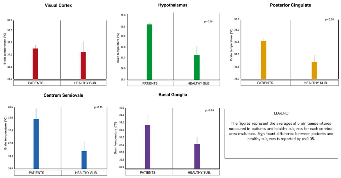

References:
552
BRAIN-0137
Poster Session
ESTABLISHMENT OF A METHOD FOR CREATING QUANTITATIVE CBF IMAGES OF OUTER AND INNER LAYERS OF THE BRAIN BY XENON-ENHANCED COMPUTED TOMOGRAPHY: APPLICATION TO DEMENTIA
Imaging Clinical
1R&D Division, Anzai Medical Co. Ltd., Tokyo, Japan
2Psychiatry, Sawa Hospital, Osaka, Japan
3The Director, Sawa Hospital, Osaka, Japan
Abstract

Seven patients with Alzheimer’s disease (AD) (77.7±9.8 years), ten patients with dementia with Lewy body (DLB) (79.4±5.0 years) and twenty healthy volunteers (73.7±6.7 years) underwent Xe-CT studies. We created the first (0-5 mm from the surface) to tenth (45-50 mm from the surface) layer images with right lateral, left lateral, superior, inferior, anterior and posterior views.
553
BRAIN-0300
Poster Session
CT PERFUSION AS AN INDEX TO ANTICIPATE INCREASE OF BLOOD SAMPLING OEF
Imaging Clinical
1Stroke Treatment, Shonan Kamakura General Hospital Stroke Center, Kamakura, Japan
Abstract
Objectives
It is important to anticipate the risk of cerebral hyperperfusion syndrome (CHS) before carotid artery stenting (CAS). CHS is likely to occur in patients with stage 2 hemodynamic failure, where oxygen extraction fraction (OEF) increases. However, OEF can not be measured in all institutions, because OEF is usually measured with PET. If some parameters of CT perfusion (CTP) widely used in many institutions have close relation to increase of OEF, it is beneficial for many institutions. In addition, OEF can be measured by blood sampling. The aim of our study was to investigate whether or not parameters of CTP had some relation to blood sampling global cerebral OEF (gcOEF) before or after CAS.
Methods
Included in our retrospective study were patients 1) who underwent elective CAS from January 2013 to December 2014, 2) who underwent CTP and blood sampling gcOEF before and after CAS. Excluded from the study were patients who underwent CAS within 30 days after last ischemic stroke. Evaluated were the relationship between gcOEF before or after CAS, usual parameters of CTP (CBF, CBV, MTT, TTP) measured in the territories of the bilateral middle cerebral artery (MCA) and peak value (PV) derived from time-density curve. Comparing the affected side parameter with the contralateral one, CBF ratio was defined as CBFa divided by CBFc, CBV ratio as CBVa divided by CBVc, MTT delay as MTTa minus MTTc, TTP delay as TTPa minus TTPc, PV ratio as PVa divided by PVc, and PV/TTP ratio as PV/TTPa divided by PV/TTPc.
Results
Twenty-three patients were analyzed. Mean age was 74.5 (SD: 8.5) years old. Twenty were male. Median pre-CAS gcOEF, post-CAS gcOEF, CBF ratio, CBV ratio, MTT delay, TTP delay, PV ratio, and PV/TTP ratio was 0.40 (quartile: 0.46), 0.42 (quartile: 0.45), 0.96 (quartile: 0.87 - 1.03), 1.05 (quartile: 1.00 - 1.12), 0.24 (-0.06 - 0.43), 0.41 (0.18 - 1.07), 1.00 (0.92 - 1.11), and 0.96 (0.89 - 1.05), respectively. There is no significant linear correlation between any CTP parameters and pre-CAS gcOEF. However, PV/TTP ratio of less 0.8 seemed to have relation to increased pre-CAS gcOEF of 0.46 or more (p = 0.05, OR: 7.5). There is a significant inverse correlation between CBF ratio and post-CAS gcOEF (p < 0.05). Among 4 patients with CBF of less than 0.83, 3 patients had increased post-CAS gcOEF of 0.45 or more (p = 0.06, OR: 8.4).
Conclusions
Pre-CAS PV/TTP ratio of less than 0.80 had probable relation to increased pre-CAS gcOEF of 0.46 or more, and pre-CAS CBF ratio of less than 0.83 had relation to increased post-CAS gcOEF of 0.45 or more.
554
BRAIN-0829
Poster Session
A MULTI-CENTRE PRECLINICAL STUDY ON THE EFFECTIVENESS OF INTERLEUKIN-1 RECEPTOR ANTAGONIST IN STROKE
Translational Studies
1Faculty of Life Sciences, University of Manchester, Manchester, United Kingdom
2Department of Pharmacology, University of Complutense, Madrid, Spain
3Laboratory of Molecular Neuroendocrinology, Institute of Experimental Medicine, Budapest, Hungary
4A. I. Virtanen Institute for Molecular Sciences, University of Eastern Finland, Kuopio, Finland
5GIP Cyceron, University Caen Lower-Normandy, Caen, France
6Institute of Experimental and Clinical Pharmacology and Toxicology, University of Lubeck, Lubeck, Germany
7Division of Clinical Neuroscience, University of Nottingham, Nottingham, United Kingdom
Abstract
Objectives
To date translation of findings from preclinical research to new treatments for stroke has proved largely unsuccessful. Amongst several things, more rigorous testing of new drug candidates in different experimental models of stroke and initiation of preclinical cross-laboratory studies have been suggested as ways to improve translation. However, to our knowledge, no drugs currently in clinical stroke trials have been investigated in preclinical cross-laboratory studies. We have identified the cytokine interleukin 1 (IL-1) as a key mediator of neuronal injury and shown that the naturally occurring IL-1 receptor antagonist (IL-1Ra) markedly reduces brain injury in a number of different experimental models and our aim here was to further investigate the efficacy of IL-1Ra in different research laboratories across Europe.
All studies used male mice (three studies with BALB/C and three using C57BL/6) with four studies using transient models of occlusion (confirmed by blood-flow measurements by laser Doppler) and four using permanent models (confirmed visually). Studies used animals of either 2.5 months (six studies) or 10 months (two studies) of age. All studies used the same dosing regimen of 100mg/kg IL-1Ra at both 30 and 180 min post-occlusion or corresponding vehicle treatment. Treatment was administered via subcutaneous injection bar one study, which administered treatment intravenously. Lesion volumes and oedema were measured by histological staining (three studies) or MRI (three studies). All lesion volumes and oedema measured by staining methods were assessed 7 days-post occlusion while MRI measurements ranged from 1 to 28 days post-stroke. Functional outcome was assessed using the corner (four studies) test (one study), assessed up to 28 days posts-stroke. Data from a total of 241 experimental animals were retrieved. Data were transferred to the project’s coordinating centre in Manchester merged into a single Microsoft Excel sheet using common field names with one row per animal for analysis. Cochrane Review Manager (version 5.2) was used to analyse the effect of IL-1Ra treatment compared to vehicle on post-stroke outcomes.
IL-1Ra reduced lesion volume, assessed using histological staining and MRI: standardised mean difference -1.84 (95% CI -2.55 to -1.12, p=<0.0001) and -1.04 (95% CI -1.96 to -0.11, p=0.03). Neurological scores showed IL-1Ra treatment to reduce neurological deficit compared to vehicle overall (-1.79, 95% CI -2.44 to -1.13, p=<0.001). Subgroup analysis found IL-1Ra treatment to be beneficial at days 1 to 28 (p=<0.001), but not shortly after surgery (p=0.11). In the corner test overall analysis found IL-1Ra treatment to be beneficial (-0.92, 95% CI -1.25 to -0.59, p=<0.001). Analysis of subgroups found IL-1Ra at days 1 (p=0.001), 2 (p=<0.001) and 7 (p=0.01) to be beneficial after stroke but not at day 28 (p=0.34).
IL-1Ra treatment was beneficial overall in terms of reducing lesion damage, neurological deficit and improve functional outcomes after experimentally induced stroke in a specific sub-population of young to middle aged male mice. Furthermore we show that multi-centre preclinical studies are feasible, but require careful planning and a clearly defined experimental design.
555
BRAIN-0357
Poster Session
DEFICIENCY OF THE STROKE-RELEVANT HDAC9 GENE IS ATHEROPROTECTIVE
Translational Studies
1Institute for Stroke and Dementia Research (ISD), Klinikum der Universität München, Munich, Germany
2Department of Neuroimmunology, Max Planck Institute of Neurobiology, Martinsried, Germany
Abstract
Effects of HDAC9 on atherogenesis were analyzed in ApoE deficient mice (ApoE-/-), an established model for atherosclerosis. We investigated atherosclerotic plaque size and immune cell infiltration in 18- and 28-week-old ApoE-/- mice by histology and immunohistochemistry. In addition, leukocyte distribution was measured by flow cytometry of the blood and the spleen.
References:
556
BRAIN-0283
Poster Session
REPOSITIONING OF THE DRUG CBG000592 FOR TREATMENT OF ISCHEMIC STROKE.
Translational Studies
1Neurology, Hospital Clínico Universitario-Clinical Neurosciences Research Laboratory, A Coruña-Santiago de Compostel, Spain
2Pharmacy, Centro de Investigación en Medicina Molecular y Enfermedades Crónicas (CIMUS), A Coruña-Santiago de Compostel, Spain
Abstract
We have discovered that compounds like oxaloacetate are able to acts as an effective blood scavengers of glutamate; however, its administration in humans has potential serious limitations as the required effective dosage to induce significant effect corresponding to that used in rats might be toxic. With the aim to solve this limitation, we have performed a screening analysis of repositioning drugs with blood glutamate scavenging activity with the aim to find a human approved drug which can be test immediately in stroke patients.
557
BRAIN-0197
Poster Session
TRANSCRANIAL CHARACTERIZATION OF CEREBRAL BLOOD FLOW DYNAMICS DURING INDIVIDUAL OBSTRUCTIVE SLEEP APNEA EVENTS BY DIFFUSE OPTICS
Translational Studies
1Medical Optics Group, ICFO-Institut de Ciències Fotòniques, Castelldefels, Spain
2Department of Pneumology, Hospital de la Santa Creu i Sant Pau, BARCELONA, Spain
Abstract

Abstract
Average CBF changes during OAE for different apnea durations. Vertical lines show the start and the end of the apneas.
References
558
BRAIN-0756
Poster Session
CEREBRAL 5-HT RELEASE CORRELATES WITH PET MEASURES OF THE 5-HT2A RECEPTOR OCCUPANCY IN THE PIG BRAIN.
Translational Studies
1Neurobiology Research Unit, Copenhagen University Hospital Rigshospitalet, Copenhagen, Denmark
2The Laboratory of Psychiatry, Copenhagen University Hospital Rigshospitalet, Copenhagen, Denmark
3Center for Excellence in Translational Medicine, University of Tartu, Tartu, Estonia
4Research Center for Advanced Imaging, Hospital of Køge and Roskilde, Roskilde, Denmark
5Department of Clinical Physiology Nuclear Medicine and PET, Copenhagen University Hospital Rigshospitalet, Copenhagen, Denmark
6Department of Neurosurgery, Aarhus University Hospital, Aarhus, Denmark
7Department of Radiology, Copenhagen University Hospital Rigshospitalet, Copenhagen, Denmark
8Department of Neurosurgery, Copenhagen University Hospital Rigshospitalet, Copenhagen, Denmark
Abstract
Objectives
Several studies have investigated PET radioligands for their ability to be displaced by endogenously released 5-HT, but the outcomes in humans have been somewhat contradictory and have not supported findings in animal experiments1. Two recent studies in humans suggested that the PET radioligands [11C]CUMI-1012 and [11C]AZ104193693 may be sensitive to endogenous changes in 5-HT levels, but this is not supported by others4. Moreover, these two studies show an increase in binding rather than the expected decrease2,3. However, the apparently conflicting outcomes may be due to differences in how the pharmacological challenges affect synaptic 5-HT levels. The aim of the present study was to measure the pharmacological challenges’ effect on 5-HT by cerebral microdialysis and to correlate this to the occupancy of the 5-HT2A receptor agonist radioligand [11C]Cimbi-36. We measured pharmacologically induced changes in 5-HT levels in the medial prefrontal cortex (mPFC) in vivo in pigs and assessed simultaneously changes in [11C]Cimbi-365 binding.
Thirteen pigs were scanned for 90 minutes in a HRRT scanner with [11C]Cimbi-36 at baseline and after serotonergic challenges aimed to increase extracellular 5-HT levels. The pharmacological interventions were 2 mg/kg escitalopram (serotonin reuptake inhibitor), escitalopram + 1 mg/kg pindolol (5-HT1A autoreceptor agonist), 0.5 mg/kg fenfluramine (serotonin releaser) or saline.
The pigs had implanted microdialysis probes bilaterally in the mPFC (MRI verified) from which extracellular fluid were collected during PET scanning and analyzed off-line for 5-HT with HPLC. We made a correlation analysis between the peak 5-HT level relative to baseline and the 5-HT2A receptor occupancy.
In two pigs, pharmacological depletion of 5-HT was done by pre-treatment with para-chlorophenylalanine (pCPA) (5-HT synthesis inhibitor) that substantially reduces 5-HT in the brain. This served to confirm that the decreased [11C]Cimbi-36 binding was not caused by direct interaction between fenfluramine and the 5-HT2A-receptor.
The extracellular 5-HT level increased to 725% following 0.5 mg/kg fenfluramine, to 337% following 2 mg/kg escitalopram + 1 mg/kg pindolol and to 171% following escitalopram alone. In pCPA pre-treated pigs, the 5-HT level was significantly reduced (p<0.01, n=2). The 5-HT2A receptor occupancy as measured with [11C]Cimbi36 correlates significantly (p=0.002, n=13) with the changes in extracellular 5-HT levels in mPFC, as measured by microdialysis (figure 1).
We here show that 2 mg/kg escitalopram iv is associated with a smaller increase in 5-HT as compared to the other challenges and without any associated change in radioligand binding. The observed correlation between changes in the extracellular 5-HT level in the pig brain and the 5-HT2A receptor occupancy indicates that [11C]Cimbi-36 is sensitive to changes in endogenous 5-HT levels, but that is only detectable on a global brain level when the 5-HT release is sufficiently high. Differences in earlier studies may thus be ascribed to the efficacy of the pharmacological interventions to change interstitial brain 5-HT levels.

References
559
BRAIN-0652
Poster Session
MODIFICATION OF FUNCTIONAL CONNECTIVITY BY ACUPUNCTURE IN THE MOUSE BRAIN
Translational Studies
1Acupuncture and Meridian Science Research Center, Kyung Hee University, Seoul, Korea
Abstract
Purpose: To identify the scientific mechanism of acupuncture therapy fromperipheral to central, the molecular event at the acupuncture point in the skinlayer and the neural activity of the brain regions for the functionalconnectivity after acupuncture needling were investigated.
Methods: Acupuncturestimulation was performed on GB34 acupuncture point of C57BL/6 mice. Afteracupuncture stimulation, changes of proteins related to tissue deformation(Rho-kinase, ERM), neurotrophins (NT-3, BDNF, NGF), cell signaling (HSP27) and initiateimmune (PRDX1, transketolase) were assessed. To investigate the correlationbetween the molecular signaling, inhibitors of ERK, ROCK, TRPV1 and A1R weretreated before acupuncture needling, then the activation of p-ERK, p-ERM,p-HSP27, PRDX1 and transketolase in the skin layer was investigated. Next, toinvestigate the whole brain neural activity after acupuncture needling, c-Fosexpression in thirty brain regions of Cortex, Cerebral nuclei, Thalamus,Hypothalamus, Hippocampus, Midbrain and Medulla was investigated and partialleast squares (PLS) analysis and network generation was performed.
Results: After acupuncture stimulation, Rho-kinase, ERM, NT-3 and HSP27 wereup-regulated and BDNF, NGF, PRDX1 and transketolase were down-regulated in skintissues at acupuncture needling point. Then we found that ERK activation workedas a trigger molecule to produce local molecular signaling. After acupunctureneedling, c-Fos positive cells were significantly increased in the brainregions of cingulate cortex area 1 (Cg1), cingulate cortex area 2 (Cg2), primarysomatosensory cortex (S1), secondary motor cortex (M2), insular cortex (Insul),piriform cortex (Pir), nucleus of solitary tract (NTS), dorsomedialperiaqueductal gray (DMPAG) and lateral periaqueductal gray (LPAG) anddecreased in the brain region of paraventricular thalamic nucleus posterior(PV) and the field CA1 of hippocampus (CA1). And these changes were inhibitedby U0126 administration. Inter-regional correlations were significantly increasedafter acupuncture needling, and inhibited by U0126 administration. Among thebrain regions, RMg, ST-DM, CA1 and NTS were determined as hub regions.
Conclusion: In conclusion, acupuncture-induced ERK expression at acupunctureneedling point plays a trigger role to acupuncture-induced cell signalingpathway, and it seem to play an important role in initiating central functionalconnectivity of acupuncture needling.
This research was supported by a grant of the Korean HealthTechnology R&D Project, Ministry of Health and Welfare, Republic of Korea(No. HI13C0540).
560
BRAIN-0698
Poster Session
MULTI-PART SURVEY: THE RISK OF BIAS IN IN VIVO STROKE RESEARCH
Translational Studies
1Centre for Clinical Brain Sciences, Univeristy of Edinburgh, Edinburgh, United Kingdom
2Faculty of Life Sciences, University of Manchester, Manchester, United Kingdom
3Division of Stroke Medicine, University of Nottingham, Nottingham, United Kingdom
4Department of Experimental Neurology, Charité Universitätsmedizin, Berlin, Germany
5Stroke Division, The Florey Institute of Neuroscience and Mental Health, Melborune, Australia
6Wellcome Surgical Institute, University of Glasgow, Glasgow, United Kingdom
7Neurovascular Research Laboratory, Vall d'Hebron Research Institute, Barcelona, Spain
8Experimental Design Analysis and Reporting, National Centre for the Replacement Refinement and Reduction of Animals in Research, London, United Kingdom
9Cerebrovascular Research team, Spanish National Research Council, Barcelona, Spain
10Department of Neurology Rudolf Magnus Institute of Neuroscience, University Medical Center Utrecht, Utrecht, Netherlands
11INSERM U919 unit, University Caen Lower Normandy, Caen, France
12Animal Welfare Division, University of Bern, Bern, Switzerland
Abstract
Objectives: The
As part of developing a framework for multicentre animal studies we performed a survey of in vivo stroke scientists. Our aim was to ascertain current practice, including measures to reduce the risk of bias, to inform the central coordination of multicentre animal studies.
Methods: We conducted an online survey in July 2014 of in vivo ischaemic stroke scientists identified through membership of the Multi-PART consortium or those that have expressed interest in the consortium. We asked respondents to state whether they randomly allocate animals to treatment groups and if applicable, the method used to perform randomisation. We also asked respondents if they blinded the conduct of surgery, animal handling, and assessment of infarct volume and behavioural deficits. Separately, we assessed the reporting of randomisation, allocation concealment and blinded assessment of outcome in in vivo stroke studies identified in systematic reviews, published after 2010 and curated in the CAMARADES database (www.camarades.info).
Results: We invited 59 laboratories to participate in the survey of which 32 (54%) responded; 23 were European-based laboratories and nine from the rest of the world. Of these, 29 (91%) stated that they randomly allocate animals to treatment groups. However, 6 of these ‘picked animals randomly from the cage’ or used ‘alternate allocation’, not considered true randomisation. Most (25) respondents induced focal ischaemia blinded to treatment allocation and of these 90% maintained blinding for the duration of the experiment. All respondents blinded the assessment of infarct volume and behavioural deficits.
Of 80 experiments in the CAMARADES database published after 2010, 23% reported allocation concealment, 36% reported randomisation and 44% reported blinded assessment of outcome.
Conclusions: Self-reporting of measures to reduce the risk of bias in leading stroke laboratories was substantially higher than in published reports. We purport that either (i) those responding to our survey may not be typical of the in vivo stroke scientist population, (ii) published reports do not adequately reflect experimental conduct and may actually underestimate the measures taken to reduce the risk of bias or (iii) reporting guidelines and increased awareness of the impact of bias has improved the quality of preclinical stroke research. In addition, our survey raised concerns of different interpretations of what is meant by randomisation. Protocols for multicentre animal studies must therefore take care to ensure that all aspects of experimental design are clearly defined and, where appropriate, provide training to address this.
The authors acknowledge support from the European Commission Seventh Framework Programme Coordination and Support Action, Grant Agreement number: 603043
561
BRAIN-0619
Poster Session
MULTI-PART BETA-TEST: TESTING THE FEASIBILITY AND FUNCTIONALITY OF MULTICENTRE PRECLINICAL ANIMAL STUDIES
Translational Studies
1Life Sciences, University of Nottingham, Nottingham, United Kingdom
2Faculty of Life Sciences, University of Manchester, Manchester, United Kingdom
3Division of Clinical Neuroscience, University of Nottingham, Nottingham, United Kingdom
4Center for Stroke Research, Charité Universitätsmedizin, Berlin, Germany
5The Florey Institute of Neuroscience and Mental Health, University of Melbourne, Melbourne, Australia
6Department of Clinical Neurosciences, University of Edinburgh, Edinburgh, United Kingdom
7Centre for Stroke and Brain Imaging Research, University of Glasgow, Glasgow, United Kingdom
8Neurovascular Research Group, Vall d'Hebron Research Institute, Barcelona, Spain
9NC3Rs, Gibbs Building, London, United Kingdom
10IIBB Department of Brain Ischemia and Neurodegeneration, Consejo Superior de Investigaciones Científicas (CSIC), Barcelona, Spain
11Department of Neurology and Neurosurgery, University Medical Center Utrecht, Utrecht, Netherlands
12INSERM UMR-S 919, Serine Proteases and Pathophysiology of the Neurovascular Unit, Caen, France
13Division of Animal Welfare, University of Bern, Bern, Switzerland
Abstract
Objectives
Multi-PART (
We are now undertaking a beta-test of this platform. We will use glyceryl trinitrate (GTN), a nitric oxide donor, in rodent models of ischaemic stroke. GTN has shown promise in patients with acute ischaemic stroke and will be further tested in the RIGHT 2 clinical trial.1,2
The aim of this beta-test is to establish the functionality, feasibility and effectiveness of this multicentre approach for preclinical testing.
Methods
We have designed an international multicentre preclinical trial to test GTN to treat stroke, across 10 participant sites from the UK, Spain, France, Germany, Australia and the USA. We will include two models of stroke: transient middle cerebral artery occlusion (MCAO) using an intraluminal filament and permanent distal (diathermy) MCAO, both in male C57BL/6 mice. A neuroscore and sensorimotor test will be used to assess functional recovery and 2,3,5-triphenyltetrazolium chloride (TTC) staining to quantify infarct volume at 48hrs post-MCAO.
Results
We will present the successes and hurdles of implementing a multicentre preclinical trial, Specifically, we will present the functionality and feasibility of the following components of the Multi-PART platform: (1) The ability to seek ethical approval from multiple regulatory authorities, (2) experimental design and sample size calculations, (3) centralised randomisation and blinding, (4) standard operating procedures, (5) centralised data collection and scoring, (6) a web-based data management system, and (7) statistical analysis and data monitoring systems.
Conclusions
Performing this beta-test will establish the functionality, feasibility and effectiveness of this multicentre approach for preclinical testing.
The authors acknowledge support from the European Commission Seventh Framework Programme Coordination and Support Action, Grant Agreement number: 603043.
References
562
BRAIN-0406
Poster Session
LOWERING ICP FOLLOWING STROKE: NK1 ANTAGONIST AS EFFECTIVE AS DECOMPRESSIVE SURGERY
Translational Studies
1Anatomy and Pathology, University of Adelaide, Adelaide, Australia
2Division of Health Sciences, University of South Australia, Adelaide, Australia
Abstract
563
BRAIN-0272
Poster Session
MULTIPLE PATHOLOGICAL CONDITION OF HYPERPERFUSION AFTER STA-MCA BYPASS FOR MOYAMOYA DISEASE
Clinical Studies
1Neurosurgery, National Cerebral and Cardiovascular Center, Osaka-Suita, Japan
Abstract
Objectives
Hyperperfusion was presumed to be a leading cause of postoperative transient neurological symptom after STA-MCA bypass for Moyamoya disease. We examined cerebral blood flow and metabolism of hyperperfusion by perioperative O15-gas PET retrospectively.
Among 35 patients (41 hemispheres) underwent STA-MCA bypass between April 2013 and April 2014, 13 patients (15 hemispheres) who underwent O15-gas PET prior to and after surgery were enrolled in this study. Preoperative PET was performed for all patients. Postoperative PET was indicated when transient neurological deficit appeared and/or postoperative SPECT showed hyperperfusion. For quantitative assessment of CBF, CMRO2, OEF and CBV, 1cm diameter of regions-of-interest delineated the area of increased CBF using Qview software. Postoperative quantitative values of each parameter were compared with those before operation.
CBF increased from 29.7 ml/100g/min before surgery to 52.67±18.1ml/100g/min after surgery except 1 case with hypoperfusion due to graft spasm. CMRO2 was unchanged from 2.9±0.9 ml/100g/min to 2.9±4.0 ml/100g/min. Change ratios of each parameter were categorized into 4 groups. Group1: increased CBF, unchanged CMRO2 (n=9, 60.0%). Group2: increased CBF, increased CMRO2 (n=2, 13.3%). Of these, 1 case revealed abnormal EEG. Group3: increased CBF, decreased CMRO2 (n=3, 20.0%). In the Group3, postoperative cerebral infarction occurred in 2 (66.7%) cases, showing vasogenic edema on MRI. Group4: decreased CBF, decreased CMRO2 (n=1, 6.6%). Repetitive MRA showed transient graft spasm at the time of PET scan.
Although the common cause of postoperative neurological symptom after STA-MCA bypass in patient with Moyamoya disease, hyperperfusion was categorized into 3 groups. Group1 was the most common type of hyperperfusion. It was suggested that multiple factors such as nonconvulsive status epilepticus and vasogenic edema may be involved in. Postoperative infarction occurred despite of increased CBF, suggesting that hypoperfusion was not the only factor of postoperative infarction.
References
564
BRAIN-0083
Poster Session
ISCHEMIC STROKE DURING TREATMENT WITH DABIGATRAN IS ASSOCIATED WITH DECREASED SEVERITY AND FAVORABLE PROGNOSIS
Clinical Studies
1Neurology and Cerebrovascular Medicine, International Medical Center Saitama Medical University, Hidaka, Japan
Abstract
Objectives: Anticoagulation therapy with warfarin is associated with a favorable prognosis in ischemic stroke (1). Dabigatran, a new oral anticoagulant, is widely used to prevent ischemic stroke in non-valvular atrial fibrillation (NVAF) patients (2). However, its association with decreased severity and favorable prognosis once ischemic stroke has occurred remain unknown. We therefore investigated whether dabigatran has a protective effect against ischemic stroke even if it occurs.
Methods: We retrospectively reviewed all patients with NVAF-associated ischemic stroke admitted to our hospital from April 2011 to December 2014, and included those who took dabigatran. We assessed whether patients were under regular use of the drug or tentative discontinuance, and classified them into 2 groups, treatment and discontinuation groups. Data on age, sex, ASCOD stroke phenotype, NVAF type (i.e., paroxysmal or persistent), comorbidities, CHADS2 score, National Institute of Health Stroke Scale (NIHSS) score on admission, modified Rankin scale (mRS) score at discharge, D-dimer, and BNP were investigated and compared between the groups.
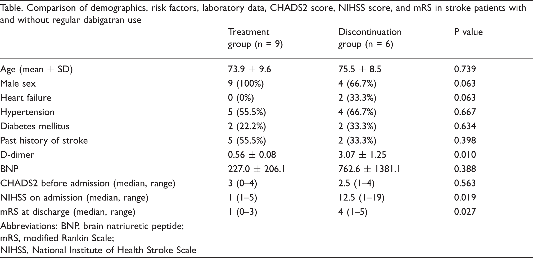
Abstract
Conclusions: Dabigatran is associated with decreased severity and a favorable prognosis even when ischemic stroke occurs. It was indicated that thrombi tended to be smaller in the treatment group.
References:
565
BRAIN-0746
Poster Session
USE OF DYNAMIC SUSCEPTIBILITY CONTRAST MAGNETIC RESONANCE IMAGING TO PREDICT THE TYPE OF STROKE AND EXTENT OF INFARCTION IN ACUTE ISCHEMIC STROKE
Clinical Studies
1neurosurgery, Suiseikai Kajikawa Hospital, Hiroshima, Japan
2neurosurgery, Tokyo Medical and Dental University, Tokyo, Japan
3neurology, Suiseikai Kajikawa Hospital, Hiroshima, Japan
Abstract
Background: Dynamic susceptibility contrast (DSC) MRI is the powerful tool to measure cerebral hemodynamics non-invasively and is widely used in the management of cerebrovascular disease. It has not been defined yet, however, whether DSC-MRI measured parameters can predict the tissue fate of ischemic brain in acute phase stroke. It also has not been well clarified whether the subtype of stroke can been distinguish by DSC-MRI measured parameters. As such information is inevitable to chose an appropriate treatment plan for acute stroke patients, we performed clinical study to clarify the role of DSC-MRI in acute stroke management.
Subjects and Methods: Thirty patients with cerebral infarction due to major vessel occlusion were included in this study. Patients were divided into 3 groups according to the subtype of stroke (atherothrombosis group, cardioembolism group, others group). All of the patients showed no recanalization for at least 3 days from onset confirmed by MRA. DSC-MRI was conducted within 24 hours of stroke onset and following parameters were calculated, referred to the counter region of the contralateral hemisphere as a control; time to peak (TTP), cerebral blood flow (CBF), cerebral blood volume (CBV), mean transit time (MTT), and time to maximum residue function (Tmax). DWI was also obtained at the same time with DSC-MRI and 3 days after onset, to identify the tissue fate of diffusion-perfusion mismatch area. The DSC parameters were compared among groups of stroke subtype, and between the areas with or without irreversible change. Parameters with a significant difference were assessed by receiver-operator characteristic (ROC) analysis to identify the optimal parameters and parameter thresholds for predicting the type of stroke and extent of infarction.
Results: There were 18, 10, and 2 patients included in atherothrombosis group, cardioembolism group, others group, respectively. Relationship between the subtype of stroke and DSC-MRI parameters were examined first. Atherothrombosis group showed significantly higher CBV ratio (CBV%) and CBF ratio (CBF%) than in cardioembolism group. When CBV% of 110% was used to distinguish between the two groups, the sensitivity was 0.966, the specificity was 0.724, and the area under the curve (AUC) was 0.904 (95%CI: 0.850-0.941)(Fig.1). Secondly we examined the fate of tissue in relation to DSC-MRI parameters in acute phase. In atheroscrelotic patients, on the other hand CBF% was the most useful parameters to predict the extent of infarction compared to other parameters [threshold 82.3%, sensitivity 0.800, specificity 0.941, and AUC 0.918 (0.749-0.977)](Fig.2). In cardioembolic stroke, a low CBV% [threshold of 87.8%, sensitivity of 0.963, specificity of 0.938, and AUC 0.968 (0.893-0.991)](Fig.3) was the most useful parameter.
Conclusions: CBV% was the most useful parameter to determine the subtype of stroke in acute ischemic stroke. CBF% was a useful parameter to predict the extent of infarction in atheroscrelotic patients. In cardioembolic stroke, CBV% was also useful to predict the fate of ischemic tissue. We concluded that DSC-MRI is a reliable method to distinguish the type of stroke and to predict the fate of ischemic tissue. Such information must be useful for the management of patients suffering from acute stroke.

566
BRAIN-0369
Poster Session
DIFFUSION TRACTOGRAPHY OF FACIAL NERVE IN CEREBELLO-PONTINE ANGLE TUMOURS
Clinical Studies
1Neurosurgery, Medical University of Gdansk, Gdansk, Poland
Abstract
Introduction. Preoperative imaging of the facial nerve (FN) in relation to cerebello-pontine angle (CPA) tumour would allow the surgeon to increase the safety of tumour resection. Tractography based on diffusion tensor imaging (DTI) is a novel method for visualisation of the white matter fibres and the cranial nerves.
Aim. To evaluate the correspondence of preoperative DTI with real FN position and assess the impact of FN imaging on reduction of facial paresis rate.
Material and methods. In 2013-2014, DTI with 3D modelling of CPA tumor was performed in 7 patients in the Neurosurgery Department of the Medical University of Gdańsk. Various factors were analysed, including age, gender, tumour size, its histopathology, and FN real and DTI location.
Results. Postoperative facial paresis was diagnosed in 3 patients (42.9%). In one patient, an eigenvector error of DTI occurred. The real position of FN corresponded with DTI only in 57% of patients. The sensitivity of 80% (95%CI:28.8-96.7%) was achieved. None of examined factors was significantly related to the agreement of the DTI with the real FN position. The correct prediction of FN location by means of DTI did not influence the reduction of postoperative facial paresis.
Conclusions. Contrary to some previous reports, sensitivity of DTI in CPA tumours has been found to be low in our study. Further prospective studies involving a greater number of subjects are required to establish a firm position of DTI in FN visualisation.
567
BRAIN-0371
Poster Session
QUANTIFICATION OF WHITE MATTER FIBRE PATHWAYS DISRUPTION IN FRONTAL TRANSCORTICAL APPROACH TO THE LATERAL VENTRICLE OR THE INTERVENTRICULAR FORAMEN IN DIFFUSION TENSOR TRACTOGRAPHY.
Clinical Studies
1Neurosurgery, Medical University of Gdansk, Gdansk, Poland
Abstract
Introduction: Pathologies occupying the interventricular foramen (foramen of Monro — FM) or the anterior part of lateral ventricle (LV) are accessed by the transcortical or transcallosal route. As severing of rostral corpus callosum has been deemed inferior to cortical incision, the approaches through various points of frontal lobe have been developed. Superior (F1), middle (F2) frontal gyrus or occasionally superior frontal sulcus are used as an entry of neurosurgical corridor. In spite of the fact that every approach to LV or FM causes its characteristic irreversible damage to white matter, to date all of transcortical routes are regarded as equivalent.
Methods: The current study compared the damage of main neural bundles between virtual trans-F1 and trans-F2 corridors by means of diffusion tensor tractography method (DTT) in 11 magnetic resonance imaging (MRI) exams from clinical series (22 hemispheres, regardless of dominance).
Results: Corpus callosum, cingulum, subdivisions I and II of superior longitudinal fasciculus (SLF I and SLF II), corticoreticular as well as pyramidal tracts crossing both approaches were subjected to surgical violation. Both approaches served a similar total number of fibres (0.94 to 1.78 [× 103]). Trans-F1 route caused significantly greater damage of total white matter volume (F1: 8.26 vs. F2: 7.16 mL), percentage of SLF I fibres (F1: 78.6% vs. F2: 28.6%) and cingulum (F1: 49.4% vs. F2: 10.6%), whereas trans-F2 route interrupted more corticoreticular fibres (F1: 4.5% vs. F2: 30.7%). Pyramidal tract (F1: 0.6% vs. F2: 1.3%) and SLF II (F1: 15.9% vs. F2: 26.2%) were marginally more vulnerable in case of the access via middle frontal gyrus. Both approaches destroyed 7% of callosal fibres. Conclusions: trans-F2 route disrupted a greater number of fibres from eloquent neural bundles (SLF II, pyramidal and corticoreticular tracts), therefore is regarded as inferior to trans-F1 one. Due to lack of up-to-date guidelines with recommendations of the approaches to LV or FM, an individual preoperative planning based on DTT should precede a surgery.
568
BRAIN-0830
Poster Session
EFFECT OF SILDENAFIL ON CEREBROVASCULAR REACTIVITY IN PATIENTS WITH BECKER MUSCULAR DYSTROPHY
Clinical Studies
1Functional Imaging Unit Department of Diagnostics, Glostrup Hospital, Glostrup, Denmark
2Neuromuscular Research Unit and Department of Neurology, Rigshospitalet, Copenhagen, Denmark
3Dept Neurology Lundbeck Foundation Centre Neurovascular Signalling, Glostrup Hospital, Glostrup, Denmark
4Neurovascular Research Unit Dept Neurology, Herlev Hospital and University of Copenhagen, Herlev, Denmark
Abstract
References
569
BRAIN-0159
Poster Session
CLINICAL CHARACTERISTICS OF CEREBRAL VENOUS THROMBOSIS IN A SINGLE CENTER IN KOREA
Clinical Studies
1Neurosurgery, Konkuk University Medical Center, Seoul, Korea
Abstract
Objective: The purpose of this study is to investigate the clinical characteristics of cerebral venous thrombosis (CVT) in a single center in Korea.
Methods: A total of 36 patients were diagnosed with CVT from August 2005 to May 2013. The patient data regarding age, sex, disease stage, pathogenesis, location, laboratory findings, radiological findings, and treatment modalities were retrospectively collected. The results were compared with those of previous studies in other countries.
Results: The patient group comprised 21 men and 15 women with a mean age of 46.9 years (ranging from three months to 77 years). The most common cause was a prothrombotic condition (8 patients, 22.2%). Within the patient group, 13 patients (36.1%) had a hemorrhagic infarction, whereas 23 (63.9%) had a venous infarction without hemorrhage. By location, the incidence of hemorrhagic infarction was the highest in the group with a transverse and/or sigmoid sinus thrombosis (n=9); however, the proportion of hemorrhagic infarction was higher in the cortical venous thrombosis group (75%) and the deep venous thrombosis group (100%). By pathogenesis, the incidence of hemorrhagic infarction was the highest in the prothrombotic group (n=6), which was statistically significant (p=0.016).
Conclusion: According to this study, CVT was more prevalent in men, and the peak age group comprised patients in the sixth decade. The most common cause was a prothrombotic condition. This finding was comparable with reports from Europe or America, in which CVT was more common in younger women. Hemorrhagic infarction was more common in the prothrombotic group (p=0.016) than in the non-prothrombotic group in this study.
570
BRAIN-0045
Poster Session
PREVENTIVE DETECTION OF AROU OF ACUTE STROKE PATIENTS
Clinical Studies
1ASU Medical & Geriatric, Princess Margaret Hospital, Hong Kong, Hong Kong China
Abstract
(2)(6)(21)(23)(30)(33), and the feasibility of an AROU assessment protocol with the use of ultrasonic bladder scan
(3)for early detection of AROU.
(4), National Institutes Heath Stroke Scales (NIHSS)(5) and Modify Barthel Index (MBI)(6). Following an in-house AROU assessment flowchart (
(3) was used to document the presence of urinary retention.
Abstract
Etiology of acute stroke patients


571
BRAIN-0748
Poster Session
MITOCHONDRIAL DNA POLYMERASEΓMUTATIONS IN CHINESE PATIENTS WITH MITOCHONDRIAL ENCEPHALOMYOPATHY
Clinical Studies
1Pathology Department, Affiliated Drum Tower Hospital of Nanjing University Medical School, Nanjing, China
2Neurology Department, Affiliated Drum Tower Hospital of Nanjing University Medical School, Nanjing, China
Abstract
Abstract

Reference:
572
BRAIN-0287
Poster Session
LOCAL RESISTANCE EXERCISE IMPROVES COGNITIVE EXECUTIVE FUNCTION IN A DOSE-DEPENDENT MANNER
Clinical Studies
1Graduate school of sport and health science, Ritsumeikan university, shiga, Japan
Abstract

References:
573
BRAIN-0821
Poster Session
COGNITION AND RESTING STATE CONNECTIVITY IN CHRONIC STROKE.
Clinical Stroke
1Physical Therapy, University of British Columbia, Vancouver, Canada
Abstract
Objectives
Although rehabilitation research tends to focus on motor function it is clear that cognition has a critical role in determining quality of life after stroke. Unlike motor deficits, which are often predicted from size and location of injury, cognitive deficits are more difficult to relate to specific lesions. Resting state functional MRI (rfMRI) allows the study of brain networks in the absence of task performance and can identify connectivity abnormalities. The current study assesses cognition and resting network activity, as identified by independent component analysis (ICA), in chronic stroke participants and healthy controls.
Methods
Neuropsychological assessment and magnetic resonance imaging, including several structural scans and 8 minutes of rfMRI, was obtained from controls (n = 7) and participants with chronic stroke (n = 7). The Montreal Cognitive Assessment was used to screen potential participants for cognitive impairment. Composite memory (California Verbal Learning Test, and Visual Reproduction Test) and non-memory (Blocks, Symbol Search, Word Association, Digit Span, and Trails) scores were compared between participants with stroke and controls; results are reported as a percentage of the max test score (mean ± SD). Standard preprocessing of rfMRI was completed in SPM 8. ICA was completed using SPM 8 based Group ICA toolbox (GIFT). Previously identified network templates were used to classify activation in the mean composite activity maps. Networks of interest were selected for further comparison between stroke and healthy control groups, specifically the default mode network (DMN), executive control network (ECN), middle frontal network (MFN), and sensory motor network (SMN).
Results
Stroke and healthy control groups were not significantly different in age (stroke, control: 67.0 ± 7.0, 62.1 ± 5.7; p = 0.179). Stroke participants were assessed at least 6 months after stroke (75.1 ± 44.3 months). The stroke and healthy groups were significantly different on the MoCA (stroke, control: 25.1 ± 1.9, 28.4 ± 1.3; p = 0.002). Stroke participants had significantly impaired memory (stroke, control: 64.6 ± 6.9, 75.4 ± 11.0; p = 0.048). However, non-memory cognition did not differ significantly between stroke and healthy controls (stroke, control: 62.1 ± 17.2, 73.7 ± 6.6; p = 0.123). Several differences existed between stroke and control participants within the resting state networks identified using ICA. Within the ECN healthy controls had greater frontal medial cortex connectivity than stroke participants (T = 6.26, p < 0.001). In the DMN healthy controls had more connectivity in the central opercular cortex than stroke participants (T = 6.83, p < 0.001). In the SMN healthy controls had greater connectivity than stroke participants in the middle temporal gyrus (T = 5.37, p < 0.001), left VI cerebellum (T = 5.33, p < 0.001), and the anterior cingulate (T = 4.77, p < 0.001).
Conclusions
In a relatively high functioning group of chronic stroke participants (i.e. independent community dwelling) memory is significantly impaired. Decreased connectivity in DMN and ECN may contribute to the cognitive deficits. Future analyses will examine the relationship between resting network activity, cognitive outcome and lesion size/location.
574
BRAIN-0788
Poster Session
THE RHEOLOGICAL PROPERTIES OF BLOOD IN ACUTE STAGE OF ISCHEMIC STROKE AND THEIR RELATION TO THE SEVERITY OF THE NEUROLOGICAL IMPAIRMENT
Clinical Stroke
1Neurology and neurosurgery, Siberian state medical university, Tomsk, Russia
2Laboratory of circulation and pharmacology, Institute of Pharmacology Russian Academy of Medical Sciences, Tomsk, Russia
Abstract

The study analyzed hemorheological parameters in the first hours after the onset of stroke in patients with different severity of disease. Patients in most acute phase of ischemic stroke had significant changes in hemorheological parameters (increased blood and plasma viscosity, plasma fibrinogen concentration, aggregation of erythrocytes) which can be described as hyperviscosity syndrome. The results showed that blood viscosity significantly different in groups with mild 5.3 [4.9; 5.8], moderate 6.1 [5.5, 6.6] and severe 6.8 [5, 7; 7.0] stroke at a shear rate of 50 s−1. Fibrinogen level was significantly different in the groups studied: mild 2.5 [2.2, 3.3], moderate 3.5 [3.1, 4.1] and severe 3.9 [3.7; 4.0] impairment. Patients with severe stroke was increased erythrocyte aggregation activity as evidenced by significantly shorter half-life of erythrocyte aggregation (T1/2 = 2.5 [1.8, 3.2] c) compared with patients with mild stroke (T1/2 = 5.8 [3.3, 7.7] c).
575
BRAIN-0274
Poster Session
STRESS AT WORK AND 16-TH YEARS RISK OF HYPERTENSION AND STROKE IN FEMALE POPULATION 25-64 YEARS IN RUSSIA: MONICA-PSYCHOSOCIAL EPIDEMIOLOGICAL STUDY
Clinical Stroke
1Collaborative laboratory of cardiovascular diseases epidemiology SB RAMS, FSBI Institute of Interna and Preventivel Medicine SB RAMS, Novosibirsk, Russia
Abstract
HR of stroke was 1.96-fold higher (95.0%CI:1.01-3.79, p<0.05) in women with job stress compared to those without stress. HR of AH over 16 years of follow-up in women with job stresswas 1.39-fold higher (95.0%CI:1.08-1.78, p=0.01) compared to those without it. There were tendencies of increasing AH and stroke in married women experienced stress at work compared to unmarried, divorced and widowed with the same stress level. AH significantly higher developed in women with university (χ2=8.23 df=1 p<0.01), college (χ2=3.98 df=1 p<0.05) and high school education (χ2=5.29 df=1 p<0.05) having job stress compared to those with elementary school education and stress at work. As the tendency there was a decline in stroke development in those with university education having job stress. With regard to occupational class higher AH rates was found for physical workers with job stress compared to pensioners without it (χ2=5.47df=1 p<0.05) and AH rates were tend to be higher in managers experienced stress at work (χ2=3.24 df=1 p=0.07). Stroke more likely developed in “physical workers” with stress at work.
576
BRAIN-0277
Poster Session
FAMILY STRESS AS PSYCHOSOCIAL PREDICTOR OF LONG-TERM RISK OF ARTERIAL HYPERTENSION AND STROKE IN FEMALE POPULATION 25-64 YEARS IN RUSSIA: MONICA-PSYCHOSOCIAL EPIDEMIOLOGICAL STUDY
Clinical Stroke
1Collaborative laboratory of cardiovascular diseases epidemiology SB RAMS, FSBI Institute of Internal and Preventive Medicine SB RAMS, Novosibirsk, Russia
Purpose: We studied the influence of family stress on risk of an arterial hypertension (AH) and stroke in female population aged of 25-64 years in Russia.
Abstract
There were tendencies of increasing AH and stroke rates in married women experienced stress in family. AH developed significantly higher in women with university and specialized secondary education compared to those having elementary school with (χ2=5.63 df=1 p<0.05; χ2=4.01 df=1 p<0.05, for university and specializedsecondary, respectively) or without stress at home (χ2=5.45 df=1 p<0.05; χ2=4.39 df=1 p<0.05, respectively). In relation to occupational class AH rates were higher in groups first-line manager (χ2=5.94 df=1 p<0.05) and physical worker (χ2=8.14 df=1p<0.01) experienced stress in family. Higher stroke rates were more likely in physical workers with family stress compared to those without it (p=0.055).
577
BRAIN-0319
Poster Session
TRANSCRANIAL OPTICAL MONITORING OF CEREBRAL HEMODYNAMICS DURING EARLY HOURS AFTER ISCHEMIC STROKE AND ITS RELATIONSHIP TO THREE MONTH OUTCOME
Clinical Stroke
1Medical Optics, ICFO-Institut de Ciències Fotòniques, Castelldefels, Spain
2Stroke Unit, Hospital Santa Creu i Sant Pau, BARCELONA, Spain
3Centre of Mathematics and Department of Mathematics and Applications, University of Minho, Braga, Portugal
4Department of Mathematics, Centre for Mathematical Research (CRM), Cendanyola, Spain
Abstract
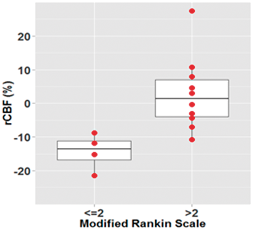
The project was funded by Fundació Cellex Barcelona, LlumMedBCN (La Caixa), Ministerio de Economía y Competitividad (PHOTOSTROKE) and LASERLAB-EUROPE.
References:
578
BRAIN-0846
Poster Session
CLINICAL RESULTS AND ANALYSIS OF OUTCOME REVASCULARIZATION PATTERN OF EXTENSIVE REVASCULARIZATION SURGERY (TODAI PROTOCOL) FOR MOYAMOYA DISEASE
Clinical Stroke
1Neurosurgery, The University of Tokyo, Tokyo, Japan
2Neurosurgery, Gunma University, Maebashi, Japan
Abstract
References
579
BRAIN-0104
Poster Session
A CASE OF SERPENTINE ICA ANEURYSM PRESENTED WITH VISUAL SYMPTOMS
Clinical Stroke
1Neurosurgery, Soonchunhyang University, Bucheon, Korea
Object: To present an interesting clinical manifestation by serpentine internal carotid artery aneurysm.
Abstract
Material and methods: 44 year-old female was visited with the decreased right visual acuity for one month. She complained the dry eye two weeks ago. Ophthalmologist diagnosed as a optic neuritis and transferred her to our hospital. Right visual acuity decreased to 0.04 (left is 1.0) and right visual filed showed defect 3/4 except right lateral upper quadrant in the neurologic examination. Brain MRI showed large aneurysm on right cavernous sinus and brain CT angiography showed fusiform dilatation of right cavernous ICA. Fundus photography showed right optic atrophy with pale neural rim on right optic disc. Similar finding was shown on transfemoral catheter angiography with brain MRI/brain CT angiography and the size was about 11.7mm*22.5mm. She passed Matas test and the collateral flow was tolerable. Vascular reserve was well maintained after Matas test on SPECT study.
Result: Coil embolization with parent artery occlusion was performed on the serpentine aneurysm. Patients didn’t show any neurologic deficit after coil embolization and visual acuity and flied defect were much improved at three month later.
Conclusion: Occlusion of aneurysm may effective for the improvement of clinical symptoms of the optic pathway compression.
580
BRAIN-0203
Poster Session
TREATMENT OF INTRACRANIAL ANEURYSMS, INCOMPLETELY-CLIPPED OR RECURRED AFTER CLIPPING
Clinical Stroke
1Neurosurgery, Korea University Ansan Hospital, Ansan-si, Korea
Abstract
Treatments to exclude aneurysm remnants of significant size identified following clipping or recurred aneurysms after clipping are often challenging to neurosurgeon. We report our experience of treatment residual or recurred aneurysms in a small series of surgically treated patients at a single institution.
Between July 2008 and June 2014, 10 patients with remnant or recurred aneurysms are surgically treated. Four aneurysms were recurred and six were remnant after clipping. Four patients were treated with repeated microsurgery and other six patients underwent endovascular coil embolizations.
Surgical morbidity happened in one patient with repeated microsurgical clipping. The endovascular coiling surgeries were successful for all six patients and the latest follow-up angiograms demonstrated no recurrence. There was no incidence for hemorrhage after treatment.
Repeated open surgery to recurred or remnant aneurysms after clipping are often technically difficult and may carry an increased risk of complications. Endovascular treatment of remnant or recurred cerebral aneurysm following prior surgical clipping can be accomplished with acceptable morbidity and mortality rates.
581
BRAIN-0401
Poster Session
THE EMBOLIC SOURCE BY DETECTING TRANSESOPHAGEAL ECHOCARDIOGRAPHY IN CRYPTOGENIC STROKE
Clinical Stroke
1Cerebrovascular Medicine, Steel Yawata Memorial Hospital, Kitakyushu, Japan
2Neurosurgery, Steel Yawata Memorial Hospital, Kitakyushu, Japan
Abstract
References:
582
BRAIN-0722
Poster Session
LOCATION OF THE CLOT, COLLATERAL SCORE AND CT ANGIOGRAPHY SOURCE IMAGES IN THE PREDICTION OF OUTCOME OF INTRAVENOUS THROMBOLYSIS
Clinical Stroke
1Medical Imaging Center, Tampere University Hospital, Tampere, Finland
2Department of Neurology, Oulu University Hospital, Oulu, Finland
3Department of Neurology, Vaasa Central Hospital, Vaasa, Finland
Abstract
References:
583
BRAIN-0468
Poster Session
A HYBRID SIGNAL PROCESSING OF RR INTERVAL FROM QTC VARIATION OF EKG AND HEART RATE VARIABILITY ASSESSMENT IN ATRIAL FIBRILLATION DURING ACUTE STROKE
Clinical Stroke
1Medical Engineering Program, Thammasat University, Pathumthani, Thailand
2Faculty of Medicine, Thammasat University, Pathumthani, Thailand
Abstract
Objectives: Abnormal QTc, tachycardia and arrhythmia with high risks for sudden cardiac death have been reported in stroke patients. Brain-heart axis is shifted affecting autonomic modulation in AF patient with stroke (1). An effective improvement of HRV analysis is needed to verify the complexity of autonomic modulation during acute stroke. Moreover, missing beats in silent AF, prolonged or shorten QTc or even ventricular tachycardia during acute stroke is still unknown mechanism . In this study, we develop a hybrid signal processing of Pan Tompkins QRS detection and Kalman Filter estimator for correction of missing beat during RR interval and then quantitate HRV by Kubios Program. We investigate autonomic changes whether QTc variation in AF stroke and non AF stroke patients.
Methods: 15 acute stroke patients with AF and those of 15 in non AF stroke (7 men, 8 women, age 65 ± 4 years old, matching control) were recruited. All subjects gave informed consent according to the Declaration of Medicine Ethic Committee (MTU-EC-IM-4-018/54). The Lead II ECG recordings (sampling rate of 1000 Hz, 5-minute long) were performed during 24 hours acute stroke, thereafter, RR intervals were analyzed by our software algorithms. AF and non AF with QTc variation in stroke patients were determined by physician. Two groups of acute stroke were assessed by HRV Kubios Program. All data in linear and nonlinear functions of HRV were analyzed.
Results: All values show significant difference between AF stroke and non AF stroke . Mean RR in AF stroke is less than those in non AF stroke. Greater parasympathetic activity is evident showing as pNN50, HF, and SD1. Decreasing sympatho-vagal balance is seen in lower level of LF/HF ratio in AF stroke. An irregularity of signals (SamEn and ApEn) in AF stroke is greater than in non AF stroke .
Conclusions: This finding suggests that a hybrid signal processing that we have developed for better RR detection is needed in QTc variation AF stroke and non AF stroke. Decreased HRV is shown in AF stroke clearly. Greater parasympathetic activity in AF stroke plays a vital role in variation of QTc.
References:
584
BRAIN-0429
Poster Session
RISK FACTORS AND OUTCOME IN STROKE PATIENTS WITH AORTIC COMPLICATED LESIONS
Clinical Stroke
1Stroke Center, Steel Memorial Yawata Hospital, Kitakyushu, Japan
Abstract
References:
585
BRAIN-0187
Poster Session
STROKE COMPLICATION ANALYSIS IN LEFT VENTRICULAR ASSIST DEVICE APPLICATION FOR DILATED CARDIOMYOPATHY PATIENTS
Clinical Stroke
1Neurosurgery, Tokyo Univesity, Tokyo, Japan
2Cardiac surgery, Tokyo Univesity, Tokyo, Japan
Abstract
Introduction:Stroke complications are common in patients with left ventricular assist devices (LVADs) and the complication affect the prognosis of them.
Methods:In this single center, retrospective study, LVAD surgeries were performed at our institution between March 2001 and March 2013. We enrolled 59 dilated cardiomyopathy cases (54 patients) without biventricular assist devices in our study. Clinical characteristics and laboratory data were analyzed. Stroke was including transient ischemic attack (TIA), and brain infarction or brain hemorrhage an imaging study.
Results:
Patients were followed-up for 472 ± 372 days. Stroke complications developed in 18 cases (TIA 1; ischemic cerebrovascular accidents 8, hemorrhagic cerebrovascular accidents 9) after LVAD implantation. Kaplan-Meier analysis revealed 1- and 2-year stroke complication rate was 31.3 and 37.6%. Multivariate analysis revealed preoperative statin administration (hazard ratio [HR] = 0.10; 95% confidence interval [CI] = 0.004-0.66; p = 0.01), preoperative albumin profile (HR = 0.35; CI = 0.13-0.92; p = 0.03) and postoperative sepsis (HR = 0.30; CI = 0.09-0.91; p = 0.03) were associated with stroke complications after LVAD surgery.
Conclusions:
We suggested preoperative statin, control of postoperative infection and improvement of nutritional status as the most important factors to reduce the risk of stroke after LVAD surgery.
586
BRAIN-0581
Poster Session
TARGETING RECOMBINANT THROMBOMODULIN FUSION PROTEIN TO RED BLOOD CELLS PROTECTS AGAINST STROKE
Cerebral Ischemia: Animal Models
1Department of Medicine Cardiology Cardiovascular Research Center, Massachusetts General Hospital, Charlestown, USA
2Department of Pharmacology Center for Targeted Therapeutics & Translational Nanomedicine ITMAT/CTSA Translational Research Center, University of Pennsylvania, Philadelphia, USA
Objectives
Abstract
Thrombomodulin (TM) is an endothelial glycoprotein that protects against thrombosis and inflammation in a domain-specific manner1. TM neutralizes cytokines via its N-terminal lectin-like domain, binds thrombin via its EGF-like domain, inhibiting its thrombotic and inflammatory effects, while facilitating thrombin’s cleavage of plasma protein C into activated protein C, a protease with anti-thrombotic and anti-inflammatory activities. We fused TM with a single chain fragment (scFv) of a monoclonal antibody to mouse red blood cells2. We hypothesized that red blood cells (RBC)-targeted scFv/TM will protects against ischemic stroke via anti-thrombotic and anti-inflammatory pathways.
The surgical procedure for the middle cerebral artery (MCA) occlusion (MCAO) was performed on adult male C57BL/6 mice under anesthesia (30% oxygen, 70% nitrous oxide, and 1.5% isoflurane). Body temperature was maintained at 36-37°C. The left common and internal carotid arteries were exposed through a neck incision and ligated. The external carotid artery was isolated and incised. Vehicle or scFv/TM solution (bolus, 200 microliters, 1mg/kg) was injected in right jugular vein 20 minutes before carotid arteries ligation. Silicon-covered nylon filament (Doccol) was advanced via the proximal external carotid artery into the internal carotid artery, occluding the MCA for 30 minutes with subsequent reperfusion for 48 hours as described3 with Laser Doppler cortical blood flow monitoring. Mice were examined for neurologic deficits by 5 point scale 48 hours after stroke. The brain was cut into 2-mm-thick coronal blocks, and stained in dark with 2,3,5-triphenyltetrazolium chloride for 1 h at 370C. The volume of infarct was calculated by indirect method as described4.
30 minutes of MCAO with 48 hours of reperfusion produced decreased cerebral infarct volume in mice injected with scFv/TM (28±20 mm3, Mean±SD, n=5) as compared with control (58±15, n=6, p< 0.05) mice. This was associated with a functional impairment in the neurologic deficit, as quantities by neurologic scoring (1.3 for mice injected with scFv/TM and 2.3 for control mice).
Stroke injury was less pronounced in mice pretreated with scFv/TM. Targeting recombinant thrombomodulin fusion protein to RBC resulted in decreased infarct volume and neurological deficit.
References
587
BRAIN-0387
Poster Session
MR-BASED T2’ MAPPING AS A SURROGATE FOR TISSUE HYPOXIA IN ACUTE ISCHEMIC STROKE: A VALIDATION STUDY AGAINST 18F-FLUOROMISONIDAZOLE (FMISO) PET IN RODENTS.
Cerebral Ischemia: Animal Models
1Department of Clinical Neurosciences, Stroke Research Group, Cambridge, United Kingdom
2Department of Clinical Neurosciences, Wolfson Brain Imaging Centre, Cambridge, United Kingdom
Abstract
BOLD imaging using T2’ may detect increased oxygen extraction fraction and as such is a potential candidate to map the penumbra in acute stroke [1]. T2’ has already yielded promising results in the clinical setting [2,3]; however, validation is still missing. Here we sought to validate T2’ imaging against FMISO, a PET tracer that provides maps of hypoxic tissue including the penumbra [4]. Specifically, we determined whether high FMISO uptake was present within T2’ lesions.
Five male Wistar rats were subjected to permanent thread MCAo, immediately followed by FMISO injection and MR (4.7 T Bruker) scanning. These sequences were repeated twice (time-points 1 and 2), interleaved with DWI. Additionally, DSC-MRI was performed in three rats. Following MR acquisition, the animal was transferred to a microPET P4 scanner (Concorde Microsystems). List-mode data were acquired from 120-150 min post-injection and measured attenuation correction was performed. Voxel data were converted to standardized uptake value (SUV) using the injected activity and animal weight. TTC staining was used at end of the protocol to map the final infarct.
T2’ was calculated voxel-wise from a multi-echo T2 and a multi-echo T2* according to:
SI(t) = SI0 e−t/qT2 or qT2*
Abstract
1/T2’ = 1/T2* - 1/T2 (with 1/T2’ = R2’)
T2’ and R2’ lesion ROIs were independently delineated by two observers on a slice-by-slice basis for both time points. Any disagreement was resolved in a consensus session. ADC lesion ROIs were automatically generated utilizing a threshold of 530*10−6 mm2/s.
There were no statistically significant differences in T2’ lesion volumes between observers 1 and 2. However mean ROI overlap between observers was 37% only. FMISO SUV was almost two-fold higher within the consensus T2’ lesion ROI compared to mirror ROI at both time-points (2-tailed p= 0.08 and 0.009, respectively). Overlap of the T2’ ROI with the ADC lesion ROI was small (≤ 10%). CBF and CBV were consistently lower and MTT and TTP higher within the T2’ ROI relative to mirror ROI at both time-points (p range: 0.27-0.01). Similar findings were obtained using the R2’ ROI. TTC staining showed an MCA infarct in each rat.
Poor inter-observer reproducibility indicates that T2’ and R2’ lesions are difficult to delineate, consistent with previous clinical data [2]. However, consensus ROIs do show the expected hypoxia and hypoperfusion characteristic of ischemic brain tissue. The small overlap with the ADC lesion is not inconsistent with T2’ reflecting mainly the penumbra given the former is expected to mainly show the ‘core’. These data suggest that the issue with T2’ imaging stems from voxel-based data processing to generate the images, which inherently creates noise due to non-linear equations. Whether this could be improved by optimizing image acquisition, image processing and/or experience of the observers should be addressed in further studies given the intrinsic validity of T2’ imaging.
References
588
BRAIN-0757
Poster Session
MIDDLE CEREBRAL ARTERY OCCLUSION WITH GOOD COLLATERALS CAUSES EARLY INTRACRANIAL PRESSURE ELEVATION POST STROKE
Cerebral Ischemia: Animal Models
1School of Biomedical Sciences and Pharmacy, University of Newcastle, Newcastle, Australia
Abstract
References:
589
BRAIN-0861
Poster Session
TREATMENT OF BRAIN ISCHEMIA BY RHEOLOGIC MODULATION OF BLOOD CIRCULATION USING DRAG REDUCING POLYMERS
Cerebral Ischemia: Animal Models
1Department of Neurosurgery, University of New Mexico School of Medicine, Albuquerque, NM, USA
2Department of Neurosurgery, Central South University, Changsha, China
3McGowan Institute for Regenerative Medicine University of Pittsburgh, Pittsburgh, PA, USA
Abstract
Objectives: Our objective was to study a previously unexplored approach for the treatment of cerebral ischemia after severe brain insults by the modulation of the rheology of blood flow by drag-reducing polymers (DRP). Nanomolar intravenous concentrations of linear, blood-soluble DRP have been shown to improve circulation and survival in animal models of the ischemic myocardium and limbs, but not yet in the brain. Using traumatic and ischemic brain injury models in rats, we examined the acute effects of DRP on the cerebral circulation and long-term recovery.
Methods: The suture permanent middle cerebral artery occlusion (pMCAO) was used as a model of ischemic injury and fluid percussion with a gas-driven device (1.5 ATA, 50 ms pulse) as a model of traumatic brain injury (TBI) in rat. Using in vivo 2-photon laser scanning microscopy we studied the acute effects of DRP (high MW polyethylene glycol) on microvascular blood flow velocity, tissue oxygenation (NADH) and blood brain barrier permeability (BBB) in the rat parietal cortex. Cortical Doppler flux, temperature, arterial pressure and blood gases and electrolytes were monitored. DRP (2 µg/ml in blood) was injected i.v. after baseline and post-sham/injury recordings. Controls were injected with saline. Neurodegeneration was evaluated by Fluoro-Jade at 24 hours and neurological motor deficit by Rotarod at 1 week after the injury.
Results: TBI/pMCAO progressively decreased microvascular flow in peri-injury/penumbra zones, induced hypoxia and BBB damage leading to neurodegeneration and neurological deficit. DRP significantly increased arteriolar blood flow volume and re-recruited collapsed capillaries (24±6.7 and 31±7.1%, p<0.05, n=10), reducing ischemia (by 16±4.6 vs 17±6.5%) and BBB damage (by 18±3.9 vs 15±5.5%), respectively (p<0.05), all data presented as Mean±SEM. Arteriolar blood flow velocity profiles, obtained by line scans at different distances from the vessel midline, revealed that DRP drastically changed blood flow velocity profile increasing near-wall velocity which resulted in a significant increase in blood flow volume. The improved microvascular flow decreased neurodegeneration at 24 hours after insults by 44.2±12.6% (pMCAO) and 37.6±11.2% (TBI), p<0.05. Rotarod tests showed that one week after insults, DRP treated rats performed better than controls with times of 75.6±16.0% vs. 47.3±15.2% (TBI) and 78.2±19.0%vs. 51.4±17.3% (pMCAO), respectively, as percent of baseline.
Conclusions: DRP improved cerebral microvascular perfusion and tissue oxygenation in both models of acute ischemia and traumatic injury resulting in improved long-term neurologic recovery. DRP increased blood flow velocity and concentration of erythrocytes near the wall of arterioles along with the previously demonstrated increase in the precapillary pressure, thereby increasing blood volume flow and the number of erythrocytes entering capillaries thus countering the effects of capillary stasis. Improved microvascular perfusion by DRP may be effectively used in the treatment ischemic stroke and traumatic brain injury.
590
BRAIN-0136
Poster Session
IN VIVO MONITORING OF CEREBRAL HEMODYNAMICS IN THE IMMATURE RAT: EFFECTS OF HYPOXIA-ISCHEMIA AND HYPOTHERMIA
Cerebral Ischemia: Animal Models
1Biomedical Engineering, Georgia Institute of Technology, Atlanta, USA
2Pediatrics/Newborn Medicine, Weill Cornell Medical School, New York, USA
3Radiology, Massachusetts General Hospital, Boston, USA
Abstract
Diffuse correlation spectroscopy was employed to quantify an average hemispheric index of cerebral blood flow (CBFi) in awake, un-anesthetized animals at baseline, pre-HI, and 0, 1, 2, 3, 4, 5, and 24 hours post-HI.
Following HI, an initial hyperemia was observed that was more prominent in the contralateral than ipsilateral hemisphere. This effect subsided within 5-10 minutes. After initiation of TH, CBFi in the HI-TH group dropped significantly below baseline levels to approximately 30% of baseline and remained depressed for the duration of TH. In contrast, during this same period, CBFi in the HI-N group was not significantly decreased from baseline levels (
Animals in the HI-TH group demonstrated significant neuroprotection relative to the HI-N group at both the 1- and 4-week post-HI study end points (p = 0.001). Reductions in CBFi after 4 hours of TH were not associated with reduced damage at 1 or 4 weeks. However, elevated ipsilateral CBFi and ipsilateral-to-contralateral CBFi ratios at 24 hours were associated with worse outcome at 1-week post-HI.
, Relative change in CBF (ΔrCBF) from baseline (a, c) and from post-ligation levels (b, d) for HI-N (solid circles) and HI-TH (open circles) animals.
591
BRAIN-0602
Poster Session
HIGH THROUGHPUT IDENTIFICATION AND QUANTIFICATION OF PERI-INFARCT DEPOLARIZATION IN MOUSE CORTEX WITH OPTICAL INTRINSIC SIGNAL IMAGING
Cerebral Ischemia: Animal Models
1Biomedical Engineering, Washington University in Saint Louis, Saint Louis, USA
2Neurology, Washington University in Saint Louis, Saint Louis, USA
3Radiology, Washington University in Saint Louis, Saint Louis, USA
Objectives
Abstract
Focal brain ischemia results in repetitive waves of electrophysiological hyperactivity followed by silence in surrounding cortex, a phenomenon known as peri-infarct depolarization (PID). Propagating at a rate of 2 – 6 mm/min [1], PIDs are hypothesized to exacerbate ischemia in the penumbra [2]. Despite the potential role that PIDs play in ischemic injury, their spatiotemporal propagation over the entire mouse cortex has not been well studied. We have developed an optical intrinsic signal (OIS) imaging system and image processing algorithms that enable high throughout identification of PIDs over majority of the mouse cortex. The automated and robust algorithm that has been developed is essential for analyzing the large datasets required to compare PIDs between different groups of mice.
Ten mice (male C57Bl6/J, 12-14 weeks old) were anesthetized with 0.75% isoflurane and imaged up to 6 hours during middle cerebral artery occlusion (MCAO). Light emitting diodes (LEDs) operating at four wavelengths (478 nm, 588 nm, 610 nm, and 625 nm) illuminated the skull, and diffuse reflected light was detected by an EMCCD camera. Images collected from light reflected at 478nm were most suitable for detecting and quantifying PIDs (Fig 1A). After mean-subtraction, PIDs were detected by applying several criteria to create an image mask over the trial: (1) an image intensity threshold, (2) a minimum image area threshold, and (3) a minimum distance of propagation threshold (Fig 1B). In addition to detecting PIDs, the algorithm also calculated the duration, source and sink, average velocity, and spatial trace of the PID. After the automated identification of PIDs, the user can inspect the intensity trace (Fig 1C), the spatial location of the mask over the trial (Fig 1D), and the trace of the PID (Fig 1D) to identify potential false positives and false negatives. The efficiency of the algorithm was determined by identifying PIDs manually and comparing these findings to the PIDs detected by the algorithm.
Our algorithm automatically detected PIDs that occurred after MCAO in mice. During the 50 hours of data analyzed, there was a total of 208 PIDs (4.16 PIDs/hour). The sensitivity and specificity of the algorithm was 0.93 and 0.97 respectively, and there was a false positive and false negative rate of 0.11 and 0.07. Using our data visualization output, a trained user can identify these false positives and negatives at around five times faster than manually watching all trials.
Our imaging system and processing algorithm efficiently identifies PIDs over mouse cortex, and provides quantitative information about the propagation of PIDs that cannot be calculated by manual inspection. This high-throughput approach to analyzing PIDs significantly reduces the need for manual user input. An exciting application of this system and detection algorithm is to identify differences in duration, number, or propagation of PIDs in mouse models of stroke. Drugs or genetic manipulations that reduce PIDs after MCAO could be tested for attenuation of ischemic brain injury.

References
592
BRAIN-0398
Poster Session
DIFFUSION MR ASSESSMENT OF SPATIOTEMPORAL EVOLUTION OF FOCAL CEREBRAL ISCHEMIA IN RATS: A NOVEL ROBUST METHOD VERSUS THRESHOLD METHOD
Cerebral Ischemia: Animal Models
1Radiology, Chungbuk National University Hospital, Cheongju-si, Korea
2Radiology, National Medical Center, Seoul, Korea
3Medical Research Institute, Chung-Ang University College of Medicine, Seoul, Korea
4National Primate Research Center, Korea Research Institute of Bioscience and Biotechnology, Cheongju-si, Korea
5Clinical Science, Philips Healthcare, Seoul, Korea
6Division of Magnetic Resonance Research, Korea Basic Science Institute, Cheongju-si, Korea
Abstract
PURPOSE
There are limitations which prevent accurate MR evaluation of spatiotemporal evolution of ADC-derived brain lesions after middle cerebral artery occlusion (MCAO) in rats. ADC values of a specific brain location differ from time to time even in a subject. Background noise, ischemic lesion, normal brain parenchyma and CSF have overlapping ADC values. This is to present our novel method for the spatiotemporal diffusion MR assessment of focal cerebral ischemia in rats after MCAO applying registration, noise/CSF removal, standardization and lesion segmentation in series.
MATERIALS AND METHODS
Immediately after right MCAO, 3T Diffusion MR scans were repeated every 10 minutes up to post-occlusion 50 minutes in 33 rats. Successful MCAO was confirmed in nine (9/33) and they were analyzed using both well-known threshold method and our robust method. Threshold method was made by removal of background noise (cut–off voxels below 150 x 10-6 mm2/sec) and CSF (above 1200 x 10-6 mm2/sec) signals. Modified method was made by following 3 steps (Fig 1): 1. Registration (registration of images with different time points and animals by 3dvolreg in AFNI) 2. Removal of background noise and CSF signals (Removal of background noise with brain mask and removal of CSF with CSF mask on minimal intensity map (Tmin map) by fslmaths within FSL) 3. ADC value standardization for relative ADC value (rADC) map (histogram standardization in contralateral normal left hemispheres by 3dROIstats of AFNI). To compare two methods, coefficients of variance (=standard deviation/mean) in normal left hemispheres were calculated. Lesion segmentation was performed with modified method (voxels below 90% of rADC) and then hemispheric lesion volumes (%HLV) and relative ADC values were calculated.
RESULTS
In every time points (Fig 2A) and in each animal (Fig 2B), coefficients of variation in normal contralateral left hemispheres were much lower with our robust method than those with threshold method respectively (p<0.001, respectively). ADC-derived abnormal lesions were developed on the first MR scan at 10 minutes
(mean %HLVs = 40.5±1.4%) and ADC values of the ischemic lesions were stable from the 1st MR during the time course for 50 minutes (mean= 69.9±1.7%) (Fig 3).
CONCLUSION
Using our novel robust method, we were able to assess spatiotemporal characteristics of focal cerebral ischemia in rats more reliably.
Steps of image processing in a robust method. Coefficient of variation (CV) in normal left hemispheres. A) CVs at different time points and B) CVs in different animals. Spatiotemporal evolution of ADC-derived hemispheric lesions volumes (%HLVs) and relative ADC values (rADCs).


593
BRAIN-0399
Poster Session
DIFFUSION LESION REVERSAL AFTER TRANSIENT MCAO IN NONHUMAN PRIMATE STROKE MODELS
Cerebral Ischemia: Animal Models
1Radiology, Chungbuk National University Hospital, Cheongju-si, Korea
2National Primate Research Center, Korea Research Institute of Bioscience and Biotechnology, Cheongju-si, Korea
3Medical Research Institute, Chung-Ang University College of Medicine, Seoul, Korea
4Radiology, National Medical Center, Seoul, Korea
5Clinical Science, Philips Healthcare, Seoul, Korea
Abstract
PURPOSEThough early diffusion lesion reversal (DLR) after recanalization treatment of acute ischemic stroke has been observed in clinical settings, still controversial is diffusion weighted imaging (DWI) lesions are truly reversible. This is to present our observation of sustained DLR after transient middle cerebral artery occlusion (MCAO)/reperfusion in nonhuman primate (NHP) models.
MATERIALS AND METHODSSeven rhesus monkeys were subjected to transient MCAO using endovascular technique to achieve focal cerebral ischemia and had been followed for 4 weeks after MCAO. Immediately after occlusion, prospective MRI scans were repeated every 10 ∼ 20 minutes. Occlusion was maintained until diffusion MRI showed plateau of lesion volume (peak volume) which was calculated by real time measurement technique using ImageJ (v1.46, NIH). Early DLR was defined as the ADC-derived lesion volume difference between peak volume and volume at 3 hours after reperfusion. We defined sustained DLR as voxels of acute ADC-derived lesion at the peak that corresponded to normal-looking brain on follow-up FLAIR MRI at 4 weeks. 3D volume analysis was used for quantitative analysis of ADC-derived lesion characteristics with AFNI software package (http://afni.nimh.nih.gov/afni/), FSL software (http://www.fmrib.ox.ac.uk/fsl) and ITKsnap (http://www.itksnap.org/pmwiki/pmwiki.php).
RESULTSPeak volume percentages of ADC-derived hemispheric lesions (%HLVs) were ranged 4.0 ∼ 21.7% (mean=10.3%). Hemispheric early DLRs were ranged 1.1 ∼ 10.5% and percentages of early DLR/Peak volume 9.9 ∼ 68.9% (mean=36.3±19.3%). Final %HLVs on FLAIR images at 4 weeks were between 0.9 and 11.0% (mean=4.8%). Sustained DLR was detected in all animals (9/9), and percentages of sustained DLR/Peak volume were 15.7 to 84.9% (mean=58.6±25.8%).
CONCLUSIONWe confirmed that sustained DLR as well as early reversal is persistent in all transient focal cerebral ischemia monkey models.
Percentages of early and sustained diffusion lesion reversal/peak volume in each animal. Representative images of diffusion lesion reversal (DLR) in animal 1. (Red, early DLR; Red+Orange, sustained DLR; White, final infarction)

594
BRAIN-0641
Poster Session
THE IMPACT OF PIAL COLLATERALS ON INFARCT GROWTH RATE IN EXPERIMENTAL ACUTE ISCHEMIC STROKE
Cerebral Ischemia: Animal Models
1Radiology, University of Chicago, Chicago, USA
2Radiology, Northwestern University, Chicago, USA
Abstract
Infarct volume growth over time. Each asymptotic funciton corresponds to one canine to derive the index of growth rate. The pial scores for each curve are listed on the left. Infarct growth rate index derived from fitted asymptotic functions has a linear relationship with pial collateral score and arterial arrival time.

Reference:
595
BRAIN-0400
Poster Session
PREFRONTAL CORTEX STROKE INDUCES DELAYED IMPAIRMENT IN SPATIAL MEMORY.
Cerebral Ischemia: Animal Models
1Anatomy, Otago Medical School, Dunedin, New Zealand
Abstract
596
BRAIN-0309
Poster Session
EFFECTS OF HYPERGLYCEMIA ON SEVERITY OF INITIAL CEREBRAL BLOOD FLOW DEFICIT, LESION GROWTH AND INFARCT SIZE INDUCED BY MIDDLE CEREBRAL ARTERY OCCLUSION IN RATS
Cerebral Ischemia: Animal Models
1Institute of Neuroscience & Psychology, University of Glasgow, Glasgow, United Kingdom
Abstract

Abstract
Mean ± SD. Blood glucose: 1h after MCAO (mmol/L). PDV, infarct volume, ADC lesion (mm3). *P<0.05 vs vehicle, unpaired t test; † P<0.05 vs vehicle, 2-way ANOVA with Bonferroni post-test.
597
BRAIN-0148
Poster Session
INTRANASAL DELIVERY OF GRANULOCYTE COLONY-STIMULATING FACTOR ENHANCES ITS NEUROPROTECTIVE EFFECTS AGAINST ISCHEMIC BRAIN INJURY IN RATS
Cerebral Ischemia: Animal Models
1Key Lab of Cerebral Microcirculation in Universities of Shandong, Taishan Medical University, Taian, China
2Taishan Vocational College of Nursing, Taian Shandong Province 271000 China, Taian, China
Abstract

References:
598
BRAIN-0040
Poster Session
EFFECT OF GABAB RECEPTOR ANTAGONISTS (CGP FAMILY) ON LEARNING AND MEMORY FORMATION IN ALBINO MICE FOLLOWING NEONATAL HYPOXIA ISCHEMIA INSULT.
Cerebral Ischemia: Animal Models
1Institute of Pure and applied biology, Bahauddin Zakariya University Multan Pakistan, Multan, Pakistan
2Institute of Molecular Biology and Biotechnology, Bahauddin Zakariya University Multan Pakistan, Multan, Pakistan
Abstract
599
BRAIN-0753
Poster Session
ROLE OF RENEXIN, A MIXED COMPOUND OF GINKGO BILOBA EXTRACT AND CILOSTAZOL, FOR SYNAPTIC PLASTICITY IN HIPPOCAMPUS OF RAT MODEL OF CHRONIC CEREBRAL HYPOPERFUSION
Cerebral Ischemia: Animal Models
1Neurology, Wonkwang University Hospital and Jeonbuk Regional Cardio Cerebrovascular Center, Iksan, Korea
2Physiology, Wonkwang University School of Medicine and Brain Research Institute at Wonkwang University, Iksan, Korea
Abstract
The impairment of cognitive function such as memory and attention has been recognized in patients with chronic mild cerebral ischemia. The hippocampal memory and long-term potentiation (LTP), a cellular correlate of learning and memory, were impaired by chronic cerebral hypoperfusion induced by permanent, bilateral occlusion of the common carotidartery (2VO). Recent animal study reported that renexin, a mixed compound of ginkgo biloba extract and cilostazol improved the deficit of spatial memory andthe lesion of the white matter in the rat 2 VO model.
Objectives: The purpose of present study wasto evaluate the effects of renexin on the impaired LTP and expression of mRNA genes involving neuronal plasticity in the rat 2 VO model.
Methods: Adult male Sprague-Dawley rats were randomly divide three experimental groups into:1) Sham without 2VO, 2) 2VO+vehicle, 3) 2VO+renexin. The permanent ligation of bilateral common carotid arteries was performed to elicit chronically lower blood flow to the brain. Animals were treated with oral administration of renexin(gingko biloba 20 mg/kg/day + cilostazol 25 mg/kg/day) or vehicle every day from 1 day after surgery for 3 weeks. We recorded LTP induced by brief high frequency stimulation to the Schaffer collateral–CA1 pathway of the hippocampus under anesthesia in vivo. Two hours after induction of LTP, the animal was sacrificed and the hippocampus was isolated for quantitative detection of mRNA for BDNF, Arc, Egr-1 and CREB using quantitative real-time RT-PCR technique.
Results: In sham-operated control group, high frequency stimulation was sufficient to induce robust LTP (110 ± 15% of control field excitatory postsynaptic potential (fEPSP) slope; n =7). Animals of 2VO+vehicle group showed a highly significant deficit in LTP induction (15 ± 3%of control fEPSP slope; n= 7; P< 0.01) 3 weeks after 2VO. On the other hand, daily oral administration of renexin showed the marked preservation of LTP induction (98 ± 17% of control fEPSP slope; n =7; P<0.01). Other parameters of synaptic excitability such as baseline fEPSP slope and input-output relation showed no significant difference between the 2VO+vehicle and the 2VO+renexin group. On the real-time RT-PCR analysis, a significant reduction of CREB, Arc and BDNF mRNA expression was observed inthe hippocampus of 2VO+vehicle group compared with that of sham group (P< 0.01). In contrast, renexin treatment increased significantly the expression of these mRNAs in the hippocampus compared with that of 2VO+vehicle group (P< 0.05).
Conclusion: These results suggest that daily oral administration of renexin can ameliorate cognitive deficit through the preservation of synaptic plasticity on the level of neural circuit in rodent model of chronic cerebral hypoperfusion.
600
BRAIN-0128
Poster Session
SPREADING DEPOLARIZATIONS MEDIATE EXCITOTOXICITY IN THE DEVELOPMENT OF ACUTE CORTICAL LESIONS
Cerebral Ischemia: Animal Models
1Neurosurgery, University of Cincinnati, Cincinnati, USA
2Anatomy and Neurobiology, University of Kentucky, Lexington, USA
Abstract
Spreading depolarizations (SD) are mass depolarizations of neurons and astrocytes that occur spontaneously in acute brain injury and mediate time-dependent lesion growth. Glutamate excitotoxicity has also been extensively studied as a mechanism of neuronal injury, although its relevance to in vivo pathology remains unclear. Here we hypothesized that excitotoxicity in acute lesion development occurs only as a consequence of SD. Using glutamate-sensitive microelectrodes, we found that SD induced by KCl in normal rat cortex elicits increases in extracellular glutamate (11.6 ± 1.3 µM) that are synchronous with the onset, sustainment, and resolution of the extracellular direct-current shift of SD (Fig 1A,B). Inhibition of glutamate uptake with D,L-threo-β-benzyloxyaspartate (TBOA, 0.5 and 1 mM) significantly prolonged the duration of the direct-current shift (148% and 426%, respectively) and the glutamate increase (167% and 374%, respectively) in a dose-dependent manner (P<0.05) (Fig 1C). These prolonged events produced significant cortical lesions as indicated by Fluoro-Jade staining (P<0.05), while no lesions were observed after SD in control conditions or after cortical injection of 1 mM glutamate (extracellular increase: 243 ± 50.8 µM) or 0.5 mM TBOA (glutamate increase: 8.5 ± 1.6 µM) without SD.

We then used an embolic focal ischemia model to determine whether glutamate elevations occur independent of SD in the natural evolution of a cortical lesion. In both the ischemic core and penumbra, glutamate increased only in synchrony with anoxic terminal SD (6.1± 1.1 µM) and transient SDs (11.8 ± 2.4 µM), and not otherwise (Fig 2). Delayed terminal SDs were also observed in two animals at 98 and 150 min after ischemic onset and induced similar glutamate elevations. Durations of SDs and glutamate increases were significantly correlated in both normal and ischemic animals (P<0.05).

These data suggest that pathologically prolonged SDs are a required mechanism of acute cortical lesion development and that glutamate elevations and the mass electrochemical changes of SD and are merely different facets of the same pathophysiologic process.
601
BRAIN-0679
Poster Session
GENDER AND AGE DIFFERENCES IN HISTOLOGICAL INJURY FOLLOWING THE CONTROLLED CORTICAL IMPACT MODEL OF TRAUMATIC BRAIN INJURY IN MICE
Cerebral Ischemia: Animal Models
1Anesthesiology, University of Colorado School of Medicine, Aurora, USA
Abstract
602
BRAIN-0559
Poster Session
NEUROPHYSIOLOGICAL RESPONSES OF RECOVERY IN PEDIATRIC MICE COMPARED TO ADULT MICE WITH TRANSIENT FOCAL CEREBRAL ISCHEMIA.
Cerebral Ischemia: Animal Models
1Anesthesiology, University of Colorado School of Medicine, Denver, USA
2Pediatrics, University of Colorado School of Medicine, Denver, USA
3Pharmacology/Anesthesiology, University of Colorado School of Medicine, Denver, USA
Abstract
603
BRAIN-0605
Poster Session
INHIBITED CAMKII ACTIVITY DECREASES HIPPOCAMPAL NEURONAL DAMAGE IN BOTH NORMOTHERMIC AND MILD THERAPEUTIC HYPOTHERMIC CA/CPR MOUSE MODEL
Cerebral Ischemia: Animal Models
1Pharmacology, University of Colorado, Denver, USA
Abstract
References:
604
BRAIN-0198
Poster Session
THE EARLY ELEVATION OF HIPPOCAMPAL BDNF BY EXERCISE AFTER CEREBRAL MICROEMBOLI REDUCES THE APOPTOSIS OF NEURON.
Cerebral Ischemia: Animal Models
1Physiology 2, Kawasaki Medical School, Kurashiki, Japan
2Rehabilitation, Kawasaki Univ Med Welfare, Kurashiki, Japan
Abstract
605
BRAIN-0780
Poster Session
BRUTON’S TYROSINE KINASE (BTK) IS AN ESSENTIAL COMPONENT FOR NLRP3 INFLAMMASOME ACTIVATION AND A POTENTIAL THERAPEUTIC TARGET FOR INFLAMMATION AFTER ISCHEMIC BRAIN INJURY
Cerebral Ischemia: Animal Models
1Microbiology and Immunology, Keio University School of Medicine, Tokyo, Japan
Abstract
Objectives
Among inflammatory cytokines, IL-1b has beenimplicated in post-ischemic inflammation after stroke. The multiple proteincomplex, inflammasome, is indispensable for converting pro-IL-1b to an activeform IL-1b p17. Thus, inhibition of inflammasome activation may remedy the stroke-inducedneurological dysfunction. In this study, we tried to find an inflammasomeinhibitor to reduce post-ischemic inflammation in the brain.
Methods
First, to select a pharmacological inhibitorthat suppresses NLRP3 inflammasome activation, we treated LPS-primed human andmurine macrophages with several candidate inhibitors, stimulated them with alumor ATP, and then determined caspase-1 activation and IL-1b secretion inmacrophages. Next, to evaluate the effects of the selected inhibitor on post-ischemicinflammation, we induced focal brain ischemia in mice by occluding the rightmiddle cerebral artery for 60 min and then intravenously administered theinhibitor.
We found that Bruton’s tyrosine kinase (BTK)inhibitors suppressed NLRP3 inflammasome activation in vitro. Importantly, BTK consisted of NLRP3 inflammasome by physicallyinteracting with both NLRP3 and apoptosis-associated speck-like proteincontaining a caspase-recruitments domain (ASC). The FDA-approved BTK inhibitor ibrutinib(PCI-32765) significantly suppressed infarct volume growth and neurologicaldamage in the brain ischemia model. Ibrutinib suppressed caspase-1 activation,thereby inhibiting IL-1b secretion of macrophages infiltrated in theinfarct lesion. These results revealed that BTK is essential for NLRP3inflammasome activation and could be a potent therapeutic target in stroke.
606
BRAIN-0609
Poster Session
THE ELECTRICAL THRESHOLD OF SPREADING DEPOLARIZATION IS REDUCED AND SPREADING ISCHEMIA IS PROLONGED IN SPONTANEOUSLY HYPERTENSIVE STROKE-PRONE RATS UNDER JAPANESE DIET
Cerebral Ischemia: Animal Models
1Dept. of Experimental Neurology, Charite University Medicine Berlin, Berlin, Germany
2Dept. of Physiology & Neurobiology, Ben-Gurion University of the Negav, Beer-Sheva, Israel
3Dept. of Medical Neuroscience, Dalhousie University, Halifax, Canada
4Center for Stroke Research Berlin (CSB), Charite University Medicin Berlin, Berlin, Germany
Abstract
References:
607
BRAIN-0129
Poster Session
RESVERATROL PRECONDITIONING INDUCES A NEW EXTENDED WINDOW OF ISCHEMIC TOLERANCE IN THE MOUSE BRAIN.
Cerebral Ischemia: Animal Models
1Neurology and Neurosurgery, University of Miami Miller School of Medicine, Miami, USA
2Human Genetics and Genomics, University of Miami Miller School of Medicine, Miami, USA
Abstract


References:
608
BRAIN-0729
Poster Session
KIR6.1 LIMITS PERI-INFARCT DEPOLARIZATION FREQUENCY DURING FOCAL CEREBRAL ISCHEMIA
Cerebral Ischemia: Animal Models
1Neurology, Washington University, St. Louis, USA
2Neurosurgery, Guangzhou First People’s Hospital, Guangzhou, China
3Cell Biology and Physiology, Washington University, St. Louis, USA
4Biomedical Engineering, Washington University, St. Louis, USA
5Radiology, Washington University, St. Louis, USA
6Radiology Physics Biomedical Engineering, Washington University, St. Louis, USA
7Neurology Radiology Biomedical Engineering, Washington University, St. Louis, USA
Abstract
Focal cerebral ischemia results in recurring spreading depression-like events initiated within the ischemic core, leading to peri-infarct depolarizations (PIDs) in the surrounding tissue [1]. PIDs are thought to exacerbate ischemia, expanding the core and further contributing to neuronal death [2]. Pathologic increases in extracellular potassium concentration due to anoxic K+ release play a critical role in PID initiation and propagation, but the molecular mechanisms driving PID phenomena are poorly understood. ATP-sensitive channels (KATP), composed of pore-forming (Kir6.1 or Kir6.2) and regulatory sulfonylurea receptor (SUR1 or SUR2) subunits, open under low ATP conditions (e.g. ischemia) and facilitate transmembrane K+ fluxes [3]. Both Kir6.1 and Kir6.2 are expressed in neural tissue, including neurons, astrocytes, and endothelial cells [3,4], and thus these channels may be critical for maintenance of extracellular K+ homeostasis during ischemia. We sought to determine the role of these channels in PID occurrence during ischemic injury.
Male mice (wild-type, Kir6.1-/-, or Kir6.2-/-, on a C57Bl6/J background) aged 12-14 weeks were anesthetized with isoflurane (2.5% induction, 1% maintenance) and middle cerebral artery occlusion (MCAO) was achieved by intra-arterial filament. The scalp was retracted and wide-field OIS imaging was performed through the intact skull for 4 hours. Light emitting diodes (LEDs) operating at four wavelengths (478 nm, 588 nm, 610 nm, and 625 nm) illuminated the skull, and diffuse reflected light was detected by an EMCCD camera running at 120Hz. PIDs were scored by manual viewing of data. PID frequency was compared between groups using ANOVA with repeated measures and Newman-Keuls multiple comparisons test.
MCAO in wild-type mice resulted in the initiation of PIDs 39±9 minutes into ischemia. PIDs continued to occur throughout the 4-hour imaging window at a consistent frequency of 2.8±1.0 PIDs/hr. In Kir6.1-/- mice, PID initiation was earlier (21±6 min vs. 39±9 min, p<0.05), and occurred with increased frequency compared to control mice (5.0±1.8 PIDs/hr vs. 2.8±1.0 PIDs/hr, p <0.05). In Kir6.2-/- mice, PID initiation time and frequency were unchanged compared to controls (34±14 min vs. 39±9 min, N.S.; 2.6±1.7 PIDs/hr vs. 2.8±1.0 PIDs/hr, N.S.).
These results demonstrate a critical role for Kir6.1 in limiting PIDs during ischemia. Given relevant activation of this channel during ischemia, it may play a role in removing excess extracellular K+. Previous studies have shown that Kir6.1 gene deletion results in increased infarct volumes [5], consistent with the idea that PIDs contribute to infarction. Kir6.1 and Kir6.2 exhibit different cell-type distributions [4], suggesting KATP channel activation in a specific cell type may be necessary for limiting PIDs. We are currently working to identify that cell type in order to better understand how KATP channels limit ischemic depolarization and neuronal death.
References:
609
BRAIN-0425
Poster Session
NO EFFECT OF CHRONIC PHOTOPERIOD DISRUPTION ON VULNERABILITY TO FOCAL CEREBRAL ISCHEMIA IN RATS
Cerebral Ischemia: Animal Models
1Institute of Neuroscience and Psychology, University of Glasgow, Glasgow, United Kingdom
2Institute of Biodiversity Animal Heath and Comparative Medicine, University of Glasgow, Glasgow, United Kingdom
Abstract

Reference:
610
BRAIN-0318
Poster Session
THE TRIALS AND TRIBULATIONS OF MODELLING STROKE IN AGED OBESE ANIMALS
Cerebral Ischemia: Animal Models
1School of Life Sciences, University of Nottingham, Nottingham, United Kingdom
2Division of Medical Sciences and Graduate Entry Medicine, University of Nottingham, Nottingham, United Kingdom
Abstract
Objectives:
Nearly 75% of all strokes occur in people over the age of 65. Despite this, animals used to model the condition are predominantly young and healthy (1). Common co-morbidities for stroke include hypertension (2) and diabetes (3), but these are rarely included in animal stroke models which currently possess little or no predictive validity. Some of these co-morbidities can be induced in aged animals by feeding a high fat diet (4, 5) and this study examines the feasibility of modelling stroke in aged, obese rats to develop a model that would better reflect the clinical situation.
12 month old Wistar-Han rats (500-650g, n=15) were maintained on a diet containing 60% fat for 8 months to induce obesity and an associated metabolic syndrome. A subset of these animals was used to determine the optimal duration of occlusion in the middle cerebral artery occlusion (MCAO) model of stroke. Remaining rats underwent this optimised procedure alongside 6-month old controls to determine effects of age and metabolic syndrome on post-stroke survival and functional deficits. Histology and blood vessel myography were performed at termination and infarct volume assessed. The development of a metabolic syndrome was analysed by serial blood samples, using multiplex ELISAs. This study was performed in accordance to the UK Animals (Scientific Procedure) Act 1986 under a project license from the UK Home Office.
In the 8 months prior to surgery rats gained a large amount of weight (750–1050g). Animals on the high fat diet did not become significantly hypertensive (106±6.2 vs. 96±5.7). However, blood samples indicated the presence of pre-diabetes and an altered inflammatory response. 30 minutes was determined to be the optimal occlusion length in this model. But full analysis of infarct volumes and vessel myography is still ongoing.
While modifications were required to provide the optimal care for these rats (including restraint, housing density and during surgery heating), this more translational, aged obese model can feasibly be used in stroke research.
References:
611
BRAIN-0107
Poster Session
RIVAROXABAN AND APIXABAN REDUCE HEMORRHAGIC COMPLICATION BY PROTECTION OF NEUROVASCULAR UNIT AFTER RECANALIZATION WITH TISSUE PLASMINOGEN ACTIVATOR IN ISCHEMIC STROKE OF RAT
Cerebral Ischemia: Animal Models
1Neurology, Okayama University, Okayama, Japan
Abstract
Objectives
This study aimed to assess the risk and benefit of tissue-type plasminogen activator treatment after oral anticoagulation with rivaroxaban or apixaban compared with warfarin or placebo.
Methods
Pretreatment with warfarin (0.2 mg/kg per day), rivaroxaban (2 mg/kg per day), apixaban (10 mg/kg per day), or vehicle (0.5% carboxymethyl cellulose sodium salt) was performed for 7 days. Transient middle cerebral artery occlusion was then induced for 120 minutes, followed by reperfusion with tissue-type plasminogen activator (10 mg/kg per 10 mL). Clinical parameters, including cerebral infarction volume, hemorrhagic volume, and blood coagulation, were examined. Twenty-four hours after reperfusion, markers for the neurovascular unit at the peri-ischemic lesion were immunohistochemically examined in brain sections, and matrix metalloproteinase-9 activity was measured by zymography.
The paraparesis score was significantly improved in the rivaroxaban-pretreated group compared with the warfarinpretreated group. Intracerebral hemorrhage was observed in the warfarin-pretreated group, and this was reduced in the rivaroxaban and apixaban-pretreated groups compared with the vehicle group. Marked dissociation of astrocyte foot processes and the basal lamina or pericytes was observed in the warfarin-pretreated group, and this was improved in the rivaroxaban and apixaban-pretreated groups. Furthermore, activation of matrix metalloproteinase-9 in the ipsilateral warfarin-pretreated brain was greatly reduced in rivaroxaban- and apixaban-pretreated rats.
This study shows a lower risk of intracerebral hemorrhage after tissue-type plasminogen activator treatment in rats with ischemic stroke that are pretreated with rivaroxaban and apixaban compared with pretreatment with warfarin. Reducing neurovascular dissociation by rivaroxaban and apixaban compared with warfarin could partly explain a reduction in hemorrhagic complications reported in clinical studies.
612
BRAIN-0110
Poster Session
PERICYTE PROTECTION BY EDARAVONE AFTER TPA TREATMENT IN RAT CEREBRAL ISCHEMIA
Cerebral Ischemia: Animal Models
1neurology, Okayama University Graduate School of Medicine Dentistry and Pharmaceutical Sciences, Okayama, Japan
Abstract
i) Background & Objectives: Pericytes play a pivotal role in contraction, mediating inflammation, and regulation of blood flow in the brain. In the present study, changes of pericytes in the neurovascular unit (NVU) were examined in relation to the effects of exogenous tissue plasminogen activator (tPA) and a free radical scavenger, edaravone.
ii) Methods: Immunohistochemistry and Western blot analyses showed that the overlap between PDGFRβ-positive pericytes and N-acetylglucosamine oligomers (NAGO)-positive endothelial cells increased significantly at 4 days after 90 min of transient middle cerebral artery occlusion (tMCAO).
iii) Results: The number of pericytes and the overlap with NAGO decreased with tPA, but recovered with edaravone 4 days after tMCAO with proliferation. Thus, tPA treatment damaged pericytes resulting in the detachment from astrocytes and a decrease in GDNF secretion. However, treatment with edaravone greatly improved tPA-induced damage to pericytes.
iv) Conclusion: The present study demonstrates that exogenous tPA strongly damages pericytes and destroys the integrity of the NVU, but edaravone treatment can extremely ameliorate such damage after acute cerebral ischemia in rats.
613
BRAIN-0257
Poster Session
MONONUCLEAR CELLS (MNCS) INCLUDING ENDOTHELIAL PROGENITOR CELLS (EPCS) PROTECT CEREBRAL ISCHEMIC DAMAGE ON MICE.
Cerebral Ischemia: Animal Models
1Neurology, Tokai University School of Medicine, Isehara, Japan
2Regenerative Medicine Science, Tokai University School of Medicine, Isehara, Japan
3Support Center for Medical Research and Education, Tokai University, Isehara, Japan
Abstract
ÐObjectivesÐ
EPCs were reported to enhance repairing andregenerating neurovascular units.[1, 2] So far many papers have reported repairing and regenerating experiments using EPCsderived from bone marrow, spleen, or peripheral blood. However, the results were not always satisfied. Recently, we succeeded to get MNCs including higher grade quality EPCs using a novel colony assay system which we have developed. [3] In the present study, we used this novel colony assay system, and evaluated effects with MNCs including EPC on ischemic stroke model in mice.
ÐMethodsÐ
We made 41 ischemic stroke model mice (10 weeks male C57BL/6 mice) with permanent middle cerebralartery occlusion (MCAO). From peripheral blood, we collected mononuclear cells, which was called peripheral blood MNCs (PB-MNCs) including primitiveEPCs, lymphocytes, and monocytes. For 5 days, we had cultured PB-MNCs, which were called quality and quantity MNCs (QQ-MNCs) including definitive EPCs, M2 macrophages and so on. We injected PBS as control, PB-MNCs,or QQ-MNCs into external carotid artery at 24 hours after MCAO. At 3 weeks after MCAO, we took the brains and investigated time-lapse physiological parameters including cerebral blood flow and immunohistochemistry against some antibodies related to vasculogenesis and inflammatory.
ÐResultsÐ
The stroke volume decreased with QQ-MNCs injected mice comparing to the control. The blood flows were no different between the control and other cell-injected mice. Positive cells with the antibodies related to vasculogenesis and inflammatory in cell-injected mice tended to increase comparing with the control.
ÐConclusionsÐ
Those results indicate QQ-MNCs possibly protected the brains from ischemic damage through thier immunity. Those QQ-MNCs could enhance repairing and regenerating neurovascular units after ischemic stroke.
ÐReferencesÐ
614
BRAIN-0366
Poster Session
AWAKE RECORDING OF PERI-INFARCT DEPOLARIZATIONS IN THE SPONTANEOUSLY HYPERTENSIVE RAT
Cerebral Ischemia: Animal Models
1Neurosurgery, Juntendo University, Tokyo, Japan
2Neurology, University of Tennessee Health Science Center, Memphis, USA
Abstract
Objectives – Peri-infarct depolarizations (PIDs) have pathophysiological significance in both experimental and clinical stroke. Anesthesia profoundly impacts PID incidence and propagation, yet awake animals have been monitored in only one prior study (1), which involved intraluminal filament occlusion in Sprague-Dawley rats, a strain with robust collateral perfusion. For comparison, the present study investigated PID number and time course after surgical middle cerebral artery occlusion (MCAO) in the more vulnerable Spontaneously Hypertensive Rat (SHR).
Methods – Male SHR were fitted with epidural electrode arrays under isoflurane anesthesia. After recovery intervals of 1 day to 5 weeks they were subjected to permanent or transient (2-hour) focal ischemia by tandem occlusion of the MCA and ipsilateral common carotid arteries (2), and PIDs were monitored for up to 3 days. An additional experiment examined the impact of maintaining anesthesia during the initial hours after MCAO. Brains were removed after recording and sectioned to determine infarct volume.
Results – Total PID number was minimally impacted by the time between electrode placement and occlusion surgery. Awake SHR exhibited 28 ± 9 PIDs (mean ± SD, n=27) during permanent occlusions, all within 4 hours following MCAO. Maintaining isoflurane anesthesia during this initial 4-hour interval significantly reduced total PID number (10 ± 4, n=6), and extended the time course for several more hours after recovery from anesthesia. A much more prolonged time course was observed through 36 hours after transient occlusions, but PID number during recirculation was highly variable (10 ± 9, n=8). PID number generally correlated with infarct size, but this relationship was dissociated by prolonged anesthesia, which reduced PID number without significantly impacting infarct size.
Conclusions – The short window of PID incidence after permanent MCAO in the SHR differs markedly from that previously described after filament occlusions in Sprague-Dawley rats (1). That study showed a biphasic time course through 24 hours after both permanent and transient occlusions, and total PID number was several-fold higher than in the SHR. Nevertheless, the present results appear to agree with a conclusion of the prior study that PID incidence correlates with the timing of infarct expansion, which progresses rapidly after permanent occlusions in the SHR. In addition, attenuated collateral perfusion and smaller penumbra volume in the SHR may reduce the probability of triggering PIDs, reducing their total number in this model. Maintained isoflurane anesthesia appears to delay recruitment of penumbra and prolong PID time course. Although without impact in the SHR, it might be predicted that such a mechanism could contribute to persistent protective effects of volatile anesthetics in strains with more robust collateral circulation.
Supported by USPHS grant R21-NS077039
615
BRAIN-0498
Poster Session
CADASIL MUTATIONS INCREASE STROKE VULNERABILITY
Cerebral Ischemia: Animal Models
1Radiology, Massachusetts General Hospital, Charlestown, USA
2Medicine, INSERM and Univ Paris Diderot, Paris, France
3Radiology and Neurology, Massachusetts General Hospital, Charlestown, USA
Abstract
Objectives:
Cerebral autosomal dominant arteriopathy with subcortical infarcts and leukoencephalopathy (CADASIL), linked to mutations in NOTCH3 expressed predominantly in vascular smooth muscle cells in adult brain, is characterized by progressive leukoaraiosis, and recurrent ischemic strokes in young or middle aged adults. Mechanisms underlying vascular dysfunction and stroke are unclear. We aimed to characterize the focal cerebral ischemia phenotype in transgenic mice expressing naturally occurring CADASIL mutations, to elucidate the mechanisms rendering the CADASIL brain vulnerable to stroke.
Methods:
We transiently (45min or 1h) occluded the middle cerebral artery by either an intraluminal filament proximally (fMCAO) or a microvascular clip distally (dMCAO) in two different CADASIL mutant mice (NOTCH3 R90C or R169C). We recorded cerebral blood flow (CBF) using laser Doppler (LDF) or speckle flowmetry (LSF), in each stroke model, respectively. We also studied cortical electrophysiology during acute stroke, tissue and neurological outcomes 24 hours after reperfusion, and gross cerebrovascular anatomy, including the diameter of major cerebral arteries, patency of posterior communicating artery and number of pial artery anastomoses. Mutants were compared with their age-matched wild-type (WT) littermates. Group sizes indicated below include WT controls of each strain.
Results:
Both R90C and R169C mutants developed significant larger infarcts (40% and 25% larger than WT, n=65 and 34, respectively, p<0.05) after filament MCAO. Outcome was not age-dependent, but appeared to be slightly worse in females. Surprisingly, neither LDF during filament MCAO nor LSF during distal MCAO showed worse CBF deficits (n=26 in R90C and n=20 in R169C), in either mutant. Cerebrovascular anatomy also did not differ between R90C mutant and WT (n=17). Interestingly, peri-infarct depolarization frequency was increased (1.5 fold) in the mutants during filament MCAO (p=0.07, n=13), a finding consistent with previous demonstration of increased susceptibility to spreading depression in non-ischemic CADASIL mutants (Eikermann-Haerter et al, Ann Neurol 2011). Presumably as a consequence of this, CBF threshold for tissue viability (figure) was significantly higher in R90C mutants compared with wild type (40±1% versus 33±2% of baseline CBF, respectively, p=0.032, n=14), suggesting increased sensitivity to ischemic injury.
Conclusions:
Our results show that outcome of focal cerebral ischemia is worse in CADASIL mutants independent of cerebral hemodynamic factors or collateral function, suggesting higher parenchymal sensitivity to ischemia possibly linked to frequent peri-infarct spreading depolarization events. The latter may be a reflection of increased susceptibility to spreading depression as a hyperexcitability phenotype in CADASIL.

616
BRAIN-0380
Poster Session
MOUSE CEREBELLAR PHOTOTHROMBOTIC STROKE MODEL
Cerebral Ischemia: Animal Models
1Translational Neuroscience Facility & Department of Physiology, UNSW Australia, Sydney, Australia
Abstract
Objective: Cerebellar strokes represent 2-3% of acute strokes worldwide, with a mortality rate 23% greater than strokes in other brain regions (Macdonell, Kalnins, & Donnan, 1987). The widely used small animal stroke models of middle cerebral artery occlusion, or global ischemia, have limited application for analysis of therapeutics directed at hindbrain strokes. Presently, hindbrain strokes in small animals involve microsphere-induced embolism via the internal carotid artery (Sekiguchi et al., 2005), or injection of autologous blood clots in the vertebral artery (Henninger et al., 2006), both of which are highly invasive and not temporally or spatially confined. Here we present a photothrombotic mouse model of cerebellar ischaemia to study neuroprotection for hindbrain strokes. We aim to determine the validity of the photothrombotic stroke model to produce a site-specific ischaemic brain injury in the cerebellum, by examining the progression of the cerebellar infarct via magnetic resonance imaging (MRI), histology and immunohistochemistry.
Methods: All experiments were approved by the UNSW animal care and ethics committee. Photothrombosis in the cerebellum was achieved by intravenous administration of rose bengal dye (40 mg/kg) followed by illumination at 561nm of the vermis region of the cerebellum in the anaesthetised mouse (129SvEv). Infarcts were examined by T2 weighted MRI (Bruker 9.4T) at days 1, 4, and 7 post-ischaemia. Following MRI, the tissue was paraffin embedded, sectioned, stained with haematoxylin and eosin (H&E), and immunostained with anti-glial fibriliary acidic protein (GFAP) antibody and anti-calbindin antibody to detect astrocytes and assess damage to Purkinje neurons respectively.
Results: The thrombus formation was determined in real-time using intravital multiphoton LSM to detect aggregation of fluorophore-conjugated anti-CD42 immunolabelled platelets. MRI scans in the days following the infarct showed development of oedema within the area delineated by the platelet labelling. This was well correlated with histological assessment of the infarct. H&E staining, and immunohistochemistry for GFAP and calbindin, delineated astrocyte infiltration, loss of Bergmann astrocyte processes, and a loss of Purkinje neurons.
Conclusion: The photothrombotic stroke model in the cerebellum is a robust model of focal ischaemia, which delivers thrombi to a specific and contained area; compared to the previous existing models of hindbrain strokes which are not spatially or temporally restrained. Hence, this model can aid investigations into novel therapeutic options targeting the hindbrain.
References:
617
BRAIN-0352
Poster Session
DELAYED INHIBITION OF VEGF SIGNALING AFTER STROKE ATTENUATES BLOOD BRAIN BARRIER BREAKDOWN AND IMPROVES FUNCTIONAL RECOVERY IN A CO-MORBIDITY DEPENDENT MANNER
Cerebral Ischemia: Animal Models
1Division of Medical Sciences, University of Victoria, Victoria, Canada
Abstract
Diabetes is a common comorbidity in stroke patients and a strong predictor of poor functional outcome. To provide a more mechanistic understanding of this clinically relevant problem, we focused on how diabetes affects blood brain barrier (BBB) function after stroke. Since the BBB can be compromised for days after stroke and thus further exacerbate ischemic injury, manipulating its function presents a unique opportunity for enhancing stroke recovery long after the window for thrombolytics has passed. Using a mouse model of type 1 diabetes, we discovered that ischemic stroke leads to an abnormal and persistent increase in vascular endothelial growth factor receptor 2 (VEGF-R2) expression in peri-infarct vascular networks. Correlating with this, BBB permeability was markedly increased in diabetic mice which could not be prevented with insulin treatment after stroke. Imaging of capillary ultrastructure revealed that BBB permeability was associated with an increase in endothelial transcytosis rather than a loss of tight junctions. Pharmacological inhibition (initiated 2.5 days post-stroke) or vascular-specific knockdown of VEGF-R2 after stroke attenuated BBB permeability, loss of synaptic structure in peri-infarct regions, and improved recovery of forepaw function. However, the beneficial effects of VEGF-R2 inhibition on stroke recovery were restricted to diabetic mice, and appeared to worsen BBB permeability in non-diabetic mice. Collectively, these results suggest that aberrant VEGF signaling and BBB dysfunction after stroke plays a crucial role in limiting functional recovery in an experimental model of diabetes. Furthermore, our data highlight the need to develop more personalized stroke treatments for a heterogeneous clinical population.
618
BRAIN-0570
Poster Session
VALIDATION OF AMIDE PROTON TRANSFER (APT) AS A MEASURE OF PH BY 31P MRS IN A PIGLET MODEL OF HYPOXIA ISCHEMIA ENCEPHALOPATHY (HIE)
Cerebral Ischemia: Animal Models
1Institute of Neurology, University College London, London, United Kingdom
2Medical Physics, University College London Hospitals, London, United Kingdom
3Institute for Woman Health, University College London, London, United Kingdom
4Center for Advance Biomedical Imaging, University College London, London, United Kingdom
Abstract
Hypoxia-ischemia (HI) in the newborn infant causes dramatic metabolic disturbances in the brain leading to alterations in tissue pH1,2. 31P MRS allows quantitative pH measurements; however this technique provides at best limited spatial information. Amide proton transfer (APT) has been shown to be sensitive to pH via the exchange rate between amide protons and water, thought to be base-catalysed3. Here, we aim at mapping regional pH changes in the piglet brain undergoing neonatal HI insult using APT.
Newborn piglets (n=16) were surgically prepared1,4 and HI was induced. 31P MRS was acquired at baseline, during, at 1 hour and at 24 hours post HI (pulse-acquire sequence, TR=10S). APT scans were also acquired pre-, 1 hour and 24 hours post HI (80 Gaussian pulses dur=50ms, FA=400°, 91% duty cycle, Turbo-Flash readout of TR=4.14ms, TE=2.09ms FA=10°, FOV=100x100mm2, matrix=128x128, thickness=4mm). Saturation frequencies: linearly spaced (77 points), range ±6ppm. The pHi values were quantified from 31P MRS2.The APT was calculated pixel by pixel as the difference between the Z-Spectrum and a linear interpolation from 3 to 4ppm. The sensitive volume of the 31P coil corresponded to the cortical watershed area of the brain. The APTp signal was averaged in this area, to match the source tissue of the pHi measurements for calibration purposes.
In animals with severe insult (n=7), the mean height of the APT peak was reduced at 1 hour post HI. Figure 1 shows the typical appearance of the Z-spectra pre and post HI. Figure 2 is a demonstration of pH-calibrated brain images where variations are clearly observed. This work demonstrates how regional brain pH can be mapped in the piglet brain undergoing HI. The disappearance of the amide peak following HI is most likely due to the amide exchange rate reduction caused by brain acidification. pHi confirms the trend seen with APT. This work highlights the advantage of spatial information of pH variations across the brain. APT has the potential to be used in the clinic for pH change mapping and assessment of treatment in newborn suffering from asphyxiation at birth.


References:
619
BRAIN-0849
Poster Session
CHARACTERIZATION OF A MODIFIED LONG TERM SURVIVAL MOUSE MODEL OF CEREBRAL ISCHEMIA
Cerebral Ischemia: Animal Models
1Anesthesiology, Duke University Medical Center, Durham, USA
2Neurosurgery, Tianjin 5th hospital, Tianjin, China
3Neurosurgery, 1st Hospital of Qinhuangdao City, Qinhuangdao City, China
4Anesthesiology, China-Japan Friendship hospital, Beijing, China
Abstract
Objectives: Current mouse models of focal cerebral ischemia present limitations for assessing long-term outcome due to a high mortality rate in the filament middle cerebral artery (MCA) occlusion (MCAO) model and insufficient behavioral deficit in the distal MCAO model. To meet the needs of preclinical stroke research, we modified a mouse cerebral ischemia model to enable long-term survival with persistent neurologic deficit.
Methods: 8-10 week old C57Bl/6J mice were anesthetized with isoflurane, intubated and ventilated. Rectal temperature was controlled at 37±0.2°C. An observer, blinded to group assignment, measured neurological deficit and infarct volume.1
Results: 
Conclusion: The tandem MCA+CCA occlusion mouse focal cerebral ischemia model allows long-term survival, tissue damage in both cortex and striatum, and persistent neurologic deficit. This model may be of use to investigators examining sustained efficacy of therapeutic intervention or delayed mechanistic responses to focal cerebral ischemia.
References:
620
BRAIN-0071
Poster Session
NEUROPROTECTIVE EFFECTS OF HUMAN NEURAL STEM CELLS OVER-EXPRESSING CHOLINE ACETYLTRANSFERASE IN A MIDDLE CEREBRAL ARTERY OCCLUSION MODEL
Cerebral Ischemia: Animal Models
1Veterinary medicine, Chungbuk National University, Cheongju, Korea
2Stem Cell R&D Center, iCellBank, Seoul, Korea
Abstract
Objectives. Ischemic stroke is one of the most-devastating brain diseases causing acute death or permanent disability. Although tissue-type plasminogen activator was approved by Food and Drug Administration for early reperfusion of the occluded vessels, oxidative injury may cause an extensive brain infarction. Accordingly, there is a need for effective neuroprotection during reperfusion, and stem cell-based therapeutic approaches should fulfill this requirement.
Methods. In the present study, we established human neural stem cells (NSCs) encoding gene of choline acetyltransferase (F3.ChAT), an acetylcholine-synthesizing enzyme, and investigated whether infusion of the F3.ChAT cells attenuate the ischemia-reperfusion brain damage in a middle cerebral artery occlusion (MCAO) model. After 2-hour occlusion with a silicone-coated thread, the middle cerebral artery of male Sprague-Dawley rats was reperfused, along with intravenous infusion of the stem cells (1?#152;106 cells/100 μl/rat). All the animal experiments were conducted according to the Standard Operation Procedures, and approved by the Institutional Animal Care and Use Committee of Chungbuk National University, Korea.
Results. In western blot analysis, F3.ChAT cells were found to produce much higher amounts of ChAT as well as neuroprotective and anti-inflammatory growth/neurotrophic factors including vascular endothelial growth factor, glial cell-derived neurotrophic factor, nerve growth factor, and ciliary neurotrophic factor than their parental F3 NSCs. Treatment with F3.ChAT cells markedly reduced the infarction volume and improved both the cognitive dysfunction (in water-maze trials) and behavioral deficits (in locomotor activity and rota-rod performance) of MCAO animals, in which F3.ChAT cells were superior to F3 cells. F3.ChAT cells not only restored microtubule-associated protein 2, a neuronal cytoskeletal protein, but also decreased pro-inflammatory interleukin-1β, glial fibrillary acidic protein, and intercellular adhesion molecule 1 in the brain tissue.
Conclusions. The results demonstrate that early intravenous infusion of NSCs expressing ChAT and growth/neurotrophic factors attenuate brain injury and restore neurobehavioral functions via neuroprotective and anti-inflammatory activities, and that F3.ChAT cells could be a candidate for the neuroprotection and functional recovery of acute stroke patients.
References.
621
BRAIN-0239
Poster Session
ISCHEMIA-INDUCED SPREADING DEPRESSION IN THE IN VIVO RETINA
Cerebral Ischemia: Animal Models
1Neuroscience, University of Minnesota, Minneapolis, USA
Abstract
Objective: To characterize retinal spreading depression (RSD) in acute branch retinal vessel occlusion and assess the contribution of RSD to tissue injury following ischemia.
Methods: Using a focal photothrombosis model of ischemia in the in vivo rat, a primary retinal vessel was occluded and the intrinsic optical signal from the retinal surface was imaged by confocal microscopy to detect RSD events. Blood flow, vessel diameter, tissue oxygen tension, or DC potential was monitored concurrently.
Results: RSD waves were generated following occlusion of a single primary arteriole or venule. Waves were typically initiated near the optic disc and spread peripherally, but waves were also initiated peripherally and spread centrally. Wave velocities range from 1.4 – 3.3 mm/min. The wave front was associated with a drop in tissue oxygen tension, constriction of primary arterioles, and a negative shift in DC potential.
Conclusions: We demonstrate, for the first time, that RSD waves are generated following occlusion of retinal vessels. The properties of in vivo RSD waves are similar to those of cortical spreading depression (CSD) waves. CSD is associated with delayed cell death following ischemic stroke and a similar RSD-mediated delayed cell death may occur in the retina following vessel occlusion.
References:
622
BRAIN-0502
Poster Session
NEUROPROTECTIVE EFFECTS OF THE INSULIN-LIKE GROWTH FACTOR IGF-I AND 17B-ESTRADIOL AFTER TRANSIENT FOCAL CEREBRAL ISCHEMIA IN THE ENDOTHELIN-1 RAT MODEL.
Cerebral Ischemia: Animal Models
1Experimental Pharmacology Center for Neurosciences, Vrije Universiteit Brussel, Brussels, Belgium
2Laboratory of Clinical Biology, UZ Brussel, Brussels, Belgium
3Department of Neurology Center for Neurosciences, UZ Brussel, Brussels, Belgium
Abstract
Introduction: Stroke is the second cause of death and the leading cause of adult disability worldwide (1-2). To date, thrombolytic therapy using recombinant tissue plasminogen activator (tPA) is the only approved therapy for acute ischemic stroke. However, only 5-10% of all patients benefit from this treatment because of the inclusion criteria (2). Targeting different processes involved in the ischemic cascade, which include excitotoxicity, oxidative stress, inflammation and cell death, could be promising for the future of stroke treatment. IGF-I is an anti-apoptotic pleiotropic factor exerting effects on different levels of the ischemic cascade (3). It has been shown in both in vitro and vivo models that the neuroprotective effects of the IGF-IR and ER are interdependent (4). This interdependency creates the possibility to increase the efficacy of IGF-I by co-treatment with estradiol.
Aim: Possible synergistic or additive effects of IGF-I and 17β-estradiol (E2) will be studied in the endothelin (Et)-1 rat model for stroke. Effects on infarct size motor/sensory deficits and glial activation will be investigated.
Material and methods: Via a guide cannula, implanted using stereotactic surgery, 200 pmol Et-1 was administered in the vicinity of the middle cerebral artery of male albino Wistar Kyoto (WKY) rats. Rats were treated with E2 (1mg/kg, s.c.) 30 min and/or IGF-I (1 or 3 mg/kg, s.c.) at 30 min or 2 h after the induction of stroke. Motor/sensory functions were determined 24 h after the insult using the neurological deficit score (NDS). Infarct size was assessed using a cresylviolet staining. To assess effects on microglial activation and astrogliosis immunohistochemistry was performed using antibodies directed against Iba-1 and GFAP.
Results: Rats treated with E2 (1 mg/kg) or IGF-I (1 and 3 mg/kg) alone showed smaller infarct sizes when compared to vehicle treated animals. Treatment with a combination of E2 and IGF-I lead to a more pronounced reduction in infarct size, only when a high dose of IGF-I (3 mg/kg) was used 30 min after stroke induction. This combination also increased the number of round shaped microglia in striatum. In contrast, the effects of 1 mg/kg IGF- I administered 2 h after stroke induction and E2 at t = 30 min were additive. Furthermore, stroke induction significantly reduced the NDS in vehicle-treated animals but not in rats treated with E2 and/or IGF-I
Conclusion: The neuroprotective effects of E2 and IGF-I are additive and a combination of these factors markedly reduces infarct size after a transient focal cerebral ischemia and increases the number of amoeboid shaped microglial cells in striatum. It remains to be established whether stimulation of microglial activation is instrumental to neuroprotection in our model.
References:
623
BRAIN-0339
Poster Session
A COMMON EXPERIMENTAL STROKE MODEL PRODUCES A BIMODAL PATTERN OF INJURY
Cerebral Ischemia: Animal Models
1School of Psychology, University of Leicester, Leicester, United Kingdom
2Centre for Core Biotechnology Services, University of Leicester, Leicester, United Kingdom
3Peninsula School of Medicine and Dentistry, University of Plymouth, Plymouth, United Kingdom
Abstract
Objectives:
Efficacy of potential stroke interventions are frequently tested using rodent focal ischemia models, however these have a poor record of detecting clinically useful drugs. Variability in lesion volume produced by transient middle cerebral artery occlusion (tMCAO) is high, whilst cost and ethical concerns result in small sample sizes. Generally, significance is tested using parametric analysis of mean lesion volume, an approach that requires a Gaussian distribution within each group. However, the small sample sizes used limit the statistical power of formal normality tests. The use of parametric analyses, if incorrect, may contribute to the poor discrimination of effective treatments affecting the progress of determining clinically viable treatments. To test the application of this approach the current study retrospectively evaluates lesion volume data distribution within 8 separate studies conducted over 10 years under identical protocols (by CL Gibson). In addition, we have begun to explore possible explanations for such a bimodal pattern of injury by focusing initially on whether such differences are attributed to variations in cerebrovascular anatomy.
Methods:
60min tMCAO was induced in male C57Bl/6j mice followed by post-mortem lesion volume analysis as detailed previously1. Osmotic mini-pumps filled with vehicle/drug were implanted 24hrs prior to MCAO, remaining in place for 2 days post-MCAO. Data was collated with lesion volume data from 8 control studies performed under identical surgical protocol over the last 10yrs. A further cohort of mice are undergoing pre-MCAO angiography MRI scans to assess vasculature patency, followed by post-MCAO T2 diffusion weighted MRI scans for lesion volume assessment in addition to post-mortem TTC staining.
Results:
Analysis of total lesion volume data within a recent study showed lesions centered on the striatum, encroachment into the cortex was only present in larger lesions. Correlation of striatal and cortical lesion volume per lesion showed no cortical component in lesions <40mm3. A single distribution of collated data from 8 identical control series spanning 10 years fitted two overlapping Gaussian curves (r2=0.95). Modified Shapiro-Wilk (Ryan-Joiner) analysis produced p<0.01 for these data, rejecting the hypothesis of normal distribution accepting the hypothesis of bimodal distribution of small (<40mm3) striatal and large (>40mm3) striato-cortical lesions.
Conclusions:
The observation that lesion volume within a single 60min tMCAO data set can be either small/striatal or large/striato-cortical is consistent with the presence of two normally distributed peaks in a larger data set collated over 10 years using an identical protocol. A similar bimodal lesion volume distribution is apparent in published data from several prior studies in various species and reversible ischemia models. Much of the variability within MCAO studies may be caused by anatomical variation within the Circle of Willis and the patency of the ipsilateral posterior communicating artery. Anatomical studies do not show correlations between cerebrovascular anatomy and lesion volume as anatomical investigation if often undertaken using non-MCAO mice. Using non-invasive MRI imaging techniques we can better determine the influence of cerebrovascular anatomy on lesion volume variation following MCAO.
References:
624
BRAIN-0642
Poster Session
ISCHEMIC INJURY PRODUCED BY PHOTOTHROMBOSIS DIFFERS IN NEONATAL AND ADULT BRAIN
Cerebral Ischemia: Animal Models
1Physiology and Pharmacology Clinical Neurosciences and Radiology, University of Calgary, Calgary, Canada
2Physiology and Pharmacology, University of Calgary, Calgary, Canada
3Experimental Imaging Centre, University of Calgary, Calgary, Canada
Abstract

625
BRAIN-0177
Poster Session
THE ROLE OF ENDOTHELIN RECEPTORS IN NEUROCHEMICAL CHANGES DURING FOCAL CEREBRAL ISCHEMIA INDUCED BY INTRAHIPPOCAMPAL INJECTION OF THE ENDOTHELIN-1 IN IMMATURE RATS
Cerebral Ischemia: Animal Models
1Department of Developmental Epileptology, Institute of Physiology v.v.i. Academy of Sciences of the Czech Republic, Prague, Czech Republic
2Department of Organic Technology, Institute of Chemical Technology, Prague, Czech Republic
3Department of Neurology University of Rochester Medical Center, Strong Epilepsy Center, Rochester, USA
4Department of Neurophysiology of Memory, Institute of Physiology v.v.i. Academy of Sciences of the Czech Republic, Prague, Czech Republic
Abstract
Cannula for drug infusion was stereotaxically implanted into the right dorsal hippocampus under isoflurane anesthesia. Microdialysis probe was implanted caudally in the close proximity of the infusion cannula and perfused with an artificial cerebrospinal fluid (1.5µl/min). Samples were taken sequentially during 2 hours after drug infusion, stored in -80°C and then analyzed by UHPLC-MS. During all experiments animals were maintained at 34.5 ± 0.5°C.
626
BRAIN-0529
Poster Session
SPONTANEOUS CHANGES IN SENSORIMOTOR FUNCTION AND BOLD FMRI MAPS IN ADULT RATS AFTER CORTICAL STROKE
Cerebral Ischemia: Animal Models
1Department of Neuroimaging, King's College London, London, United Kingdom
2Wolfson Centre for Age Related Diseases, King's College London, London, United Kingdom
Abstract
Most ischemic strokes occur in the middle cerebral artery (MCA), inducing infarcts affecting the sensorimotor cortex, causing persistent hemi-plegia or -paresis. Neurotrophin-3 (NT3) is a growth factor shown to increase sprouting of neurons after axonal injury. We have used diathermy of the middle cerebral artery (MCA) and occlusion of the common carotid arteries (CCA) to model stroke in young rats over 12 weeks. Motor (horizontal ladder, cylinder), sensory (bilateral tactile stimulation) and fine movement (staircase) behavioral tests were conducted at baseline then performed weekly after stroke surgery. Stroke rats (n=25) were anaesthetised and a craniotomy performed to expose, then occlude the MCA using coagulating bipolar forceps. The ipsilesional CCA was ligated, and the CCA on the distal side was occluded for 60 minutes. Treatment using AAV-NT3 or AAV-GFP was given into disabled bicep and tricep brachii 4 weeks after stroke to determine if there would be a beneficial effect. Lesion volume was measured 24 hours after stroke using T2-weighted MRI under isoflurane anesthesia. Functional MRI (fMRI) was then performed using an EPI sequence during stimulation of the left, right or both paws simultaneously via platinum electrodes placed in the forepaw at baseline then 1, 4, 8 and 12 weeks after stroke. Resulting images were analysed in SPM8 to obtain group mean probabilities of activation and region-of-interest analyses. Results from all behavioural tests show no difference between stroke animals treated with AAV-NT3 compared to AAV-GFP, possibly due to the treatment being given too late to take effect. AAV treatment was delivered at 4 weeks, and NT3 was still present in the blood serum 8 weeks later in the group given AAV-NT3 (56.8±5.0pg/mg) compared with 7.13±1.7pg/mg in the AAV-GFP group (mean±SEM, Kruskal-Wallis p<0.001). fMRI shows a decreased BOLD activation in the somatosensory cortex of the affected hemisphere 1 week after stroke, shown in group maps by the loss of significance in the red areas of the heat map (Figure 1). Negative BOLD in blue on these maps is of particular interest to us as they may suggest a role of inhibitory mechanisms after lesion.
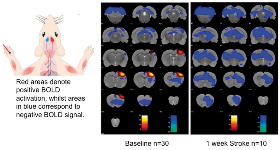
Group analysis is being conducted at 8 and 12 weeks to determine whether there is a difference between treatment groups. Region-of-interest analysis to date indicates a decline in activity in the lesioned sensory cortex forelimb area one week after stroke (Figure 2), and analysis of other regions and timepoints is ongoing.

The behavioral results, after showing the expected initial deficit from stroke, indicate a recovery of both treatment groups back towards baseline. This spontaneous recovery of young animals has been previously observed in other young animal stroke models. Whilst treatment of these stroke animals with AAV-NT3 has not shown a functional improvement, we need to further elucidate time points at which this treatment can be effective, as previous work in our lab has shown a functional recovery when AAV-NT3 is delivered at 24 hours. We hope that the fMRI data acquired will enable us to understand more about mechanisms of spontaneous recovery seen in this study.
627
BRAIN-0479
Poster Session
A SPECIFIC MULTI-NUTRIENT INTERVENTION AS THERAPEUTIC APPROACH FOR STROKE
Cerebral Ischemia: Animal Models
1Anatomy & Geriatric Medicine, RadboudUMC, Nijmegen, Netherlands
2European Institute for Molecular Imaging, (EIMI), Münster, Germany
3Anatomy, RadboudUMC, Nijmegen, Netherlands
4Nutricia Research, Nutricia Advanced Medical Nutrition, Utrecht, Netherlands
5Radiology and Nuclear Medicine, RadboudUMC, Nijmegen, Netherlands
6Geriatric Medicine, RadboudUMC, Nijmegen, Netherlands
Abstract
Objectives
Occlusion of the middle cerebral artery (MCAo) is among the most common causes of ischemic stroke in human. Cerebral ischemia leads to brain lesions existing of an irreversibly injured core and ischemic boundary zone, the penumbra, containing damaged but potentially salvageable tissue. Using a transient MCAo mouse model we investigated the effect of a specific multi-nutrient intervention as a therapeutic approach to counteract impairments of cerebral (structural+functional) connectivity, cerebral blood flow (CBF), cognition, motor function and neurodegeneration.
Methods
Male C57BL/6j mice (3 months) were subjected to transient (30 min) right MCAo using the intraluminal filament model. Success of surgery was verified intraoperatively via Laser Doppler. Before tMCAo, baseline measurements of motor parameters (open field test, rotarod, grip test, pole test) and spatial learning and memory (Morris Water Maze) were performed and repeated after tMCAo. Starting directly after tMCAo, animals were fed either a control diet or the specific experimental diet. At 14 and 35 days after tMCAo, MRI scanning was conducted to identify stroke location and size (RARE+DWI), and functional and neuronal connectivity and CBF measures (rsfMRI, DTI, MRS, FAIR-ASL) on the 11.7T magnet (Bruker BioSpec). Directly following the MR measurements, brains were collected and immunohistochemical analyses were performed.
Data processing is ongoing and the final results will be presented. First results show that at 7 days after tMCAo, all mice showed a decreased CBF in the occluded cerebral hemisphere compared to the left control hemisphere. Mice fed experimental diet had a higher cortical CBF in the ischemic hemisphere than control fed mice. 35 days post tMCAo, experimental diet fed animals had a restored cortical and hippocampal CBF in the ischemic hemisphere. On control diet, cortical and hippocampal CBF remained decreased in the right occluded cerebral hemisphere compared to the left control hemisphere. The first functional benefits of the experimental diet were noted in the open field, where locomotor activity was improved at 16 days after tMCAo as compared to animals on the control diet.
No therapeutic intervention is available for stroke yet. Our present data show that a specific dietary intervention has beneficial effects on the cerebral hemodynamics after tMCAo. In addition, the diet may reduce some of the functional consequences of tMCAo. Together our data indicate that specific multi-nutrient interventions may be able to counteract harmful effects of ischemic stroke, protecting against neuronal and connectivity loss and accompanying functional deficits.
628
BRAIN-0356
Poster Session
MIR-181 ANTAGOMIR REDUCES FOCAL CEREBRAL ISCHEMIA INJURY IN OVARIECTOMIZED FEMALE MICE
Cerebral Ischemia: Animal Models
1Anesthesia, Stanford University, Stanford, USA
Abstract
References:
629
BRAIN-0213
Poster Session
SYSTEMIC INFLAMMATION AND EXACERBATED BRAIN DAMAGE TO TRANSIENT FOCAL ISCHEMIA IN TYPE 2 DIABETIC MICE
Cerebral Ischemia: Animal Models
1Department of Neurology, Juntendo University, Tokyo, Japan
2Department of Neurology, Juntendo Urayasu Hospital, Chiba, Japan
Abstract
References
630
BRAIN-0262
Poster Session
Z-AJOENE AMELIORATES NEURONAL DAMAGE INDUCED BY CEREBRAL TRANSIENT ISCHEMIA IN THE GERBIL HIPPOCAMPUS.
Cerebral Ischemia: Animal Models
1Department of Anatomy and Cell Biology, College of Veterinary Medicine and Research Institute for Veterinary Science Seoul National University, Seoul, Korea
2Department of Anatomy and BK21 Center for Advanced Medical Education, College of Medicine Inha University, Incheon, Korea
3Department of Anatomy, College of Veterinary Medicine Kangwon National University, Chuncheon, Korea
Abstract
Objective
Z- and E- ajoene isomers are found in oil-macerated garlicproducts1. We investigated their neuroprotective effects againstischemic damage in the gerbil hippocampus.
Methods
Male Mongolian gerbils were divided into 2 groups: sham-operated(control) and ischemia-operated groups. The latter group was further dividedinto 3 subgroups, treated with vehicle (corn oil), E-ajoene, or Z-ajoene. Vehicle(corn oil), or Z- or E-ajoene (25mg/kg)2 was orallyadministered 30 min prior to the induction of transient forebrain ischemiaby occlusion of the common carotid arteries for 5 min.
One day after ischemia/reperfusion (I/R), I/R-inducedhyperactivity significantly reduced in the E- and Z-ajoene-treated groups,compared to that in the vehicle-treated group. Five days after I/R, the numberof cresyl violet-positive neurons in the E- and Z-ajoene-treated groupsincreased, compared to that in the vehicle-treated group. Reactive gliosis inthe CA1 region of E- and Z-ajoene-treated groups reduced, compared to that inthe vehicle-treated group. These neuroprotective effects were more prominent inanimals treated with Z-ajoene, than in those treated with E-ajoene. Inaddition, Z-ajoene significantly decreased lipid peroxidation, as indicated by4-hydroxy-2-nonenal levels in hippocampal homogenates, compared to thatobserved in the vehicle-treated group at a range of time points after I/R.
These results suggest that Z-ajoene protects I/R-induced delayed neuronal death and gliosis byreducing lipid peroxidation in the gerbil hippocampal CA1 region.
References
631
BRAIN-0263
Poster Session
NEUROPROTECTIVE EFFECTS OF VALERIANA OFFICINALIS IN THE GERBIL HIPPOCAMPAL CA1 REGION AFTER TRANSIENT FOREBRAIN ISCHEMIA
Cerebral Ischemia: Animal Models
1Department of Anatomy and Cell Biology, College of Veterinary Medicine and Research Institute for Veterinary Science Seoul National University, Seoul, Korea
2Department of Anatomy and BK21 Center for Advanced Medical Education, College of Medicine Inha University, Incheon, Korea
3Department of Biomedical Laboratory Science, College of Biomedical Sciences Soonchunhyang University, Asan, Korea
Abstract
Objective
As a medicinal plant, the roots of Valeriana officinalis havebeen used as a sedative and tranquilizer1,2. In the present study,we evaluated the neuroprotective effects of valerian root extracts (VE) on thehippocampal CA1 region of gerbils after 5 min of transient cerebral ischemia.
The male gerbils were divided into four groups as follows; 1)sham-operated (Control group), 2) vehicle-treated ischemia (Vehicle group), 3)25 mg/kg VE-treated ischemia (VE25 group), and 4) 100 mg/kg VE-treated ischemiagroup (VE100 group). Vehicle or VE was orally administered to gerbils once aday for 3 weeks before the ischemic surgery.
The administration of 100 mg/kg VE (VE100 group) significantlyreduced the ischemia-induced hyperactivity in spontaneous motor activity 1 dayafter ischemia/reperfusion. Four days after ischemia/reperfusion, animalstreated with VE showed abundant cresyl violet-positive neurons in thehippocampal CA1 region when compared to the vehicle or 25 mg/kg VE-treatedgroups. In addition, VE treatment markedly decreased microglial activation inthe hippocampal CA1 region 4 days after ischemia. Compared to the other groups,the VE100 group showed the lowest level of lipid peroxidation during the first24 h after ischemia/reperfusion.
Pre-treatment with VE has protective effects againstischemic injury in the hippocampal pyramidalneurons by decreasing microglialactivation and lipid peroxidation.
References
632
BRAIN-0413
Poster Session
THE ROLES OF HAX-1 AFTER TRANSIENT FOREBRAIN ISCHEMIA IN MICE
Cerebral Ischemia: Animal Models
1Neurosurgery, Yamanashi University, Chuo, Japan
2Neurosurgery, Stanford University, Stanford, USA
Abstract
633
BRAIN-0802
Poster Session
OPTICAL IMAGING OF NEURAL ACTIVITY, OXYGENATION AND BLOOD FLOW DYNAMICS DURING THE PROGRESSION OF ACUTE STROKE
Cerebral Ischemia: Animal Models
1Biomedical Engineering, Columbia University, New York City, USA
2Radiology, Columbia University, New York City, USA
3Neuroscience, Columbia University, New York City, USA
Abstract
In this study, we use high-speed optical imaging of the bilaterally-exposed cortex of Thy1-GCaMP mice during localized, optically-induced unilateral acute stroke. We chart the progression of spatiotemporal changes in blood flow, hemoglobin oxygenation, and spontaneous neural activity over 1-3 hours. We additionally presented somatosensory stimulation on both the affected and unaffected sides. Our goal is to characterize the minute-by-minute progression of acute stroke in terms of metabolic support, vascular reactivity and the downstream effects on neural function and recoverability. Our results also allow us to probe the properties of neurovascular coupling during stroke progression to assess the potential of functional MRI and resting state fMRI techniques as tools for clinical evaluation.
References
634
BRAIN-0475
Poster Session
COMBINATION TREATMENT WITH U0126 AND RT-PA PREVENTS THE DETRIMENTAL EFFECTS INDUCED BY DELAYED RT-PA TREATMENT AND PROVIDES POTENT NEUROPROTECTION AFTER STROKE
Cerebral Ischemia: Cellular and Molecular
1Serine Proteases and Pathophysiology of the Neurovascular Unit, INSERM UMR-S 919, Caen, France
2Clinical Sciences, Lund University, Lund, Sweden
Objectives
Abstract
Only one approved treatment for ischemic stroke is available, i.e. the thrombolytic agent recombinant tissue type plasminogenactivator (rt-PA). The rt-PA approach has many limitations, recanalization efficacy is far from optimal and it can only be administered within 4.5 hours after stroke onset in order to reduce risk of hemorrhagic transformation and neurotoxicity. Elevated metalloproteinase (MMP) plasma levels have been reported to correlate with the frequency of hemorrhagic transformation following stroke1. We have shown that inhibitors of the Mitogen–activated protein kinasekinase extracellular signal–regulated kinase kinase (MEK) 1/2 pathways have a beneficial effect on stroke outcome2. Besides reducing infarct size and improving neurological outcome, they also reduce the MMPs following experimental stroke. By adding a MEK1/2 inhibitor we hypothesize that we can improve the safety of delayed rt-PA treatment. The aim of this study was to investigate if the combination therapy of a MEK/1/2 inhibitor and rt-PA can prevent the detrimental effects of delayed rt-PA treatment in stroke.
Thromboembolic stroke was induced by local injection of thrombin directly into the right MCA of C57 black/6J mice. Combination treatment with a specific MEK1/2 inhibitor U0126 and rt-PA was administrated at 4 h post stroke-onset. The efficiency of rt-PA to induce thrombolysis was measured by laser Doppler. After 24 h, all animals were euthanized and matrixmetalloproteinase (MMP)-9 and phosphorylated extracellular signal regulated kinase (ERK) 1/2 protein levels were investigated by immunofluorescence. Presence of hemorrhage was verified by histology and infarct volume was measured using MRI.
Delayed administration of rt-PA (4 h post-stroke onset) had deleterious effect versus control group. An increased infarct volume, presence of hemorrhage and enhanced MMP-9 protein levels was observed in the MCAO group treated with rt-PA alone compared to control animals. Combination treatment with U0126 and rt-PA prevented hemorrhagic transformation and significantly decreased infarct volume, and MMP-9 protein levels compared to rt-PA alone and control group. Furthermore, p-ERK1/2 protein expression was blocked after combination treatment compared to control animals.
Combination therapy with rt-PA and U0126 decreases cerebral infarct volume, MMP-9 protein levels and prevents rt-PA induced hemorrhagic transformation. These results suggest that a MEK1/2 inhibitor is a promising adjuvant strategy to alleviate the detrimental side effects of delayed rt-PA treatment. These results provide new insights into how we may improve the only FDA approved treatment for ischemic stroke.
Figure: We observe a significantly increase in the infarct volume after delayed rt-PA treatment (4 h post-stroke onset). Combination treatment with U0126 and rt-PA at 4 h post stroke onset significantly decreased infarct volume compared to vehicle treated animals and rt-PA alone. Data are presented as mean ± SEM. 
References
635
BRAIN-0358
Poster Session
EFFECT OF HDAC9 DEFICIENCY ON ISCHEMIC STROKE OUTCOME
Cerebral Ischemia: Cellular and Molecular
1Institute for Stroke and Dementia Research (ISD), Klinikum der Universität München, Munich, Germany
Abstract
References:
636
BRAIN-0442
Poster Session
PHOSPHOINOSITIDE 3-KINASE GAMMA DEFICIT ENHANCES NEUROTOXIC EFFECTS OF MICROGLIA AFTER TRANSITORY FOCAL CEREBRAL ISCHEMIA
Cerebral Ischemia: Cellular and Molecular
1Institute of Molecular Cell Biology, Jena University Hospital, Jena, Germany
2Department of Neurology, Jena University Hospital, Jena, Germany
3Institute of Pathology, HELIOS Klinikum, Erfurt, Germany
4Department of Genetics Biology Biochemistry, University of Torino, Torino, Italy
Abstract
Objectives: Phosphoinositide 3-kinase γ (PI3Kγ) is linked to neuroinflammation and phagocytosis. This study was conducted to investigate the specific role of lipid kinase-dependent and -independent function of PI3Kγ in evolvement of brain damage induced by focal cerebral ischemia/reperfusion.
Methods: PI3Kγ wild-type, knockout (PI3Kγ-/-), and kinase-dead (PI3KγKD/KD) mice as well as wildtype chimeras containing PI3Kγ-/--bone marrow and PI3Kγ-/- chimeras containing wildtype-bone marrow were subjected to middle cerebral artery occlusion (45 minutes) followed by 6h, 12h and 48h as well as 7d of reperfusion and immunohistochmical (IHC) investigations were performed. Additionally, primary microglial cells derived from respective mouse genotypes were used for mechanistic analysis of PI3Kγ effects on matrix metalloproteinase-9 (MMP-9) release, phagocytic activity and cAMP content and signaling after oxygen/glucose deficit (OGD) challenge.
Results: Enlargement of brain infarction was more pronounced in PI3Kγ-/- mice compared to wild-type and PI3KγKD/KD mice as well as chimeras 48 h after reperfusion, whereas early infarct demarcation (6 and 12 h after reperfusion) was similar. IHC analysis revealed a reduced amount of PMN invasion within infarct core of brains derived from PI3Kγ-/- mice, compared to wild-type mice, and a distinct immunoreactivity for MMP-9 in Iba-1 positive cells. Furthermore, number of Galectin-3/MAC-2-positive microglial cells indicating activated phagocytosis1 was reduced in brain slices of PI3Kg-/- mice. Cell culture studies revealed enhanced MMP-9 secretion in supernatants derived from microglia of PI3Kγ-deficient mice after 2h OGD and 48h recovery. Furthermore, PI3Kγ-deficient microglial cells showed a markedly diminished phagocytic activity already under normal conditions and a failed phagocytic activation throughout the observed recovery period of 48h following OGD. Lastly, PI3Kγ-deficient microglia exhibited strongly increased cAMP levels and an activated cAMP signaling verified by CREB phosphorylation in comparison with wild-type microglia or cells expressing PI3KγKD/KD. This effect was enhanced after OGD/Reox challenge.
Conclusions: Our data suggest kinase-independent control of cAMP phosphodiesterase activity by PI3Kγ as a crucial mediator of microglial cAMP control, MMP expression, activation of phagocytic activity and subsequent attenuation of infarct enlargement following focal brain ischemia/recirculation. The results identify the suppressive effect of PI3Kγ on cAMP as a critical mediator of ischemia-induced immune cell functions.
References
637
BRAIN-0766
Poster Session
G PROTEIN-COUPLED ESTROGEN RECEPTOR SIGNALLING PROVIDES NEUROPROTECTION IN FEMALE MICE POST-STROKE
Cerebral Ischemia: Cellular and Molecular
1Dept. of Pharmacology, Monash University, Clayton, Australia
Abstract
638
BRAIN-0639
Poster Session
STAT3 IS A POSITIVE REGULATOR OF ENDOTHELIAL FUNCTION IN THE BRAIN
Cerebral Ischemia: Cellular and Molecular
1Anesthesiology and Perioperative Medicine & Knight Cardiovascular Institute, OHSU, Portland, USA
2Department of Medicine, Norwalk Hospital affiliated with Yale University School of Medicine, Norwalk, USA
3Anesthesiology and Perioperative Medicine, OHSU, Portland, USA
Abstract
Objectives
Signal Transducer and Activator of Transcription 3 (STAT3) is protective to the brain following ischemic injury1,2,3. Ablation of STAT3 specifically in neurons does not alter infarct size. We therefore hypothesized that endothelial STAT3 is responsible for protection from ischemic brain injury.
Endothelial- specific STAT3 knock-out mice (STAT3 floxed; Tie2Cre) were subjected to 1 hour middle cerebral artery occlusion (MCAO) and infarct volume determined by 2,3,5-triphenyltetrazolium chloride (TTC) staining. Primary mouse brain microvascular ECs were isolated and cultured from 2 month old male C57BL6 mice. ECs were subjected to varying durations of OGD and p-STAT3 levels determined by Western blot. STAT3 was pharmacologically inhibited by AG490 and Stattic; cell death was determined by Calcein/PI labeling, barrier integrity by transendothelial electrical resistance (TEER) measurement and FITC-Ficoll 70 flux across the cell monolayer and nitric oxide response to acetylcholine by nitrate/nitrite fluorometric assay.
Our in vivo studies show that cerebral infarct volume is increased following MCAO in mice with conditionally attenuated STAT3 specifically in endothelial cells compared to control littermates. We found that STAT3 protein levels and phosphorylation are regulated by OGD; decreasing immediately following OGD and increasing again during reoxygenation. Pharmacological attenuation of STAT3 signaling induces EC death, while sub-lethal inhibition of STAT3 results in endothelial dysfunction, assessed by increased permeability and decreased resistance of the endothelial monolayer as well as a reduced nitric oxide response to acetylcholine.
STAT3 is a positive regulator of endothelial function and integrity in the brain, playing an important role in determining susceptibility to ischemic damage. Since endothelial dysfunction is a contributing factor, as well as an outcome of pathological states, regulation of endothelial STAT3 may have important consequences in cerebrovascular disease.
Acknowledgements
We thank Dr X-Y Fu at Indiana University–Purdue University Indianapolis for the STAT3 floxed mice and Dr W Fleming at OHSU for the Tie2-Cre mice.
This work was supported by a Pacific Mountain Affiliate Postdoctoral Fellowship from the American Heart Association to CMD.
References
639
BRAIN-0139
Poster Session
FIRST ESTIMATION OF NONLINEAR DYNAMICS OF GLOBAL CEREBRAL ISCHEMIA
Cerebral Ischemia: Cellular and Molecular
1Physioloy, Wayne State University, Detroit, USA
2Physics and Astronomy, Wayne State University, Detroit, USA
Abstract

640
BRAIN-0311
Poster Session
HYPOXIC PRECONDITIONING PROTECTS AGAINST WHITE MATTER INJURY AFTER NEONATAL HYPOXIA-ISCHEMIA THROUGH AMELIORATING MICROGLIAL-MEDIATED INFLAMMATION
Cerebral Ischemia: Cellular and Molecular
1State Key Laboratory of Medical Neurobiology and Institute of Brain Sciences, Fudan University, Shanghai, China
2Center of Cerebrovascular Disease Research, University of Pittsburgh Medical Center, Pittsburgh, USA
Abstract
641
BRAIN-0485
Poster Session
CEREBROVASCULAR RESPONSES IN SPONTANEOUSLY HYPERTENSIVE RATS AFTER ISCHEMIC STROKE
Cerebral Ischemia: Cellular and Molecular
1Clinical Experimental Research, Glostrup Research Institute, Glostrup, Denmark
2Experimental Vascular Research, Clinical Sciences, Lund, Sweden
Abstract
Objectives:
Stroke is the third most common cause of death in Europe why it is global and socio-economic important. Furthermore, hypertension is known to be an important controllable risk factor of stroke (1). Despite numerous animal models, new drugs that appear effective in ischemic stroke models have not worked in clinical trials. A main reason proposed for this is that the majority of pre-clinical studies are performed in healthy rodents. It is previously shown that cerebrovascular endothelin type B receptors are upregulated after ischemic stroke mediating a significant contraction of the occluded blood vessel. In addition, increased expression of endothelin type B receptors in lethal stroke patients has been observed (2). We hypothesize that hypertension enhances the cerebrovascular damages that are associated with reduced cerebral blood flow after ischemic stroke. In this project, vascular changes in the cerebral blood vessels after ischemic stroke were investigated in spontaneously hypertensive rats (SHR).
Middle cerebral artery occlusion (MCAO) was induced in 12-week-old male SHR (Permit number: 2014-15-0201-00042). After occlusion of the right middle cerebral artery (MCA) for 2 hrs the filament was withdrawn to allow reperfusion. The contractile response to different agonists were investigated in the MCAs after 48 h of reperfusion in the wire-myograph. The endothelin type B receptor agonist Sarafotoxin 6c (S6c) was added with and without the endothelium inhibitors L-NAME and Indomethacin, and the endothelin type A receptor agonist Endothelin-1 (ET1) was added with and without the endothelin type B receptor antagonist BQ788. In addition, potassium and endothelium responses were examined.
The contractile response to ET1 was increased in the occluded MCA compared to the non-occluded MCA. The response was mediated by endothelin type A (ETA) receptors confirmed by an endothelin type B receptor antagonist BQ788. There was no contractile response to S6c in any groups with or without the endothelium inhibitors L-NAME and Indomethacin. The potassium and endothelium responses were significant increased in the occluded MCA compared to the non-occluded MCA.
MCAO in SHR increased the contractile response to ET1 in the occluded MCA mediated by ETA receptors. There was no difference in the contractile response to S6c. Interestingly, an enhanced endothelium response was demonstrated in the occluded MCA perhaps indicating a protective mechanism. The role of the endothelium in SHR after ischemic stroke will be further investigated.
References:
642
BRAIN-0769
Poster Session
A NEW THERAPEUTIC STRATEGY AGAINST TOXIC NASCENT PEPTIDE CHAIN AGGREGATION AFTER BRAIN ISCHEMIA
Cerebral Ischemia: Cellular and Molecular
1Shock Trauma and Anesthesiology Research Center, University of Maryland School of Medicine, Baltimore, USA
Abstract
643
BRAIN-0774
Poster Session
THE BETA/PIX – RAC1 - COFILIN PATHWAY AFTER BRAIN ISCHEMIA
Cerebral Ischemia: Cellular and Molecular
1Shock Trauma and Anesthesiology Research Center, University of Maryland School of Medicine, Baltimore, USA
Abstract
644
BRAIN-0461
Poster Session
CEREBROVASCULAR ENDOTHELIN-1 HYPERREACTIVITY IS ASSOCIATED WITH TRANSIENT RECEPTOR POTENTIAL CANONICAL CHANNELS 1 AND 6 ACTIVATION AND DELAYED CEREBRAL HYPOPERFUSION AFTER FOREBRAIN ISCHEMIA IN RATS
Cerebral Ischemia: Cellular and Molecular
1Dept. of clinical experimental research, Glostrup research Institute, Glostrup, Denmark
2Dept. of Radiology, Copenhagen University Hospital, Herlev, Denmark
Abstract
Here, we aimed to investigate the association between enhanced ET-1-mediated vasoconstriction in cerebral arteries and calcium channel plasticity in the post-ischemic hypoperfusion phase after induced forebrain ischemia in rats.
645
BRAIN-0069
Poster Session
EFFECT OF TH2 IMMUNITY ON STROKE OUTCOME
Cerebral Ischemia: Cellular and Molecular
1Department of Pharmacology, Monash University, Clayton, Australia
2Department of Physiology, National University of Singapore, Singapore, Singapore
Abstract
Stroke is the world's second leading cause of mortality and over a third of survivors are left with major neurological injury making it the sixth leading burden of disease worldwide. Following an ischemic stroke, immune cells including macrophages, lymphocytes, neutrophils and dendritic cells infiltrate the ischemic brain hemisphere and contribute to secondary brain damage. Growing evidence shows the importance of T lymphocytes, in particular T regulatory cells, in the brain following stroke where they appear to modulate tissue injury. This is likely to involve the direction of non-T regulatory CD4+ T cells (T helper (Th) cells) towards a Th2-type anti-inflammatory response and away from a Th1 pro-inflammatory response. Commonly, the C57Bl/6 mouse strain is known to exhibit a Th1-prone, pro-inflammatory type response to injury, whereas the FVB strain is relatively Th2-prone, or anti-inflammatory, in its immune response. We tested the hypothesis that stroke outcome is more severe in C57Bl/6 than FVB mice. We compared functional outcomes and quantitated immune cell numbers in brains of the two strains of mice following stroke. C57Bl/6 and FVB mice (n=127) were subjected to sham surgery or 1 h occlusion of the right middle cerebral artery followed by 23 h of reperfusion. C57Bl/6 mice displayed greater functional deficits than FVB mice after stroke, as assessed by neurological scoring and hanging grip test (n=28-34, P<0.01). Total numbers of CD45+ leukocytes tended ∼2-fold greater in the brains of C575Bl/6 than FVB mice despite a similar infarct size. Moreover, there were marked differences in the composition of leukocyte types in the ischemic hemispheres of the two mouse strains after stroke. Compared with sham-operated mice, fold-increases in cell numbers after stroke in C57Bl/6 vs FVB mice (n=5-8) included: neutrophils, 6.7 vs 96.9; CD3+ cells, 3.1 vs 1.6; CD4+ cells, 4.7 vs 1.9; CD8+ cells, 0.8 vs 2.8; macrophages, 2.2 vs 7.9; dendritic cells, 3.5 vs 4.8. We then further tested whether acutely inducing a Th2 immune response in C57Bl/6 mice might improve functional outcome following stroke. These differing immune cell compositions may have contributed to the differential functional outcomes of C57Bl/6 and FVB mice following stroke. Finally, we used the Th2 cytokine, interleukin-4 (IL-4), to induce a Th2 immune profile, with mice receiving either mouse recombinant IL-4 (5 μg i.p.) or vehicle (1% bovine serum albumin) 24 h before stroke. This treatment resulted in ∼35% smaller infarct volumes than in vehicle-treated mice (P<0.05, n=17-24). Thus, a Th2 immune profile leads to milder brain injury after stroke, and acute administration of a Th2-promoting cytokine can provide neuroprotection.
646
BRAIN-0674
Poster Session
EXPRESSION, DISTRIBUTION AND CELLULAR LOCALIZATION OF SIRTUINS IN THE NORMAL AND ISCHEMIC BRAIN OF RAT
Cerebral Ischemia: Cellular and Molecular
1neurology, Yonsei University. Medical Center, Seoul, Korea
Abstract
Objectives:
Sirtuins (SIRTs) are nicotinamide adenine dinucleotide-dependent histone deacetylases. To date, seven sirtuins (SIRT1-SIRT7) have been identified in mammals. While basic knowledge on the cellular expression and anatomical localization of sirtuins in the brain is essential to understand the role of sirtuins in brain diseases, only limited information is available on this issue.1, 2 We investigated the distributions and cellular localization of sirtuins in the normal brain as well as ischemic brain of the rat.
Methods:
Male Sprague-Dawley rats were used. Normal whole brain samples of rats were obtained through transcardiac perfusion under inhalation anesthesia. Cerebral ischemia was induced by 2 h of middle cerebral artery occlusion and 22 h of reperfusion using nylon thread. In paraffin or frozen sections, immunohistochemistry was performed and whole brain was examined into regions: olfactory bulb, cerebrum and cerebellum. To examine the cell types that express sirtuins, double immunofluorescence staining was performed using antibodies for each sirtuin and those for specific cell markers such as NeuN or β-III tubulin (neuron), myelin basic protein (MBP, myelin), CNPase (oligodendrocyte), glial fibrillary acidic protein (astrocyte), and collagen type IV or RECA-1 (vessels).
Results:
SIRT1, SIRT3 and SIRT5-7 were colocalized with neuronal markers and SIRT2 was colocalized with myelin markers. SIRT6 and SIRT7 were also partially colocalized with oligodendrocyte markers. SIRT4 was expressed in vessels. As a result, SIRT1 and SIRT5-7 were widely distributed and highly expressed in the olfactory bulb, cerebrum, and cerebellum. SIRT1-3 and SIRT5 were not detected in the corpus callosum and internal capsule, while SIRT6 and SIRT7 showed signals in them. SIRT2 was highly expressed in the glomerular layer and anterior olfactory nucleus of olfactory bulb, the cerebral cortex, hippocampus, and the granular layer of cerebellum. SIRT3 showed extensive distribution in the olfactory bulb and cerebellum except for the molecular layer of cerebellum. SIRT3 was also expressed in the cerebral cortex, hippocampus and thalamus. SIRT4 showed wide distribution through whole brain except for the molecular layer and purkinje layer of cerebellum. After induction of cerebral ischemia, SIRT2, SIRT4 and SIRT7 showed increased expression in the ischemic and peri-infarct region. However, expression of SIRT1 and SIRT6 was decreased in the ischemic regions. Expression of SIRT3 and SIRT5 was not changed after cerebral ischemia.
Conclusions:
Distribution and cellular localization in the rat brain were different among sirtuins. Expression in the ischemic brain was also different. Findings of this study might provide with basic information for studying pathophysiologic role of sirtuins in the brain.
References:
647
BRAIN-0412
Poster Session
HSP70 PROTECTS THE BRAIN FROM STROKE BY INTERFERING WITH DYNAMIN-FAS MEDIATED SIGNALING IN NEURONS
Cerebral Ischemia: Cellular and Molecular
1Neurology, San Francisco Veterans Affairs UCSF, San Francisco, USA
2Anatomy, Yonsei University College of Medicine, Seoul, Korea
Abstract
Objective: The 70 kDa heat shock protein (Hsp70) is known to protect brain cells in animal and cell culture models of cerebral ischemia. We carried out proteomic screening of mice subjected to middle cerebral artery occlusion (MCAO), which identified dynamin as a major downregulated gene in Hsp70-overexpressing transgenic mice (Tg) compared to wildtype (Wt). Dynamin is a protein involved in endocytosis, and is expressed in neurons. Dynamin has been found to transport the death receptor Fas to the cell surface where it can be bound by its ligand (FasL) and lead to apoptosis.
Methods: We subjected groups of wildtype, Hsp70 transgenic, and Hsp70 knockout (Ko) mice to cerebral ischemia using distal MCAO (dMCAO). Brains were assessed for dynamin and Fas expression 3d following dMCAO as well as for infarct size and neurological behavior 14d post dMCAO. To explore the significance of dynamin in brain ischemia, we then injected mice with a dynamin inhibitor (dynasore).
Results: We found that dynamin is upregulated by ischemia and colocalizes primarily to neurons. Further, Hsp70 overexpression protects the brain following dMCAO as evidenced by decreased infarct volume (n=6/group, P<0.05), improved neurobehavioral outcomes (P<0.05), and decreased brain dynamin and Fas expression relative to wildtype and KO groups. Dynasore also protects against ischemic brain damage and improves neurological outcome out to 14 days post dMCAO (n=7∼9/group; **P<0.05). Dynasore (Dyna) treatment alsoattenuated surface expression of Fas in experimental stroke (P<0.01).
Condclusion: Our results showed that Hsp70 disrupts apoptotic pathways after experimental stroke in mice by downregulating dynamin, which may be involved in Fas trafficking to the cell surface, thus triggering apoptosis . These findings suggest a new molecular target for treating stroke.
648
BRAIN-0859
Poster Session
THE ROLE OF α-SYNUCLEIN IN ISCHEMIC BRAIN DAMAGE
Cerebral Ischemia: Cellular and Molecular
1Neurological Surgery, University of Wisconsin School of Medicine and Public Health, Madison, USA
Abstract
References
649
BRAIN-0462
Poster Session
EXPRESSIONAL CHANGES IN AN ENRICHED FRACTION OF THE CEREBRAL MICROVASCULATURE AFTER TRANSIENT GLOBAL CEREBRAL ISCHEMIA – A PROTEOMIC SCREEN
Cerebral Ischemia: Cellular and Molecular
1Clinical Experimental Research, Glostrup Research Institute, Glostrup, Denmark
2Biochemistry and Molecular Biology, University of Southern Denmark, Odense, Denmark
Abstract
650
BRAIN-0799
Poster Session
SUMO2/3 CONJUGATION AS AN ENDOGENOUS NEUROPROTECTIVE MECHANISM
Cerebral Ischemia: Cellular and Molecular
1Department of Experimental Neurology, Center for Stroke Research Berlin (CSB) Charité-Universitätsmedizin, Berlin, Germany
2Department of Experimental Neurology, Clinic for Neurology Charité-Universitätsmedizin, Berlin, Germany
3Department of Psychiatry, Cluster of Excellence NeuroCure Charité-Universitätsmedizin, Berlin, Germany
Abstract
Objectives
Small ubiquitin-like modifier (SUMO) conjugation to a broad range of target proteins was identified in animal models of hibernation and ischemia as an endogenous brain protective mechanism.1,2
SUMOylation predominantly targets chromatin-associated transcription factors and epigenetic modifiers and thereby modifies transcriptional profiles.3 The maintenance of SUMO-conjugation levels in post-mitotic neurons during the course of multiple and multicellular damaging cascades following an ischemic insult may therefore increase the survival rate of neurons and improve the outcome after stroke.
We investigated the loss of SUMO2/34 and the SUMO-deconjugating enzyme sentrin-specific isopeptidase (SENP) 7 in primary cultures of cortical neurons following a combined oxygen- and glucose deprivation (OGD) damaging protocol. The impact of SENP7 mutations on SUMO-modified proteins was studied by fluorescence microscopy localization and kinetics assays. Further, we assessed neurite outgrowth after SENP7 depletion in retina explants to evaluate its influence on regeneration.
The loss of free SUMO2/3 in cortical neurons reduced cell viability and survival already in case of sub-threshold OGD where almost all control neurons maintained healthy morphology. While SENP7 depletion in cortical neurons improved their survival during OGD, the number of neurites and the length of neurite outgrowth was reduced in retina explants.
SUMO2/3 conjugation to nuclear proteins is an important mechanism to modify cellular transcriptional activities. Insufficient amounts of SUMO2/3 limit the cellular stress tolerance of cortical neurons to OGD. However, maintenance of the pool of SENP7-cleavable SUMO2/3 conjugation levels enhances survival after OGD. Loss of SENP7 had a detrimental effect in a neurite outgrowth assay in retina explants. This illustrates the complex and intricate interplay between endogenous neuroprotection and endogenous regeneration and recovery after stroke.
References
651
BRAIN-0286
Poster Session
THE CHOROID PLEXUS IN BRAIN LYMPHOCYTE INVASION AFTER STROKE
Cerebral Ischemia: Cellular and Molecular
1Institute for Stroke and Dementia Research (ISD), Klinikum der Universität München, Munich, Germany
2Center for Neuropathology and Prion Research, Ludwig-Maximilians-Universität, Munich, Germany
3Theodor Kocher Institute, Universität Bern, Bern, Switzerland
Abstract
652
BRAIN-0755
Poster Session
IS THE ISCHEMIC PENUMBRA AT THE CORE OF EARLY INTRACRANIAL PRESSURE ELEVATION FOLLOWING EXPERIMENTAL STROKE?
Cerebral Ischemia: Cellular and Molecular
1School of Biomedical Sciences and Pharmacy, University of Newcastle, Newcastle, Australia
Abstract
Objectives
Intracranial pressure (ICP) elevation after ischemic stroke may cause secondary damage. It has historically been accepted that ICP elevation following stroke is primarily caused by edema and therefore ICP only increases significantly after large strokes. Little is known about ICP in patients who have small infarcts. We have shown that ICP increases dramatically 24 hours after temporary intraluminal middle cerebral artery occlusion (MCAo) in animals with small infarcts and little edema. Preliminary data from our lab suggests that a change in CSF volume may contribute to the ICP elevation. This may be explained by choroid plexus ischemia, a known complication of the thread occlusion model. However, we have recently discovered that 24 hour ICP elevation can also occur in a cortical photothrombotic (PT) stroke model that induces submaximal infarction without choroid plexus damage. Animals with completed infarction had no ICP rise. Our aims were to use a cortical PT stroke model to: 1. Test whether the ICP elevation 24 hours post stroke occurs in the absence of choroid plexus ischemia. 2. Determine whether ICP elevation is associated with submaximal infarction (ischemic penumbra).
PT stroke was induced in male Wistar rats (n= 16) by intravenous infusion of 10 mg/kg Rose Bengal followed by illumination of the right parietal bone using either low light exposure (2 min at 0.13 W/cm2) or the standard light exposure (20 min at 0.3 W/cm2), to produce submaximal or maximal infarct, respectively. Epidural intracranial pressure was measured at baseline and 19 – 25 hours following stroke. Infarct and edema volumes were measured histologically.
Animals subjected to low light exposure had smaller infarcts than the standard light exposure group (9.3 ± 8.3 mm3 v. 43 ± 18.8 mm3, respectively, p<0.01). Smaller infarcts associated with low light exposure were restricted to the upper layers of the cortex, without damage to the choroid plexus or ventricles. Oedema volumes were 0.2 ± 0.4 mm3 v. 2.82 ± 3.8 mm3, p>0.05. ICP increased by 12.8 ± 2.5 mmHg from baseline only in animals exposed to low light (p<0.01), with no significant rise in the standard light group (1.9 ± 0.9 mmHg).
ICP elevation occurred approximately 24 hours after small cortical PT stroke. This indicates that early ICP elevation observed in previous studies is not simply a product of choroid plexus ischemia that may occur in the thread occlusion MCAo model. Significant ICP elevation was only observed in animals subjected to low light exposure, which has been shown to induce submaximal infarcts with a penumbra-like region at risk. PT strokes induced with standard light exposure were larger and clearly demarcated, consistent with reported absence of penumbra in this model. This group was not associated with ICP elevation. It is well known that that many molecular processes occur within the penumbra. We propose that the ICP elevation seen at 24 hours following stroke is triggered by an as yet unidentified active molecular mechanism in the ischemic penumbra.
653
BRAIN-0492
Poster Session
LONG NON-CODING RNA FOSDT PROMOTES ISCHEMIC BRAIN DAMAGE BY INTERACTING WITH CHROMATIN MODIFYING PROTEINS
Cerebral Ischemia: Cellular and Molecular
1Neurological Surgery, University of Wisconsin School of Medicine and Public Health, Madison, USA
Abstract
654
BRAIN-0517
Poster Session
REVERSAL OF IN VIVO ISCHEMIC LONG-TERM POTENTIATION CAUSED BY CARDIAC ARREST AND CARDIOPULMONARY RESUSCITATION IMPROVES SYNAPTIC FUNCTION
Cerebral Ischemia: Cellular and Molecular
1Anesthesiology, University of Colorado, Denver, USA
2Pharmacology, University of Colorado, Denver, USA
Abstract
Objective: Experience-dependent memory acquisition, in vitro tetanic stimulation and simulated ischemia results in long-term potentiation (LTP) of hippocampal CA1 synapses. The goal of this study was to determine whether in vivo cerebral ischemia results in iLTP of CA1 synapses. We hypothesized that iLTP resulting from CA/CPR prevents physiological LTP.
Methods: Cardiac arrest (8 minutes) followed by cardiopulmonary resuscitation (CA/CPR) or sham surgery was performed on adult (8-12 week) male mice. Electrophysiology was performed in hippocampal slices from sham and 7 and 30 days after CA/CPR. Field excitatory postsynaptic potentials (fEPSP) resulting from Schaffer collateral stimulation were recorded and rising slope was analyzed. Theta burst stimulation (TBS) was used for LTP induction and low frequency stimulation for reversal of LTP (depotentiation). Western blot analysis of synaptic isolations were performed from hippocampi collected from shams and 7 and 30 days after CA/CPR.
Results: TBS resulted in LTP of fEPSP to 164±9.8% (n=7, P<0.05) of baseline in sham controls, however no increase was observed at 7 (106±15.9% n=4) and 30 days (109±13.9% n=7) after CA/CPR. A depotentiation stimulus had no effect in sham controls (110±5.1%, n=6), but caused a 55% reduction in fEPSP in control slices that received LTP induction and 23% reduction in slices from CA/CPR mice. Importantly, in slices form CA/CPR mice, LTP induction protocol applied subsequent to depotentiation stimulus resulted in physiological LTP to 126.9±11.8% (n=3) of the original baseline, an effective increase of approximately 40%. Glutamate receptor subunit GluR1 phosphorylation and expression in synaptic fractions were increased at 7 and 30 days after CA/CPR.
Conclusions: Depotentiation in slices from CA/CPR mice suggests CA1 synapses are in a chronically potentiated state. Increased GluR1 phosphorylation and expression after CA/CPR also suggest in vivo ischemia causes iLTP. Our ability to induce physiological LTP after depotentiation in slices from CA/CPR suggests that reversing iLTP allows for normal synaptic plasticity. The results of this study are significant as they demonstrate in vivo iLTP and suggest a physiological stimulus has the potential to reverse synaptic impairments in the chronic phase after cerebral ischemia and reset synaptic function.
655
BRAIN-0378
Poster Session
ABERRANT ACTIVATION OF APOPTOSIS SIGNAL-REGULATING KINASE 1 MEDIATES PRO-INFLAMMATORY AND NEUROTOXIC MICROGLIAL RESPONSES AFTER ISCHEMIC/REPERFUSION BRAIN INJURY
Cerebral Ischemia: Cellular and Molecular
1Neurology, University of Pittsburgh, Pittsburgh, USA
2Division of Pharmaceutical Sciences, Duquesne University, Pittsburgh, USA
3State Key Laboratory of Medical Neurobiology and Institute of Brain Sciences, Fudan University, Shanghai, China
Abstract

656
BRAIN-0325
Poster Session
CASPASE-1 ACTIVATION LINKS DAMP-MEDIATED PERIPHERAL MONOCYTE ACTIVATION AND PYROPTOTIC LYMPHOCYTE CELL DEATH AFTER STROKE.
Cerebral Ischemia: Cellular and Molecular
1Institut für Schlaganfall- und Demenzforschung (ISD), Klinikum der Universität München, München, Germany
Abstract
657
BRAIN-0653
Poster Session
PINEAL HORMONE MELATONIN WORKS VIA NEUROPROTECTIVE NOVEL MECHANISM; INTERLEUKIN-4 (IL-4) DEPENDENT M2 MICROGLIAL POLARIZATION, AFTER FOCAL CEREBRAL ISCHEMIA/REPERFUSION (FI/R)
Cerebral Ischemia: Cellular and Molecular
1Neurology, Department of Neurology University of Pittsburgh, Pittsburgh, USA
Abstract
Objectives;
Microgliarepresent rational but difficult therapeutic targets for stroke due totheir diverse phenotypes that playdual-faced protective (M2 phenotype) and toxic (M1 phenotype) effects. Previousstudy has shown that subcutaneous melatonin injection increased the level ofIL-4, the best known M2 inducing cytokine, in the blood. In this study, weinvestigated the role of melatonin in microglia M2 polarization and its effecton long-term recovery after stroke.
Focal cerebralischemia was induced for 60 min FI/R. Animals were randomly assigned to receiveeither melatonin or vehicle treatmentat 2h after stroke. Brains were assessed for cerebral tissue loss at 3 and 14days of reperfusion. Neurological performance was analyzed up to 14 days afterischemia. Markers for microglia polarization were assessed usingimmunofluorescent staining and RT-PCR. Invitro experiments using primary microglia and in-transwell microglia-neuroncoculture were done to confirm the effect of melatonin on microglialinflammatory responses and its effect on microglia-potentiated neuronal injuryupon OGD.
Melatoninsignificantly reduced infarct volume and attenuated sensorimotor deficits 3-14day after FI/R. IL-4 deficiency, abolished melatonin-afforded long termprotection. Melatonin-treated mice showed significantly reduced expression ofinflammatory cytokine and chemokines, which is accompanied by significantlyincreased expression of M2 markers and decreased expression of M1 markers inmicroglia. In primary microglial cultures, melatonin inhibited LPS (a M1inducer)-induced production of NO and TNFα, confirming that melatonin hasdirect anti-inflammatory effect on microglia. Furthermore, melatoninameliorated the neurotoxic effect of M1 microglia on OGD neurons, and thiseffect was absent in IL-4 deficient microglia.
Melatoninmay represent an innovative therapeutic strategy that shifts microgliapolarization toward a protective M2 phenotype in an IL-4-dependent manner andthus enhance long-term recovery after stroke.
658
BRAIN-0112
Poster Session
SERIAL CHANGE FOR PHOSPHODIESTERASE 3A AND 3B EXPRESSION AFTER TRANSIENT FOCAL ISCHEMIA IN MICE BRAIN
Cerebral Ischemia: Cellular and Molecular
1Department of Neurology, Juntendo University Urayasu Hospital, Urayasu, Japan
2Department of Neurosurgery, Juntendo University School of Medicine, Tokyo, Japan
3Department of Neurology, Juntendo University School of Medicine, Tokyo, Japan
4Department of Neuroendovascular Therapy, Juntendo University School of Medicine, Tokyo, Japan
Abstract
659
BRAIN-0450
Poster Session
DETERMINATION OF MICRO-RNA PROFILES IN EXTRACELLULAR VESICLES ISOLATED FROM SERUM OF HUMAN STROKE PATIENTS.
Cerebral Ischemia: Cellular and Molecular
1Institute of Cardiovascular and Medical Sciences, University of Glasgow, Glasgow, United Kingdom
2School of Medicine, University of Glasgow, Glasgow, United Kingdom
3Institute of Neuroscience and Psychology, University of Glasgow, Glasgow, United Kingdom
Abstract
Stroke is the 3rd leading cause of death in the UK and the leading cause of long-term adult disability. There is only one licensed pharmacological intervention, intravenous delivery of thrombolytic recombinant tissue plasminogen activator within 4.5 hours of stroke, beyond which time there are significant haemorrhagic risks. An alternative intervention is needed and through their ability to alter the expression of multiple genes involved in stroke pathophysiology miRNAs offer a novel therapeutic intervention. miRNA expression is altered in experimental stroke and in patients with stroke. Recently, active transport of miRNA in extracellular vesicles (EV), such as exosomes, has been demonstrated pre-clinically between cells in atherosclerosis. We hypothesised that miRNAs packaged in EV would differ between patients with stroke and patients without stroke, raising the potential for novel miRNAs to be used as biomarkers or therapeutic agents for modulation.
We recruited 169 patients with suspected stroke and a blood sample was taken at 48h post-stroke. All participants gave full informed consent and the study was approved by the Scotland A Research Ethics Committee. A miRNA microarray was performed (Openarray™ platform) on samples from 39 patients (n=10 non-stroke, n=29 with stroke – further subdivided by TOAST classification into large artery (n=9), cardioembolic (n=10) or small vessel disease (n=10) stroke). Validation of results was performed using samples from 169 patients. EVs were isolated from 200 µL serum before RNA was extracted and the concentration determined using nanodrop spectrophotometry. Taqman™ real-time quantitative polymerase chain reaction was used to determine the expression levels of specific miRNA(s).
The microarray identified 26 miRNAs that were significantly dysregulated between stroke vs non-stroke patients or between specific TOAST subtypes and non-stroke control. Of these, changes in 17 miRNA were validated in the larger cohort: levels of miRNAs -17 (relative quantification, RQ vs non-stroke=1.74*), -20b (RQ, vs non-stroke=1.95*), -27 (RQ, vs non-stroke=1.83*), -30a-5p (RQ, vs non-stroke=1.67*), -93 (RQ, vs non-stroke=1.80*), -199a-3p (RQ, vs non-stroke=1.91*) and hsa-let-7e (RQ, vs non-stroke=1.61*) were significantly increased in stroke vs non-stroke patients (*p<0.05 by unpaired Student’s t-test). Furthermore, differences between TOAST subtypes were shown with small vessel disease consistently having the highest levels of miRNA. There was no significant difference observed in the expression of 10 miRNAs including miRNAs-520b (RQ, vs non-stroke=1.09), -660 (RQ, vs non-stroke=1.21) and -218 (RQ, vs non-stroke=1.57) between non-stroke and stroke patients. Bioinformatic analysis highlighted a number of important target genes implicated in stroke pathophysiology for each miRNA including genes involved in the regulation of apoptosis, angiogenesis and cell migration.
We have identified and validated changes in EV packaged miRNA expression in patients with stroke across differing stroke subtypes. This will direct future studies looking into paracrine signalling in the setting of stroke and the modulation of specific miRNAs as a novel therapy in the setting of experimental stroke.
660
BRAIN-0275
Poster Session
NEURONAL SOLUBLE FAS LIGAND DRIVES M1-MICROGLIA POLARIZATION AFTER ISCHEMIA
Cerebral Ischemia: Cellular and Molecular
1Department of Neurology, the Affiliated Drum Tower Hospital of Nanjing University Medical School, Nanjing, China
Abstract
661
BRAIN-0288
Poster Session
DOUBLE NEGATIVE T LYMPHOCYTES FROM GLD MICE UP-REGULRATE M2 MICROGLIA IN ISCHEMIC STROKE THROUGH FAS/FASL
Cerebral Ischemia: Cellular and Molecular
1Department of Neurology, the Affiliated Drum Tower Hospital of Nanjing University Medical School, Nanjing, China
Abstract
662
BRAIN-0105
Poster Session
HYPERGLYCEMIA AND POST-STROKE SEIZURES
Cerebral Ischemia: Cellular and Molecular
1Anatomy & Cell Biology, Indiana University School of Medicine, Indianapolis, USA
Abstract
Seizures are the most common neurological sequelae of stroke and are considered as a major cause of epilepsy. Diabetes mellitus has been identified as an independent predictor of acute seizures in stroke patients. However, the pathogenesis of post-stroke seizures in diabetes is not fully understood. It is well known that abrupt increase of neuronal excitability causes seizures/epilepsy. Potassium currents are important to maintain the baseline membrane potential and regulate neuronal excitability. Accumulating evidence indicates that down regulation of A-type potassium current (IA) is associated with seizures after stroke by increasing neuronal excitability. The present study tested the hypothesis that down-regulation of IA contributes to seizure generation after ischemia in diabetes and explored its underlying mechanisms.
Transient forebrain ischemia was produced in adult Wistar rats using the 4-vessel occlusion (4-VO) method. Hyperglycemia (>300 mg/dL) was induced by injection of glucose solution (i.p.) 15 min prior to ischemia. The seizure activity was defined using the Racine scale of III - V. The expression of potassium channels was analyzed by immunohistochemical staining and Western blotting. The alteration of IA and excitability of hippocampal neurons was examined using patch-clamp techniques in brain slices.
The incidence of seizures after ischemia significantly increased in hyperglycemic rats (100%) as compared to the normoglycemic ones (0%). The neuronal damage in the cortex and hippocampus after ischemia was about the same in these two groups of animals, suggesting that neuronal injury is not the major contributor to seizure generation in hyperglycemia. The brain water content after ischemia in hyperglycemic rats was significantly higher than the control ones, indicating the severe edema in hyperglycemic brains. However, administration of Mannitol (1.5 g/Kg, i.v.) reduced the brain edema but the seizure rate remained the same after ischemia (100%), suggesting that brain edema is not the major cause of post-stroke seizure in hyperglycemia. On the other hand, immunohistochemistry and protein analysis showed a significant reduction of IA channel subunit Kv4.2 expression in hyperglycemic rats with seizures. Electrophysiological data also showed that the current density of IA in hippocampal neurons was reduced in hyperglycemic rats after ischemia and the neuronal excitability was increased.
There results demonstrate that hyperglycemia facilitates seizures generation after ischemia. The down-regulation of IA in hippocampal neurons might contribute to the post-ischemic seizure generation in hyperglycemia.
663
BRAIN-0144
Poster Session
20-HETE IS DIRECTLY INVOLVED IN NEURONAL INJURY IN AN IN VITRO MODEL OF ISCHEMIA
Cerebral Ischemia: Cellular and Molecular
1ACCM, Johns Hopkins School of Medicine, Baltimore, USA
2Biochemistry, University of Texas Southwestern Medical Center, Dallas, USA
Abstract
664
BRAIN-0208
Poster Session
INVOLVEMENT OF MIR-181C IN CLINICAL ACUTE ISCHEMIC STROKE AND EXPERIMENTAL STROKE
Cerebral Ischemia: Cellular and Molecular
1Cerebrovascular Diseases Research Institute, Xuanwu hospital of Capital Medical University, Beijing, China
Abstract
665
BRAIN-0226
Poster Session
COMBINED LATE TISSUE PLASMINOGEN ACTIVATOR AND ISCHEMIC POSTCONDITIONING: POSSIBLE ROLE OF REPERFUSION REDUCTION IN NEUROPROTECTION FOLLOWING EMBOLIC STROKE
Cerebral Ischemia: Reperfusion
1Physiology, Rafsanjan University of Medical sciences, Rafsanjan, Iran
Abstract
666
BRAIN-0623
Poster Session
ANGIOTENSIN-(1-7) INCREASES TISSUE SALVAGE FOLLOWING FOCAL CEREBRAL ISCHAEMIA WITH REPERFUSION.
Cerebral Ischemia: Reperfusion
1Glasgow Experimental MRI Centre (GEMRIC) Institute of Neuroscience & Psychology College of Medical Veterinary & Life Sciences, University of Glasgow, Glasgow, United Kingdom
2BHF Glasgow Cardiovascular Research Centre Institute of Cardiovascular & Medical Sciences College of Medical Veterinary & Life Sciences, University of Glasgow, Glasgow, United Kingdom
Abstract

References:
667
BRAIN-0361
Poster Session
ISCHEMIC INJURY MONITORED WITH A NOVEL MICROPERFUSION DEVICE UNVEILS THE ROLE OF PH IN I/R INJURY
Cerebral Ischemia: Reperfusion
1Department of Chemistry and Biochemistry Institute of Arctic Biology, University of Alaska Fairbanks, Fairbanks, USA
2Institute of Arctic Biology, University of Alaska Fairbanks, Fairbanks, USA
Abstract
668
BRAIN-0368
Poster Session
TOLERANCE TO ISCHEMIA IS MODULATED IN PART VIA TARGETING NITRIC OXIDE SIGNALING PATHWAY BY AN ENDOGENOUS FACTOR, NEUROGLOBIN
Cerebral Ischemia: Reperfusion
1Department of Chemistry and Biochemistry/Institute of Arctic Biology, University of Alaska Fairbanks, Fairbanks, USA
Abstract
Reference:
669
BRAIN-0810
Poster Session
PERICYTES ON MICROVESSELS PREVENT COMPLETE REPERFUSION AFTER RETINAL ISCHEMIA
Cerebral Ischemia: Reperfusion
1Institute of Neurological Sciences and Psychiatry, Hacettepe University Faculty of Medicine, ANKARA, Turkey
2Center for Visual Science, University of Rochester, Rochester NY, USA
Abstract
Background: During cerebral ischemia, pericytes constrict microvessels, which do not relax after recanalization, causing an incomplete reperfusion. Since the retina contains the highest density of microvascular pericytes of any organ, retinal reperfusion after ischemia may also be impaired by pericyte-induced microvascular constrictions.
Aim: To analyze whether pericytes constrict retinal microvessels during in vivo ischemia and hinder reperfusion.
Methods: We used wild type (n=35) and NG2-DsRed (n=9) mice. We induced clot formation in the central retinal artery by topical 20%-FeCl3 application over the artery for 3 minutes. After 60 minutes of ischemia, we infused tissue plasminogen activator (tPA) through tail vein to induce recanalization. We monitored the retinal blood flow by laser speckle contrast imaging. We also imaged retinal pericytes in vivo under conditions of normal perfusion, ischemia, and reperfusion animals by using a two channel adaptive optics scanning laser ophthalmoscopy (AOSLO). In one channel, fluorescent NG2-DsRed pericytes were imaged while in a second channel imaged, simultaneously, blood cell movement was recorded in the near infrared regime. After, we labeled whole mount retinas ex vivo with markers for pericytes (NG2, alpha-SMA) and vessels (Claudin-5, lectin) and, stereologically counted the constrictions and labeled cells.
Results: We found a significantly higher number of microvessel constrictions and a decreased microvessel diameter in ischemic (n=5) retinae compared to the sham group (n=6). Constrictions were not restored after recanalization of the retinal artery (n=3). We confirmed these ex vivo observations with in vivo retinal imaging (n=9). Intravitreal injections of a Ca2+-channel antagonist, amlopidine (1mg/ml) 60 minutes before ischemia prevented ischemia-induced microvascular constrictions (n=3, P<0.05), suggesting a role for Ca2+ overload during ischemia. There was a significant co-localization between pericytes and microvascular constrictions (P<0.05). We did not observe any diameter changes in upstream retinal macrovessels between cohorts (P>0.05).
Conclusions: Incomplete microcirculatory reperfusion caused by ischemia-induced persistent contractions of pericytes causing microvascular constrictions are one of the problems facing recanalization therapies.
670
BRAIN-0553
Poster Session
IRON OVERLOAD ANTICIPATES THE SIDE EFFECT OF HEMORRHAGIC TRANSFORMATION AFTER EXPERIMENTAL ISCHEMIA.
Cerebral Ischemia: Reperfusion
1Farmacology, Universidad Complutense de Madrid School of Medicine, Madrid, Spain
2Anatomy, Universidad Autónoma de Madrid School of Medicine, Madrid, Spain
3Neurology, Hospital Dr. Josep Trueta, Girona, Spain
4Neurology, Hospital Germans Trias i Pujol, Barcelona, Spain
Abstract
Bibliography:
671
BRAIN-0465
Poster Session
REAL-TIME TRANSCRANIAL OPTICAL MONITORING OF MICROVASCULAR CEREBRAL BLOOD FLOW AND OXYGENATION IN ACUTE ISCHEMIC STROKE AFTER RECOMBINANT TISSUE PLASMINOGEN ACTIVATOR TREATMENT
Cerebral Ischemia: Reperfusion
1Medical Optics, ICFO-Institut de Ciències Fotòniques, Castelldefels, Spain
2Stroke unit, Hospital de Sant Pau i de la Santa Creu, Barcelona, Spain
Abstract
Background
Diffuse correlation spectroscopy (DCS) and near-infrared diffuse optical spectroscopy (NIRS) measure local microvascular cerebral blood flow (CBF) and oxygenation, respectively, at the bedside [1]. Recombinant tissue plasminogen activator (rt-PA, alteplase) is used as a treatment for acute ischemic stroke (AIS) patients within 4.5 hours of onset but with variable outcomes [2]. Studies suggest that local tissue reperfusion predicts better the outcome after rtPA thrombolysis than recanalization [3]. Therefore, we hypothesize that outcomes will be improved with individualized treatments based on local tissue microvascular perfusion and metabolism. Unfortunately, these variables are difficult to monitor in the stroke unit. We have, for the first time, introduced a hybrid NIRS/DCS device to the emergency room and tested whether the effect of rtPA on cerebral perfusion can be assessed by NIRS/DCS at the bedside in patients with AIS.
AIS patients with middle cerebral artery occlusion were recruited. After rtPA injection, continuous DCS and continuous-wave near-infrared spectroscopy data in both frontal lobes were obtained for the first two hours. We have evaluated the hemodynamic temporal profile in both hemispheres by averaging a 6-minute window every 15 minutes, beginning 15 minutes after treatment initiation. The results were evaluated in relation to the presence of recanalization on follow-up transcranial Doppler and clinical status.
To date, five patients (median NIHSS -National Institute of Health Stroke Scale- 19) were measured with complete arterial recanalization. At 24 hours of the stroke onset, all patients improved significantly (median NIHSS 3). Good signal-to-noise ratio was obtained for all optical variables. Significant increases in CBF, oxy- and total hemoglobin concentrations were observed 90 minutes following rtPA treatment in the ipsilateral, but not in the contralateral hemisphere (Fig.1).
Bedside NIRS/DCS showed the capability for monitoring continuously the effect of reperfusion therapy on AIS in the emergency room. More patients are being recruited and non-treated subjects are being included as a control group. We will explore the feasibility of the method, the specifics of the individual responses and their relationship to the outcome. We will discuss our findings and speculate on the advantages and disadvantages of the method.
The project was funded by Fundació Cellex Barcelona, LlumMedBCN (La Caixa), Ministerio de Economía y Competitividad (PHOTOSTROKE), LASERLAB-EUROPE and Fondo de Investigaciones Sanitarias (Spanish Ministry of Health).
References
672
BRAIN-0734
Poster Session
THE EFFECT AND SAFETY OF EARLY STA-MCA BYPASS IN ACUTE ISCHEMIC STROKE
Cerebral Ischemia: Reperfusion
1Neurosurgery, Uijeong-Bu St. Mary's Hospital, Uijeongbu, Korea
Abstract
673
BRAIN-0448
Poster Session
MILD HYPOTHERMIA PROTECTS THE BRAIN FROM ISCHEMIA/REPERFUSION BY ATTENUATING P63/P73-INDUCED APOPTOSIS THROUGH IASPP
Cerebral Ischemia: Reperfusion
1Cerebrovascular Diseases Research Institute, Xuanwu Hospital of Capital Medical University, Beijing, China
2Department of Neurosurgury, Beijing Tongren Hospital Capital Medical University, Beijing, China
3Cerebrovascular Diseases Research Institute, Xuanwu hospital of Capital Medical University, Beijing, China
4Department of Neurosurgury, Xuanwu Hospital of Capital Medical University, Beijing, China
Abstract
References
674
BRAIN-0444
Poster Session
EFFECTS OF GHRELIN ON NITRIC OXIDE PRODUCTION, HYDROXYL RADICAL METABOLISM DURING CEREBRAL ISCHEMIA AND REPERFUSION IN MICE
Cerebral Ischemia: Reperfusion
1Department of Neurology, Saitama Medical University, Moroyama Saitama, Japan
Abstract
Microvascular, bilateral cerebral blood flow (CBF), oxy- (HbO2) and deoxy-hemoglobin (Hb) and total hemoglobin concentration (THC) changes are shown from the frontal lobes. The time axis shows the time elapsed from the rtPA treatment onset. (*) indicates significant (p<0.05) change from the baseline (time=15 minutes after rtPA injection).
References:
675
BRAIN-0636
Poster Session
BRAIN INJURY FOLLOWING RECURRENT CEREBRAL ISCHEMIA: EFFECT OF RECOVERY TIME BETWEEN ISCHEMIC INSULTS
Cerebral Ischemia: Reperfusion
1Physiology and Pharmacology Clinical Neurosciences and Radiology, University of Calgary, Calgary, Canada
2Physiology and Pharmacology, University of Calgary, Calgary, Canada
3Clinical Neurosciences, University of Calgary, Calgary, Canada
4Experimental Imaging Centre, University of Calgary, Calgary, Canada
Abstract
(Supported by the Canadian Institute for Health Research)
References:

676
BRAIN-0087
Poster Session
EFFECTS OF RAPAMYCIN ON CEREBRAL OXYGEN SUPPLY AND CONSUMPTION DURING REPERFUSION AFTER CEREBRAL ISCHEMIA
Cerebral Ischemia: Reperfusion
1Neuroscience & Cell Biology, Rutgers Robert Wood Johnson Medical School, Piscataway, USA
2Anesthesiology, Rutgers Robert Wood Johnson Medical School, Piscataway, USA
Abstract
Activation of the mammalian target of rapamycin (mTOR) leads to cell growth and survival. We tested the hypothesis that inhibition of mTOR with rapamycin would increase infarct size and decrease microregional O2 supply/consumption balance after cerebral ischemia-reperfusion. This was tested in isoflurane anesthetized rats after middle cerebral artery blockade for 1 hour and reperfusion for 2 hours with and without rapamycin (20 mg/kg once daily for two days). Regional cerebral blood flow was determined using a C14-iodoantipyrine autoradiographic technique. Regional small vessel arterial and venous oxygen saturations were determined microspectrophotometrically. The control group ischemic-reperfused cortex has a similar blood flow and O2 consumption to the contralateral cortex. However, microregional O2 supply/consumption balance was significantly reduced in the ischemic-reperfused cortex. Rapamycin significantly increased cerebral O2 consumption and reduced O2 supply/consumption balance. This was associated with an increased cortical infarct size (13.5 ± 0.8% control vs 21.5 ± 0.9% rapamycin). This suggests that mTOR is important for not only cell survival, but also for the control of oxygen balance after cerebral ischemia-reperfusion.
677
BRAIN-0735
Poster Session
COMBINED PROTECTIVE EFFECTS OF HYPOTHERMIA AND HYPOXIC POSTCONDITIONING IN RAT ORGANOTYPIC HIPPOCAMPAL SLICE CULTURES
Preconditioning and Post-Conditioning
1Pharmacology School of Medical Sciences, University of New South Wales, Sydney, Australia
Abstract
Objectives: Hypoxic-ischemic (HI) brain injury in newborns is associated with high morbidity and mortality rates, with many survivors suffering neurological impairments. Hypothermia is currently the standard of care used for newborns that suffer from mild to moderate HI and can reduce the mortality and disability in these infants [1]. Recent studies have demonstrated the neuroprotective potential of combining hypothermia with other treatments such as erythropoietin and melatonin in animal models of HI [2, 3]. Hypoxic postconditioning or exposure to mild hypoxia following an injury has been shown to result in neuroprotection against brain damage through a variety of mechanisms [4, 5]. Here, we aimed to examine the effects of hypothermia and hypoxia as monotherapies, as well as in combination, against glutamate-induced injury in organotypic hippocampal slices obtained from neonatal rats.
Methods: Organotypic hippocampal slices were obtained from Sprague Dawley rat pups (postnatal day 7), cultured for 12-14 days and exposed to glutamate (10mM for 24h) to induce injury. Hypothermia (33°C, with a delay of 30min, 3h or 24h for 6, 24 or 72h) or hypoxia (8, 10 and 12% oxygen, 1h/day for 5 days or 2h/day for 3 days) were applied post-injury as monotherapies to determine which conditions were protective. The degree of cell death was determined using propidium iodide staining in the CA1 and dentate gyrus in the hippocampal slices. Once protective conditions were established, a combination treatment strategy was applied. All experiments were performed in duplicate with slices obtained from n=3 different animals.
Results: There was no protection observed with 6 or 24h exposure to hypothermia. However, 72 hours of hypothermia was shown to be neuroprotective when applied to slices post-injury. In addition, all of the hypoxic treatments (8, 10 and 12% oxygen) were able to protect the hippocampal slices against injury. Combination treatment, using hypothermia (33°C for 72h, with a delay of 30min after injury and hypoxia (8% and 10% oxygen for 1h/day for 5 days) was able to reduce cell death levels following glutamate-induced injury in both the CA1 and dentate gyrus regions of hippocampal slices.
Conclusions: Here, we have demonstrated that there is the potential for neuroprotection resulting from combination treatment with hypothermia and hypoxic postconditioning in hippocampal slices. While we did not observe a synergistic neuroprotective effect with our combined treatments, our results suggest that that the strategy of mild hypoxia with hypothermia may extend the therapeutic time window for hypoxia and is worthy of further investigation. Our future studies will examine protective mechanisms involved in this combination treatment strategy.
References:
678
BRAIN-0364
Poster Session
PERI-INFARCT DEPOLARIZATIONS IN THE SPONTANEOUSLY HYPERTENSIVE RAT. EFFECT OF PRECONDITIONING BY ANESTHESIA AND CORTICAL LESIONS
Preconditioning and Post-Conditioning
1Neurology, University of Tennessee Health Science Center, Memphis, USA
2Neurosurgery, Juntendo University, Tokyo, Japan
Abstract
Objectives – Ischemic preconditioning appears to include distinct components contributed by prior anesthesia exposure and cortical injury (1). Recent results showed that these have differing effects on peri-infarct depolarizations (PIDs) during subsequent stroke (Zhao and Nowak, submitted manuscript). Isoflurane preconditioning reduced PID incidence when monitored by perfusion imaging under the same isoflurane anesthesia, without affecting PID number recorded under alpha-chloralose, whereas the latter was additionally impacted in animals with prior cortical lesions. The current studies were designed to evaluate the persistence of anesthetic preconditioning effects on PIDs, and to assess the impact of lesion preconditioning on PID incidence in awake animals.
Methods – Spontaneously Hypertensive Rats (SHR) were preconditioned by producing small 2-5 mm3 cryogenic cortical lesions (CL), or by the corresponding sham surgery under isoflurane anesthesia. Focal ischemia was produced the next day by tandem occlusion of the middle cerebral artery (MCA) and ipsilateral common carotid artery. PIDs were detected by speckle contrast perfusion imaging during 4 hours under isoflurane anesthesia, or were recorded for 24 hours in the awake state via electrode arrays implanted at varied intervals prior to occlusion.
Results – PID-associated hyperemic flow transients recorded under isoflurane anesthesia were significantly reduced from 8.5 ± 2.4 (mean ± SD, n=12) in naïve rats to 3.3 ± 1.3 (n=9) after isoflurane exposure the previous day (P < 0.05, one-way ANOVA). Recovery was variable at 1 week (6.6 ± 4.1, n=10), but the control range was regained after 2 weeks or longer (7.2 ± 2.6, n=16). Electrophysiologically recorded PIDs were more numerous in awake rats, but occurred within the same 4-hour window after occlusion. PIDs numbered 25 ± 4, 21 ± 6, 32 ± 9 and 29 ± 11 at 1 day and 1, 3, and 5 weeks, respectively. There was no effect of the interval between electrode placement and occlusion surgery for individual time points, although pooled results for ≤ 1 week (23 ± 5, n= 10) vs. ≥ 3 weeks (31 ± 10) achieved significance (P < 0.03, unpaired t-test). With chronic electrode placement, PID incidence was reduced from 28 ± 4 (n=8) in sham-operated rats to 21 ± 8 (n=7) after CL preconditioning (P < 0.05).
Conclusions – Prior isoflurane exposure reduces PID incidence through a 1-week interval, as typically described for infarct reduction by anesthetic preconditioning. This effect is only observed when PIDs are monitored under the same isoflurane anesthesia, indicating that some aspect of the response to this agent is altered by repeated exposure and mediates the preconditioning effect. Previous results suggest that this involves increased penumbral CBF (1). Minimal impact of prior isoflurane exposure is detected in rats allowed to recover from anesthesia immediately after occlusion surgery. In contrast, lesion-preconditioning effects on PID incidence continue to be observed during awake recordings.
1) Zhao L, Nowak Jr TS (2006) J. Cereb. Blood Flow Metab. 26:1128-1140
Supported by the Ganey Fund, Department of Neurology, University of Tennessee Health Science Center, and by USPHS grant R21-NS077039
679
BRAIN-0589
Poster Session
POST-STROKE LIMB CONDITIONING INDUCED BENEFITS OCCUR THROUGH MODULATING PERIPHERAL IMMUNITY
Preconditioning and Post-Conditioning
1Research, Burke-Cornell Medical Research Institute, White Plains, USA
Abstract

680
BRAIN-0154
Poster Session
THE EFFECTS OF CAPILLARY TRANSIT TIME HETEROGENEITY (CTH) ON BRAIN OXYGENATION
Cerebral Metabolic Regulation
1Center of Functionally Integrative Neuroscience & MINDLab, Aarhus University Hospital, Århus C, Denmark
Abstract
We recently extended the classical flow-diffusion equation, which is traditionally used to infer brain tissue oxygenation from cerebral blood flow, to take capillary transit time heterogeneity (CTH) into account1. This modelling has generated new hypotheses for understanding Alzheimer’s disease2 and stroke3. However, it is based on several simplifying assumptions which we seek to relax here.
We present a new model4 where we explicitly incorporate the effects of oxygen metabolism on tissue oxygen tension and extraction efficacy, and compare the predictions obtained with the new and the original model.
Additionally, we evaluate the dependence on the specific form of the capillary transit time distribution (CTD).
Based on a three compartments (hemoglobin, plasma, tissue) model, and assuming that the metabolism in tissue is governed by Michaelis-Menten kinetics, the mean oxygen extraction fraction and tissue oxygen tension over the capillary network are computed by summing the contribution of each capillary weighted by the assumed CTD.
To assess the extent to which the type of CTD affects the overall influence of CTH on flow-metabolism, we analyzed the model with four different transit time distributions.
We tested the model assuming that the maximum metabolic rate of oxygen vmax is kept constant, and in a second step that it slightly increases during cerebral activation, proportionally to stimulation intensity.
Figure 1 compares contour plots of CMRO2 maps obtained with the new and original models.
Our model predicts that for large CTH values, a blood flow increase fails to cause significant improvements in oxygen delivery, and can even lower it; a condition of malignant CTH.
During activation, increases in CMRO2 are found to be smaller in the new model4 than in the original model1, due to the high degree of saturation in oxygen consumption already in the resting state.
A small (10%) increase in vmax during activation leads to an increase in CMRO2 about twice as large as with vmax kept constant.
These results are found to be largely insensitive to the choice of CTD, in particular when considering physiological values.
Explicitly modelling oxygen metabolism to alleviate the assumption of a constant extravascular oxygen tension assumed in Ref.1 did not qualitatively change the conclusions of the original model. Furthermore, increases in CMRO2 were smaller in the new model than in the original model, resulting in neurovascular coupling in better agreement with experimental data, especially when vmax increases slightly during activation. Both micro- and macro-heterogeneity of oxygen tension may lead to an apparent increase in vmax, but the potential mechanism and extent to which it increases has yet to be determined.
The relation between MTT, CTH and CMRO2 was robust across CTDs: the malignant CTH phenomenon is hence an inherent property of oxygen extraction.

References:
681
BRAIN-0705
Poster Session
BRAIN GLUCOSE AND KETONE METABOLISM IN ADULTS DURING MODERATE DIET-INDUCED KETOSIS: A DUAL TRACER QUANTITATIVE PET-MRI STUDY
Cerebral Metabolic Regulation
1Physiology, Université de Sherbrooke, Sherbrooke Qc, Canada
2Research Center on Aging, University Institute of Geriatrics of Sherbrooke, Sherbrooke Qc, Canada
3Sherbrooke Molecular Imaging Center, Université de Sherbrooke, Sherbrooke Qc, Canada
4Nuclear Medicine and Radiobiology, Université de Sherbrooke, Sherbrooke Qc, Canada
Abstract
(2) Castellano CA et al. J Alzheimer Dis 2015; .43(4): 1343-1353.
(3) Krikorian R et al. Neurobiol Aging 2012; 33(2): 425.
682
BRAIN-0691
Poster Session
MAGNETIC RESONANCE IMAGING FINDINGS IN HYPOGLYCEMIC ENCEPHALOPATHY
Cerebral Metabolic Regulation
1Department of Neurology, Brain Research Institute Niigata University, Niigata, Japan
Abstract
To clarify the relationship between prognosis and magnetic resonance imaging (MRI) findings in hypoglycemic encephalopathy.
Hypoglycemia causes neurological symptoms due to a decrease in blood glucose levels and generally refers to blood glucose levels of ≤ 60 mg/dL. In most cases, hypoglycemia rapidly improves with glucose administration; however, severe cases in which neurological symptoms do not improve with glucose administration are referred to as 'hypoglycemic encephalopathy”. Features of brain MRI findings in hypoglycemic encephalopathy reportedly include, (1) lesion distribution unrelated to vascular territories, (2) the possibility of various lesion distributions, (3) no cerebellum/brain stem impairment. However, the relationship between these findings and prognosis is unclear.
Subjects comprised patients who underwent brain MRI at least once after being diagnosed with hypoglycemic encephalopathy between 2005 and 2011. Medical records were obtained from retrospectively investigate age, sex, cause of hypoglycemia, neurological findings on admission, body temperature, minimum blood glucose, duration of hypoglycemia, corrected blood glucose levels, lactic acid levels, brain MRI findings and prognosis (Glasgow outcome scale; GOS).
Of the 165 patients diagnosed with hypoglycemic encephalopathy, 44 (27.6%) underwent brain MRI. High signal intensity lesions were observed on diffusion weighted imaging (DWI) in 26 (59.6%) patients and 25 (56.8%) patients had a poor prognosis (GOS 1–4). Prognosis tended to be poorer in cases with high signal intensity lesions on DWI than those without (P=0.084). Lesion sites included the cerebral cortex (18 patients), cerebral white matter (15 patients), the internal capsule (10 patients), the basal ganglia (5 patients), the hippocampus (8 patients) and the splenium of corpus callosum (2 patients). Lesions were observed in multiple these sites in 16 patients. High signal intensity lesions on DWI were unilateral in 8 (30.8%) patients and no significant difference in prognosis was observed between patients with unilateral and bilateral lesions (P=0.272). With regards to the relationship between lesion site and prognosis, prognosis was poor in 1 subject where a lesion was noted in the internal capsule (10%), whereas prognosis was poor in all patients where lesions were noted in the basal ganglia and hippocampus. Although prognosis was poor in 1 (25%) of the 4 patients where a lesion was noted in the white matter only, prognosis was poor in 9 (90%) of the 10 patients where lesions were noted in the white matter and the cortex. Results also suggested that a high body temperature contributes to the appearance of high signal intensity lesions on DWI (P=0.066).
A relationship was observed between lesion site and prognosis as the appearance of high signal intensity lesions on DWI was suggestive of a poor prognosis. Moreover, high body temperature may be related to the appearance of high signal intensity lesions on DWI.
683
BRAIN-0808
Poster Session
FITTING 13C MRS DATA TO EVALUATE ALTERNATIVE METABOLIC PATHWAY MODELS FOR SYNAPTIC TRAFFICKING OF NEUROTRANSMITTER GLUTAMATE
Cerebral Metabolic Regulation
1Radiology, Weill Cornell Medical College, New York City, USA
Abstract

References
684
BRAIN-0291
Poster Session
HYPEROXIC GAS-INDUCED CHANGES IN TWO MODELS OF GLIOMA: AN MRI STUDY TO ASSESS BLOOD VOLUME AND OXYGEN SATURATION IN RATS.
Cerebrovascular Regulation
11 CNRS UMR 6301 ISTCT 2 Université de Caen Basse-Normandie 3 CEA DSV/I2BM 4 Normandie Univ, CERVOxy Group, Caen, France
Abstract
Carbogen increased mean ΔSMRIO2 values by 1.8±5.0% and 4.3±4.2% in the contralateral brain of rats implanted with U87 and U251 cells, respectively. Interestingly, the U87 tumor was responsive to the hyperoxic gas with 1.5±4.0% of difference. In contrast, the U251 tumor seems unresponsive to Carbogen (0.0±3.0%) in the animals so far studied (Figure 1B).
References: 1. Miralbell R et al., J Clin Oncol 
685
BRAIN-0390
Poster Session
INVESTIGATING THE PHYSIOLOGICAL ORIGIN OF SPONTANEOUS CHANGES IN CEREBRAL HAEMODYNAMICS FOLLOWING BRAIN INJURY.
Cerebrovascular Regulation
1Institute of Neurology, University College London, London, United Kingdom
2Neurocritical Care, University College Hospitals Queen Square, London, United Kingdom
3Medical Physics and Bioengineering, University College London, London, United Kingdom
Abstract
Objectives: Cerebral haemodynamic regulation is frequently impaired following acute brain injury; this is associated with cerebral hypoxia-ischaemia and worse patient outcome. Strategies that optimise cerebral perfusion by manipulation of systemic physiology are associated with improved outcome[1]. Various cerebral autoregulation (CA) indices have been used to guide such treatment – most notably Mx and PRx, which are correlations between arterial blood pressure (ABP) and transcranial Doppler monitored cerebral blood flow velocity (FV) and intracranial pressure (ICP) respectively [2]. Non-invasive near infrared spectroscopy derived CA indices have also been reported - TOx and THx are derived as correlations between ABP and tissue oxygenation index (TOI), a measure of cerebral haemoglobin oxygen saturation, and tissue haemoglobin index (THI), a measure of cerebral total haemoglobin, respectively [2]. In addition to ABP, cerebral haemodynamic regulation is influenced by arterial carbon dioxide (CO2), arterial oxygen saturation (SaO2) and neuronal metabolic activity. These are not assessed by standard indices of CA, which consider only ABP.
The aim of this study is to measure the influence of systemic variables (ABP, CO2 and SaO2) on changes in cerebral variables (TOI, THI, ICP and FV). We hypothesise that all the systemic variables, not only ABP, will have significant influence on monitored cerebral physiology, and describe additional dimensions of the physiological state.
Data were collected on 20 mechanically ventilated brain injured patients over 60 minutes following ethics approval and representative consent. TOI and THI (NIRO 100, Hamamatsu Photonics KK), FV and ICP, and ABP, end tidal CO2 and SaO2 were monitored continuously. The influence of the systemic variables on the cerebral variables was examined using multivariate analysis (canoncial correlation analysis), generating a linear model to explain variation in the cerebral physiology by variance in ABP, CO2 and SaO2. For robustness, subsequent analysis was performed only in patients in whom canonical analysis explained more than 10% of the variance (R2 >10%)
The multivariate models were significant in all 20 patients (chi-squared p
3Medical Physics & Bioengineering, UCL Centre for Medical Image Computing, London, United Kingdom
4Medical Physics & Bioengineering, UCLH NHS Foundation Trust, London, United Kingdom
Perinatal hypoxia-ischemia (HI) can cause catastrophic alteration of brain metabolism and physiology1, resulting in neonatal encephalopathy. Metabolic changes detected using magnetic resonance spectroscopy (MRS) have been used as a reliable predictor of clinical outcome2. Similarly, abnormalities in cerebral blood flow (CBF), reflecting cerebral physiology, have been linked to HI3,4. Therefore this study aims to investigate the added value of combining thalamic MRS with automatic regional CBF analysis as a potential biomarker of outcome in HIE.
Fourteen term neonates (GA 38-41 weeks) with neonatal encephalopathy were included. All neonates underwent therapeutic hypothermia and were scanned between the 3rd and 7th day of life. The protocol included: T1, T2 and diffusion-weighted imaging, MRS (TR/TE=2288/288ms) and pseudo-continuous arterial spin labeling (pCASL5 label duration=1.7s,PLD=1.5-2s). Thalamic lactate to N-acetylaspartate (Lac/NAA) peak area ratio was calculated. Subjects were divided into two groups based on Lac/NAA: below 0.292 (likely favorable outcome; n=9) and above 0.29 (high risk of poor outcome; n=5). Raw ASL images were corrected for motion before averaging and CBF quantification6,7. CBF was calculated in automatically defined regions, based on both atlas based probabilistic tissue segmentation of 5 tissue classes8 and joint multi-atlas label propagation with fusion of 50 neonatal brain regions9. T-tests and F-tests were computed to investigate the statistical differences in mean and variance of CBF values between low- and high-risk groups.
Examples of CBF maps and analysis of regional CBF are shown in Figure 1 and 2 respectively. Boxplots show the range, median and interquartile range. In the low risk group, all CBF values are within a tight range for all brain regions (8-47ml/100g/min), whereas in the high- risk group, the range is much broader: (7-128ml/100g/min). In this group, CBF in the lentiform nuclei and parasagittal cortex is especially high. There were statistical differences for CBF in the lentiform nuclei between the high- and low-risk groups (t-test, F-test<0.05) but not in the parasagittal cortex (t-test=0.07,F-test<0.05).
This study presents a detailed analysis of regional CBF from ASL in conjunction with Lac/NAA peak area ratio. ASL was used here as it provides physiological information complementing structural scans noninvasively. Generally, an increase in CBF accompanies an increase in Lac/NAA, most notably within the deep grey matter nuclei and parasagittal cortex. This may relate to the loss of autoregulation in severely affected neonates, and this information has the possibility to further separate this high risk group of patients from the moderately affected, which is difficult using current imaging and clinical criteria. It is expected that long-term follow-up data of these neonates will enable correlation between CBF findings and final clinical outcome.
The proposed automated method of segmentation and parcellation increases the objectivity of ROI placement, making it user-independent. It is less labour-intensive and time-consuming, and therefore more amenable to implementation in clinical practice.


REFERENCES:
687
BRAIN-0381
Poster Session
ASTROCYTES CONTRIBUTE TO THE CEREBRAL BLOOD FLOW RESPONSE TO HYPERCAPNIA
Cerebrovascular Regulation
1Department of Psychology, University of Sheffield, Sheffield, United Kingdom
2Radcliffe Dept. of Medicine, University of Oxford, Oxford, United Kingdom
3Djavad Mowafaghian Centre for Brain Health, University of British Columbia, Vancouver, Canada
4Department of Neuroscience and Pharmacology and Center for Healthy Aging, University of Copenhagen, Copenhagen, Denmark
5Department of Neuroscience, Mount. Sinai School of Medicine, New York, USA
6Cancer Research UK and Medical Research Council Oxford Institute for Radiation Oncology Dept. of Oncology, University of Oxford, Oxford, United Kingdom
Abstract
Objectives
Although astrocyte [Ca2+]i transients can evoke cerebral blood flow (CBF) changes1, their role in CBF regulation has been questioned2,3. Based on the signalling pathways which occur within astrocytes, we investigated whether astrocytes contribute to hypercapnia-evoked CBF responses.
All procedures were approved by the relevant Institutional Animal Care and Use Committees and treatment of animals complied with the Animals (Scientific Procedures) Act, 1986. [Ca2+]i responses to hypercapnia (10% CO2) in vivo in C57/BL6 mice and astrocyte [Ca2+]i-evoked vasodilation in hippocampal slices from Sprague-Dawley rats were measured using 2-photon laser scanning microscopy. Evoked CBF responses were measured in vivo using laser Doppler flowmetry or laser speckle contrast imaging in Wistar rats. Intracerebral injection of buthionine sulfoximine lowered glutathione (GSH) levels in vivo. GSH levels and PgE2 release were measured by biochemical assay.
In vivo, hypercapnia selectively increased astrocyte [Ca2+]i and evoked COX-1-dependent CBF increases. In vitro, astrocyte [Ca2+]i transients resulted in PgE2 formation (via a COX-1 and GSH-dependent pathway) and evoked GSH- and COX-1-sensitive vasodilations. In vivo, hypercapnia-evoked CBF responses were attenuated when GSH levels were lowered.
These data indicate a novel role for astrocytes in regulating CBF responses to hypercapnia in vivo. Hypercapnia evokes an increase in astrocyte [Ca2+]i transients, activating COX-1 and resulting in PgE2 release. Lowering GSH levels reduces astrocyte release of PgE2 and, therefore, vasodilations in vitro and hypercapnia-evoked CBF increases in vivo. Hence, regulation of CBF may be impaired in CNS pathologies in which GSH levels are depleted.
References
688
BRAIN-0199
Poster Session
CHANGES IN EFFECTIVE DIFFUSIVITY FOR OXYGEN DURING NEURAL ACTIVATION AND DEACTIVATION ESTIMATED FROM CHANGES IN CAPILLARY DIAMETER MEASURED BY TWO-PHOTON LASER MICROSCOPE
Cerebrovascular Regulation
1Advanced Clinical Research Center, Fukushima Medical University, Fukushima, Japan
2Molecular Imaging Center, National Institute of Radiological Sciences, Chiba, Japan
Abstract
References:
689
BRAIN-0626
Poster Session
CHARACTERIZATION OF CHANGE IN CEREBRAL MICROVASCULAR NETWORK IN RESPONSE TO HYPERCAPNIA THROUGH CAROTID ARTERY INJECTION OF FLUORESCENT DEXTRAN
Cerebrovascular Regulation
1Medical Biophysics, University of Toronto, Toronto, Canada
2Physical Sciences, Sunnybrook Research Institute, Toronto, Canada
Abstract

References
690
BRAIN-0557
Poster Session
BRAIN MICROVASCULAR ENDOTHELIAL CELLS EXPRESS CONSTITUTIVELY ACTIVE NEURONAL NITRIC OXIDE SYNTHASE DISTINCT FROM THE ISOFORM EXPRESSED IN NEURONS
Cerebrovascular Regulation
1Pharmacology, Tulane University School of Medicine, New Orleans, USA
Abstract
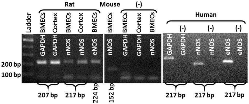
691
BRAIN-0816
Poster Session
ACUTE HYPONATREMIA IMPAIRES FUNCTION OF LARGE CONDUCTANCE CALCIUM-ACTIVATED POTASSUIM CHANNELS (BKCA) IN THE MIDDLE CEREBRAL ARTERY OF THE RAT.
Cerebrovascular Regulation
1Laboratory of Experimental Neurosurgery, Mossakowski Medical Research Centre Polish Academy of Sciences, Warsaw, Poland
2Department of Biophysics, Warsaw University of Life Sciences SGGW, Warsaw, Poland
3Department of Experimental and Clinical Physiology, Medical University of Warsaw, Warsaw, Poland
Abstract
Middle cerebral artery myocytes were isolated from 9 MCAs according to the method described by Holland et al.2 Conventional patch-clamp electrophysiology was used to measure whole cell BKCa currents using an Axopatch 200B amplifier (Axon Instruments).
1. Aleksandrowicz M, Kozniewska E. Disturbed regulation of the isolated middle cerebral artery in acute hyponatremia. Folia Neuropathol 2013; 51: 227-234.
2. Holland M, Langton PD, Standen NB, Boyle JP. Efect of the activator, NS1619, on rat cerebral artery smooth muscle. Br J Pharmacol 1996; 17(1): 119-129.
692
BRAIN-0190
Poster Session
EFFECTS OF PHYSICAL EXERTION AND HEAT ON CEREBROVASCULAR RESPONSE IN PROFESSIONAL FIREFIGHTERS
Cerebrovascular Regulation
1Kinesiology & Health Studies, University of Regina, Regina, Canada
2Medicine, University of Saskatchewan, Regina, Canada
3Health & Exercise Sciences, University of British Columbia Okanagan, Kelowna, Canada
Abstract
Firefighting is a physically demanding occupation and a number of factors can compromise the firefighter’s health during duty, including inadequate cerebral blood flow (CBF) regulation. We hypothesized that dynamic cerebral pressure–flow relationships will be altered in Professional Firefighters in response to acute physical exertion in the heat. Seven firefighters (age=30.8 ± 10.3 yrs) performed 30 minutes of treadmill walking at 65% of heart rate maximum in full turn-out gear in an environmental chamber at 39°C. Testing involved: 5 minutes squat-stand maneuvers at a point estimate at 0.05 Hz (the driven frequency) performed before and after the 30 minute treadmill test. CBF velocity was monitored in the middle cerebral artery (MCAv) using transcranial Doppler, non-invasive continuous blood pressure (BP) was recorded continuously using finger plethysmography, and 3-lead electrocardiography recorded heart rate. Transfer function analysis (TFA) was used to determine the autoregulatory metrics. The results showed that the driven TFA metrics were not significantly different (p<0.05) pre- to post-exercise for coherence (0.991 ± 0.007 vs 0.966 ± 0.037), gain (0.62 ± 0.15 vs 0.582 ± 0.199 cm/s/mmHg) and phase (0.634 ± 0.20 vs 0.612 ± 0.32 Rad), and power spectral density for both MCAv (22954.1 ± 11271.2 vs 24958.1 ± 13909.4 cm/s2) and BP (56121.0 ± 16177.9 vs 62612.6 ± 23002.4 mmHg2). In conclusion, the cerebral pressure – flow relationship in FF was unaltered pre- to post- moderate intensity exercise in the heat.
693
BRAIN-0658
Poster Session
INTERACTION OF INTRACRANIAL PRESSURE AND CEREBRAL PERFUSION PRESSURE IN ISCHEMIC INJURY OF THE HEALTHY RAT BRAIN.
Cerebrovascular Regulation
1Department of Traditional Chinese and Western Medicine, Xiangya Hospital Central South University, Changsha Hunan, China
2Neurosurgery, University of New Mexico, Albuquerque, USA
3Neurology, University of New Mexico, Albuquerque, USA
4College of Pharmacy, University of New Mexico, Albuquerque, USA
5Pathology and Laboratory Medicine, VA University Health Care System, Pittsburgh, USA
6Neurosurgery, University of New Mexico, Pittsburgh, USA
Abstract
In previous studies in healthy rats, we showed that a progressive increase in ICP from 10 to 30 and 50 mmHg induces microvascular shunting (MVS), characterized by tissue hypoxia, edema and increased blood brain barrier permeability even at normal to high CPP. Thus, high ICP may cause ischemic injury by MVS in the healthy brain. Our aim was to determine whether high ICP as a function of CPP causes ischemic injury in the healthy brain
Abstract
* = P <0.05 compared to ICP 10 and 30 mmHg.
Legend: Sum and average Fluorojade stained neurons in 18 sections taken from one section of the rat brains at the level of the striatum. Cerebral perfusion pressures at intracranial pressures of 10, 30 and 50 mmHg were 80, 55 and 50 mmHg, respectively.
694
BRAIN-0847
Poster Session
ACUTE INCREASES IN SYMPATHETIC TONE DURING COLD PRESSOR TEST RESULTS IN INCREASED GLOBAL BRAIN BLOOD FLOW
Cerebrovascular Regulation
1Pharmacology and Physiology, Rutgers Biomedical Health Sciences, Newark, USA
2War Related Illness and Injury Study Center, New Jeresy VA Hospital, East Orange, USA
3War Related Illness and Injury Study Center, New Jersey VA Hospital, East Orange, USA
Abstract
695
BRAIN-0625
Poster Session
CEREBRAL AUTOREGULATION IN PATIENTS WITH SEVERE BRAIN INJURY
Cerebrovascular Regulation
1Department of Neurorehabilitation TBI Unit, Glostrup University Hospital, Glostrup, Denmark
2Department of Anaesthesia, Rigshospitalet University of Copenhagen, Copenhagen, Denmark
3Department of Paediatrics, Copenhagen University Hospital Hvidovre, Hvidovre, Denmark
4Coordinating Research Centre, Frederiksberg Hospital, Copenhagen, Denmark
Abstract
696
BRAIN-0764
Poster Session
CEREBRAL OXYGENATION DURING TILT IN SEVERE BRAIN INJURY
Cerebrovascular Regulation
1Department of Neurorehabilitation TBI Unit, Glostrup University Hospital, Glostrup, Denmark
2Department of Anaesthesia, Rigshospitalet University of Copenhagen, Copenhagen, Denmark
3Department of Pediatrics, Copenhagen University Hospital Hvidovre, Hvidovre, Denmark
4Coordinating Research Centre, Frederiksberg Hospital, Copenhagen, Denmark
Abstract
697
BRAIN-0520
Poster Session
CHRONIC ANGIOTENSIN IV ADMINISTRATION RESCUES CEREBROVASCULAR DEFICITS IN A MOUSE MODEL OF ALZHEIMER’S DISEASE
Cerebrovascular Regulation
1Laboratory of Cerebrovascular Research, Montreal Neurological Institute McGill University, Montreal, Canada
Abstract
References: 1. Wolozin B et al., Alzheimers.Dement, 2008, 4: T118; 2. Ongali B et al., Neurobiol.Dis, 2014, 68: 126-136; 3. Braszko JJ et al., JRAAS, 2006, 7(3): 168-174; 4. Braszko JJ et al., J.Neurosci, 1988, 27(3): 777-783; 5. Albiston AL et al., Neurobiol.Learn.Mem, 2010, 93(1): 19-30; 6. Royea J et al., CANAD Abstract, 2014; 7. Deipolyi AR et al., Neurobiol.of.Aging, 2008, 29: 253-66
698
BRAIN-0360
Poster Session
INTRACRANIAL PRESSURE IS A PHYSIOLGICAL STRESSOR THAT DETERMINES SYMPATHETIC NERVOUS SYSTEM ACTIVITY
Cerebrovascular Regulation
1Neurosurgery, University Hospital, Toulouse, France
2I2MC, INSERM UMR-1048, Toulouse, France
3Neurology, University Hospital, Toulouse, France
4Neurosurgery, University Hospital, Cambridge, United Kingdom
5Pharmacology, University Hospital, Iowa City Iowa, USA
Abstract
Objectives:
Intracranial pressure (ICP) is a determinant of cerebral perfusion pressure (CPP) that is the difference between arterial blood pressure (ABP) and ICP. Raised ICP reduces CPP and blood delivery to the brain. Massive ICP rise is also known to produce an increase in ABP, bradycardia and respiratory irregularities termed the Cushing response1. This mechanism is a terminal event occurring in extreme pathological conditions of brainstem ischaemia leading to a sympatho-adrenal response2. However, it is still debated whether the Cushing response is an acute pathological response to brain ischemia or part of an important physiological reflex mechanism for ABP regulation3. Indeed clinical and experimental studies suggest that modest ICP increase modulates systemic hemodynamics probably via the sympathetic nervous system (SNS)4-7. We hypothesize that modest ICP changes drive sympathetic activity. Using state-of-the-art technique to measure SNS, we performed two different sets of experiments in human subjects and mice. In both species during controlled hydrostatic modest ICP increase and decrease, SNS was measured directly by microneurography and indirectly by heart rate variability analysis (HRVA).
In 10 patients, ICP was measured and increased during lumbar infusion study. Signed consent was always obtained. Heart rate and ABP were non-invasively monitored. Muscle sympathetic nerve activity (MSNA) was recorded by a microelectrode in the right fibular nerve.
In 15 anesthetized mice, ICP was measured and intraventricular infusion was performed. Heart rate and ABP were invasively monitored. Renal sympathetic nerve activity (RSNA) was recorded by a microelectrode at the level the left kidney’s nerves.
In human and mice, HRVA allows calculating high frequency (HF) and low frequency (LF) bands. LF and LF/HF ratio represent sympathetic indices, whereas HF is an index of parasympathetic activity 8
Table 1 gives the human and animal results. Strikingly, modest increase in ICP was associated with a parallel increase in MSNA in humans; blood pressure was stable. Similarly, modest rise in ICP increased RSNA in mice. In both species ICP drop significantly reduced MSNA and RSNA. Spectral analysis of heart rate variability confirmed that raising ICP augments sympathetic indices in humans and mice.
Using gold-standard measurement of SNS, we demonstrate that ICP drives efferent SNS outflow. ICP is not only a determinant of CPP but also a physiological stressor that influences and reversibly modulates SNS activity. We demonstrate a new physiological link between ICP and SNS activity which may represent an important highly regulated circuit. Our findings strongly suggest the presence of a novel intracranial baroreflex that might participate to the physiology of the heart-brain cross-talk, but also to the pathophysiology of various sympathetically-driven diseases.

References:
699
BRAIN-0850
Poster Session
RUGBY PLAYERS WITH CONCUSSIONS DEMONSTRATE INCREASED BLOOD PRESSURE AND ESTIMATES OF INTRACRANIAL PRESSURE IMMEDIATELY POST CONCUSSION WHILE CEREBRAL AUTOREGULATION REMAINS INTACT
Cerebrovascular Regulation
1Pharmacology & Physiology, Rutgers - New Jersey Medical School, Newark, USA
2War Related Illness & Injury Study Center, Dept of Veteran Affairs, East Orange, USA
Abstract
700
BRAIN-0854
Poster Session
EFFECT OF MENSTRUAL CYCLE ON CEREBRAL BLOOD FLOW REGULATION IN YOUNG WOMEN
Cerebrovascular Regulation
1War Related Illness & Injury Study Center, Dept of Veteran Affairs, East Orange, USA
2Pharmacology & Physiology, Rutgers New Jersey Medical School, Newark, USA
3Pharmacology & Physiology, Rutgers - New Jersey Medical School, Newark, USA
Abstract
701
BRAIN-0437
Poster Session
MECHANISTIC PATHWAYS UNDERLYING ALTERED NEUROVASCULAR COUPLING IN BRAIN METASTASIS
Cerebrovascular Regulation
1CRUK and MRC Oxford Institute for Radiation Oncology Department of Oncology, University of Oxford, Oxford, United Kingdom
2MIRcen UMR9199 CEA CNRS, University of Paris Sud, Fontenay-aux-Roses, France
Abstract
Objectives: It is recognised that astrocytes form a physical bridge between neurons and the cerebrovasculature. Astrocytic end-feet help maintain endothelial tight junctions to support the blood–brain barrier (BBB) and release vasoactive molecules that regulate vascular tone. In secondary cancer (metastasis) to the brain, astrocytes are displaced from the vasculature and become activated, and we have shown that neurovascular coupling is compromised in rat model of brain metastasis (Serres et al., abstract submitted). The aim of this study, therefore, was to characterise the effects of astrocyte activation on astrocyte-vascular structure, vascularity and production of vasoactive molecules, using in vivo magnetic resonance imaging (MRI) and immunofluorescent microscopy.
Methods: Two cohorts of BD-IX rats were injected intracortically in one node of the whisker barrel cortex pathway (the barrel field somatosensory cortex) with either (i) a lentivirus expressing ciliary neurotrophic factor (Lv-CNTF) known to switch astrocytic phenotype to an activated state, or (ii) a metastatic N-ethyl-N-nitrosourea-induced mammary adenocarcinoma cell line (ENU1546). Lv-CNTF injected animals were studied 6 weeks after intracortical injection, and ENU1546 injected animals were studied 1 week after injection. All animals underwent T1- and T2-weighted MRI to follow macroscopic structural changes, and post-gadolinium T1-weighted MRI to assess BBB integrity. Immunofluorescent microscopy was performed post-mortem to identify activated astrocytes (GFAP), neurons (NeuN), blood vessels (CD31), cyclooxygenase-1 and 2 (COX-1/2), inducible isoform of nitric oxide synthase (iNOS), glutathione (GSH), cytochrome p450 a precursor of 20-hydroxyeicosatetraenoic acid (20-HETE), alpha-smooth muscle actin (α-SMA) and proteoglycans of the basement membrane (β-dystroglycan).
Results: In both cohorts, persistent activation of astrocytes, revealed by strong upregulation of GFAP, was observed in the injected cortex, which was not associated with BBB disruption as assessed by MRI. Disruption of the dystroglycan-laminin interaction was observed in the area of astrocyte activation causing dissociation of astrocytes from blood vessels, as shown by a lower immuno-colocalisation of blood vessel, astrocyte and β-dystroglycan, for both Lv-CNTF and ENU1546 injected animals (Fig1). Enlargement of blood vessels was also observed in both models. Together with structural changes in the astrocyte-vessel complex, upregulation of iNOS, COX-1, COX-2 and GSH were observed in activated astrocytes. Whilst upregulation of cytochrome p450 was observed in α-SMA-positive arterioles found in the area of astrocyte activation.
Conclusion: Our findings suggest that astrocyte-vessel dissociation, leading to enlargement of blood vessels, together with dysregulation of signalling pathways controling vessel diameter likely underlie disruption of neurovascular coupling in brain metastasis. Upregulation of vasoconstrictory molecules (p450/20-HETE) at the arteriole end of the vascular bed may lead to reduced basal blood flow as observed Co-localization of β-dystroglycan (green), astrocyte (blue) and blood vessel (red) is reduced in the area of either Lv-CNTF or brain metastasis induced astrocyte activation.
702
BRAIN-0868
Poster Session
EFFECTS OF INTERNAL JUGULAR VEIN STENOSIS ON VENOUS COLLATERAL FLOW USING MAGNETIC RESONANCE IMAGING
Cerebrovascular Regulation
1The MRI Institute of Biomedical Research, Detroit, MI, USA
2University of Ferrara, Ferrara, Italy
Abstract
Objectives: Jugular venous structure and flow anomalies have been noted in a subset of multiple sclerosis (MS) patients using MRI1, 2. This study was to investigate effects of internal jugular vein stenosis on venous collateral outflow in a large cohort of MS and healthy controls (HC) using MRI. All analyses are retrospective from an imaging database.
Methods: A group of 569 MS subjects and 95 HC subjects were imaged with 3T scanners. Magnetic resonance (MR) venographic imaging and phase contrast flow quantification at two different neck levels (C5/C6 and C2/C3) were exclusively to image anatomy and flow of the extracranial vessels. MR venography was used to classify venous vessels into three categories: Type I: Primary (jugular vein), Type II: Paraspinal, and Type III: Superficial veins. Presence, type, and location of jugular anomalies were also evaluated based on criteria previously established1,2. Venous flow normalized to arterial brain inflow was calculated for individual and bilateral totals. The effects of stenosis type on venous collateral flow subtotals were assessed. Student t-tests were performed to compare means between groups. Chi-square tests were used to check differences in prevalence of jugular anomaly between the HC and MS groups.
Results: Forty-six of the 95 HC did not have venography data for anatomic evaluation. Vertebral artery inflow showed good consistency from the upper neck level to the lower neck level for the entire data set (Figure 1). Thirty-eight of the 49 HC classified as non-stenotic, while 11/49 HC classified as stenotic; of the MS cohort, 213/569 classified as non-stenotic while 346/569 showed stenosis. For normalized IJV flow, differences were seen in stenotic (M=0.51, SD=0.21) and non-stenotic (M=0.77, SD=0.12) HC groups (p<0.05), as well as stenotic (M=0.52, SD=0.21) and non-stenotic (M=0.72, SD=0.12) groups (p<0.05). When grouping both MS and HC groups, differences in Type I, II, and III flows were seen between non-stenotic and stenotic groups, with the stenotic group showing elevated Type II, and III flow compared to the non-stenotic groups (p<0.05).
Conclusions: Consistency in scanning and conservation of arterial inflow between neck levels was established based on the vertebral artery flow. Anatomical jugular stenosis was more prevalent in the MS cohort compared to HC (X2=28.9, p<0.05). For cases with anatomical jugular stenosis in both MS and HC, flow is redirected to Type II and III venous collaterals while maintaining similar total normalized venous flow to the non-stenotic group.
References:
703
BRAIN-0523
Poster Session
IMPACT OF AUTONOMIC MODULATION DURING CEREBROVASCULAR REACTIVITY IN SMALL ARTERY STROKE
Cerebrovascular Regulation
1Stroke Unit, Thammasat University Hospital, Pathumthani, Thailand
2Faculty of Medicine, Thammasat University, Pathumthani, Thailand
Abstract
Abstract—Autonomic modulation involves in brain-heart axis havereported in AF stroke and hypothermia treated patients. Predominant activity ofparasympathetic nerve and significant increase in mean cerebral blood flowvelocity during cerebrovascular reactivity (CVR) in healthy was evident. Cerebrovascularreactivity by breath holding induces vasodilation. Recently, many reports suggest that endothelialdysfunction may be involved in the pathogenesis of small artery stroke. In this study, weexamine the impact of ANS modulation by heartrate variability (HRV) to cerebral reserve function in small artery stroke.
In conclusion, nonlinear HRV with frequencydomain is proposed in CVR assessment which it needed tocompromise the lower cerebral blood volume velocity in small artery stroke.
Reference:
704
BRAIN-0080
Poster Session
HYPERCAPNIA-INDUCED CEREBRAL VASODILATION IS MEDIATED PREDOMINANTLY BY ENOS
Cerebrovascular Regulation
1Neurosurgery, Ludwigs-Maximilians-Universität, München, Germany
2Anesthesiology, Ludwigs-Maximilians-Universität, München, Germany
3Institute for Stroke and Dementia Research, Ludwigs-Maximilians-Universität, München, Germany
Abstract
CO2-reactivity is an important feature of cerebral autoregulation. Increase of CO2 partial pressure (pCO2) induces a CPP independent increase of cerebral perfusion. Lack of CO2-mediated vasoreactivity is an important feature of the pathophysiology of several cerebral insults, especially subarachnoid hemorrhage; after SAH, impaired CO2-reactivity is associated with worse neurological outcome. The mechanism of CO2-elicited hyperemia has not been completely elucidated. There is consensus that nitric oxide (NO) is heavily implicated, however, the role of the individual NOS-isoforms in inducing arterial dilation is not completely clear. The present study was initiated in order to evaluate the CO2-elicited CBF-responses of the cerebral microcirculation in mice deficient for eNOS, nNOS, and iNOS.
eNOS-/-, iNOS-/-, nNOS-/- mice, and wildtype C57BL/6 littermates were anesthetized, intubated, and mechanically ventilated with 30% oxygen in room air. Temperature, endtidal pCO2, and respiration parameters were continuously monitored and kept within physiological range. MAP measured via femoral artery catheter and CBF measured via bitemporal Laser-Doppler-Flowmetry over MCA territory were continuously monitored. The cerebral microcirculation was studied using in-vivo-epifluorescence microscopy under baseline conditions, during, and after a 30-minute hyperkapnic stimulus (addition of 10% CO2). At the end of the experiments, ex vivo blood gas analysis was performed.
As previously described, eNOS-deficient mice had an elevated arterial blood pressure under baseline conditions. All other parameters obtained were comparable in the 4 groups examined. As expected, increasing the CO2-concentration to 10% led to an immediate and significant increase in CBF in control animals; MAP remained stable during CO2 inhalation (iCO2). The CBF-increase was mainly mediated by dilation of small cerebral arterioles (10-20µm) since these vessels had the biggest change in diameter compared to baseline. After cessation of iCO2 CBF returned quickly to baseline. iNOS-transgenic animals showed a CO2 vessel reactivity similar to wildtype controls while this response was significantly blunted in nNOS-transgenic mice and completely absent in eNOS-deficient animals. Interestingly, iCO2 tented to reduce CBF in eNOS KO mice.
The vasodilation elicited by hypercapnia is mediated by precapillary arterioles (diameter 10-20µm) and predominantly by eNOS and, to a lesser degree, by nNOS while iNOS is not involved in this process. The current study clarifies the role of the individual NOS isoforms for hypercapnia-induced vasodilatation and further deepens our understanding on this fundamental process in the cerebral microcirculation.
705
BRAIN-0775
Poster Session
BEHAVIOR OF RED BLOOD CELLS IN INTRAPARENCHYMAL CAPILLARIES DURING CORTICAL SPREADING DEPRESSION OBSERVED WITH HIGH-SPEED CAMERA CONFOCAL FLUORESCENCE MICROSCOPE IN ANESTHETIZED MICE
Cerebrovascular Regulation
1Neurology, Keio University School of Medicine, Tokyo, Japan
2Brain Science Inspired Life Support Research Center, University of Electro-Communications, Chofu, Japan
3Molecular Imaging Center, National Institute of Radiological Sciences, Chiba, Japan
4Neurology, Osaka City University Graduate School of Medicine, Osaka, Japan
Abstract
[Objective] Cortical spreading depression (CSD) involves mass depolarization of neurons and glial cells, followed by sustained suppression of spontaneous neuronal activity. CSD induces marked increases in glucose utilization and metabolism, resulting in severe tissue hypoxia (1), accompanied with marked constriction, followed by dilation and subsequent mild constriction of pial arteries (2), and suppression of red blood cell (RBC) velocity (3). Since changes of capillary diameter are limited, control of RBC velocity in capillaries is essential to meet local neuronal requirements. To further understand the microcirculatory response during CSD, we observed and analyzed the temporal changes of RBC velocity and its distribution in individual capillaries by two-dimensional spatial analysis.
[Methods] Male Tie 2-green fluorescent protein transgenic mice, in which fluorescent vascular endothelial cells can be specifically identified, were used (N=13). Under urethane anesthesia and artificial ventilation, intraparenchymal images (approximately 50 µm from the surface) were obtained using a high-speed camera laser-scanning confocal fluorescence microscope at 125 fps for 60 sec through a cranial window (4) made on the parieto-temporal cortex, with simultaneous recording of DC potential and regional cerebral blood flow (rCBF) by laser-Doppler flowmetry. A fluorescein isothiocyanate (FITC)-labeled RBC suspension was intravenously injected, and the velocity of RBCs was automatically measured with our original Matlab domain software (KEIO-IS2) (5). CSD was induced by application of KCl to the cortical surface through an additional hole.
[Results] RBC velocity in individual capillaries at rest fluctuated irregularly, but we confirmed that the average was stable for at least 10 to 12 sec before KCl application. We took the averaged RBC velocity for 30 sec before KCl application as baseline and calculated % change from the baseline in each capillary during and after passage of CSD (in total; n=122). Application of KCl elicited repeated transient depolarization, seen as DC potential deflection and delayed hyperperfusion (Figure A-C; middle and lower panels). During the first CSD passage, RBC velocity was significantly decreased approximately in concurrence with the trough of DC potential and a transient drop of rCBF (FigureA). Subsequent CSD did not induce temporal changes in RBC velocity, which were rather high during CSD passage, whereas rCBF was increasing without a transient drop (Figure B and C). RBC velocity remained high (+6 to +22%) after passage of CSD, whereas rCBF significantly decreased by 13 to 19% (post-CSD oligemia). The changes in RBC velocity were heterogeneous in space, though the average velocity was well maintained.
[Conclusion] We demonstrated temporal change of RBC velocity in intraparenchymal capillaries and its spatial heterogeneity during CSD. This model is useful to investigate regulatory mechanisms of cerebral capillary flow and coupling between neurons and nearby capillaries.
[References] (1) Takano T, et al., Nat Neurosci 10: 754-62: 2007. (2) Unekawa M, et al., J Cereb Blood Flow Metab, in press (doi:10.1038/jcbfm.2014.250). (3) Unekawa M, et al., Microcirculation 19: 166-74: 2012. (4) Tomita Y, et al., J Cereb Blood Flow Metab, 25: 858-67: 2005. (5) Tomita M, et al.,Microcirculation 15: 163-174: 2008.

706
BRAIN-0375
Poster Session
SUPPLY-DEMAND MISMATCH TRANSIENTS TRIGGER PERI-INFARCT DEPOLARIZATIONS IN ISCHEMIC PENUMBRA
Cerebrovascular Regulation
1Neurology and Radiology, Massachusetts General Hospital Harvard Medical School and Charité-Universitätsmedizin Berlin, Berlin, Germany
2Neurology and Radiology, Massachusetts General Hospital Harvard Medical School and Leiden University Medical Center, Leiden, Netherlands
3Radiology, Massachusetts General Hospital Harvard Medical School, Boston, USA
4Radiology and Pharmacology, Massachusetts General Hospital Harvard Medical School and Gazi University Faculty of Medicine, Ankara, Turkey
5Neurosurgery, Charité-Universitätsmedizin Berlin, Berlin, Germany
6Athinoula A Martinos Center for Biomedical Imaging, Massachusetts General Hospital Harvard Medical School, Boston, USA
7Neurology, Charité-Universitätsmedizin Berlin, Berlin, Germany
8Radiology and Neurology, Massachusetts General Hospital Harvard Medical School, Boston, USA
Abstract
References:
707
BRAIN-0396
Poster Session
THE EFFECT OF RAPAMYCIN ON COLLATERALS IN RODENT CEREBRAL ISCHEMIA USING NOVEL MULTI-SCALE DYNAMIC IMAGING MODALITIES
Cerebrovascular Regulation
1Neuroscience and Neuroengineering Research Center, Med-X Research Institute and School of Biomedical Engineering, Shanghai, China
2Pharmacology and Neuroscience, University of North Texas Health Science Center, Fort Worth, USA
Abstract
Abstract
SRA image of collateral circulation (A) and (B) between posterior cerebral artery and middle cerebral artery after MCAO in rats.The statistical bar graphs (C, D) showed the diameter of collateral a and b 0.5, 1, 3 and 6 hours after MCAO in control and rapamycin treated rats. Bar: 1 mm. TPLSM image of arterioles before (A) and (B) after rapamycin treatment in living normal mice. Bar:10µm.

References
708
BRAIN-0169
Poster Session
EFFECT OF MAXIMAL BREATH HOLD APNEA ON PIAL ARTERY PULSATION AND SUBARACHNOID WIDTH IN HUMAN
Cerebrovascular Regulation
1Institute of Human Physiology, Medical University of Gdansk, Gdansk, Poland
2Department of Hypertension and Diabetology, Medical University of Gdansk, Gdansk, Poland
3Department of Radiology Informatics and Statistics, Medical University of Gdansk, Gdansk, Poland
4Department of Computer Communications Faculty of Electronics Telecommunications and Informatics, Gdansk University of Technology, Gdansk, Poland
Abstract
Objectives
Little is known about intracranial pressure (ICP)-cerebral hemodynamic interplay during maximal breath-hold apnea. A recently developed method based on near-infrared transillumination/backscattering sounding (NIR-T/BSS) non-invasively measures changes in pial artery pulsation (cc-TQ) as well as subarachnoid width (sas-TQ) in humans [1,2,3,4]. Changes in sas-TQ correlate with ICP to a considerable extent [1], whereas cc-TQ reflects the functional status of the pial artery [1,3,4]. We tested the complex response of the pial artery and subarachnoid width to apnea using this method.
The pial artery and subarachnoid width response to maximal breath-hold (91.1±23.1 s) was studied in 20 healthy volunteers. The cc-TQ and sas-TQ were measured using NIR-T/BSS; cerebral blood flow velocity (CBFV), pulsatility index (PI) and resistive index (RI) were measured using Doppler ultrasound of the left internal carotid artery; heart rate (HR) and beat-to-beat systolic (SBP) and diastolic (DBP) blood pressure were recorded using a Finometer; end-tidal CO2 (EtCO2) was measured using a medical gas analyzer. The relationship between spontaneous oscillations in BP and cc-TQ at frequencies between 0.5 Hz and 5 Hz was assessed using wavelet transform analysis.
With reference to baseline recordings apnea evoked a multiphasic response in BP, cc-TQ and sas-TQ (Figure 1, 2). First, SBP declined (-15.2%, P<0.01), which was accompanied by an increase in cc-TQ (17.1%, P<0.01) and sas-TQ (47.6%, P<0.001). Directly after these changes, SBP exceeded baseline values (14.4%, P<0.001), which was associated with a decline in cc-TQ (-12.3%, P<0.001) and the return of sas-TQ to baseline. During these initial changes CBFV first borderline increased (P<0.01) which was followed by its normalization (P=NS). However, toward the end of the apnea, BP, cc-TQ and CBFV increased (29.2%, P<0.001; 46.7%, P<0.01 and 73.3%, P<0.001, respectively) while PI, RI and sas-TQ declined (-42.5%, P<0.01; -21.2%, P<0.001 and -26.7%, P<0.01, respectively). A multivariate analysis showed that changes in sas-TQ were linked to variations in EtCO2, HR and SBP. Increase in wavelet coherence between augmented BP and cc-TQ oscillations was observed by the end of apnea (P<0.05; Figure 3).
Apnea is associated with ICP swings, closely reflecting changes in EtCO2, HR and peripheral BP. The baroreflex influences the pial artery response. Apnea increases the contribution of cardiac activity to BP and cc-TQ oscillations. Baroreflex and ICP changes are involved in a complex and intricate mechanism aiming at the stabilization of CBF.


References
709
BRAIN-0585
Poster Session
ELECTRICAL COMMUNICATION IN THE CEREBRAL ARTERIES: IMPORTANCE TO GLOBAL BLOOD FLOW REGULATION AND STROKE INJURY
Cerebrovascular Regulation
1Physiology and Pharmacology, University of Calgary, Calgary, Canada
Abstract
710
BRAIN-0530
Poster Session
PERICYTE CONTROL OF THE MICROCIRCULATION IN ISCHEMIC BRAIN
Cerebrovascular Regulation
1Anesthesiology and Perioperative Medicine, Oregon Health & Science University, Portland, USA
2Pathology, Oregon Health & Science University, Portland, USA
Abstract
Objective: Despite successful recanalization of the occluded cerebral artery using tissue plasminogen activator or endovascular devices, more than half of stroke patients still end up with a poor clinical outcome1. This may be due to prolonged changes within the microvasculature contributing to microvascular perfusion deficits. We investigated whether pericyte constriction is associated with impaired microvascular blood flow in a mouse model of ischemic stroke with reperfusion. We then evaluated the role of epoxyeicosatrienoic acids (EETs), previously shown to regulate microvascular perfusion in brain2, in the regulation of pericyte tone in the cerebral microcirculation.
Methods: The cortical microvasculature in the penumbra of Tie2-GFP/NG2-DsRed mice was imaged with two-photon microscopy through a chronic thin skull window before, 6 and 24 hours after 1-hour middle cerebral artery occlusion (MCAO). The resultant z-stacks were analyzed with Imaris FilamentTracer to detect capillary diameter changes, and red blood cell (RBC) velocity and flux were assessed by capillary line scans. At 24 hours of reperfusion, animals were fixed and stained with Hoescht dye to detect evidence pericyte death in the ischemic penumbra. In a separate set of animals, the following agents were microinjected into the cortex through a craniotomy to investigate their roles in pericyte contractility: ATP, the EETs antagonist 14,15-Epoxyeicosa-5(Z)-enoic acid (14-15-EEZE) or vehicle.
Results: After MCAO, the majority of capillaries in the ischemic penumbra underwent a bidirectional response with the majority of capillaries experiencing a loss of tone, while others experienced constriction. BBB integrity was compromised surrounding pericytes that lost tone after stroke. Application of 14-15 EEZE resulted in robust pericyte contraction, suggesting that endogenous EETs tonically promote pericyte relaxation, whereas ATP elicited more subtle and inconsistent pericyte contraction. Regardless of diameter change, the majority of capillaries scanned exhibited markedly reduced RBC velocity and flux after MCAO. Pericyte cell death was evident in the ischemic core but not in the ischemic penumbra.
Conclusion: Capillaries in the ischemic penumbra undergo either dilation or constriction, which seem to reflect different responses to ischemic injury. RBC velocity and flux are compromised in the penumbra regardless of dilation or constriction while BBB permeability is associated with loss of pericyte tone and capillary dilation. Based on the constrictor activity of the EETs antagonist 14,15-EEZE, we propose that endogenous EETs mediate pericyte relaxation at baseline, which is lost after ischemia.

References:
711
BRAIN-0163
Poster Session
SKULL OPTICAL CLEARING FOR IMPROVING CEREBROVASCULAR IMAGING
Cerebrovascular Regulation
1Wuhan National Lab for Electronics, Huazhong University of Science & Technology, Wuhan, China
Abstract
Various optical imaging methods for visualization of both structural and functional architecture with high temporal-spatial resolution have shown tremendous advantages in investigation of cortical neurons and microvasculature. However, the applications usually depend on cranial window by craniotomy because the turbid skull reduces the imaging contrast and resolution. Fortunately, we developed an innovative skull optical clearing solution (SOCS), which could make the skull transparent within 20-30 minutes. With the optical clearing cranial window, it is easy to monitor the blood flow with the laser speckle contrast imaging and blood oxygenation with the multispectral imaging. Not only the resolution, but the signal to noise ratio and sensitivity were significantly improved. The transparent cranial window was introduced in an acoustic resolution photoacoustic microscopy (AR-PAM) or optical resolution photoacoustic microscopy (OR-PAM) for mapping the cerebral vasculature. The results demonstrated that the former’s signal level can be effectively elevated under the case of retaining the resolution. For the latter, the resolution can be increased by more 2 times. For the both modes PAMs, the signal from deeper can be obtained. The skull optical clearing technique is hopeful to provide a simple and effective method for visualizing cerebral microvascular structure and function.
712
BRAIN-0482
Poster Session
EFFECT OF ADRENALINE ON CEREBRAL BLOOD FLOW IN HEALTHY RATS AND RATS WITH INTRACRANIAL HEMORRHAGE
Cerebrovascular Regulation
1Biology, Saratov State University, Saratov, Russia
Abstract
In the next stage the role of adrenergic effects in the regulation of cerebral blood flow in neonatal rats under normal conditions and during the development of stress-induced cerebral hemorrhage with the use of OCT was examined. The results were recorded in 4 hours and 24 hours after stress.
The diameter of arteries increased, large-diameter arteries were insensitive to the effects of the drug under the influence of adrenaline. Blood flow velocity in the cerebral arteries was significantly reduced. Sagittal vienna decreased in response to the injection of adrenaline. However, the velocity of blood flow and perfusion in it did not significantly change. Loss of reactivity to epinephrine was observed either 4 hours after the stress or a day later in stressed rats.
The research supported by grants No 14-02-00526-a, MD-2216.2014.4
Reference:
713
BRAIN-0582
Poster Session
TUMOR EFFECTS ON BOLD FMRI SIGNAL IN BROCA'S AREA
Neurovascular Coupling
2Radiology, WVU, Morgantown, USA
2Bio-Enginnering, University of Washington, Seattle, USA
3Neurosurgery, Huashan Hospita, Shanghai, China
4Neuoral surgery, WVU, Morgantown, USA
5Neural Surgery, Huashan Hospita, Shanghai, China
Abstract
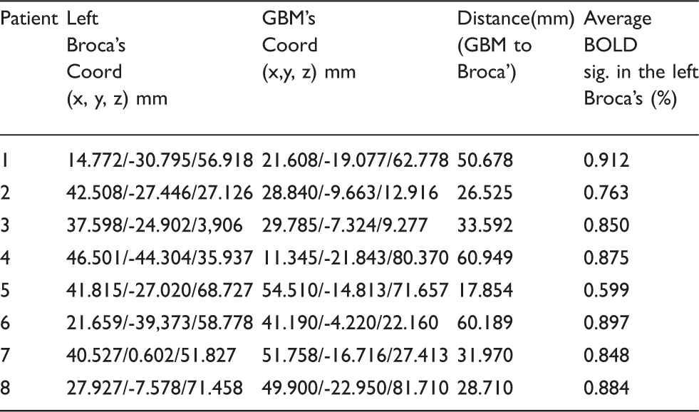
714
BRAIN-0680
Poster Session
MAPPING LARGE-SCALE FUNCTIONAL CONNECTIONS WITH CHR2-EVOKED HEMODYNAMIC SIGNALS
Neurovascular Coupling
1Radiology, Washington University in Saint Louis, Saint Louis, USA
2Neuroscience, Washington University in Saint Louis, Saint Louis, USA
3Anesthesiology, Washington University in Saint Louis, Saint Louis, USA
Abstract

References
715
BRAIN-0058
Poster Session
IN VIVO OPTOGENETIC MANIPULATION OF CELLS WITHIN THE NEUROVASCULAR UNIT LEADS TO LOCAL CHANGES IN NEURAL ACTIVITY
Neurovascular Coupling
1Neuroscience, Brown University, Providence, USA
Abstract
Changes in neural activity can drive vasodilations in local blood vessels, a process measured by functional magnetic resonance imaging. Despite substantial investigation of the relationship between neural and vascular dynamics, the potential impact of vascular responses on local neural activity is unknown. Here, using in vivo two-photon imaging of neural calcium responses in sensory cortex, we found that sensory-evoked changes in neural activity lead and follow vasodilations. To determine if vasodilations drive changes in local neural activity, we developed optogenetic control of cerebral vascular responses (vasOpto) to shape vascular dynamics similar to the vasodilations that occur during sensory processing. VasOpto-evoked dilations led to rapid changes in neural activity and these changes predicted the dynamics observed with natural sensory-driven vasodilations in a significant portion of neurons. Our results capture a previously inaccessible dimension of sensory processing and may reveal new strategies to treat neurological and vascular disease.
716
BRAIN-0219
Poster Session
DYNAMIC MAGNETISATION TRANSFER MRI: THE EFFECT OF CARDIAC PULSATION ON CELLULAR WATER EXCHANGE
Neurovascular Coupling
1Institute of Neuroscience, Trinity College Dublin, Dublin, Ireland
Abstract
Objectives
MRI methods can measure dislocation of water molecules [1]. They can differentiate between flow and diffusion but not between flow and movement. This is the reason why signal changes in diffusion-weighted imaging which coexist with the cardiac pulse are considered generally related to movement [2]. Here, we show a method to measure the intra-extracellular water exchange with a low sensitivity for movement. Because of insensitivity to movements, signal changes in the tissue related to the cardiac pressure wave are detectable.
The MRI sequence consists of a magnetisation transfer (MT) preparation phase followed by single-slice echo-planar imaging sequence. MT effect was introduced by a negative (-5236.8 ± 894.18Hz) and a positive off frequency (6982.5 ± 894.18Hz Hz) RF pulse. Imaging parameters were voxel size of 3.5 x 3.5 x 3 mm, TR = 60 ms, TE = 11 ms, FA = 35 degrees and 2000 repetitions. For comparison, a control sequence with the same read out parameter but without the MT preparation was used.
12 subjects were scanned with the protocols approved by the Trinity College ethics committee and the Irish Department of Health.
A comparison between the time series acquired, both with and without MT preparation, is shown in the figure. For both cases, a voxel was extracted to showcase the difference between each time series. Only for the MT preparation, strong cardiac peaks are resolved while for the control sequence physiological noise is dominant. Peaks can be observed in all regions except the periventricular area whereby white matter, in general, shows higher peaks than grey matter. These results are consistent over all subjects.

Time series extracted from a voxel with MT preparation (red) and without MT preparation (blue).
To our knowledge, the observation of a cardiac related MT change has not yet been reported, therefore the mechanism behind this contrast is extremely interesting. Most likely, the pool of free water that exchanges with the macromolecules [3], is removed or separated by the pressure wave with the effect that the proton exchange between the pools is interrupted. Our explanation is that the pulse wave compresses cells with the result that water is pumped out of the cells. Hypothetically, this could then help to facilitate the lymphatic clearance of extracellular solutes and fluid from the brain parenchyma.
This would be consistent with new findings that astroglial AQP4 water channel play an important role in the interstitial solute clearance [4].
References
717
BRAIN-0186
Poster Session
SIMULTANEOUS VISUALIZATION AND ANALYSIS OF CELLS AND BLOOD VESSELS IN A WHOLE MOUSE BRAIN
Neurovascular Coupling
1Wuhan National Laboratory for Optoelectronics, Huazhong University of Science & Technology, Wuhan, China
Abstract
References:
718
BRAIN-0566
Poster Session
FUNCTIONAL ACTIVATION OF VISUAL CORTEX AND THE EFFECT ON CYTOCHROME C OXIDASE OXIDATION STATUS
Neurovascular Coupling
1Neurocritical Care, National Hospital for Neurology and Neurosurgery University College London Hospitals, London, United Kingdom
2Medical Physics and Bioengineering, University College London, London, United Kingdom
Objectives
Abstract
Neurovascular coupling results in typical task evoked functional hyperaemia. This is exploited in functional imaging because direct measurement of metabolic activity is problematic1. Cytochrome c oxidase (CCO), the terminal electron acceptor in the mitochondrial respiratory chain, exhibits task evoked changes in oxidation status, and its measurement likely reflects metabolic changes at the mitochondrial level. Using in-house broadband functional near-infrared spectroscopy optimised for detection of CCO in adults we have previously demonstrated different patterns of CCO change during frontal cortex activation2, which may reflect differences in neurovascular coupling or metabolism. The V1 visual cortex is defined histologically into two areas (blob, interblob) by their CCO concentration – and it is thought that this is an adaption to deliver a greater capacity for oxidative metabolism in the blob region. However the nature of flow-metabolism coupling in this region remains disputed. The aim of this work is to examine the CCO response to functional activation in blob/interblob regions.
Optodes of an in-house broadband spectrometer2 were placed over left frontal (control) and left visual cortex. The visual stimulation paradigm consisted of an isoluminant chromatic challenge (red-green 4Hz flicker - blob stimulation) and luminance challenge (black and white checkerboard - interblob stimulation). Each epoch consisted 20sec periods of chromatic, black, luminance and black, repeated 20 times. Concentration changes in oxy-[HbO2] deoxy-haemoglobin [HHb] and the oxidation status of CCO [oxCCO] were derived by fitting the broadband spectra between 780-900nm at a 3.5cm source detector spacing. Results were averaged across the repeated blocks and the [oxCCO] change at time 20 and 40sec compared using Students t-test. Because the partial pathlength of light is likely to vary between the two brain regions, derived concentration changes may not reflect actual differences. Therefore we compared the gradient of [Hbdiff] ([HbO2]-[HHb], marker of oxygen delivery) to [oxCCO] using linear regression, for each challenge.
8 healthy volunteers were recruited following ethics approval and written consent. Figure 1 demonstrates the changes in [HbO2], [HHb] and [oxCCO] observed across the group. The chromatic and luminance challenge generated significant elevations in [oxCCO] 0.06 micromol/L (p<0.001, 95%CI 0.04-0.08) and 0.11 micromol/L (p<0.001, 95%CI 0.08-0.15) respectively in the visual cortex. No such changes were seen in the frontal brain region. There was a difference in the ([oxCCO]/[Hbdiff] gradient between the two challenges in the visual cortex - chromatic 0.17 (99%CI 0.15 – 0.19) and luminance 0.10 (99%CI 0.08-0.12).
We have demonstrated differences in [oxCCO] dynamics between two different visual stimulation paradigms in different regions of visual cortex. This might reflect differences in metabolism or oxygen delivery between blob and interblob regions proposed by other investigators, and is also consistent with our previous observations and mathematical modeling of [oxCCO] dynamics2. Further work is required to elucidate the physiological significance of these data, but our findings suggest that [oxCCO] may have unique potential to provide a window on cellular energetics in the investigation of neurovascular coupling.

References
719
BRAIN-0218
Poster Session
ROLE OF NEUROTRANSMITTERS ON THALAMOCORTICAL REGULATION OF THE ACTIVATED BARREL CORTEX: A FUNCTIONAL MR SPECTROSCOPY STUDY.
Neurovascular Coupling
1CIBM, Ecole polytechnique fédérale de Lausanne, Lausanne, Switzerland
Objectives: Our aims were to explore the role of neurotransmitters on the neuro-metabolic and vascular regulation of the barrel cortex (S1BF) by the thalamus.
Abstract
A strong trend towards a negative correlation of BOLD percent changes between S1BF and the thalamus was found. At Rest, BOLD changes in S1BF were strongly and negatively correlated to GABA levels (Pearson’s coefficient=-0.75, p=0.013). In S1BF, Glu levels were correlated at rest with BOLD changes but not during stimulation. ΔGlu were strongly and negatively correlated to ΔBOLD (Pearson’s coefficient=-0.70. p=0.024).

References:
720
BRAIN-0426
Poster Session
CORTICAL SPREADING DEPRESSION AND IN VIVO CALCIUM IMAGING IN TRANSGENIC MICE WITH FAMILIAL HEMIPLEGIC MIGRAINE TYPE 1
Neurovascular Coupling
1TransNeuro, Institute of Neuropharmacology, Copenhagen, Denmark
2Department of Human Genetics, Leiden University Medical Centre, Leiden, Netherlands
Abstract
Familial hemiplegic migraine type 1 (FHM1) is a rare autosomal dominantly inherited subtype of migraine with prolonged aura of visual and sensory symptoms. The disease is caused by a mutation of the CACNA1A gene that encodes the α1A-subunit of voltage-gated Ca2+ channel in presynaptic terminals. The migraine aura is most likely caused by cortical spreading depression (CSD), a transient slowly propagating depolarization wave, which is strongly dependent on the interaction between glutamate and pre- and postsynaptic glutamate NMDA receptors. Due to increased action-potential-evoked Ca2+ influx in presynaptic terminals there is an increased probability of glutamate release at pyramidal cell synapses in FHM1; in comparison inhibitory neurotransmission at fast-spiking interneuron synapses remained unchanged. However, it is incompletely understood to what extent this expected enhancement of excitatory neurotransmission is reflected in Ca2+ activities in cortical pyramidal cells and glia under physiological conditions and CSD in vivo. It is also unclear how the basal facilitation is reflected in the mechanisms that couple rises in synaptic activity to changes in the cerebral blood flow (CBF) and the cerebral metabolic rate of oxygen (CMRO2).
We here explored the effect of somatosensory stimulation and CSD on neuronal and glial Ca2+ activities and the corresponding changes in CBF and CMRO2 in FHM1 with the R192Q mutation as compared to wild type mice in vivo. We report that in the mouse somatosensory cortex of FHM1 mice, afferent stimulation produced smaller, not larger, rises in neuronal and glial Ca2+ activities than in WT mice. In comparison, Ca2+ rises and the DC potential changes induced by CSD were similar for FHM1 and WT mice. CSD produced a larger increase in CMRO2 in FHM1 than in WT mice, but a similar rise in CBF. This indicated a mismatch between O2 use and supply that was reflected in protracted tissue anoxia in FHM1 mice, while WT animals only displayed a reduced level of tissue O2. FHM1 mice showed a slower recovery of cerebral blood flow to pre-CSD baseline values than wild-type after a CSD, and following the CSD, evoked CBF and Ca2+ responses were moderately decreased in WT mice and severely affected in FHM1 mice.
This study shows that the R192Q mutation leads to impairment in control of activity-.dependent rises in neuronal and glial Ca2+ activities in vivo. This impairment is not reflected on the overall EPSP amplitudes, but in impaired neurovascular coupling in FHM1 mice. Collectively, the data support the notion that affection of selective Ca2+ -dependent regulation of neurovascular coupling may underlie several key features of FHM1 such as the prolonged aura in the human phenotype. Specifically, the protracted anoxia and the abolished neurovascular coupling response during and following CSD provide a potential mechanism for the severe and prolonged neurological deficits in FHM patients. This underscores that genetic factors affecting the supply of substrates for brain energy metabolism and in turn brain ion homeostasis contribute to the phenotypic diversity of human migraine syndromes.
721
BRAIN-0322
Poster Session
NEW MODEL FOR THE BOLD HEMODYNAMIC RESPONSE EVOKED BY BRIEF NEURAL ACTIVATION IN HUMAN CEREBRAL CORTEX
Neurovascular Coupling
1Neuroscience, Baylor College of Medicine, Houston, USA
Abstract
Objective
The BOLD signal evoked by short stimulation, the hemodynamic response function (HRF), is the basis for linear analysis of most functional magnetic resonance imaging (fMRI) experiments. We propose a new model that includes only CBF and CMRO2 responses as drivers of the BOLD HRF, excluding venous cerebral blood volume (CBV) changes based on many recent experimental findings [1-4]. The CBF response is modeled using a linear network as a biomechanical model of the pial vasculature [5]. A stereotypical parametric temporal form is assumed for the CMRO2 response. We test this model against measurements of the BOLD HRF in human visual cortex evoked by brief stimulation using high-resolution fMRI to avoid contamination from large pial blood vessels.
The BOLD signal depends on changes in oxygen saturation (SO2) in capillaries and veins. To model longitudinal oxygen transport, we assume a uniform cylindrical geometry for the capillary and finely grid its length. We then use differential mass balance, including the processes of blood flow, hemoglobin dissociation, and oxygen diffusion into extravascular tissue, to obtain a continuous SO2 spatial profile, Fig 1A. Venous oxygen saturation is assumed spatially constant from the value at the distal end of the capillary.
The CBF response was modeled using a linear network model to describe the upstream arterial impulse response produced by prompt arterial dilation. We simulated CMRO2 response using a gamma-variate-function kernel.
In experiments, stimulus was a 2-s pulse of 4-Hz flickering dots followed by a 26-s blank period to let the HRF evolve and subside. High-resolution fMRI data (0.9-mm voxels) was obtained in 7 subjects (3-shot spiral acquisition, 8 slices, 1.5-s/volume). Each session produced ∼85 HRF responses that were averaged together throughout the activated gray matter in prescribed portions of areas V1—3.
The BOLD model successfully fit the experimental data, Fig. 1B. The model explains the early latency as competition between CBF and CMRO2, Fig 1C. After the CMRO2 demand peaks, the CBF continues to increase for a few seconds while the CMRO2 decreases. The late-time behavior of the HRF is explained by the underdamped oscillatory CBF response, and there is significant variability of the undershoot between subjects.
The model predicts the largest contribution (68%) on the BOLD signal is from the intravascular venous compartment, followed by intravascular capillary (22%), then extravascular venous (10%). There is minimal contribution (< 1%) from the extravascular capillary compartment.
We developed a new BOLD model based on prompt arterial dilation evoked by brief neural stimulation in human cortex. The model provides a simple, self-consistent biomechanical mechanism for an underdamped CBF response, and integrates it with oxygen transport. Our theory successfully fits measurements of the HRF evoked in early visual cortex, and explains the HRF time course as competition between CBF and CMRO2, a much simpler picture that appropriately neglects blood volume effects for brief stimulation periods.

Reference
722
BRAIN-0801
Poster Session
HEMODYNAMIC AND NEURONAL RESTING STATE FUNCTIONAL CONNECTIVITY MAPPING IN THE AWAKE MOUSE BRAIN
Neurovascular Coupling
1Biomedical Engineering, Columbia University, New York City, USA
2Neurobiology and Behavior, Columbia University, New York City, USA
Abstract

References
723
BRAIN-0494
Poster Session
SIMVASTATIN RESCUES IMPAIRED HIPPOCAMPAL NEUROVASCULAR COUPLING BY NORMALIZING VASCULAR REACTIVITY TO NEURONAL AND ASTROCYTIC STIMULATIONS IN APP TRANSGENIC MICE
Neurovascular Coupling
1Department of Pharmacology, Université de Montréal, Montreal, Canada
2Montreal Neurological Institute, McGill University, Montreal, Canada
Abstract
724
BRAIN-0659
Poster Session
SPONTANEOUS OPTICAL NEURAL, HEMODYNAMIC AND METABOLIC SIGNALS TO ACCESS RESTING-STATE FUNCTIONAL CONNECTIVITY IN NORMAL AND DISEASED BRAIN
Neurovascular Coupling
1Wuhan National Laboratory for Optoelectronics, Huazhong University of Science and Technology, Wuhan, China
Abstract
Resting-state functional connectivity (RSFC) of spontaneous hemodynamic fluctuations is widely used to investigate large-scale functional brain networks. However, there is still a lack of the direct comparison between the functional connectivity based on high-resolution neural activity signal and that based on hemodynamic signals to understand the neural mechanisms underlying RSFC. Moreover, it is necessary to investigate whether the RSFC based on hemodynamic signal can reflect the functional brain networks during those conditions that neurovascular uncoupling may occur. Here we investigated the RSRC of cortical networks based on spontaneous optical neural and hemodynamic signals accessed by optical imaging of voltage-sensitive dyes and optical spectral imaging in normal and diseased brain of mice.
725
BRAIN-0628
Poster Session
LOCAL UP-REGULATION OF TISSUE PO2 BY CAPILLARY DILATIONS - A SIMULATION STUDY
Neurovascular Coupling
1Institute of Fluid Dynamics, ETH Zurich, Zurich, Switzerland
2Institute of Pharmacology and Toxicology, University of Zurich, Zurich, Switzerland
Abstract
Objectives. Neurovascular coupling is essential in healthy brain function and has fundamental implications in medical imaging. Recently, it has been shown that pericytes play an active role in neurovascular coupling at the capillary level [1]. However, the influence of changes in capillary diameter on oxygen transport is still unknown. We aim at quantifying the impact of capillary dilations on tissue oxygenation using a new oxygen transport model.
Methods. The hemodynamics were modeled using a red blood cell (RBC) transport model, which is able to track individual RBCs [2]. The resulting RBC trajectories were used in the oxygen transport model by Lücker et al. [3] with resolved, moving RBCs. The unsteady advection-diffusion-reaction equations for oxygen partial pressure (PO2) and hemoglobin saturation were solved using a finite-volume method. This new framework enables us to capture the influence of erythrocyte-based flow phenomena on tissue oxygenation with unprecedented detail.
Results. We ran simulations in a simple capillary network (d = 4µm) contained in a rectangular tissue domain (150µm x 80µm x 40µm, Fig. 1A) with an average inflow tube hematocrit of 0.3. The active capillary was dilated by a factor of 1.2 (d = 4.8 µm) and results were compared to the baseline case (Fig. 1B). CMRO2 was set to the same value in both simulations. Oxygen tension was averaged for 3 seconds after a steady state had been reached. At 20µm above the dilated capillary, the time-averaged tissue PO2 increased by >2.9mmHg compared to baseline. Oxygen tension below the passive capillary stays almost constant. The oxygen extraction fraction decreases by 2.5% due to the higher tissue PO2. We also discuss transient dilations and the interplay between arteriolar and capillary dilations.
Conclusions. Capillary dilations are an effective potential mechanism for local up-regulation of tissue PO2. This is due to the higher number of RBCs in the dilated capillaries [2], which increases the oxygen availability in that vessel. Therefore, we hypothesize that dilations induced by pericytes have the ability to partly compensate local CMRO2 increases. Although the simulated tissue PO2 increase was moderate, it was caused by the dilation of a single capillary segment. Future work will focus on multiple capillary dilations in larger networks and on the conjugate effects of transient capillary and arteriole dilations.
A: Schematic of the computational domain with dilation of the active capillary. B: Time-averaged PO2 profiles along the dashed line shown in A. Solid line: baseline case with d = 4µm; dashed line: dilated active capillary with d = 4.8µm.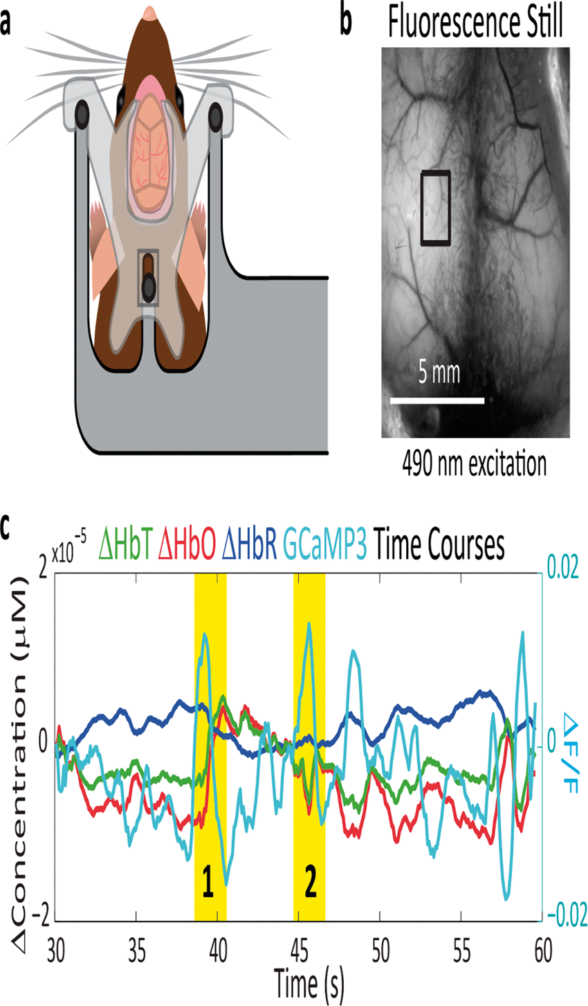
References.
726
BRAIN-0503
Poster Session
COMPARING STIMULUS-EVOKED AND RESTING-STATE NEUROVASCULAR COUPLING WITH SIMULTANEOUS ELECTROPHYSIOLOGY, WIDE-FIELD NEURONAL GCAMP AND HEMODYNAMIC IMAGING
Neurovascular Coupling
1Biomedical Engineering, Columbia University, New York City, USA
2Department of Neuroscience, Columbia University, New York City, USA
Objectives
Abstract
Functional magnetic resonance imaging (fMRI) measures local changes in hemodynamics as a surrogate for neuronal activity. Resting state functional connectivity captures spontaneous fluctuations in the fMRI blood oxygen level dependent (BOLD) signal, and infers neuronal network connectivity from regional hemodynamic synchronization [1]. These methods assume that hemodynamics faithfully reflect neuronal activity, and that neurovascular coupling is static and predictable in both stimulus-evoked and resting state conditions.
Here, we test these assumptions by imaging both neural activity and hemodynamics simultaneously in the mouse brain both during somatosensory stimulation and resting state conditions.
All experiment procedures were approved by the Columbia University Institutional Animal Care and Use Committee.
Wide-field multi-spectral optical intrinsic signal imaging (MS-OISI) was used to image the exposed cortex of mice expressing genetically encoded fluorescence calcium sensor GCaMP under the Thy1 promoter targeting pyramidal neurons located at cortical layers II/III and V [2]. By using this method, we can simultaneously acquire cortical neuronal and hemodynamic activity with high temporal and spatial resolution. Local electrophysiology was simultaneously acquired from the hindpaw region of the somatosensory cortex in urethane anesthetized mice (n=9), with and without hindpaw stimulation.
Because both the excitation and emission light for GCaMP fluorescence are contaminated by the changing hemodynamics, detected fluorescence was corrected by linear regression of its log.
To test whether stimulus-evoked and resting state neurovascular coupling is equivalent, a hemodynamic response function (HRF) based model was developed that predicted stimulus-evoked hemodynamics from GCaMP fluorescence. This model was then used to predict expected resting state hemodynamics from spontaneous GCaMP activity.
After eliminating hemodynamic cross-talk, GCaMP fluorescence was found to agree well with multi-unit activity (MUA) measured by electrophysiology, both with and without stimuli.
The hemodynamic response evoked by stimulation was found to consist of two parts: (1) A linear component that can be predicted by length of stimulation or neuronal activity and (2) a constant component which is present at the beginning of the response, whose waveform is independent of stimulus duration.
Our HRF model-based comparison revealed epochs of neurovascular uncoupling during resting-state recordings, whereas other periods were well-predicted. Objectively generating a best-fit HRF function from resting state data revealed that compared to the HRF driven by stimulation, the HRF at resting-state has a significantly smaller amplitude and faster decay, although time to peak was similar between the two.
Wide-field GCaMP recordings can provide accurate representations of MUA after correction for hemoglobin absorption. Stimulus-evoked and resting state neurovascular coupling have similar, but different HRF properties. Additionally, resting state recordings revealed epochs of apparent neurovascular de-coupling. Understanding factors leading to these mismatches between neural activity and hemodynamics could provide improved methods for analysis of resting state fMRI data, and could provide clues to the basis of altered resting state functional connectivity in pathological states.

References
727
BRAIN-0561
Poster Session
NEUROVASCULAR COUPLING IN PRETERM NEONATES WITH INTRA-VENTRICULAR HEMORRHAGE: COMBINED HIGH DENSITY EEG-NIRS STUDY
Neurovascular Coupling
1Neuropediatrics, Centre Hospitalier Universitaire, Amiens, France
2brain development, Neurospin, Gif/Yvette, France
3NICU, Centre Hospitalier Universitaire, Amiens, France
Can we detect auditory neurovascular impairment during the early phase of infancy? Continuous measurements during sleep were performed in healthy (n=12) and Intra Ventricular Hemorrhage (IVH grade III & IV, n=7) preterm neonates (28-32 weeks GA) using functional Near-Infrared Spectroscopy (fNIRS) in conjunction with EEG. We have shown that the preterm brain is able to discriminate a change of phonemes (ba vs ga) and a change of voices (male vs female) (Mahmoudzadeh, 2013). The dynamic of the responses reveals a structured network evolving differently in time and space (temporal/frontal lobes areas, left/right hemispheres). The study described here aims also to investigate the impact of the IVH on auditory hemodynamic responses. While EEG disclosed active language neural network, fNIRS revealed much weaker auditory hemodynamic responses, showing neurovascular coupling impairment. The present data confirm the existence of neurovascular coupling in healthy premature brain. It also shows that IVH premature neonates have lack of local mechanisms that allocate blood oxygen and to the active neurons. These results demonstrate that particular regions of the cortex, critical for language acquisition and processing, contain innate language specific representations in early infancy. In addition, the approaches we developed provide early diagnosis of auditory neurovascular coupling impairment in IVH preterms which is known to induce learning disabilities.

728
BRAIN-0563
Poster Session
MULTIMODAL IMAGING OF NEUROVASCULAR COUPLING RELATIONSHIP BETWEEN SPONTANEOUS EEG, CMRO2 AND HEMODYNAMIC RESPONSES IN PRETERM INFANTS
Neurovascular Coupling
1medicine, University of Picardie Jules Verne, Amiens, France
2NICU, centre hospitalier Universitaire, Amiens, France
3Neuropediatrics, centre hospitalier Universitaire, Amiens, France
Abstract
Objectives
We previously demonstrated [Roche, 2007] that spontaneous hemodynamic response and electroencephalography (EEG) simultaneously recorded from human preterm infant are coupled with the burst of activity, characteristic of the discontinuity EEG pattern observed at this period of the development. On the other hand, our studies [Mahdmoudzadeh et al., 2013; 2014 submitted] have reported that the preterm brain can exhibit distinct hemodynamic responses to external stimulation under auditory task, which may indicate a neurovascular coupling relationship. One relevant and interesting question would be the neurovascular coupling relationship between spontaneous EEG and CMRO2/hemodynamic responses in
the resting premature brain.
We used the simultaneous acquisition of electroencephalography (EEG, 8 channels) and functional optical imaging (Near infrared spectroscopy-Diffuse correlation spectroscopy) as only potentially executable- noninvasive multimodal imaging technique for measuring the functional activity of the preterm brain. Continuous measurements during sleep were performed in healthy and Intra Ventricular Hemorrhage (IVH grade II), preterm neonates (28-35 weeks GA). The Global Mean field Power (GMFP) were first calculated from the continuously recorded EEG signals. Then an HRF, which was modeled as a gamma probability density function, was convoluted with the power of EEG (extracted by GMFP); then correlation between convolved EEG power signal and CMRO2 was calculated.
To answer the neurovascular coupling relationship question, we analyzed the correlation between CMRO2 and EEG signal linking spontaneous CBV/CBF and EEG signals measured on preterm infants. The results indicate that an EEG power change is fairly correlated (∼0.3) with CMRO2 in healthy preterm while in pathological case it is considerably less correlated (∼0.07).
Given that EEG measures the electrical activity of neural populations while NIRS and DCS measures their relative hemodynamics via change in blood oxygenation and in blood flow signal respectively. Simultaneous EEG/NIRS/DCS offers multi-modality imaging to investigate the neurovascular relationship. There is still substantial debate regarding the relationship between local neuronal activity and hemodynamic changes (Logothetis and Wandell, 2004; Sirotin and Das, 2009). That is, our results shed light on a more comprehensive understanding of the specific mechanisms underlying neurovascular coupling. Perhaps the most compelling reason for EEG/NIRS/DCS is that temporally specific variations in the EEG fluctuations can be captured and correlated with the functional optical hemodynamic signal. Many aspects of neurovascular coupling and brain maturation are essentially characterized by EEG signals fluctuations, however there is a potential that many of the associated neural-vascular correlates can be measured via multimodal neurovascular imaging noninvasively (scalp EEG/NIRS/DCS).
729
BRAIN-0524
Poster Session
FUNCTIONAL ULTRASOUND IMAGING OF INTRINSIC CONNECTIVITY IN THE LIVING RAT BRAIN WITH HIGH SPATIOTEMPORAL RESOLUTION
Neurovascular Coupling
1Wave Physics for Medicine, Institute Langevin ESPCI CNRS UMR7587 INSERM U979, PARIS, France
2Brain Plasticity Unit, ESPCI-ParisTech UMR8249, PARIS, France
Abstract
Objectives
The brain dynamically integrates and coordinates responses to internal and external stimuli across multiple spatiotemporal scales through large-scale functional networks. Assessment of its functional connectivity (FC), through the measurement of regionally correlated, spontaneous, low frequency (0.01–0.1 Hz) fluctuations in blood oxygen level dependent (BOLD) signals with functional magnetic resonance imaging (fMRI), particularly during resting-state/task-free periods (resting-state fMRI), has greatly advanced our understanding of the functional organization of the human brain1. Here we propose a novel, highly resolved connectivity mapping approach using ultrafast functional ultrasound (fUS)2. This technique enables imaging of cerebral microvascular haemodynamics deep in the anaesthetized rodent brain, through a large thinned-skull cranial window, with pixel dimensions of 100µm x 100µm in-plane and a millisecond time resolution allowing unambiguous cancellation of low-frequency cardio-respiratory noise. Both seed-based and singular value decomposition analysis of spatial coherences in the low-frequency (<0.1 Hz) spontaneous fUS signal fluctuations reproducibly report, at different coronal planes, overlapping high-contrast, intrinsic functional connectivity patterns3.
The experiment was carried out using a 15MHz ultrasound probe on 20 anesthetized rats (metomidine/ketamine) with a thinned skull. The concept of functional ultrasound relies on ultrafast Doppler based on acoustic plane-wave transmission. The brain was insonified with a succession of ultrasound plane waves at 500Hz. Their backscattered echoes were recorded and beamformed to produce an echographic image every 2ms. Ten-minute duration acquisitions were performed at three anterior/posterior coordinates: Bregma +0.84, -0.6 and -2.16 mm.
Functionally correlated contralateral cortical areas activated by electrical stimulation of the right or left sciatic nerve were identified using fUS (see figure 1a) then the spontaneous fUS signal of this coronal slice was measured without stimulation. We found that these functionally similar regions were highly correlated at rest (correlation coefficient>0.8) (figure 1b).

In addition, different regions of interest can be defined using the spatial referential frame of the Paxinos (figure 2a) and a functional connectivity (FC) matrix can be computed (figure 2b). This FC matrix was found highly reproducible (r=0.85+/-0.03, p<0.001, N=6), all the coefficient >0.2 were reproducible (p<0.05).This matrix showed strong bilateral correlations in cortical brain areas. By contrast, cerebral structures such as the CPu and septum showed no correlation with the ‘sensory-motor resting-state network’ as found by Schwarz et al3.

We also used singular value decomposition based data treatment to retrieve anticorrelated FC patterns revealing distinct functional networks (figure 3).

The same type of results can be extracted from the anterior/posterior coordinate (bregma=+0.84, -2.16mm).
fUS imaging was able to detect FC patterns of distinct neuro-anatomical systems with 100µm spatial resolution. These patterns are similar to major functional networks described in humans by resting-state fMRI, such as the lateral task-dependent network putatively anticorrelated with the midline default-mode net-work. These results introduce fUS as a powerful novel neuroimaging method, which could be extended to portable systems for three-dimensional functional connectivity imaging in awake and freely moving rodents.
References
730
BRAIN-0284
Poster Session
THE EFFECT OF GLUCOSE SUPPLY ON NEUROVASCULAR COUPLING IN ANESTHETIZED RATS
Neurovascular Coupling
1Department of Neurosurgery Universitätsklinikum Aachen RWTH University, Translational Neurosurgery and Neurobiology, Aachen, Germany
2Department of Neurosurgery Technical University Munich, Experimental Neurosurgery, Munich, Germany
Abstract
Objectives:
Physiological stimulation leads to increased neuronal activity in the brain, accompanied by increases in local cerebral blood flow (CBF) to ensure supply of oxygen and glucose. In resting state the vast majority of ATP is metabolized via oxidative phosphorylation, but during activation the ratio changes towards a larger contribution of non-oxidative aerobic glycolysis (Fox et al., 1988). We tested the hypothesis whether the level of glucose supply changes the balance between oxidative and non-oxidative metabolism during activation. It may be suggested that during reduced glucose supply, a change in favor of oxidative phosphorylation, which uses glucose more effectively than aerobic glycolysis, occurs.
Methods:
Electrical forepaw stimulation (1.8 mA, 3 Hz, 16s) was used in ALPHA-chloralose-urethane anesthetized male Wistar rats to induce changes in neuronal activity in the brain. Changes in CBF and regional blood oxygenation (CBO) were recorded by a combined microfiber optical hemoglobin spectroscopy and laser-Doppler flowmetry probe under different glucose levels over a time period of 3h. Cerebral metabolic rate of oxygen (CMRO2) was calculated (Leithner et al., 2010) and somatosensory evoked potentials (SEPs) were recorded. CBF and CMRO2 at rest as well as SEPs, averaged CBF and CBO responses during stimulation and the time required to return to resting values after end of stimulation under highest (>22.2mmol) resp. lowest glucose levels (<0.8mmol) were expressed as % changes from prior values under euglycemia (= baseline values, normalized to 100%, data expressed as mean +/- SD) and compared to baseline values by paired Students t-test.
To exclude time related effects, a time-control group under euglycemia was tested (n=6), which showed a slight decrease in resting CBF and an attenuation of SEP amplitudes after 3h by 32+/-12%.
Results:
During hyperglycemia (n=10), resting CBF and CMRO2 remain unaltered, whereas CBF (154 +/- 60%, p=0.02) and CMRO2 responses (132 +/- 47%, p=0.06) were increased during unchanged SEPs.
Under hypoglycemia (n=8) resting CBF (120%, +/- 22) was increased. Similar to control group, during stimulation CBF and CMRO2 responses remained unaltered, whereas SEPs were slightly attenuated. After end of stimulation the time to return to resting values for CBF was significantly prolonged (134%, +/- 38), whereas time to return for CMRO2 remained unchanged (119+/- 27%, p=0.09).
Conclusions:
During hyperglycemia the absence of SEP attenuation shown in time control might be responsible for the hyperglycemia induced increase in CMRO2 and CBF. Under severe hypoglycemia our results give no evidence of increased oxidative phosphorylation during neuronal activation. However, we found a prolonged increase of blood flow after the end of stimulation, which may suggest that during low glucose supply more time is required to get adequate amount of glucose from the blood to meet activation induced demand.

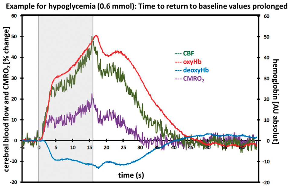
References:
731
BRAIN-0595
Poster Session
TONIC BLOOD FLOW CONTROL BY ASTROCYTES
Neurovascular Coupling
1Physiology and Pharmacology, Hotchkiss Brain Institute, Calgary, Canada
Abstract
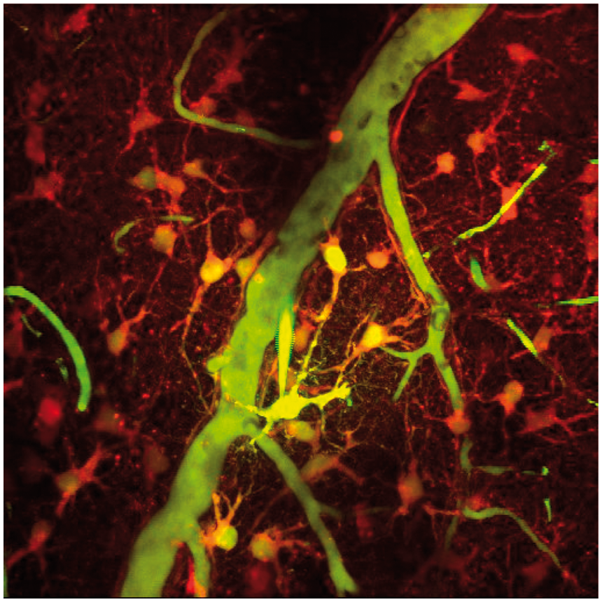
Objectives: Local brain blood flow control requires intricate communication by neurons and astrocytes to vascular contractile cells that make fine adjustments to arteriole diameter to control blood perfusion. Neurovascular coupling accounts for phasic, activity-dependent blood flow control but it remains unclear whether local brain cells can control blood flow tonically to provide steady-state arteriole diameter regulation. We propose a novel role of astrocytes in helping to set the basal arteriole diameter of cerebral vessels, thereby helping to set the perfusion rate to meet the ongoing metabolic demands of the brain.
Methods: Two-photon imaging and patch clamp electrophysiology was performed in acute slices of rat sensory-motor cortex. Animals were IV injected with an FITC dextran to label the vasculature, and brain slices were incubated in the Ca2+ indicator Rhod-2/AM for imaging. In-vivo experiments were performed on mice expressing the genetic Ca2+ indicator GCaMP3 in astrocytes using the GLAST promoter.
Results: Using intracellular delivery of the Ca2+ chelator BAPTA into astrocytes, we observed a vasoconstriction upon the arrival of BAPTA to the endfeet. This result suggests that astrocytes are providing tonic vasodilation which is determined by the astrocyte resting Ca2+ concentration. Further experiments indicate that astrocytes are tonically releasing prostaglandins, mediated by the constitutive action of COX-1. Application of the COX-1 specific inhibitor SC560 will itself cause vasoconstriction and will also occlude the astrocyte BAPTA induced constriction, suggesting the two are part of the same pathway.
Conclusions: We propose that astrocytes help regulate the steady-state diameter of cerebral arterioles, thus ensuring that the supply of blood to the brain is sufficient to meet its ongoing metabolic demands.
732
BRAIN-0809
Poster Session
THE INFLUENCE OF ENDOTHELIAL DYSFUNCTION ON NEUROVASCULAR COUPLING
Neurovascular Coupling
1Biomedical Engineering, Columbia University, New York City, USA
Abstract
Objectives:
Several cell-types have been implicated in mediating the hyperemic response to neuronal activity, including neurons1, astrocytes2 and pericytes3. Recent studies have added endothelial cells to this list by demonstrating the presence of conducted vasodilation along arterioles in brain4 during functional hyperemia. Light-induced reactive-oxygen species (ROS) mediated transection of the vascular endothelium was shown to interrupt conducted vasodilation in the brain4, altering both the amplitude and timing of the hemodynamic response to stimulus. These results demonstrate that normal neurovascular coupling is reliant on endothelial mechanisms of vasodilation.
In addition to extrinsic ROS damage, disease states that cause intrinsic ROS up-regulation are known to cause impaired endothelial vasoactivity. These conditions include systemic cardiovascular diseases like diabetes and hypertension, but also neurodegenerative diseases including Alzheimer’s5, all of which are associated with long-term cognitive decline. In addition to other mechanisms for neuronal damage, we hypothesize that these conditions would cause alterations in dynamic coupling between neural activity and functional hemodynamics, which may have a direct effect on neuronal health. Here, we explore the effect of pharmacological modulation of endothelial function on neurovascular coupling.
Sprague-Dawley rats were anesthetized with 3% isoflurane, ventilated via tracheostomy and the right femoral artery and vein were cannulated for blood pressure monitoring and drug delivery respectively. A craniotomy was performed over the right somatosensory cortex and the dura was reflected. The pial surface was resealed with ACSF in agar under a glass coverslip. Anesthesia was switched to IV alpha-chloralose for stimulus-evoked imaging using multispectral optical intrinsic signal imaging (MS-OISI6). Square-wave hindpaw electrical stimuli at 3 Hz and 0.3 ms pulse-width of varying durations were used. Data was converted to show changes in total hemoglobin concentration (Δ[HbT]), averaged from 1 hour before and after intravenous bolus injection of either Ketorolac, a non-blood brain barrier (BBB)-permeable non-selective COX inhibitor, and Enalaprilat, a non-BBB permeable ACE inhibitor.
Both Ketorolac and Enalaprilat potentiated hemodynamic response at the central region of activation (Figure 1). Towards the periphery of the responding region, Enalaprilat had a large effect on the Δ[HbT] response, while Ketorolac had little effect. The spatial differences between the effects of these drugs can be explained by their properties. Ketorolac inhibits slowly conducted endothelial vasodilation, which stays restricted to the region of neuronal activation. Enalaprilat has a large effect on the mean arterial pressure, and changes basal tone of the pial arteries, which may potentiate the hemodynamic response in a spatially uniform manner. Further experiments targeted at slow and fast components of conducted vasodilation are in progress. The current data suggests that selective modulation of endothelial function has significant effects on the hyperemic response to stimulus implying that endothelial disease models could similarly affect neurovascular coupling and brain health.

References:
733
BRAIN-0730
Poster Session
NEUROVASCULAR COUPLING DURING CEREBROVASCULAR REACTIVITY IN SMALL ARTERY STROKE BY NITRIC OXIDE, HYDROGEN PEROXIDE AND HEART RATE VARIABILITY
Neurovascular Coupling
1physiology, Faculty of Medicine Thammasat University, Pathumthani, Thailand
2Medical Engineering Program, Faculty of Engineering Thammasat University, Pathumthani, Thailand
Abstract

References:
734
BRAIN-0299
Poster Session
DIAMETER CHANGES TO CEREBRAL BLOOD VESSELS DURING NEURAL ACTIVATION AND DEACTIVATION MEASURED BY TWO-PHOTON MICROSCOPY IN AWAKE MICE
Neurovascular Coupling
1Molecular Imaging Center, National Institute of Radiological Sciences, Chiba, Japan
2Advanced Clinical Research Center, Fukushima Medical University, Fukushima, Japan
Abstract
Objectives:
Neural activation monitored with positron emission tomography (PET) has been reported to a cause larger increase in cerebral blood flow (CBF) than cerebral blood volume (CBV) in humans [1]. It has also been reported that neural activation evokes larger increases in red blood cell (RBC) velocity than RBC concentration in mice when measured with Laser-Doppler flowmetry (LDF) [2]. Crossed cerebellar diaschisis (CCD) caused by contralateral supratentorial lesions can be considered to be a model of neural deactivation, and hemodynamic changes in CCD measured with PET in humans have been reported to show similar percentage decreases in CBF and CBV [3]. Recently, we developed a mouse model of CCD induced by middle cerebral artery occlusion (MCAO) that also showed similar percentage decreases in CBF and RBC concentration [4]. On the other hand, changes to the diameter of microvessels during neural activation and deactivation are still unclear. Using two-photon microscopy, we investigate microvessels diameter changes in these mouse models of neural activation and deactivation.
Two-photon microscopic imaging was performed on mouse models of neural activation and deactivation (C57BL/6Jmice, 7-9weeks, N=5). Neural activation was induced by air-puff stimulation and a permanent occlusion of the MCAO was used to mimic deactivation. Microvessels were fluorescently labeled with sulforhodamine 101 and changes to the diameter of arterioles, capillaries and venula were measured in cerebral cortex (activation) and cerebellar cortex (deactivation). Baseline diameters of arterioles, capillaries and venula in cerebral cortex were 17 ± 20µm, 6 ± 7µm and 17 ± 17µm, respectively (mean ± SD). Baseline diameters of the same vessels in cerebellar cortex were 16 ± 12µm, 6 ± 6µm and 14 ± 13µm, respectively. The two-photon imaging was performed on awake animals immobilized using a hand-made fixation apparatus [5].
Using two-photon imaging of mouse brain in the awake state, vasodilation and vasoconstriction were observed during neural activation and deactivation, respectively. Percentage changes in diameter during whisker stimulation were +19 ± 3%, +8 ± 4%, and +3 ± 1% for arterioles, capillaries and venula, respectively. Percentage changes in diameter during CCD were -23 ± 3%, -11 ± 3%, and -2 ± 7% for arterioles, capillaries and venula, respectively.
In the case of microvessels, the degree of vasoconstriction under neural deactivation was similar to the degree of vasodilation under neural activation. These results only partially agree with our previous results obtained PET and LDF [1], [2], [3], [4]. PET and LDF measure hemodynamic changes in a broad brain region that includes large arteis and veins which deffers from situation measured with two-photon imaging in this study. Therefore, changes in microvessels may different from changes in large vessels during the neural activation and deactivation.
References
735
BRAIN-0340
Poster Session
EXPERIMENTAL NEUROVASCULAR UNCOUPLING PROMOTES COGNITIVE IMPAIRMENT IN MICE: IMPLICATIONS FOR BRAIN AND CEREBROMICROVASCULAR AGING
Neurovascular Coupling
1Reynolds Oklahoma Center on Aging, University of Oklahoma Health Sciences Center, Oklahoma City, USA
2Advanced Medical Resonance Center, Oklahoma Medical Research Foundation, Oklahoma City, USA
3Department of Medical Physics and Informatics, University of Szeged, Szeged, Hungary
Abstract
Increasing evidence shows that vascular risk factors such as aging, hypertension, diabetes mellitus, and obesity promote cognitive impairment, however, the underlying mechanisms are not well understood. Cerebral blood flow (CBF) is adjusted to neuronal activity via neurovascular coupling (NVC) and this mechanism is known to be impaired in the aforementioned pathophysiological conditions. To establish a direct, causal relationship between impaired NVC and cognitive decline, we induced neurovascular uncoupling pharmacologically in mice by inhibiting the synthesis of vasodilator mediators involved in NVC.
Methods
Male C57BL/6J mice (n=20 in each group) were assigned into two groups: 1) the pharmacological treatment was carried on for 7 days and included the administration of MS-PPOH (20 mg/kg/day, s.c.), a specific inhibitor of EET-producing epoxidases. Indomethacin (7.5 mg/kg/day, p.o.) and L-NAME (100 mg/kg/day, p.o.) were administered in drinking water to inhibit cyclooxygenase-derived vasodilator metabolites and NO synthase activity respectively; 2) vehicle control. A battery of behavioral test was performed to characterize the effect of pharmacologically-induced neurovascular uncoupling on learning and memory, sensory-motor function, gait and locomotion. NVC was assessed by measuring changes in CBF (laser Doppler flowmetry), extracellular glucose (amperometry) and evoked filed potentials in the whisker barrel in response to contralateral whisker stimulation. Arterial spin labeling magnetic resonance imaging was used to measure changes in basal CBF. To determine whether pharmacological treatments result in primary neuronal dysfunction, extracellular recordings were performed from acute hippocampal slices to assess synaptic function and long-term potentiation (LTP).
Results
Mice treated with MS-PPOH+L-NAME+Indomethacin exhibited a ∼75% decline in functional hyperemia in response to neuronal activation and impaired performance on the Y maze, the elevated plus maze and the novel object recognition tests, indicating impaired spatial working memory and novelty-seeking behavior. Neurovascular uncoupling in mice was associated with impaired motor coordination (rotarod) and static force production (grip strength), whereas olfactory ability and gustatory motivation (buried food retrieval) and gait function (CatWalk) were unaffected. The pharmacological treatments did not alter basal CBF, evoked field potentials and LTPs.
Conclusions
Selective experimental disruption of NVC per se leads to impairment of cortical function, including cognitive decline, recapitulating some of the neurological symptoms and signs observed in brain aging and pathophysiological conditions associated with accelerated cerebromicrovascular aging.
736
BRAIN-0244
Poster Session
CORTICAL ARTERIOLES MODULATE ASTROCYTES CA2+ DYNAMICS
Neurovascular Coupling
1Physiology and Pharmacology, Hotchkiss Brain Institute, Calgary, Canada
Abstract
Objective
Recent in vivo evidence conducted in anesthetized or slightly sedated animals questions the direct role of astrocyte intracellular Ca2+ in mediating functional hyperemia as spatial and temporal profile of astrocyte Ca2+ transients are poorly associated with the onset of functional hyperemia (1). Our objective was to uncover the spatiotemporal basis of the communication between astrocytes and the vasculature in fully awake, behaving mice.

A craniotomy over the barrel cortex with the dura removed was performed. A custom build two-photon microscopy was used to image the vasculature and astrocytes Ca2+ from either C56Bl/6 mice or GLAST-cre-LSL-GCaMP3 mice or TEK-cre-LSL-ArchT3 mice. A cannula was inserted into the tail artery for dye and drug injections.
We found that 5-second whiskers' stimulation induced fast vasodilatory responses in penetrating arterioles while generated a delayed onset of endfoot and cell-wide Ca2+ transients. Interestingly, the onset of these Ca2+ transients was typically observed at the peak of the sensory induced vasodilation and toward the end of the vibrissae stimulation. Thus, we tested if the vasculature was communicating back to the astrocytes and consequently modulating astrocytes Ca2+. We showed that intraluminal perfusion of acetylcholine, but not the mGluR5 agonist DHPG, caused vasodilation that was followed by an increase in astrocytes Ca2+ fluctuations. Activation of endothelial expressed archaerhodopsins with brief yellow laser induced fast vasodilation followed by an initiation of endfoot Ca2+ transient.

Our data redefine the uni-directional communication between astrocytes and the vasculature. We introduce a potential role of the vasculature as the modulator of astrocytes Ca2+ transients and propose that astrocytes act as responders to changes in blood flow.
Reference
737
BRAIN-0427
Poster Session
DOPAMINE AND ALPHA2-ADRENOCEPTORS INTERACT TO MODULATE THE VASCULAR RESPONSE IN THE DORSAL HIPPOCAMPUS AFTER MESOLIMBIC DOPAMINE ACTIVITY
Neurovascular Coupling
1Laboratory for Circuit and Behavioral Physiology, RIKEN BSI, Wako-shi, Japan
Abstract

References:
738
BRAIN-0490
Poster Session
COUPLING OF SPONTANEOUS AND SENSORY EVOKED HEMODYNAMIC SIGNALS TO NEURAL ACTIVITY IN THE BARREL CORTEX OF AWAKE MICE
Neurovascular Coupling
1Engineering Science and Mechanics, Penn State University, University Park, USA
Abstract
Spontaneous hemodynamic signals, measured in the absence of overt sensory stimulation, are used as indicators of neural activity in resting-state MRI experiments. However, it is not clear whether spontaneous hemodynamic signals reflect ongoing neural activity in the same way that sensory evoked hemodynamic signals do. To determine if coupling of neural activity and hemodynamic signals is the same for both sensory-evoked and spontaneous activity, we examined neurovascular coupling in the barrel cortex of awake, head-fixed mice during several behaviors (passive sensation, volitional whisking, and rest).
We simultaneously measured local neural activity, cerebral blood volume (CBV), and behavior in awake, head-fixed mice. Blood volume changes were quantified by illuminating the vibrissa cortex with 530 nm light through a reinforced thinned-skull window. The local field potential (LFP) and multi-unit activity (MUA) were measured via a tungsten stereotrode implanted in the imaged area of the vibrissa cortex. Whisker position was tracked to detect volitional movements. The vibrissae were also stimulated with brief puffs of air during data acquisition. The data were then categorized into three behaviors: sensory evoked, volitional whisking, and rest. We calculated a hemodynamic response function (HRF), which quantitatively relates the measured CBV to the local neural activity for each behavior. We then used these HRFs to determine what fraction of the spontaneous and sensory evoked changes in CBV could be explained by neural activity.
Whisker stimulation and volitional whisking were accompanied by increases in the MUA, CBV, and gamma band power of the LFP, though responses to whisker stimulation were substantially stronger. During resting behavior, neural activity and CBV were weakly correlated. These correlations were not disrupted by transection of the facial nerve, indicating that they could not be attributed to undetected whisker movements. HRFs calculated from each of the three behaviors were similar, indicating that neurovascular coupling was conserved across behavioral state. However, there were substantial differences in the variance of the hemodynamic signal captured by the HRFs across behaviors. Sensory evoked hemodynamics, both on average and for individual trials, were better predicted by local neural activity than hemodynamic changes measured during volitional whisking or resting behaviors. The prediction error, for all behaviors, could be attributed to the presence of additive, ongoing CBV fluctuations that were not associated with any measured neural activity. This uncorrelated signal obscures neurally-evoked hemodynamics to a greater degree during volitional whisking and resting behaviors since they are accompanied by smaller neural increases than sensory-evoked activity.
Neurovascular coupling was similar across all three behavior types that we monitored. However, the measured hemodynamics were not equally representative of local neural activity across behaviors. This discrepancy can be attributed to the presence of a separate component of the hemodynamic signal which was uncorrelated to the measured neural activity. Thus, spontaneous fluctuations in CBV cannot be directly related back to local neural activity.
739
BRAIN-0556
Poster Session
HUMAN UMBILICAL CORD PERIVASCULAR CELL (HUCPVC) THERAPY FOR TRAUMATIC BRAIN INJURY: TARGETING THE NEUROVASCULAR UNIT.
Neurovascular Unit
1Research, Create Fertility Centre, Toronto, Canada
2Critical Care, St. Michael's Hospital, Toronto, Canada
3Obstetrics & Gynaecology, University of Toronto, Toronto, Canada
Abstract
White matter sparing after traumatic brain injury (TBI) is an important predictor of survival and outcome. Mesenchymal stem cells derived from the bone marrow (BMSCs) are currently being used in clinical trials for treatment of neurodegenerative diseases including stroke[1]. MSCs demonstrate great potential to provide therapeutic benefit, and have thus far proven to be safe in early trials[2]. Accordingly, we were interested in evaluating the potential of human umbilical cord-derived perivascular cells (HUCPVCs), a less differentiated type of MSC, on their therapeutic potential in TBI. HUCPVCs have several advantages over BMSC therapies including ease of procurement and greater rates of expansion. HUCPVCs are a rich source of pericyte-like cells, and are positive for the pericyte markers CD146, NG2, and PDGFRβ[3].
References:
740
BRAIN-0228
Poster Session
AMYLOID BETA INTERFERES WITH ENDOTHELIAL PROGENITOR CELL ADHERENCE VIA MMP UPREGULATION
Neurovascular Unit
1Radiology, Massachusetts General Hospital, Charlestown, USA
Abstract
741
BRAIN-0231
Poster Session
ASTROCYTIC HMGB1 INDUCES AN ANGIOGENIC RESPONSE IN ENDOTHELIAL PROGENITOR CELLS
Neurovascular Unit
1Radiology, Massachusetts General Hospital, Charlestown, USA
Abstract
742
BRAIN-0086
Poster Session
PROGRANULIN MEDIATES NEUROVASCULAR PROTECTION VIA MULTIPLE THERAPEUTIC EFFECTS IN EXPERIMENTAL ACUTE ISCHEMIC STROKE
Neurovascular Unit
1Neurology, Brain Research Institute Niigata University, Niigata, Japan
2Veterinary Physiology, Graduate School of Agricultural and Life Sciences The University of Tokyo, Tokyo, Japan
3Center for Integrated Human Brain Science, Brain Research Institute Niigata University, Niigata, Japan
Abstract
Objectives; In central nervous system, progranulin (PGRN), a glycoprotein growth factor, is considered to play crucial roles in maintaining physiological functions, and mutations in PGRN gene cause TAR DNA-binding protein-43 (TDP-43)-positive frontotemporal lobar degeneration.1 In addition, PGRN is known for its role in biological processes such as anti-inflammation and wound healing.2 Although several studies reported that PGRN plays protective roles against ischemic brain injury,3,4 it remains unknown the precise mechanisms by which PGRN exerts protective effects on the ischemic brain injury.
Methods; We determined the temporal changes of expression and localization of PGRN after ischemia as well as therapeutic effects of PGRN on ischemic brain injury using in vitro and in vivo models.
Results; First, we demonstrated a dynamic change of PGRN expression in ischemic Sprague-Dawley rats, including increased levels of PGRN expression in microglia within the ischemic core, and increased level of PGRN expression in survived neurons as well as induction of PGRN expression in endothelial cells within the ischemic penumbra. Second, we demonstrated that PGRN could protect against acute focal cerebral ischemia by variety of mechanisms including via attenuation of blood-brain barrier disruption, suppression of neuroinflammation, and neuroprotection: we found that PGRN may regulate vascular permeability via vascular endothelial growth factor (VEGF), that PGRN may suppress neuroinflammation after ischemia via anti-inflammatory interleukin-10 (IL-10) in microglia, and that neuroprotective effect of PGRN may be explained in part by inhibition of cytoplasmic redistribution of TDP-43 using PGRN knock-out mice (C57Bl/6 background). Finally, we demonstrated the therapeutic potential of PGRN against acute focal cerebral ischemia using a rat autologous thromboembolic model with delayed tissue plasminogen activator (tPA) treatment. Intravenously administered recombinant PGRN reduced volumes of cerebral infarct and edema, suppressed hemorrhagic transformation, and improved motor outcome (P = 0.007, 0.038, 0.007, and 0.004, respectively).
Conclusions; PGRN may be a novel therapeutic target that provides neurovascular protection, anti-neuroinflammation, and neuroprotection related in part to VEGF, IL-10, and TDP-43, respectively. We demonstrated for the first time that intravenous administration of recombinant PGRN with tPA treatment showed therapeutic effects on the volumes of cerebral infarct and edema, hemorrhagic transformation, and prognosis.
References;
743
BRAIN-0573
Poster Session
HUMAN UMBILICAL CORD PERIVASCULAR CELLS (HUCPVCS) EXPRESS AND SECRETE NEUROTROPHIC FACTORS AND PROTECT SYMPATHETIC AXONS FROM DEGENERATION
Neurovascular Unit
1Research, Create Fertility Centre, Toronto, Canada
2Obstetrics and Gynecology, University of Toronto, Toronto, Canada
Abstract
The vascular system and the nervous system are mutually dependent during development[1, 2]. Vascular remodeling takes place after the establishment of peripheral nerves, and in turn, requires neural input for proper patterning. Axons provide vascular endothelial growth factor (VEGF) which is necessary for arteriole development and vascular remodelling in the skin [2]. Blood vessels also provide molecular cues to guide the development of neurons.
References:
744
BRAIN-0718
Poster Session
DIFFERENTIAL NEUROVASCULAR SUSCEPTIBILITY TO OXYGEN-GLUCOSE DEPRIVATION AND RESPONSE TO CYTOPROTECTION
Neurovascular Unit
1Neurology, Cedars-Sinai Medical Center, Los Angeles, USA
Abstract

745
BRAIN-0317
Poster Session
PERICYTES INTERACT WITH OLIGODENDROCYTE PRECURSOR CELLS IN PERIVASCULAR REGION IN CEREBRAL WHITE MATTER
Neurovascular Unit
1Neurology and Radiology, Massachusetts General Hospital, Charlestown, USA
2Regenerative Medicine, Institute of Biomedical Research and Innovation, Kobe, Japan
3Neurology, Kyoto University, Kyoto, Japan
4Stroke and Cerebrovascular Diseases, National Cerebral and Cardiovascular Center, Osaka, Japan
Abstract
746
BRAIN-0175
Poster Session
EFFECTS OF NMDA RECEPTOR ANTAGONIST MEMANTINE ON NO PRODUCTION, HYDROXYL RADICAL METABOLISM AND ISCHEMIC CHANGE OF HIPPOCAMPAL CA1 DURING CEREBRAL ISCHEMIA AND REPERFUSION IN MICE
Oxidative Mechanisms
1Department of Neurology, Saitama Medical University, Saitama, Japan
Abstract
Results: (1) Blood pressure: There were no significant differences between the groups. (2) Cerebral blood flow (CBF): There were no significant differences between the groups. (3) NO2-; Memantine group (120.9±5.00%; mean±SD) showed significantly higher than that of the control group (88.5±18.0) after repurfusion 60 minutes (p<0.05). (4) NO3-; Memantine group (97.2±10.1%; mean±SD) showed significantly higher than that of the control group (65.3±21.0) at ischemia (p<0.05). (5) 2,3-DHBA; Memantine group (90.7±2.90%; mean±SD) showed significantly lower than that of the control group (99.5±2.66) at ischemia, after repurfusion 20, 80-120 minutes (p<0.05). (6) Survival rate in CA1 area: There were no significant differences between the groups.
747
BRAIN-0862
Poster Session
FRET PROBES VISUALIZE ERK AND JNK PHOSPHORYLATION AND CONSEQUENTLY CELL DIVISION OR CELL DEATH IN HT22 CELLS.
Oxidative Mechanisms
1Neurosurgery, Graduate School of Medicine The University of Tokyo, Bunkyo City, Japan
2Neurosurgery, The University of Tokyo Hospital, Bunkyo City, Japan
3Biological Sciences, The University of Tokyo HospitalGraduate School of Science The University of Tokyo, Bunkyo City, Japan
Abstract
Backgraound
Sufficient data exist that Oxidative stress plays critical role in neuronal cell death under ischemic conditions. The immortalized mouse hippocampal HT22 cell in vitro model is widely used for evaluating the mechanisms of oxidative stress-associated neuronal cell death. The HT22 cells do not possess glutamate receptors, and treatment with high levels of glutamate can inhibit the synthesis of intracellular glutathione (Li et al., 1997). Typically, cystine is rapidly reduced to cysteine, which is necessary for the formation of glutathione (GSH) and then, reactive oxygen species (ROS) levels become increased .
It has been clearly demonstrated that the activation of Mitogen-activated protein Kinase (MAPK): while phosphorilation of ERK or JNK is detected during the death of HT22 cells by Western blotting assay, MAPK activity apparently decides the fate of cell (survival or death) under stress. Western blotting reflects the average status of a specific cell population and we cannot understand the spatiotemporal dynamics of signaling molecules within a single cell. To overcome these limitations, the Förster resonance energy transfer (FRET) biosensors, which quantifies molecular interactions in biology and chemistry, can be used. We applied the FRET probe that is able reflect the interaction between proteins, and by using ERK-FRET probes, we made possible measurement of intensity as real-time visualization of ERK or JNK phosphorylation in a single cell nucleus.
ÐMaterials and methodsÐ
Plasmids of ERK-FRET probes and JNK-FRET utilizing a PiggyBac transposon system to generate HT22 cell lines with stable expression of FRET biosensors (Aoki et al., 2012) were used. Cells were seeded onto 35 mm glass-based dishes. At 24-36 hr after seeding, we exchanged medium to CO2-independent medium and time-lapse FRET images were obtained by epifluorescence microscope (Olympus IX83) We induced oxidative stress in the HT22 cell line with glutamate of varied concentration (0∼10mM)
ÐResultÐ
In the cells whith JNK-FRET probe transfer, FRET efficiency were elevated according to level of phosphorylation of JNK. All cells with JNK activation died and opposite, those without JNK activation survived. We confirmed cell death by Propidium Iodide staining.On the other hand, in cells with ERK-FRET probe induction, phosphorylation of ERK was observed just before cell division.
ÐDiscussionÐ
We developed a new model for real-time visualization of MAPK activity under oxidative stress in HT22 cells and confirmed that phosphorylation of ERK can be seen in cell proliferation, to differ from phosphorylation of JNK, which is seen in cell death.

748
BRAIN-0278
Poster Session
NATURAL BIOACTIVE COMPOUNDS FROM RESOURCE ANIMALS: ANTIOXDATIVE AND CURATIVE EFFECT IN DISEASE MODEL CELLS
Oxidative Mechanisms
1Department of Biotechnology, Hoseo University, Asan, Korea
2Department of Biomedical Laboratory Science, Soonchunhyang University, Asan, Korea
3The Animal Genomics and Breeding Center, Han-Kyong National University, Anseong, Korea
Abstract
Neuronal cell death is the suggested cause of neurodegeneration in many disease, the molecular mechanism remains uncertain. We recently suggested that the cell cycle profile and its regulatory factors in lysosomal storage disease cells. We found G1/G0 cell cycle arrest, with overexpression of p21, sphingosine, glucosylceramide, and sulfatide. We also observed enhanced levels of α-synuclein oligomers and gangliosides GM1, GM2, and GM3 and reduced levels of sphingomyelin and autophagy. We report here that one of the pathogenic mechanism of neuronal cell death in lysosomal storage disease (Batten disease and Gaucher disease) is mediated by cellular stress, also upregulate the production of reactive oxygen species (ROS), destabilize mitochondrial membrane potential (MMP). Furthermore, we demonstrate that the levels of cell death indicators (ROS, MMP etc) in disease model cells are markedly decreased by bioactive compounds from resource animal. Our results provide that the availability of suitable bioactive compounds for testing and validation. This work was carried out with the support of 'Cooperative Research Program for Agriculture Science & Technology Development (Project No. PJ01104601)” Rural Development Administration, Republic of Korea.
749
BRAIN-0540
Poster Session
COBALT CHLORIDE-INDUCED-CHEMICAL HYPOXIA PROVOKES CHANGES IN SUBCELLULAR LOCALIZATION OF HEMEOXYGENASE ISOFORMS.
Oxidative Mechanisms
1Patologia Vascular Cerebral, Instituto Nacional de Neurologia y Neurocirugia, Mexico City, Mexico
2Laboratorio de Neurobiologia Celular y Molecular, Instituto Nacional de Neurologia y Neurocirugia, Mexico City, Mexico
Abstract
Objectives: The hemeoxygenases (HO)-1 and-2 are microsomal oxidoreductase enzymes that remove oxidant heme to biliverdin, carbon monoxide and Fe2+. Non-enzymatic roles have been proposed for both enzymes. HO-1 is an inducible isoform; under hypoxic and hyperoxic stress migrates to the nucleus. HO-1 also increases in mitochondrial fractions by different stimuli. HO-2 is not an inducible enzyme, however its expression is differentially regulated by hypoxia. C-terminal HO-2-deleted mutants have been observed in the nucleus suggesting its translocation. The objective of this work was to determine the behavior of HO isoforms in hipoxia mimicking conditions at transcript and protein levels as well as in the subcellular localization (nucleus, mitochondria and cytoplasm) using the CoCl2 induced-chemical hypoxia model in PC12 cells.
Methods: Cell death and viability with increasing CoCl2 concentrations (0.1-1.0 mM) for 24 or 48h were determined by SubG0 and MTT reduction assays. Stabilization, nuclear translocation and binding activity of HIF-1α were quantified by ELISA in nuclear fractions. ROS production was meeasured by DCF-DA fluorescence. HO-1 and HO-2 transcript levels were determined by real-time PCR and protein levels were measured by western blot and ELISA in cytoplasm, nucleus and mitochondria purified fractions.
Results: CoCl2 affected cell viability and cell death in a concentration- and time-dependent manner. A significant reduction in cell viability was observed. At 0.5 mM CoCl2 at 24 and 48h the percentage was 65 and 50% respectively, while at 1.0 mM CoCl2 a considerable reduction was observed (30% at 24h and 10% with 48h). However, cell death determination by SubG0 shows that at 24h there is only a 5 and 15% of dead cells at 0.5 and 1.0 mM CoCl2; while at 48h 22 and 40% dead cells were observed. Cells incubated 24h at 0.5 and 1.0 mM CoCl2 showed stabilization, nuclear translocation and binding activity of HIF-1α about 6- and 4-fold at 0.5; while 48h incubation induced an increase of 19- and 35-fold at 0.5 and 1.0 mM CoCl2 respectively. In addition to HIF-1α protein stabilization, CoCl2 mimics other hypoxia responses, including ROS generation. HO-1 transcript increased 210–fold with 0.5 mM CoCl2 and 80-fold at 1.0 mM (24h). Transcript levels were maintained until 48h. In contrast, HO-2 transcript only decreased in both CoCl2 concentrations at 24h. Subcellular fractionation evidenced that HO-1 was localized in nucleus and mitochondria and had dependent increase on CoCl2 concentration and incubation time. HO-2 was detected in cytoplasm, showing a decrease with CoCl2 0.5 and 1.0 mM at 24 and 48h by WB and ELISA. Surprisingly HO-2 was detected in nucleus decreasing with CoCl2 treatment. In addition, the presence of mitochondrial HO-2 increased with CoCl2 at 48h.
Conclusions: HO system responds differentially in a CoCl2-induced chemical hypoxic stress in PC12 cells. HO-1 and HO-2 proteins are translocated to nucleus and mitochondria as result of chemical hypoxia stress. Understanding these roles and finding therapeutic tools to specifically alter them could lead to clinical interventions that prevent hipoxic oxidative injury, tumor progression, and cell proliferation.
750
BRAIN-0779
Poster Session
EVALUATION OF SCAVENGING ACTIVITY AGAINST MULTIPLE SPECIES OF FREE RADICALS BY ELECTRON SPIN RESONANCE SPECTROSCOPY
Oxidative Mechanisms
1Neurophysiology, Oita University Faculty of Medicine, Yufu, Japan
2Anesthesiology, Oita University Faculty of Medicine, Yufu, Japan
Abstract
Evaluation ofantioxidative activity of antioxidants is important to understand theirmechanism of neuroprotective activity. Although measurement of lipid peroxideis often employed for the evaluation, their direct free radical scavengingactivity is not necessarily always measured, and, especially, data concerningtheir concentration-response relationship against multiple free radical species remain sparse. The authors haveevaluated direct scavenging activity of antioxidants against multiple speciesof free radicals by electron spin resonance (ESR) spectroscopy. Here theauthors illustrate an example of evaluation of multiple free radical scavengingactivity by presenting the data of edaravone, a powerful free radical scavengerand the only drug currently available in clinical use for the treatment ofcerebral infarction.
Free radicalscavenging activity of edaravone was evaluated by ESR spectroscopy using spintrapping method. The spectrum of its dose-response relationships to multiplefree radical species was estimated.
Edaravone scavengedthe following six kinds of free radicals with the IC50 as indicated:hydroxyl radical (IC50 = 0.4 mM), superoxide anion (4 mM), ascorbylfree radical (8 mM), tert-butyl peroxyl radical (0.3 mM), DPPH (5 μM),nitrous oxide (0.04 mM). No radical scavenging activity was observed againstthe methyl radical.
The estimated dose-response relationship of radicalscavenging activity by edaravone varied among free radical species examined.Since the IC50 varied among different species of free radicals, itis suggested that it is not sufficient to demonstrate antioxidative activity orscavenging activity against a single species of free radical to understand itstherapeutic mechanism. When evaluating antioxidative activity of antioxidants,it is necessary to evaluate scavenging activity against multiple species offree radicals to fully understand their neuroprotective mechanism againstoxidants
751
BRAIN-0115
Poster Session
FOCAL ISCHEMIC INJURY WITH COMPLEX MIDDLE CEREBRAL ARTERY IN STROKE-PRONE SPONTANEOUSLY HYPERTENSIVE RATS WITH LOSS-OF-FUNCTION IN NADPH OXIDASES
Oxidative Mechanisms
1Laboratory for Neurochemistry, National Hospital Organization Hizen Psychiatric Center, Saga, Japan
2Department of Functional Pathology, Shimane University School of Medicine, Izumo, Japan
3Division of Laboratory Animal Research, Shinshu University, Matsumoto, Japan
Abstract
References
752
BRAIN-0313
Poster Session
CHRONOLOGICAL CHANGES OF GLUCOCORTICOID RECEPTORS IN THE HIPPOCAMPUS OF STREPTOZOTOCIN-TREATED TYPE 1 DIABETIC RATS
Oxidative Mechanisms
1Department of Biomedical Laboratory Science, Soonchunhyang University, Asan, Korea
2Department of Anatomy and Cell Biology College of Veterinary Medicine, Seoul National University, Seoul, Korea
3Laboratory of Developmental Biology and Genomics, Seoul National University, Seoul, Korea
Abstract
Type 1 diabetes is a common metabolic disorder accompanied by increased blood glucose levels along with glucocorticoid and cognitive deficits. The disease is also thought to be associated with environmental changes in brain and constantly induces oxidative stress in patients. Therefore, glucocorticoid-mediated negative feedback mechanisms involving the glucocorticoid receptor (GR) binding site are very important to understand the development of this disease. Many researchers have used streptozotocin (STZ)-treated diabetic animals to study changes in GR expression in the brain. However, few scientists have evaluated the hyperglycemic period following STZ exposure. In the present study, we found GR expression in the hippocampus varied based on the period after STZ administration for up to 4 weeks. We performed immunohistochemistry and Western blotting to validate the sequential alterations of GR expression in the hippocampus of STZ-treated type 1 diabetic rats. GR protein expression increased significantly until week 3 but decreased at week 4 following STZ administration. GR expression after 70 mg/kg STZ administration was highest at 3 weeks post-treatment and decreased thereafter. Although STZ-induced increase in GR expression in diabetic animals has been described, our data indicate that researchers should consider the sequential GR expression changes during the hyperglycemic period following STZ exposure.
This work supported by a grant (20120261) funded by Soonchunhyang University
753
BRAIN-0314
Poster Session
TIME-DEPENDENT CHANGES OF CALBINDIN D-28K AND PARVALBUMIN IMMUNOREACTIVITY IN THE HIPPOCAMPUS OF STREPTOZOTOCIN-TREATED TYPE I DIABETES
Oxidative Mechanisms
1Department of Life Science and Biotechnology, Soonchunhyang University, Asan, Korea
2Department of Biomedical Laboratory Science, Soonchunhyang University, Asan, Korea
Abstract
The hippocampus is affected by various stimuli that include hyperglycemia, depression, and ischemia. Calcium-binding proteins (CaBPs) have protective roles in the response to such stimuli. However, little is known about the expression of CaBPs under diabetic conditions. This study was conducted to examine alterations in the physiological parameters with type 1 diabetes induced with streptozotocin (STZ) as well as
time-dependent changes in the expression of two CaBPs changes of were being evaluated. Rats treated with STZ (70mg/kg) had high blood glucose levels (>21.4 mmol/L) along with increased food intake and water consumption volumes compared to the sham controls. In contrast, body weight of the animals treated with STZ was significantly reduced compared to the sham group. CB-specific immunoreactivity was generally increased in the hippocampal CA1 region and granule cell layer of the dentate gyrus (DG) 2 weeks after STZ treatment, but decreased thereafter in these regions. In contrast, the number of PV-immunoreactive neurons and
fibers was unchanged in the hippocampus and DG 2 weeks after STZ treatment. However, this number subsequently decreased over time. These results suggest that CB and PV expression is lowest 3 weeks after STZ administration, and these deficits lead to disturbances in calcium homeostasis.
Abstract
This research supported by the Soonchunhyang University Research Fund
754
BRAIN-0440
Poster Session
LACTATE REGULATES PLASTICITY-RELATED GENE EXPRESSION IN NEURONS THROUGH A MECHANISM INVOLVING POTENTIATION OF THE NMDA RECEPTOR.
Molecular Mechanisms
1Brain Mind Institute, Ecole Polytechnique Fédérale de Lausanne, Lausanne, Switzerland
2Division of Biological and Environmental Sciences and Engineering, KAUST, Thuwal, Saudi Arabia
Abstract
The effects of L-lactate involve potentiation of NMDA receptor activity, as they are prevented in the presence of specific inhibitors of NMDA receptors (MK801, APV and the glycine site blocker L-689.560). Consistent with this observation, L-lactate potentiates NMDA receptor-mediated currents (induced by co-application of glutamate and glycine) and the ensuing increase in intracellular calcium. The NMDA receptor downstream signaling cascade Erk1/2 is critically involved in L-lactate effects since induction of IEGs expression by L-lactate is prevented in the presence U0126, an Erk1/2 kinases inhibitor.
All effects of L-lactate are mimicked by NADH, suggesting that changes in redox state of neurons following conversion of L-lactate to L-pyruvate are crucial in the effect of L-lactate.
References:
755
BRAIN-0170
Poster Session
ELAV/HU PROTEINS ARE DEFICIENT IN CONTROL HIPPOCAMPAL CA1: CONSEQUENCES FOR ISCHEMIC INJURY.
Molecular Mechanisms
1Department of Physiology, Wayne State University, Detroit, USA
Abstract
Objectives: Translation arrest (TA) after global cerebral ischemia and reperfusion (I/R) persists in vulnerable CA1 but reverses in resistant CA3. TA correlated with the absence of ELAV/Hu granules in CA1 [1]. ELAV/Hu proteins bind and regulate adenine and uridine rich (ARE) mRNAs. We tested the hypothesis that ELAV proteins facilitate ARE-mRNA binding to polysomes during reperfusion by assessing: (1) ELAV-polysome interactions, (2) ELAV binding partners, and (3) binding of ARE- mRNAs on polysomes. We expected levels of polysome-bound ELAV proteins to correspond to levels of ARE-mRNAs on polysomes.
Methods: Male Long Evans rats were subjected either to sham surgery (non-ischemic control, NIC) or to 10 min normothermic global cerebral ischemia by two-vessel occlusion plus hypotension and 8 hr reperfusion (8R). Hippocampal CA1 and CA3 were dissected. Five animals were pooled per region per replicate. Polysome pellets were isolated by sucrose pad, followed by LC-MS proteomics to determine polysome-RNA binding protein (RBP) profiles. To determine ELAV binding partners, homogenates of CA1 and CA3 were immunoprecipitated (IP) with anti-ELAV antisera and bound proteins subject to LC-MS proteomics, and IP-Western validation. To determine polysome-bound ARE-mRNAs, RNA was extracted from polysome pellets and then analyzed on Rat Gene 2.0 ST Arrays (Affymetrix). ARE-mRNAs were determined by searching the ARE Database.
Results: Proteomics validated that polysome pellets consisted of ∼25% ribosomal subunit protein. A complex pattern of polysome-RBP association was found between NIC and 8R CA1 and CA3. The main finding was lack of ELAV proteins associated with NIC CA1 polysomes. Following ELAV IP, LC-MS detected different RBPs associated with each group. Again, the main finding was, surprisingly, ELAV proteins were not detected in NIC CA1. This result was validated by IP-Western where the ELAV proteins HuD, HuC and HuB were undetectable in NIC CA1. However, ARE-containing mRNAs accounted for 9% and 4% of all differentially expressed mRNAs at 8R CA1 and CA3 polysomes, respectively. The largest differentially expressed ARE-mRNA, hsp70, was detected on polysomes of both CA1 and CA3. Significantly, ELAV proteins were detected in 8R CA1, and the mRNAs for these proteins increased in 8R CA1 compared to 8R CA3, suggesting ELAV proteins are translated during reperfusion in CA1 but not CA3.
Conclusions: ELAV proteins were undetectable in control CA1 and our data suggests they are translated in CA1 during reperfusion. Lack of ELAV proteins would put CA1 neurons at a disadvantage compared to CA3 neurons who already express them at the onset of ischemia. These results suggest that a differential in the control state contributes to outcome following brain I/R. Therefore, in addition to inhibition of damage, and bolstering injury-induced stress responses, accounting for intrinsic differences in the control state of neurons may contribute to developing successful neuroprotection.
References: Jamison et al (2008). Neuroscience (154):504–520.
756
BRAIN-0781
Poster Session
INDICATIONS OF INCREASED BRAIN GLYCOLYSIS AND CITRIC ACID CYCLE ACTIVITY IN RATS ON A HIGH FAT KETOGENIC DIET: AN NMR STUDY WITH [1-13C]-GLUCOSE AND [2,4-13C2]-BETA-HYDROXYBUTYRATE.
Molecular Mechanisms
1Clinical Axis, Research Centre on Aging, Sherbrooke, Canada
2Physiology and Biophysics, Université de Sherbrooke, Sherbrooke, Canada
3UMR5536 CNRS, Université de Bordeaux, Bordeaux, France
Abstract
Introduction: The neuroprotective effect of mild ketosis induced by a very high fat ketogenic diet (KD) remains enigmatic. Few studies have directly compared brain metabolism of carbon-13 (13C)-enriched forms of glucose and ketones in rats on a KD.
Objective: The primary aim of the present study was to investigate how the KD affects 13C enrichment in brain metabolites following infusion of [1-13C]-glucose (13C-Glc) or [2,4-13C2]-β-hydroxybutyrate (13C-β-HB).
Methods: Four week old male Sprague-Dawley rats consumed a control diet (CTL; low fat) or underwent 48 h fasting followed by the KD for 7 days. All rats were then anesthetized with chloral hydrate i.p. and infused via the tail vein with ∼2 mmol of 13C-Glc or 13C-β-HB for one hour (n=6/diet group and labeled substrate; n=24 in total). After the infusions, rats were euthanized with focused brain microwaves and the brain and the liver surgically removed and put immediately in liquid nitrogen. Brain metabolites were extracted into perchloric acid and 13C and 1H-NMR spectra were obtained.
Results: Specific enrichments (SEnr) of brain amino acids were similar between CTL and KD rats after 13C-Glc infusion. 32% higher SEnr of brain lactate in the KD group after 13C-Glc infusion suggested higher brain glycolysis on the KD. After 13C-β-HB infusion, SEnr was higher in brain lactate-C3 (+31%), alanine-C3 (+26%), glutamate-C4 (+52%), glutamine-C4 (+54%), gamma-aminobutyric acid (GABA)-C2 (+57%) and aspartate-C3 (+30%) in the KD group only. Homonuclear couplings on C4, C2 and C3 of glutamate were higher after 13C-β-HB infusion in the KD group, suggesting greater citric acid cycle efficiency on the KD. Regardless of the infused substrate, brain GABA/glutamate was 11% higher in KD group. The branched-chain amino acids, leucine and isoleucine, accumulated in the liver and brain in the KD group but were not detected in the CTL group.
Conclusions: Under the present conditions, the KD appears to have increased both glycolysis and citric acid cycle activity in the brain. The increase in liver leucine and isoleucine on the KD could in part be the result of metabolic competition between hepatic metabolism of branched chain amino acids and free fatty acids, because both are ketone precursors. Higher brain GABA/glutamate could contribute to the mechanism by which the KD is neuroprotective and reduces seizures in epilepsy.
757
BRAIN-0583
Poster Session
AGONISTS OF PROTEASE-ACTIVATED RECEPTOR-1 MODULATE CELL DEGENERATION FROM RAT BRAIN.
Molecular Mechanisms
1Physiology, Pirogov Russian National Research Medical University, Moscow, Russia
2Human and animals physiology, Lomonosov Moscow State University, Moscow, Russia
3Institut Inflammation und Neurodegeneration, Otto-von-Guericke-Universität Medizinische Fakultät, Magdeburg, Germany
Abstract
Ischemic and hemorrhagic stroke, traumatic brain injury accompanied by glutamate (Glu)-induced toxicity, as well as the appearance in the brain tissue of thrombin and activated protein C (APC) as a result of damage to the vessel wall and the blood-brain barrier permeability disorders.
758
BRAIN-0744
Poster Session
FACTOR(S) WITHIN CEREBROSPINAL FLUID POST-STROKE CAUSE INTRACRANIAL PRESSURE TO RISE IN HUMANS AND ANIMALS
Molecular Mechanisms
1School of Biomedical Sciences and Pharmacy, University of Newcastle, Newcastle, Australia
2Department of Neurology, Second University Hospital of Harbin Medical University, Harbin, China
Abstract
759
BRAIN-0279
Poster Session
PRETREATMENT BY FIBRATES REDUCED M. SMEGMATIS INFECTION IN HUMAN CELLS THROUGH PPAR INDEPENDENT PATHWAY
Molecular Mechanisms
1Department of Biotechnology, Hoseo University, Asan, Korea
2Department of Biomedical Laboratory Science, Soonchunhyang University, Asan, Korea
3The Animal Genomics and Breeding Center, Han-Kyong National University, Anseong, Korea
Abstract
Tuberculous meningitis, Mycobacterium tuberculosis infection in the meninges, which is the most common form of tuberculosis in the central nervous systmem. Mycobacterium tuberculosis exploits immune privilege to grow and divide in pleural macrophages. Fibrates are associated with immune response and control lipid metabolism through glycolysis with β–oxidation of fatty acids. In this study, we investigated the effect of pretreatment using fibrates on the immune response during M. Smegmatis infection in U937 cells, a human leukemic monocyte lymphoma cell line. The protein expression of tumor necrosis factor α (TNFα), an inflammatory marker, and myeloid differentiation primary response gene 88 (MyD88), a toll like receptor (TLR) adaptor molecule, in the infected group increased at 1 h and 6 h after M. smegmatis infection into U937 cells. Acetyl coenzyme A acetyl transferase-1 (ACAT-1), Peroxisome proliferatoractivated receptor α (PPAR α), TNF α, and MyD88 were observed to be decreased in U937 cells treated with fibrates at 12 and 24 h after treatment. More than 24 h pretreatment with fibrate exhibited similar expression levels of ACAT-1 and PPAR α between infected vehicle control and infected groups which were pretreated with fibrate for 24h. However, upon exposure with M. smegmatis, the cellular expression of the TNF α and MyD88 in infected groups pretreated with fibrate for 24h was shown to be significantly decreased, compared to the infected vehicle group. Thus, we suggested that fibrate pretreatement reduced the occurrence of M. smegmatis infection in U937 cells through PPAR α independent pathway. Further studies need to be done to confirm the findings on pathophysiology and immune depense mechanism of U937 with fibrates during M. tuberculosis infection. This work was carried out with the support of 'Cooperative Research Program for Agriculture Science & Technology Development (Project No. PJ01104601)” Rural Development Administration, Republic of Korea.
760
BRAIN-0632
Poster Session
PRIOR EXPOSURE OF DIABETIC RATS TO RECURRENT HYPOGLYCEMIA EXACERBATES CEREBRAL ISCHEMIC DAMAGE VIA INCREASED INTRA-ISCHEMIC ACIDOSIS.
Molecular Mechanisms
1Neurology, University of Miami Miller School of Medicine, Miami, USA
Abstract

761
BRAIN-0661
Poster Session
TARGETING PROTEIN INTERACTIONS LINKING NEURONAL ENERGY METABOLISM AND ACUTE AND CHRONIC NEURODEGENERATION
Molecular Mechanisms
1Biological Sciences, Sunnybrook Research Institute University of Toronto, Toronto, Canada
Abstract
Neurons are intolerant of inadequate energy supply in either an acutely or chronically disturbed metabolic environment. The high energy demand of the brain predisposes it to a variety of diseases if energy supplies are interrupted. Here, we investigate the intricate connection of glucose metabolism and the regulation of cell death pathways for neuronal viability or neuronal degeneration.
We have developed a novel biophysical cell-based imaging tool to study biochemistry in live neurons to expedite research on acute and chronic neurodegenerative diseases that is amenable to combined chemical biology screening and RNAi mediated protein knock-down analyses in neurons.
We have previously characterized the glucose phosphorylating enzyme hexokinase II (HKII) as a crucial regulator of neuronal fate after metabolic disturbances. As a prototypic mechanistic example of the interdependence of these major cellular pathways, we hypothesize that disturbed regulation of the protein interaction network of HKII promotes neurodegeneration. Thus, we investigate the molecular pathophysiology of neuronal cell death after metabolic impairment.
This research will permit mechanistic insight into the role of HKII in controlling neuronal survival and death in acute and chronic neurodegeneration. At the same time, we envision that this specific mechanism will provide paradigmatic insight into the regulation of neuronal fate through disturbed glucose metabolism, thus allowing exploitation of the glucose metabolism pathways for novel treatment approaches of stroke and neurodegenerative diseases.
762
BRAIN-0538
Poster Session
INVESTIGATING METAL BINDING AND THE RESULTING CONFORMATIONAL CHANGES AND AGGREGATION OF MONOMERIC WILD-TYPE ALPHA-SYNUCLEIN AND A PHOSPHORYLATION MIMIC
Molecular Mechanisms
1Biomedical Research Centre, Sheffield Hallam University, Sheffield, United Kingdom
Abstract
References:
763
BRAIN-0057
Poster Session
TRANSMISSION ELECTRON MICROSCOPY OF RETINAL PIGMENT EPITHELIUM IN VIGABATRIN-TREATED MICE: ENHANCED MITOCHONDRIAL NUMBERS ASSOCIATED WITH GABA-ELEVATION AND HOMOCARNOSINE-DEPLETION
Molecular Mechanisms
1Experimental and Systems Pharmacology, Washington State University, Spokane, USA
2Clinical Chemistry, VU University Medical Center, Amsterdam, Netherlands
Abstract
The antiepileptic drug (AED) vigabatrin (VGB, g-vinyl GABA; SabrilR; LundbeckCorp.) elevates central nervous system gamma-aminobutyric acid (GABA) viairreversible inhibition of GABA-transaminase, the enzyme penultimate to succinicsemialdehyde dehydrogenase (SSADH) in GABA metabolism. VGB is employed as monotherapy for infantilespasms and adjuvant therapy for secondary generalized and complex partialseizures, and it is historically employed in patients with inherited SSADHdeficiency. Unfortunately, VGB has awell-defined retinal toxicity that significantly impacts its long-termuse. In recent work we documented that rapamycin(sirolimus; an mTor inhibitor) alleviated anomalies of mitophagy in the SSADH-deficientmouse, the latter manifesting elevation of GABA (Lakhani et al 2014). In the current study we investigated whether asimilar pathophysiological mechanism between VGB-induced GABA elevations and mitochondrialabnormalities exist and whether mTor inhibition can alleviate these abnormalities.
We administered VGB (35 and 250 mg/kg/d) and VGB plus Torin1 (5 mg/kg/d) to mice for 7 days collecting tissues for biochemical andtransmission electron microscopy (TEM) analysis. GABA and homocarnosine were quantified byisotope dilution mass spectrometry, and redox parameters (malondialdehyde(MDA), glutathione (GSH)) determined using spectrometric assays. TEM micrographs taken at 5.0K magnificationwere analyzed to quantify mitochondrial abundance in the retina, liver,parietal cortex, and hippocampus.
VGB induced a dose-dependent increase in GABA in the brainand eye, and brain homocarnosine analysis revealed that VGB-relatedGABA-elevation inversely correlated with the level of homocarnosine (Pearsonr=-0.73, p<0.0001; Figure 1). TEMmicrographs (5.0K resolution) revealed significantly increased mitochondrialabundance along the retinal pigmented epithelium (vehicle 60 ± 20 mitochondria/micrograph;VGB (35 mg/kg) 73±18; p<0.01; Figure 2). Mitochondria were also significantly elevated in hippocampus and liverof VGB-treated subjects. MDA levels were3.1-fold elevated with VGB intervention (p<0.05) and total GSH levels showeda trend toward depletion in eye, indicative of oxidative stress associated withenhanced mitochondrial number.
We addressed the hypothesis that rapalog intervention (mTorinhibition) would counteract GABA-associated mitochondrial elevations throughapplication of torin 1 (5 mg/kg) concomitantly with VGB (35 mg/kg) for 7days. Although torin 1 did not improvemitochondrial accumulation in the eye or liver, it corrected this accumulationin the hippocampus. Our data highlightthe potential of adjuvant rapalog intervention with VGB that couldsignificantly mitigate the retinal toxicity of this AED and significantlyimprove its safety and utility profile.
Correlation between GABA and homocarnosine in total brain extracts of VGB-treated mice Average mitochondrial number in 5.0K micrographs for vehicle, VGB 35 mg/kg/d, and VGB and Torin 5 mg/kg/d groups in the retina and hippocampus (number of micrographs counted in parentheses)

References
764
BRAIN-0089
Poster Session
PHARMACOKINETIC EVALUATION AND METABOLITE IDENTIFICATION OF THE GHB RECEPTOR ANTAGONIST NCS-382 IN MOUSE AND HUMAN: TOWARDS A NOVEL THERAPY FOR SUCCINIC SEMIALDEHYDE DEHYDROGENASE DEFICIENCY.
Pharmacology and Therapeutics
1Experimental and Systems Pharmacology, Washinton State University, Spokane, USA
Abstract
References.
765
BRAIN-0403
Poster Session
TARGETED TEMPERATURE MANAGEMENT WITH A1 ADENOSINE AGONISTS IN CONJUNCTION WITH AMBIENT TEMPERATURE CONTROL IN RATS
Pharmacology and Therapeutics
1Institute of Arctic Biology & Department of Chemistry & Biochemistry, University of Alaska Fairbanks, Fairbanks, USA
2SOM-Cell and Devopmental Bio, University of Colorado Denver - Anschutz Medical Campus, Fairbanks, USA
Abstract
766
BRAIN-0304
Poster Session
MONOACYLGLYCEROL LIPASE INHIBITORS REDUCE INFARCT VOLUME AND IMPROVE FUNCTIONAL OUTCOME IN A RAT MODEL OF FOCAL CEREBRAL ISCHEMIA
Pharmacology and Therapeutics
1Laboratory of Functional and Molecular Imaging, National Institute of Neurological Disorders and Stroke, Bethesda, USA
2School of Medicine, University of Maryland, Baltimore, USA
Abstract
References: Long JZ et al (2009) Nat Chem Biol 5:37-44; Nomura DK et al (2011) Science 334: 809-813; Niphakis MJ et al (2013) ACS Chem Neurosci 4:1322-1332.
767
BRAIN-0138
Poster Session
NARINGIN REVERSES MEMORY DEFICITS AND RESTORES DECREASED PHOSPHO-GSK3B IN THE HIPPOCAMPUS IN INTRACEREBROVENTRICULAR STREPTOZOTOCIN INDUCED ALZHEIMER’S DISEASE MODEL.
Pharmacology and Therapeutics
1Pharmacology, All India Institute of Medical Sciences, New Delhi, India
2Department of Biomedical Sciences, University of Delhi, New Delhi, India
Abstract
768
BRAIN-0333
Poster Session
RAPAMYCIN RESTORES CEREBRAL BLOOD FLOW AND BLOOD BRAIN BARRIER INTEGRITY IN COGNITIVELY HEALTHY APOE4 CARRIERS
Pharmacology and Therapeutics
1Sanders-Brown Center on Aging, University of Kentucky, Lexington, USA
2Physiology, University of Texas Health Science Center at San Antonio, San Antonio, USA
3Geriatric Medicine, University of Oklahoma Health Science Center, Oklahoma City, USA
Abstract
Objectives:
The ϵ4 allele of the apolipoprotein E gene (APOE4) is the major genetic risk factor for Alzheimer’s Disease (AD). Cross sectional studies in healthy APOE4 carriers show that cerebrovascular dysfunction was observed in carriers years before any clinical changes in memory occur, suggesting that cerebrovascular dysfunction is an initial event in AD. Restoration of cerebrovascular integrity is thus critical to slow down the progression of AD. We have recently showed that rapamycin, an anti-aging intervention, can restore cerebral blood flow (CBF), vascular density and memory in rodents modeling human AD (1). In the study, we will determine whether rapamycin administered before memory decline will restore cerebrovascular functions in mice expressing the human APOE4 gene.
Methods:
One month old of female wild-type (WT, C57BL/6, N= 6) and Tg APOE4 mice (N= 12) were purchased from the Jackson Laboratory. All WT and six of the APOE4 mice were fed with control diet, whereas the other six APOE4 mice were fed with 14 ppm rapamycin (APOE4-Rapa). Diet was given for 6 months. MR experiments were performed on a Bruker 7T magnet. CBF MRI was acquired using a continues arterial spin labeling technique and was measured longitudinally on each mouse at 1 (baseline), 2, 4, and 7 month of age. We used manganese (Mn)-enhanced MRI to determine blood brain barrier (BBB) integrity at final time point (7 mo) as Mn would not penetrate BBB in normal condition. Manganese (II) chloride tetrahydrate from Sigma-Aldrich was dissolved in saline and injected intraperitoneally. BBB integrity was determined be comparing the impaired and normal area (ratio) of certain brain region. We used Morris Water Maze to test the mice’s memory (N = 15 per group) at 7 months of age. We used one-way, repeated measures ANOVA to determine the difference of the measured indices between the three groups. Post-hoc testing was performed by Newman-Keuls test.
Results:
One month old of APOE4 mice showed significant reduction (20%) in CBF compared to the WT (baseline measurement). With rapamycin treatment, however, APOE4 mice began to show restored CBF in one month. The treatment efficacy was more prominent over time - the CBF in APOE4-Rapa were significantly higher than those of APOE4 controls, but had no difference compared to that of WT after 6 months of treatment. Without rapamycin, APOE4 mice showed BBB impairment in temporal lobe, the area highly associated with AD pathology. The BBB integrity has also been restored by rapamycin, with 45% reduction of impairment area relative to the non-treated group. Despite the cerebrovascular dysfunctions found in the young control APOE4 mice, they did not show significant cognitive impairments at 7 months of age, compared with WT and APOE4-Rapa groups.
Conclusion:
We used multi-metric MRI Methods to demonstrate that rapamycin can restore vascular integrity in transgenic APOE4 mice before they had cognitive impairment. Preserved cerebrovascular functions are crucial for cognitively healthy APOE4 carriers to prevent/slow down the onset of AD. Rapamycin shows promise for future prevention and treatment of AD.
References:
769
BRAIN-0402
Poster Session
ANGIOTENSIN RECEPTOR BLOCKER CANDESARTAN AS A POTENTIAL THERAPEUTIC TREATMENT IN A MOUSE MODEL OF ALZHEIMER’S DISEASE
Pharmacology and Therapeutics
1Montreal Neurological Institute, McGill University, Montreal QC, Canada
Abstract
References:
(
770
BRAIN-0072
Poster Session
SYNERGISTIC USE OF GENIPOSIDE AND GINSENOSIDE RG1 BALANCE MICROGLIAL TNF-A AND TGF-β1 FOLLOWING ISCHEMIC INJURY
Pharmacology and Therapeutics
1experiment center, Institute of Basic Theory China Academy of Chinese Medical Sciences, Beijing, China
2morphology, The Experimental Research Center China Academy of Chinese Medical Sciences, Beijing, China
3Research Department, China Academy of Chinese Medical Sciences, Beijing, China
Abstract
Objectives
Ischemia-activated microglia are like a double-edged sword, characterized by both neurotoxic and neuroprotective effects. Activated microglia can threaten the survival of neural cells through release of proinflammatory and cytotoxic factors, including tumor necrosis factor (TNF)-α, interleukin (IL)-1β and IL-6 [1,2]. Activated microglia have also been reported to possess neuroprotective/neurotrophic by the production of anti-inflammatory cytokines such as IL-10 and transforming growth factor (TGF)-β, which have neuroprotective effects in traumatic injury and stroke [3,4]. Microglial cells are the major source of TNF-α and TGF-β1. In this paper, we compared the single pair of proinflammatory cytokine (TNF-α) and anti-inflammatory cytokine (TGF-β1) changes in microglia treated in combination or individually with geniposide and ginsenoside Rg1. We also detected the whole genome-wide mRNA of OGD-injured microglia to explore the synergic effect of geniposide and ginsenoside Rg1.
BV2 microglial cells were divided into 5 groups: control, oxygen–glucose deprivation, geniposide-treated, ginsenoside-Rg1-treated, and combination-treated. A series of assays was used to detect the effects of geniposide and ginsenoside Rg1 on: (i) cell viability via the Cell Counting Kit-8; (ii) NO content by Griess Reagent; (iii) expression (content) of TNF-α and TGF-β1 by western blotting (ELISA); and (iv) gene expression profiles by next-generation sequencing technology.
Compared with the model group, we observed an obvious improvement in cell viability after co-treatment with geniposide and ginsenoside Rg1, which was prior to ginsenoside Rg1 or geniposide monotherapy. Compared with the model group, there was a marked reduction in NO release after combination of geniposide and ginsenoside Rg1 (p < 0.01), which was prior to geniposide or ginsenoside Rg1 monotherapy (p < 0.05).(Figure 1)

TNF-α content was markedly reduced in the culture media after co-treatment with geniposide and ginsenoside Rg1 (p < 0.01), which was prior to geniposide or ginsenoside Rg1 treatment alone (p < 0.05). As for TNF-α expression, the effect of co-treatment with geniposide and ginsenoside Rg1 was the same as geniposide treatment alone, with an obvious reduction. The protein level and expression of TGF-β1 in the geniposide group were unaffected relative to the model group. However, ginsenoside Rg1 monotherapy increased TGF-β1 expression and content significantly (p < 0.05), with the same effect as combination of geniposide and ginsenoside Rg1. (Figure 2)

Most of the genes in the TNF-α pathway in the control and geniposide individual group were clustered, which indicated that the expression patterns of the 2 groups were similar to a certain extent. In the TGF-β pathway, the expression pattern of the ginsenoside Rg1 individual group was more similar to that of the control group. Next-generation sequencing also showed that the Fcg-receptor-mediated phagocytosis pathway played a key regulatory role in the balance of TNF-α and TGF-β1 when co-treated with geniposide and ginsenoside Rg1. (Figure 3)

Our findings indicate that combined use of geniposide and ginsenoside Rg1 has a synergistic effect, which is mainly characterized by the balance of proinflammatory cytokine TNF-α and anti-inflammatory cytokine TGF-β1. This synergistic effect may correlate with the most clearly changed genes and the FcgR-mediated phagocytosis pathway.
771
BRAIN-0282
Poster Session
RELATIONSHIP BETWEEN INTRA-ARTERIAL ADMINISTRATION OF STEM CELLS, BIODISTRIBUTION AND ANIMAL SURVIVAL
Stem cells and Cell Therapy
1Neurology, Hospital Clínico Universitario-Clinical Neurosciences Research Laboratory, A Coruña-Santiago de Compostel, Spain
Abstract

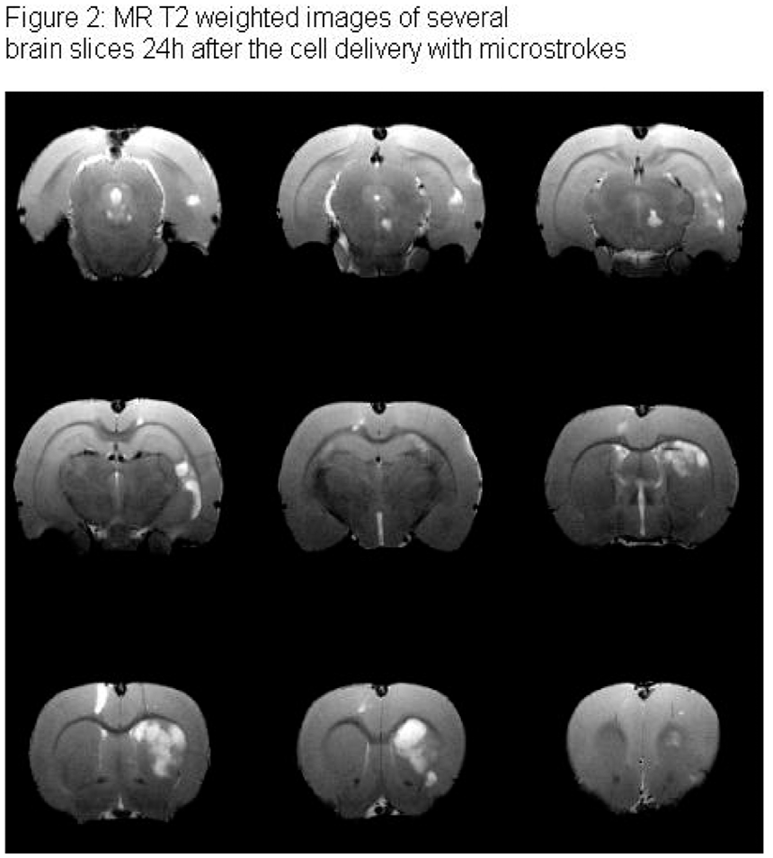

772
BRAIN-0760
Poster Session
HUMAN AMNION EPITHELIAL CELLS REDUCE INFARCT VOLUME, SPLENIC ATROPHY AND LUNG INFLAMMATION FOLLOWING ISCHEMIC STROKE IN MICE.
Stem cells and Cell Therapy
1Department of Pharmacology, Monash University, Clayton, Australia
2The Ritchie Centre, Monash Institute of Medical Research, Clayton, Australia
Abstract
773
BRAIN-0063
Poster Session
MYOBLAST-MEDIATED CO-DELIVERY OF VEGF AND PDGF AFTER EXPERIMENTAL ENCEPHALOMYOSYNANGIOSIS IMPROVES COLLATERALIZATION AND FUNCTIONAL OUTCOME IN A MODEL OF CHRONIC CEREBRAL HYPOPERFUSION
Stem cells and Cell Therapy
1Neurosurgery, Charité Berlin, Berlin, Germany
2Institute for Surgical Research and Gene Therapy Group, University Hospital Basel, Basel, Switzerland
3Baxter Laboratory for Stem Cell Technology, Stanford University, Stanford, USA
Abstract
Objectives
The myoblast-mediated transfer of angiogenic genes is a cell-based approach for induction of therapeutic collateralization.1 In chronic cerebral hypoperfusion, we recently demonstrated successful implantation and fusion of primary monoclonal mouse-myoblasts in the non-ischemic temporal muscle of an experimental encephalomyosynangiosis (EMS)2 with hemodynamic rescue and improved collateralization through myoblast-mediated supplementation of vascular endothelial growth factor-A (VEGF).3 However, blood vessel stability depends on the coordinated and balanced interaction of multiple signaling pathways in the endothelial and perivascular cells. In the present study, we therefore investigated the effect of myoblast-mediated co-delivery of platelet derived growth factor-BB (PDGF) in addition to VEGF supplementation in an EMS mouse model of chronic cerebral hypoperfusion.
Monoclonal mouse myoblasts expressing a reporter gene alone (control) or in combination with VEGF, PDGF or VEGF/PDGF were implanted into the temporal muscle of an EMS before permanent ipsilateral ICAO in C57/BL6 mice. Exogenous gene factor expression was confirmed by real-time PCR and western blot analysis. Hemodynamic impairment was quantified by laser speckle imaging and cerebrovascular reserve capacity (CVRC) measurements throughout the observation period of 84 days. On days 21, 42 and 84, transpial collateralization, vessel density and vessel maturity at the muscle/brain interface of the EMS were assessed by in-vivo FITC-lectin perfusion and immunohistochemical staining. Further, cortical stroke volume and neuronal cell death after 60-minute MCAO and 23-hour reperfusion were determined by MRI and immunohistochemical analysis.
By day 21, co-expression of VEGF/PDGF resulted in an improved CVRC (VEGF/PDGF 37±15%; PDGF 27±8%; VEGF 30±11%; control 24±10%; p<0.05 for VEGF/PDGF vs. day 0), which was paralleled by significantly improved microvascular remodeling and higher pericyte coverage (VEGF/PDGF 68±11%; PDGF 63±9%; VEGF 47±6%; control 50±5%; p<0.05 for VEGF/PDGF vs. VEGF and control) of the collateral and parenchymal vasculature at the target site of VEGF/PDGF supplementation. Functional and morphological findings were in line with an attenuated cortical stroke volume (VEGF/PDGF 34±12%; PDGF 47±13%; VEGF 41±12%; control 50±10%; p<0.05 for VEGF/PDGF vs. control) and decreased neuronal cell death (NeuN cells: VEGF/PDGF 391±145/mm2; PDGF 239±88/mm2; VEGF 189±119/mm2; control 181±95mm2; p<0.05 for VEGF/PDGF vs. all groups).
The myoblast-mediated co-delivery of VEGF and PDGF after indirect experimental revascularization improved morphological and functional outcome compared to delivery of VEGF alone. In the future, an appropriate translational approach could facilitate indirect revascularization procedures in patients suffering from chronic cerebral hypoperfusion with better protection from ischemic stroke.
References
774
BRAIN-0150
Poster Session
STEREOTACTIC TRANSPLANTATION OF STROMAL CELL-DERIVED FACTOR-1 GENE MODIFIED ENDOTHELIAL PROGENITOR CELL EFFECTIVELY ATTENUATED ISCHEMIC BRAIN INJURY IN MOUSE MODEL
Stem cells and Cell Therapy
1Med-X Research Institute, Shanghai Jiao Tong University, Shanghai, China
2Department of Neurology Ruijin Hospital, Shanghai Jiao Tong University, Shanghai, China
Abstract
Abstract
EPC-SDF-1α transplantation improved neurobehavioral outcomes and reduced ischemia-induced brain atrophy. (
Referencesš
775
BRAIN-0327
Poster Session
WHITE MATTER REPAIR AFTER ADIPOSE TISSUE-DERIVED MESENCHYMAL STEM CELL ADMINISTRATION IN SUBCORTICAL ISCHEMIC STROKE
Stem cells and Cell Therapy
1Department of Neurology and Stroke Center Neuroscience and Cerebrovascular Research Laboratory, La Paz University Hospital Neuroscience Area of IdiPAZ Health Research Institute Autónoma University of Madrid, Madrid, Spain
2Department of Neurology Clinical Neurosciences Research Laboratory, Hospital Clínico Universitario Health Research Institute of Santiago de Compostela (IDIS) University of Santiago de Compostela, Santiago de Compostela, Spain
3Laboratory for Imaging and Spectroscopy by Magnetic Resonance, Institute of Biomedical Research Alberto Sols, Madrid, Spain
Abstract
This study was supported by research grants PS12/01754, INVICTUS SpanishNeurovascular Network RD12/0014/0006 and Research Institute Carlos III,Ministry of Science and Innovation of Spain.
776
BRAIN-0418
Poster Session
SPATIOTEMPORAL RESPONSE OF NEURAL PROGENITORS IN THE ADULT SUBVENTRICULAR ZONE TO EXOGENOUS GROWTH FACTOR STIMULI.
Stem cells and Cell Therapy
1Neurosurgery, Graduate School of Medicine The University of Tokyo, Tokyo, Japan
2Cellular Neurobiology, Graduate School of Medicine The University of Tokyo, Tokyo, Japan
3Developmental Biology, Cincinnati Children’s Hospital Research Foundation, Cincinnati, USA
Abstract
Objectives
We have previously demonstrated anatomical and functional neuroregeneration in the ischemic brain by recruitment of endogenous neural progenitors with the intraventricular infusion of growth factors (GFs). Now, there is cumulative evidence that this approach is feasible as a method of brain repair. However, outcomes of studies of GF treatment are often divergent and sometimes disappointing. Then optimizing conditions to induce the maximal response of endogenous progenitors by exogenous GF stimuli is an important issue.
We performed time-course analysis to investigate the changes in the cell profile of the subventricular zone (SVZ) in intact adult male C57/BL6J mice administered an intraventricular infusion of high-dose growth factor (EGF) and fibroblast growth factor-2 (FGF2) for different durations. First, we treated animals with GF for 0-14 days and investigated the response of SVZ progenitors and neuroblasts soon after the therapy. For that purpose, we used the following antibodies: Tuj1, Ki67, Pax6, Sox2. Second, we treated animals with GF for 7 or 14 days. Then, animals were allowed to survive 2, 6, 10 and 28 days after the end of the therapy and were investigated in the same manner. Finally, we labeled progenitors with BrdU and tracked them in the olfactory bulb and SVZ.
EGF/FGF2 therapy promoted proliferation of SVZ progenitors up to 200% of the control level. Interestingly GF therapy arrested neuroblast production, an effect that was sustained as long as EGF/FGF2 therapy was continued. Withdrawal of EGF/FGF2 led to differentiation of the proliferated SVZ progenitors into neuroblasts with a transient but significant increase up to 150% of the control level.
Our study revealed the mechanism of action underlying the effects of GF treatment. Our findings can help to achieve the maximal response of endogenous progenitors to in vivo GF treatment.
777
BRAIN-0191
Poster Session
TRANSPLANTATION OF NEURAL STEM CELLS THAT OVEREXPRESS SOD1 ENHANCES AMELIORATION OF NEURONAL DAMAGE FROM INTRACEREBRAL HEMORRHAGE IN MICE
Stem cells and Cell Therapy
1Neurosurgery, Yamanashi University, Chuo, Japan
2Neurosurgery, Stanford University, Stanford, USA
Abstract
778
BRAIN-0858
Poster Session
IINTRA-ARTERIAL STEM CELL TREATMENT REDUCES INJURY IN A REPRODUCTIVELY SENESCENT RAT MODEL OF STROKE
Stem cells and Cell Therapy
1Neurology, University of Miami, Miami, USA
2Interdisciplinary Stem Cell Institute, University of Miami, Miami, USA
3Interdisciplinary Stem Cell Institute Department of Medicine, University of Miami, Miami, USA
4Cerebral Vascular Disease Research Laboratories Department of Neurology, University of Miami, Miami, USA
Abstract
References:
779
BRAIN-0395
Poster Session
COMPARISON OF NEURAL DIFFERENTIATION BY NOGGIN ALONE OR NOGGIN PLUS SB431542
Stem cells and Cell Therapy
1Stroke Division, The Florey Institute of Neuroscience and Mental Health-Austin Campus, Melbourne, Australia
Abstract
Objectives: Many studies focus on the pathways that specify the final neural sub-types that can be derived from human embryonic stem cells (hESC). However, few studies now compare the viability and fate of the intermediates and final cellular products produced by different initial induction methods. We compared two of the most commonly used induction methods– Noggin and Noggin plus SB431542 (Noggin+SB). The cell growth condition, lineage differentiation, and survival rate were compared at different time points.
Methods: hESC was treated with either Noggin for 14 days, or Noggin+SB for 11 days as previously described. After one week (1w) or two weeks (2w) of proliferation as neurospheres, the cells were dissociated into single cells and seeded on 48 well plates. Neural differentiation was then induced as described previously. The cells were grown for up to 21 days. Cell samples were collected at 7, 14, 21 days in vitro (DIV) for immunohistochemical staining using Nestin and MAP2. Cell death was determined by propidium iodide staining. HCA-vision software was used to perform the quantification of immunohistochemical staining.
Results: hESC induced by Noggin+SB appeared to differentiate at a faster rate than the cells induced by Noggin alone. Mature clusters appeared at 4 DIV in 1w neurosphere, and 8 Div in 2w neurosphere, comparing to the Noggin, in which they appeared at 14 Div in 1w, and 21 Div in 2w. However, in the dual induction there were significantly higher cell loss, almost double, compared to the Noggin alone induction.
Regardless of which induction method was used, 2w neurospheres gave rise to a lower percentage of Nestin positive cells compared to 1w neurospheres. 2w neurospheres induction by Noggin alone generated a greater percentage of Nestin positive cells than the Noggin+SB method (6.48% vs. 0.05% respectively) did. The same trend was observed in the 1w neurospheres under the two induction methods (7.85% and 3.61% Nestin expression, respectively).
The percentage of Nestin positive cells in the 2w Noggin group decreased at a much slower rate, with 4.58% of neurons still expressing Nestin at 21 DIV. 2w neurospheres gave rise to a relatively low proportion of MAP2 positive neurons using both induction methods. Noggin+SB induction showed slightly lower expression of MAP2 (32.73%) compared to Noggin alone (40.26%) in 2w neurospheres, but a greater MAP2 expression (62.56% compared to 48.01%) in 1w neurospheres.
Conclusions: 2w neurosphere showed a slower but steadier differentiation than 1w neurospheres in both induction methods. Noggin in combination with SB431542 caused a faster differentiation, quickly losing Nestin positive cells and forming mature cell networks at an earlier stage. However, not all of these cells contribute to MAP positive cells population. Moreover, this faster differentiation appears to lead to greater cell death following the dual induction method, suggesting Noggin alone may be the preferred induction method.

780
BRAIN-0484
Poster Session
EVALUATION AND QUANTIFICATION OF 18F-FPEB AS RADIOLIGAND FOR PET IMAGING OF THE METABOTROPIC GLUTAMATE RECEPTOR 5 IN RAT BRAIN.
BrainPET: Novel Radiotracers
1Pathology and Imaging: Division of Nucleaur Medicine & Molecular Imaging, KU Leuven, Leuven, Belgium
2Pathology and Imaging: Laboratory for Radiopharmacy, KU Leuven, Leuven, Belgium
Abstract
18F-FPEB is a negative allosteric modulator for the metabotropic glutamate receptor 5 (mGluR5), a high interest target for PET imaging in, among others, addiction and depression. We characterized the pharmacokinetics of 18F-FPEB and evaluated its ability to quantify mGluR5 in rat brain.
Nine male Wistar rats (281±18.5 g) were scanned on a FOCUS-220 system for 120 min, following a bolus injection of 18F-FPEB (37.6±5.6 MBq, Specific activity > 35.0 GBq/µmol). Arterial blood samples were collected and parent intact fraction measured by HPLC to obtain the metabolite-corrected plasma input function. Blocking experiments were performed using MTEP to assess specificity (n=3). Time-activity curves were extracted for the striatum, nucleus accumbens, hippocampus, cortex, thalamus, and cerebellum. BPND (VT/VT,Ref - 1) of 18F-FPEB was used as outcome measure and compared to simplified methods of quantification using the cerebellum as reference region. Metabolite formation was also evaluated in vitro by incubation with blood, plasma and brain homogenate (n=3/condition). Additionally, ex vivo brain metabolite formation was assessed in perfused brain homogenates at 10, 30, 60, and 120 minutes post-injection using HPLC.
Intact 18F-FPEB comprised 68 ± 7% at 3 minutes after injection, declining to 44±9% at 10 min, 26±6% at 40 min, 19±5% at 60 min, and 16±5% at 120 min after tracer injection. 18F-FPEB was metabolized into two polar radiometabolite fractions in vivo. However, metabolites were not found in neither the ex vivo perfused brain homogenates at any time point, nor in the in vitro samples of blood, plasma or brain.
PET images showed an 18F-FPEB activity distribution consistent with mGluR5 localization in rat brain, including no uptake in the cerebellum (1). In vivo blocking studies with 20 mg/kg MTEP showed that more than 96±3% of brain activity was mGluR5 specific. A two tissue compartment model was used to determine the BPND for the striatum (11.7±1.5), nucleus accumbens (10.6±2.0), hippocampus (9.0±1.2), cortex (7.2±1.0) and thalamus (4.0±0.9). SRTM, Logan Non-Invasive, and MRTM0 were tested as reference models with BPND as outcome parameter. Time stability was evaluated for 3 scan-durations: 60, 90, and 120 minutes. Each reference model showed high correlation with the 2TC BPND (Spearman r >0.90). This was maintained for shorter scans (Figure A). For the 120 minutes data there was a significant mean underestimation of the BPND by MRTM0 (0.77), SRTM (0.66), and Logan NI (0.86), but this bias became insignificant for the shorter scan-durations. Bland-Altman analysis showed a coefficient of repeatability of 24.2 percent difference, independent of scan duration (Figure B). Test-retest studies (n=6) showed good reproducibility for MRTM0 (ICC = 0.83), SRTM (ICC = 0.86) and Logan NI (ICC = 0.81).
Due to its favorable reversible kinetics, high specificity and absence of brain radiometabolites 18F-FPEB seems a highly useful tracer for in vivo visualization of the mGluR5 in rat brain. Moreover, reference tissue models allow non-invasive rapid scanning with acceptable test-retest.
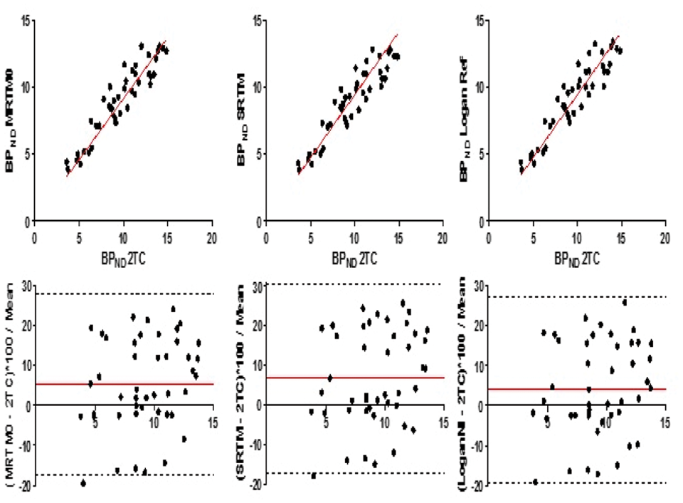
781
BRAIN-0416
Poster Session
EFFECT OF LIGAND SIZE ON UPTAKE AND WASHOUT OF EPHA2 TARGETED THERANOSTICS FROM GLIOBLASTOMAS USING 64CU-PET
BrainPET: Novel Radiotracers
1Australian Institute for Bioengineering and Nanotechnology, University of Queensland, Brisbane, Australia
2Digital Productivity, CSIRO, Brisbane, Australia
3Radiation Oncology, Genesis Cancer Care Lake Macquarie Private Hospital, Newcastle, Australia
4Dept. Pharmacology, University of Newcastle, Newcastle, Australia
Abstract
Fig. 1. Box plots of (A) uptake in brain and tumour, and (B) washout in tumour for YSA, scFv and mAb tracers.
782
BRAIN-0316
Poster Session
MAPPING MAO-B WITH FLUORINE-18 LABELED DEUTERATED FLUORODEPRENYL (18F-FLUORODEPRENYL-D2). DEVELOPMENT, IN VITRO AUTORADIOGRAPHY AND IN VIVO EVALUATION IN NON-HUMAN PRIMATES.
BrainPET: Novel Radiotracers
1Clinical Neuroscience, Karolinska Institutet, Stockholm, Sweden
2Global Drug Discovery, Bayer HealthCare AG, Berlin, Germany
3Clinical R&D Neurosciences, Piramal Imaging GmbH, Berlin, Germany
Abstract

References:
783
BRAIN-0749
Poster Session
[18F]NIFENE NICOTINIC ACETYLCHOLINE RECEPTOR BINDING IN HUMANS
BrainPET: Novel Radiotracers
1Medical Physics, University of Wisconsin-Madison, Madison, USA
2Radiological Sciences, University of California-Irvine, Irvine, USA
3Waisman Center, University of Wisconsin-Madison, Madison, USA
4Cardiovascular Medicine, University of Wisconsin-Madison, Madison, USA
Abstract
[18F]Nifene binding distribution. Each row has been selectively thresholded to highlight regional uptake in DVR images: (top row) midbrain, (middle row) cortex, (bottom row) thalamus.
784
BRAIN-0541
Poster Session
QUANTIFICATION OF INCREASED CEREBRAL 64CU UPTAKE IN TAU-P301L TRANSGENIC MOUSE MODEL OF ALZHEIMER’S DISEASE WITH 64CUCL2-PET/CT
BrainPET: Novel Radiotracers
1Radiology, University of Texas Southwestern Medical Center, Dallas, USA
2Pediatrics and Radiology, Wayne State University, Detroit, USA
Abstract
785
BRAIN-0728
Poster Session
HIGH FAT DIET DECREASES BINDING OF N-11C-METHYL-JNJ-31020028 TO NEUROPEPTIDE Y2 RECEPTORS
BrainPET: Novel Radiotracers
1Department of Nuclear Medicine & PET-Centre, Aarhus University and Hospital, Århus C, Denmark
2Center of Functionally Integrative Neuroscience, Aarhus University, Århus C, Denmark
3Department of Psychiatry, Oxford University, Oxford, United Kingdom
Abstract
Objectives:
Neuropeptide Y (NPY) is a potent orexigenic agent expressed in both the central and peripheral nervous system. In the brain, NPY increases food intake and body weight leading to metabolic changes promoting energy storage. The activation of NPY2 receptors, the most prominent NPY receptor in the CNS, has been linked to the induction of satiety, and polymorphisms in this gene have been implicated in human obesity. We have recently developed a novel positron-emitting radioligand based on the NPY2 receptor antagonist, JNJ-31020028, and have used the radiotracer for positron emission tomography brain imaging in pigs. Here we examine changes in the binding potential (BPND) of NPY2 receptors in response to diet-induced obesity.
Four average weight (25kg) adult female Gottingen minipigs were anesthetized and scanned at baseline with N-[11C]methyl-JNJ-31020028 in a Siemens PET/CT scanner. Minipigs were then fed an unlimited, high-fat, palatable diet for 10-12 weeks which resulted in doubling of their body weight (51kg), prior to rescanning with N-[11C]methyl-JNJ-31020028. PET data were registered to an average minipig MRI atlas and processed using Pmod software package 3.6. The BPND was obtained using the Logan graphical analysis, with corpus callosum as a region of non-displaceable binding.
On average in the four pigs, JNJ-31020028 BPND was significantly reduced in the striatum, thalamus and amygdala as a result of the high-fat diet.
Data demonstrate the use of this novel tracer in longitudinally examining physiological processes and reinforce the idea of NPY2 antagonism as a potential beneficial treatment of obesity.
786
BRAIN-0476
Poster Session
CHARACTERIZATION OF [11C]LU AE92686 AS A PHOSPHODIESTERASE 10A PET RADIOLIGAND IN THE MONKEY BRAIN
BrainPET: Novel Radiotracers
1Department of Clinical Neuroscience, Karolinska Institutet, Stockholm, Sweden
2Division of Synaptic Transmission, H. Lundbeck A/S, Valby, Denmark
3Division of Discovery Chemistry and DMPK, H. Lundbeck A/S, Valby, Denmark
4Neuroscience Research Unit, Pfizer Inc., Cambridge MA, USA
Abstract
References: [1] Nishi et al. 2011, [2] Kehler et al. 2014.
Figure: PET summation images (0-123 min) for (A) substantia nigra and (B) striatum before and after MP-10 pretreatment.
787
BRAIN-0467
Poster Session
UNIFIED THEORY OF SOKOLOFF'S MODEL AND RENKIN-CRONE'S MODEL
BrainPET: Novel Modeling and Methods
1Division of Nuclear Medicine, National Center for Global Health and Medicine, Tokyo, Japan
2Department of Computational Systems and Biology Faculty of Biology-Oriented Science and Technology, Kinki Univiersity, Kinokawa, Japan
Abstract
k* 1 of deoxyglucose (DG) model developed by Sokoloff et al.[1] does not include cerebral blood flow (F), although influx rate constant from arterial plasma into brain tissue (K 1 ) is a product of the value of F and first-pass extraction (E =1-exp(-PS/F))[2,3].
Some people might think that K 1 of DG is independent of F, because DG goes through transporter competing with glucose. However, it is natural that k* 1 of Sokoloff's model does not include F, because the model itself does not consider F from first to last. Their model represents that tissue acquires deoxyglucose from the silent blood pool of capillary.
We have corrected their DG model considering flowing blood in capillaries.
Our model show here that k* 1 of Sokoloff's model is identical as the permeability surface area product (PS). Certainly K 1 of DG can be approximated to PS, because E of DG is low. However, our proposed model predicts that K 1 of DG should be around 15% accurate than the conventional model under the normal condition [4].
Although K 1 is less dependent on flow, we would like to emphasize the fact that K 1 of DG is flow-dependent in principle and a product of E and F, like all the other tracers.
[1] Sokoloff L, Reivich M, Rosies MHD, Patlak CS, Pettigreow KD, Sakurada O
et al. The [14C]deoxyglucose method for the measurement of local cerebralglucose utilization: Theory, procedure, and normal values in the conscious andanesthetized albino rat. J Neurochem 1977;
Abstract
[2] Renkin EM. Transport of potassium-42 from blood to tissue in isolated mammalian
skeletal muscles. Am J Physiol 1959;
Abstract
[3] Crone C. The permeability of capillaries in various organs as determined by
use of the ‘indicator diffusion’ method. Acta Physiol Scand 1963;
Abstract
[4] Hasselbalch SG, Knudsen GM, Holm S, Hageman LP, Capaldo B, Paulson OB.
Transport of n-Glucose and 2-Fluorodeoxyglucose Across the Blood-Brain Barrier
in Humans. J Cereb Blood Flow Metab 1996;
788
BRAIN-0737
Poster Session
UNCOVERING HIDDEN PHYSIOLOGICAL PARAMETERS BASED ON THE REALISTIC TISSUE SIMULATION
BrainPET: Novel Modeling and Methods
1Department of Bio and Brain Engineering, Korea Advanced Institute of Science and Technology, Daejeon, Korea
2Graduate School of Medical Science and Engineering, Korea Advanced Institute of Science and Technology, Daejeon, Korea
Abstract
References: [1] Barros LF et al.A quantitative overview of glucose dynamics in the gliovascular unit. Glia 2007;55:1222-1237.
789
BRAIN-0242
Poster Session
COMPUTATION OF FULL PARAMETRIC IMAGES FOR REVERSIBLE TRACER OF FLT WITHIN REASONABLE TIME
BrainPET: Novel Modeling and Methods
1Medical Physics, Kagawa University, Kitagun, Japan
2Clinical Radiology, Kagawa University Hospital, Kitagun, Japan
3Neurological Surgery, Kagawa University, Kitagun, Japan
4Radiology, Kagawa University, Kitagun, Japan
Abstract
Objectives: PET enables to quantify biomedical functional images such as rate constants of uptake (K1), washout (k2), phosphorylation (k3) and dephosphorylation (k4), by applying the two tissue compartment model. However there are, so far, no feasible computational strategies for imaging all rate constants within reasonable computation time. We proposed a new method to compute all those rates within a reasonable time, namely, < 1 min.
Methods: A set of differential equations for the reversible two tissue compartment model was converted to one formula expressed as tissue and plasma curve terms involving convolution integral and derivative terms. Applicability was tested by applying clinical data with 18F-FLT PET (ECAT HR+) for patients with glioma (n=15). Then the images of those rates were obtained. To test validity, regions of interest (ROI) were placed on regions with glioma on a summed image. Parametric values were extracted from the obtained rate images. Also, time activity curve was extracted from the placed ROIs and those rate values were computed by the non-linear fitting method (Gauss-Newton method). The obtained rate values were compared between the methods.
Results: Computation time was around 30 s and quality of generated images was reasonably acceptable (Figure). Obtained values for K1 were 0.16±0.04 and 0.17±0.05 ml/min/g for image and ROI based methods, respectively. Those were 0.15±0.08 and 0.16±0.08 min−1 for k2, 0.050±0.034 and 0.043±0.026 min−1 for k3, 0.019±0.010 and 0.014±0.007 min−1 for k4, respectively. Paired t-test showed no significant difference between the methods. Regression analysis showed correlations as: r=0.75, 0.66, 0.85 and 0.64 for K1, k2, k3 and k4, respectively.

790
BRAIN-0374
Poster Session
REFERENCE TISSUE DUAL TIME POINT QUANTIFICATION OF IRREVERSIBLE TRACER KINETICS APPLIED TO [18F]FDOPA
BrainPET: Novel Modeling and Methods
1NGMB, University Medical Center Groningen, Groningen, Netherlands
2Neurology, University Medical Center Groningen, Groningen, Netherlands
3Medical Physics, PUCRS, Porto Alegre, Brazil
4Nuclear Medicine, KULeuven, Leuven, Belgium
Abstract
Objectives
Imaging with PET and [18F]FDOPA is a commonly used method for the diagnosis and assessment of Parkinson’s disease (PD) progression. Two well established quantitative endpoints for its quantification are the influx constant Kiocc, which requires a full dynamic scan, and the striatal-to-occipital ratio (SOR), which can give information on the disease severity with a short static scan. Aim of this study was to develop a method for quantification of irreversible tracer kinetics using two time points and a reference tissue as input function and validate it for [18F]FDOPA.
Methods
Starting from the Patlak equation with a reference tissue as input function, an approximation can be deduced in which the influx constant can be obtained from only two time points instead of a full dynamic scan. To validate the approximation, a total of 21 subjects were included in the study, divided into three groups, 5 healthy controls, 5 confirmed PD patients and 11 subjects with probable PD. Subjects fasted for at least 4h before the start of the scan and were given 2.5mg/kg of carbidopa orally on arrival. One hour after the carbidopa dose, subjects underwent a 2h dynamic 3D PET scan. Dynamic PET data were spatially normalized to MNI space and TACs of the caudate, putamen and occipital cortex were obtained using the Hammers atlas. The Kiocc was estimated by a Patlak graphical analysis over the 40 to 120 min time interval using the non-specific uptake in the occipital region as input function and considered as gold standard. Next to the Patlak graphical analysis, Kiocc was estimated with a dual time point reference (DTPref) approach using two PET frames, a 10min frame starting at 40 min post injection and a 15min frame starting 105min post injection (DTPref40-120). SORs were obtained for each striatal structure using the last 15 min time frame of the dynamic PET scans.
Results
As shown in the graph, linear regression between the DTP approximation for Kiocc and the values obtained from the Patlak graphical analysis yielded a regression slope of 1.014 with R2 = 0.98. Linear regression between Patlak derived Kiocc and SOR yielded an R2 = 0.69.

Conclusions
We validated a reference tissue dual time point quantification method for [18F]FDOPA which demonstrated excellent correlation with a Patlak analysis using a reference tissue. These findings suggest the usefulness of the DTP approach for determining the same quantitative endpoint as a graphical Patlak analysis of a full dynamic scan. Therefore, DTP imaging of [18F]FDOPA is able to give the same information for both diagnosis and disease progression while significantly reducing acquisition time, thus increasing patient throughput and comfort.
References
791
BRAIN-0435
Poster Session
EVALUATION OF PERINIDAL CEREBRAL BLOOD FLOW AND METABOLISM USING A NOVEL QUANTITATIVE 15O-PET METHOD IN PATIENTS WITH ARTERIOVENOUS MALFORMATION
BrainPET: Novel Modeling and Methods
1Neurosurgery, National Cerebral and Cardiovascular Center, Osaka-Suita, Japan
2Investigative Radiology, National Cerebral and Cardiovascular Center, Osaka-Suita, Japan
3Radiology, National Cerebral and Cardiovascular Center, Osaka-Suita, Japan
4Neurosurgery, Faculty of Medicine Fukuoka University, Fukuoka, Japan
Abstract
Objectives
We describe a dual-tracer basis function method (DBFM) to avoid the need to assume a fixed fractionation of blood volume as being arterial and venous, a common assumption in dual-tracer autoradiography (DARG) and other three-step approaches[1] [2] [3]. Assuming a fixed fractionation may introduce systematic errors when imaging pathologic tissues, such as in patients with arteriovenous malformation (AVM), a disorder often characterized by increased arterial blood volume. In AVM patients, we compared the quantitative values of 15O-PET parameters derived using the present DBFM and those derived using the previously proposed DARG methods.
Methods
Seven patients with cerebral AVM were examined using 15O-PET scans based on findings of both DBFM and DARG methods, and three patients also underwent 123I-IMP SPECT imaging to obtain cerebral blood flow (CBF) baseline/control values. These three sets of images were calibrated using MR images. Two sets of region-of-interest circles (ROI) were established: (1) within 20 mm of the nidus, which we refer to as perinidal ROI (ROI-p), and (2) more than 20 mm away from the nidus, which we refer to as the remote ROI (ROI-r). A quantitative value of the regional CBF (rCBF), cerebral metabolic rate of oxygen (rCMRO2), and oxygen extraction fraction (rOEF) were obtained and were analyzed using linear regression analysis.
Results
We observed a higher positive correlation between the DBFM method and DARG method in ROI-r (rCBF: r=0.97, rCMRO2: r=0.96, and rOEF: r=0.78) than in ROI-p (rCBF: r=0.63, rCMRO2: r=0.32, and rOEF: r=0.56). In ROI-p, quantitative values of rCBF and rCMRO2 tended to be overestimated more when using DARG than when using DBFM. In ROI-r, significant positive correlations were also observed in values of rCBF between the 123I-IMP SPECT method and DBFM method (r=0.70), or in values of between the 123I-IMP SPECT method and the DARG method (r=0.67), but these correlations were lower for ROI-p values, although relatively remarkable in the DARG method (123I-IMP SPECT and DBFM: r=0.49, and 123I-IMP SPECT and DARG: r=0.33).
Conclusions
Our findings demonstrate the validity of the present DBFM method as a possible way to more accurately evaluate perinidal regions in AVM patients using not only CBF but also CMRO2 and OEF.
References
792
BRAIN-0568
Poster Session
QUANTIFICATION OF [11C]RO15-4513 SPECIFIC BINDING AND SELECTIVITY IN VIVO.
BrainPET: Novel Modeling and Methods
1Centre for Neuropsychopharmacology, Imperial College London, London, United Kingdom
2Pharma Research and Early Development, Roche, Basel, Switzerland
3Centre for Imaging Sciences, Imanova Ltd, Londond, United Kingdom
Abstract
Objectives
[11C]Ro15-4513 is a partially-selective inverse-agonist radioligand at the GABAAR-α5 receptor subtype with lower but significant affinity for α1, and to a lesser extent α2 and α3 (Myers et al. 2012). As such, the proportion of the [11C]Ro15-4513 specific signal in vivo in humans that corresponds to α5 remains an open question. In this work data obtained using a GABAAR-α5-selective blocking agent (RG1662) is used to enable partitioning of the specific [11C]Ro15-4513 signal across different brain regions.
Methods
Previously acquired [11C]Ro15-4513 PET data from 8 healthy male volunteers were provided, at baseline and after administration of 4 separate doses of the α5-selective negative allosteric modulator RG1662. Plasma concentrations of RG1662 were acquired at the start of each scan, as well as full arterial plasma sampling of radioactivity and metabolites for [11C]Ro15-4513. A two-tissue compartmental model (2TCM) was identified as the most appropriate kinetic model and was used to estimate VT in pre-specified regions of interest (ROIs) in the brain. VT data were fitted on a regional basis to a single site competition model which also included an α1 term across all subjects and allowed for the estimate of the specific α5 component of the signal:

where V’ = VT + Vα1.
Abstract
VND and Vα1were then estimated by employing regional estimates of α1 density from previous studies using [11C]flumazenil (Lassen et al. 1995) and determining the maximal regional correspondence between [11C]Ro15-4513 Vα1 and [11C]flumazenil BP ND .
These data also allowed for an evaluation of reference tissue methods and kinetic partitioning of the specific binding with spectral analysis.
Results
The 2TCM provided good fits for time-activity curves in each ROI with baseline VT estimates ranging from about 1 in the pons to 10 in the nucleus accumbens. The competition binding model produced good fits to the VT data (Figure 1a) and gave an RG1662 EC50 estimate of 427 ng.ml−1 with corresponding estimates of occupancy of between 22% and 79% in the dose interval 15-1250 mg. Regional levels of the specific α5 component of the signal were also determined (Vα5 ranged from 0.11 to 6.58). Estimation of the VND by maximising the relationship of V α1 with α1 literature estimates (see Figure 1b) indicated a VND of 0.78. The individual Vα5, Vα1and VND components of the total volume of distribution were then calculated in all ROIs (see Figure 1c). This demonstrates that for cortical regions approximately 80% of the specific signal corresponds to α5 whilst for subcortical regions the proportion is higher.
Strong relationships were seen between 2TCM outcome measures and those derived from the simplified reference tissue model and spectral analysis, though biases were introduced using both methods.
Conclusions
Analysis of the in vivo selectivity of [11C]Ro15-4513 specific binding using competition data provides an important characterisation of this ligand and enables assessment of the strengths of different analysis methods.
Figure 1:

References
793
BRAIN-0345
Poster Session
VOXEL-WISE QUANTIFICATION OF [11C]CURB IN HUMAN BRAIN WITH THE HIGH RESOLUTION RESEARCH TOMOGRAPH (HRRT)
BrainPET: Novel Modeling and Methods
1RIC, Centre for Addiction and Mental Health, Toronto, Canada
Abstract
References:
794
BRAIN-0527
Poster Session
EARLY DETECTION OF PRE-SYMPTOMATIC AD IN A COGNITIVELY HEALTHY POPULATION
BrainPET: Novel Modeling and Methods
1Radiology and Radiological Sciences, Vanderbilt University Medical Center, Nashville, USA
2Biostatistics, Vanderbilt University Medical Center, Nashville, USA
Abstract

795
BRAIN-0367
Poster Session
HYBRID DECONVOLUTION APPROACH FOR MODEL-FREE ESTIMATION OF NON-SPECIFIC BINDING IN PET STUDIES WITHOUT REQUIRING A REFERENCE REGION
BrainPET: Novel Modeling and Methods
1Molecular Imaging and Neuropathology, New York State Psychiatric Institute and Columbia University, New York, USA
2Biostatistics, Columbia University, New York, USA
Abstract

796
BRAIN-0270
Poster Session
MODEL SELECTION CRITERIA FOR DYNAMIC PET STUDIES
BrainPET: Data Acquisition and Analysis
1Radiology and Nuclear Medicine, VU University Medical Center, Amsterdam, Netherlands
Abstract
Objectives: Several criteria exist to identify the optimal kinetic model for quantification of tracer uptake or binding. These criteria are all based on the squared difference between observed (measured) and fitted data, the number of fit parameters, the number of frames, and weighting factors applied to residuals. Despite similarities between these criteria, the best criterion for kinetic model selection is under debate and different criteria are being used in the literature. Therefore, the purpose of the present study was to evaluate several commonly used criteria, such as Akaike Information Criteria (AIC), AIC unbiased (AICunb), model selection criteria (MSC), Schwartz Criterion (SC) and the F-Test for 4 different tracers.
Methods: Four tracers with different kinetic properties were evaluated: [11C]flumazenil (FMZ), [18F]DPA-714, [11C]GMOM and [11C]phenytoin (PHT). Previously, different plasma input tracer kinetic models have been selected for these 4 radiotracers. For each tracer, PET data from 5 subjects were included, randomly selecting patients and healthy controls. Time activity curves (TACs) were extracted using PVElab and Hammers template. TACs were then analysed using six plasma input models: single-tissue reversible plasma input model (1T2K), two-tissue irreversible plasma input model (2T3K), two-tissue reversible plasma input model (2T4K) with and without blood volume fraction correction (VB). Parameter boundaries were applied based on previously estimated values. Preferred models across TACs and subjects were then obtained using the various model selection criteria. The F-test requires direct comparison of two models. Therefore, for each subject, the AIC preferred model was compared with the other models using the F-test. The percentage of TACs disagreeing with AIC was calculated per radiotracer.
Results: For all 4 radiotracers strong agreement was seen across the different model selection criteria (Figure 1). The F-Test confirmed the AIC preferred model in 90% of all cases. The poorest regional agreement was found when comparing AIC with AICunb for [11C]PHT, with a 16% difference in model preference. [1-1C]PHT also showed the poorest agreement between AIC and SC (9%). Despite these regional disagreements, for [11C]PHT, the 1T2K_VB model was selected as the preferred model by all model selection criteria. For all tracers AIC and MSC gave exactly the same model preferences.
Conclusion: AIC and MSC yielded identical results. AICunb showed some regional disagreement when compared with AIC. AICunb has a stronger penalty for number of fit parameters and might therefore favour models with a smaller number of fit parameter more frequently in case of ‘noisy’ TACs (small VOIs). Overall, the 5 tested model selection criteria preferred the same model across all subjects. In conclusion, all model selection criteria tested performed similarly with only minor, non-relevant differences in practice.
Abstract
Model preference (percentage of all TACs) per selection criterion for the 4 brain PET radiotracers.
797
BRAIN-0535
Poster Session
[18F]DPA-714 BRAIN UPTAKE OVER A TWO YEARS PERIOD: STABILITY IN HEALTHY CONTROL AND EVOLUTION IN THE EARLY STAGE OF ALZHEIMER'S DISEASE
BrainPET: Data Acquisition and Analysis
1Laboratoire Imagerie Moléculaire In Vivo (IMIV) UMR 1023 Inserm/CEA/Université Paris Sud - ERL 9218 CNRS CEA/I2BM/Service Hospitalier Frédéric Joliot, CEA, Orsay, France
2CEA/I2BM/Service Hospitalier Frédéric Joliot, Sainte Anne Hospital Paris, Orsay, France
3Langage and Memory Neurology Unit, Sainte Anne Hospital Paris, Paris, France
4Neurospin I2BM DSV CEA, CATI Multicenter Neuroimaging Platform, Gif sur Yvette, France
Abstract
Neuroinflammation is thought to play a crucial role in the early stages of AD. 18 kDa Translocator Protein is expressed at a low level in healthy brain and is up-regulated during inflammatory processes that may occur in neurodegenerative diseases. It is considered as a promising target for the early imaging of microglial activation and can be measured by PET imaging using [18F]DPA-714 radioligand. It has been shown that TSPO ligand uptake was highly dependent on their affinity status based on a genetic polymorphism.
Our goal was to quantify the stability of [18F]DPA-714 brain uptake in healthy volunteers and its evolution in AD patients over a two years period, according to their TSPO-affinity status
Nine subjects were analyzed including: 4 AD patients at the Mild Cognitive Impairment stage (MCI), as defined by a typical progressive amnesic deficit of the hippocampal type and a positive amyloid PET imaging ([11C]-PiB), and 5 healthy controls, with negative amyloid PET imaging. All subjects underwent a first [18F]DPA-714 PET imaging at baseline and after two-years follow up. PET scans were acquired on a HRRT scanner (Siemens) for 90 minutes. Dynamic scans were corrected for head motion and co-registered with3D T1-weighted MRI. Relative [18F]DPA-714 brain uptake was measured, using cerebellum as pseudo reference region, in 76 anatomical regions previously segmented on MRI
Analyses of the TSPO polymorphism showed that 4 controls and 3 AD patients were mixed or high binders (TSPO+ groups), and 1 control and 1 AD patient were low binders (TSPO- group-). At baseline, AD-TSPO+ patients showed a higher [18F]DPA-714 uptake than controls-TSPO+ subjects, with standardized global cortical uptake of 1.40 and 1.22, respectively.
At follow up, [18F]DPA-714 uptake remained stable in the control-TSPO+ group (mean change over all regions of 6.4%) whereas it increased in all AD-TSPO+ patients (+ 20% on average).
In AD patients, the highest increase was found in the parietal region (mean increase of +26%). No difference at baseline and no longitudinal change was found in the AD and the control TSPO- groups.
[18F]DPA-714 brain uptake did not change significantly over 2 years in healthy volunteers. AD patients at the MCI stage showed a higher [18F]DPA-714 standard uptake at baseline and during follow up, suggesting an early inflammatory involvement in AD pathogenesis
798
BRAIN-0701
Poster Session
QUANTIFICATION OF ACETYLCHOLINESTERASE USING 11C-PMP PET IN NORMALS: COMPARISON OF 3 DIFFERENT STRATEGIES
BrainPET: Data Acquisition and Analysis
1Laboratory for cognitive neurology, KU Leuven, Leuven, Belgium
2Dept. Radiology and Nuclear Medicine, VU University Medical Centre (VUMC), Amsterdam, Netherlands
3Dept. Of experimental psychology, University of Oxford, Oxford, United Kingdom
4Psychiatry, KU Leuven, Leuven, Belgium
5Medical Imaging and Clinical Oncology, University of Stellenbosch, Cape Town, South Africa
6Radiopharmacy, KU Leuven, Leuven, Belgium
7Nuclear Medicine and Molecular Imaging, KU Leuven, Leuven, Belgium
Abstract
Objectives: To compare different strategies to quantify AChE in cortical regions using 11C-PMP PET.
Dynamic images were processed using an automatic pipeline that includes realignment, coregistration with a structural MRI and the corresponding GM segmentation, warping to MNI and the use of an atlas to define VOIS (AAL atlas). Time-activity curves were extracted in each cortical VOI intersected with the subject-specific GM map. We applied 3 different strategies to determine k3 as measure for AChE: (1) using an irreversible 2-tissue compartment model including a blood volume component and with arterial blood sampling to determine the metabolite corrected plasma input function (2T3k_Vb) [1]; (2) using a reference region with a high rate of ACh hydrolysis (REF) [2] and (3) using shape analysis (SA) [1]. The last two approaches do not require blood sampling.
Comparisons were performed using a Bland-Altman approach and variability in cortical regions was assessed. Test-retest values for each method were determined based on the subjects who received a repeat scan.
Comparing REF and 2T3k_Vb, we found that both methods weakly correlated (r=0.32, p<10−6) while REF showed larger bias for larger values of k3. The comparison between 2T3k_Vb and SA showed a weaker correlation (r=0.04, p>0.1) but the bias for higher values was smaller. The variability of k3 values for each method is shown in Figure 1A: 2T3k_Vb showed the smallest variability (interquartile range (IQR) = [0.0197; 0.0238]/min) and the values of k3 were significantly lower compared to the other methods (ANOVA, p < 0.05). The method 2T3k_Vb also showed the lowest amplitudes of the test-retest values (IQR = [-4.75; 9.45]%) (Figure 1C) and the test-retest values were significantly smaller compared to the other two methods (ANOVA, p < 0.05). If we limit our analysis to the baseline scans, the variability of the methods is similar as taking all scans together (Figure 1A and 1B).

References
799
BRAIN-0677
Poster Session
TEST-RETEST VARIABILITY OF [123I]CLINDE-SPECT BINDING IN HEALTHY HUMAN SUBJECTS
BrainPET: Data Acquisition and Analysis
1Neurobiology Research Unit and Center for Integrated Molecular Brain Imaging, Rigshospitalet, Copenhagen, Denmark
2Department of Neuroanaesthesiology, Rigshospitalet, Copenhagen, Denmark
3UFR de Médecine, Université François Rabelais de Tours, Tours, France
4Department of Radiology, Rigshospitalet, Copenhagen, Denmark
Abstract
Objectives
Neuroimaging of translocator protein (TSPO) unveils neuroinflammation and is a potential marker of immunotherapy in neurological disorders. [123I]CLINDE-SPECT has shown a strong regional TSPO expression within stroke and glioma patients [1] and both cortical and subcortical increase of TSPO binding in anti-NMDA receptor encephalitis [2]. Here we report the results of test-retest [123I]CLINDE-SPECT studies in healthy subjects.
Methods
Ten healthy volunteers (4 females) were scanned twice with [123I]CLINDE-SPECT, with an interval of 49±39 days. The rs6971 TSPO polymorphism was genotyped from genomic DNA using TaqMan® SNP Genotyping Assay (Applied Biosystems, USA, C_2512465_20). Structural MRI was acquired with a 3T Siemens Prisma scanner. Dynamic SPECT (Philips IRIX triple head scanner) images were acquired 90 min post [123I]CLINDE bolus injection. Arterial blood samples were obtained during scanning, and were corrected for metabolites using HPLC. SPECT and MRI were co-registered using interactive image overlay, and 11 anatomical VOIs were automatically delineated using probability maps [3]. The mean standard uptake value (mSUV) weighted by frame length was calculated as an outcome measurement of [123I]CLINDE both in plasma and the brain. The SUV was defined as the time activity curve normalized by injected dose per kg body weight. Within-subject Mean sum of Squares (MSW) to measure reproducibility, Intra-class Correlation Coefficient (ICC) to measure reliability, Coefficient of Variance (COV), and Percentage Difference (PD=2*(test-retest)/(test+retest)*100%)) across 11 VOIs were calculated.
Results
Large PD was observed in plasma measurements between test and retest: -17.9%±26.5% of the mSUV in plasma; 16.1%±19.4% of the mSUV in parent fraction; and -1.6%±29.9% of mSUV in plasma parent compound. For mSUV of 11 brain VOIs Pearson's correlation coefficients between test and retest of each subject were 0.936±0.04 (0.8617-0.9861). Test-retest outcome of brain binding is shown in Table 1. These matrices were also calculated separately for each genotype group: mixed-affinity binders (MAB) (n=4), and high-affinity binders (HAB) (n=6), and no significant difference was observed for MSW, ICC (paired-sample t-test) and PD (two-sample t-test). However, COV of MAB was 23.7% higher compared to HAB (p<0.001, paired-sample t-test), indicating lower brain uptake (mSUV) for MAB. An approximately 1.4 fold difference of CLINDE binding was observed between HAB and MAB in the cerebellum (p<0.001), which was the VOI with the highest ICC value, see Figure 1.
Conclusions
Like other second-generation TSPO ligands, [123I]CLINDE binding is influenced by the TSPO polymorphism. The test-retest percentage difference was lower than that of the first-generation TSPO ligand [11C]PK11195-PET [4]. The ICC measurements were also much better than [11C]PK11195 [4] and comparable to [11C]DPA-713 [5]. The instability of plasma measurement will be further investigated; more data are to be acquired in healthy individuals to further validate the use of [123I]CLINDE.
Mean SUV of cerebellum grouped by genotype, MAB: mixed-affinity binder; HAB: high-affinity binder.
References
800
BRAIN-0608
Poster Session
SURFACE-BASED MODELLING OF MOLECULAR IMAGING MARKERS IN PERI-INFARCT CORTEX
BrainPET: Data Acquisition and Analysis
1Department Neurology and Neurosurgery, Montreal Neurological Institute, Montreal, Canada
2Department of Neurology and Neurosurgery, Montreal Neurological Institute, Montreal, Canada
3Department of Neurology, Technical University Munich, Munich, Germany
4McConnell Brain Imaging Center, Montreal Neurological Institute, Montreal, Canada
5Department of Neurology, Jewish General Hospital, Montreal, Canada
Abstract
Assessing molecular imaging markers like GABA-A receptor density in peri-infarct cortex is of interest for acute stroke treatment and stroke recovery studies alike (Heiss, 2010). Traditional analysis methods of the peri-infarct region, are based on volumetric dilations, which cut-across cortical regions with distinct vascular territories. We developed a surface-based automatic analysis method which respects the individual sulcal anatomy and vascular territories and compared it to a volumetric approach in 18F-FMZ data from patients with ischemic stroke.
9 patients with cortical ischemic stroke underwent PET and MR imaging within two weeks after the stroke and again at 6 month follow-up. T1 and flair MR images were acquired on a Siemens Sonata 1.5 Tesla Scanner (Siemens MedicalSolutions, Erlangen, Germany) using a 3D fast-field echo scan and MPRAGE sequences. Surface mesh representations of the cortical gray matter were extracted from the T1 images using CIVET, version 2.0 (Kim, et al. 2005).
PET images were acquired with [18-F]-flumazenil on the ECAT HRRT PET scanner in list mode and reconstructed with filtered back-projection. PET images were partial-volume corrected using the idSURF algorithm (Funck, et al. 2014). Parametric non-displaceable binding (BPnd) images were created using the Logan plot method (Logan et al., 1996).
Infarct masks were drawn on the individual flair MR images for each subject for initial and follow-up scans (Fig.1.A). Surface infarct masks were produced by interpolating the volumetric infarct masks with the surface representations from CIVET (Fig.1.B). Geodesic distances from the infarct masks were calculated on the surfaces (Fig.1.C) and segmented into 3mm rings, from 0 to 21mm from the infarct (Fig.1.D, example of one ring).
In addition, volumetric distance rings were created using a series of 1mm voxel-wise dilations of the original, volumetric infarct mask.
Parametric BPnd images were then analyzed, once using surface-based and then volumetric rings in the affected and at the mirror positions of the non-affected hemisphere. A two-way repeated measures ANOVA was used to analyze the BPnd values observed with the surface-based and volumetric methods, respectively. A second two-way repeated measures ANOVA was used to test the difference between the two methods, once for the initial and once for follow-up scans
(Fig.1)

BPnd was significantly different between the peri-infarct regions in the affected hemisphere and the non-affected hemisphere both when using the surface-based (p<0.01) and volumetric (p<0.05) methods. The two methods were significantly different at the follow-up (p<0.01) (Fig.2). Pariwise multiple comparisons revealed that the significant difference was due to distance rings between 9mm to 21mm away from the infarct (all p<0.01).
(Fig. 2) Difference between non-affected and affected scans, at initial and follow-up for volumetric and surface-based methods. Surface and volumetric methods were significantly different at follow-up.

The sensitivity of BPnd measurements in the peri-infarct region can be increased by using a surface based analysis method, which respects anatomical and vascular territories rather than volume-based methods.
References
801
BRAIN-0598
Poster Session
PREDICTION OF THE LIKELIHOOD FOR SUBSEQUENT PHENOCONVERSION IN RBD PATIENTS WITH METABOLIC NETWORK ACTIVITIES: AN EXPLORATORY STUDY WITH FDG PET
BrainPET: Data Acquisition and Analysis
1Center for Neurosciences, Feinstein Institute for Medical Research, Manhasset, USA
2PET Center, Huashan Hospital, Shanghai, China
3Department of Neurology, Huashan Hospital, Shanghai, China
Abstract
Objectives:
Idiopathic rapid eye movement behavior disorder (RBD) represents a significant risk factor for the development of Parkinson’s disease (PD), dementia with Lewy bodies (DLB) and multiple system atrophy (MSA). It has been shown recently that elevated baseline activity of PD-related brain network (PDRP) in RBD patients is predictive of eventual onset of PD and DLB after 4.6 years of clinical follow-up [1]. In this study we evaluated whether it is possible to predict the status of phenoconversion in RBD patients from baseline activities of both metabolic brain networks associated with PD and MSA (i.e. PDRP and MSARP) described previously.
Methods:
We used FDG PET images acquired in China on a Siemens Biography PET/CT scanner in two cohorts of patients with a diagnosis of idiopathic RBD confirmed by polysomnography [2]. The study was approved by the IRB for which the subjects had signed written informed consent. Network scores were computed in all subjects using PDRP and MSARP validated previously [3-4]. The combined sample of 27 independent patients from the two RBD cohorts were classified into three different subgroups using the subject scores of PDRP and MSARP. Group membership was estimated empirically using either the strict criteria (PD: PDRP > 1.5 and MSARP < 0.0; MSA: by MSARP > 1.5 and PDRP < 0.0) or the liberal criteria (PD: PDRP > 1.5 and MSARP < 1.5; MSA: MSARP > 1.5 and PDRP < 1.5).
Results:
PDRP scores were higher than MSARP scores (P < 0.005) and correlated negatively with each other (R > 0.54, P < 0.01) in each of the two RBD cohorts. Analysis in the 27 patients showed that 9-10 (33-37%) and 1-2 (4-7%) subjects would convert to PD/DLB and MSA respectively over a period of 3-5 years from baseline. These findings were in agreement with previous reports based on prospective clinical follow-up over similar periods [5-6].
Conclusions:
We demonstrated the feasibility of predicting the rates of phenoconversion in RBD subjects by analyzing bivariate distribution of both brain network activities associated with parkinsonism. The accuracy of this prediction model needs be confirmed in this cohort with longer follow-up by clinicians blinded to the imaging outcomes.
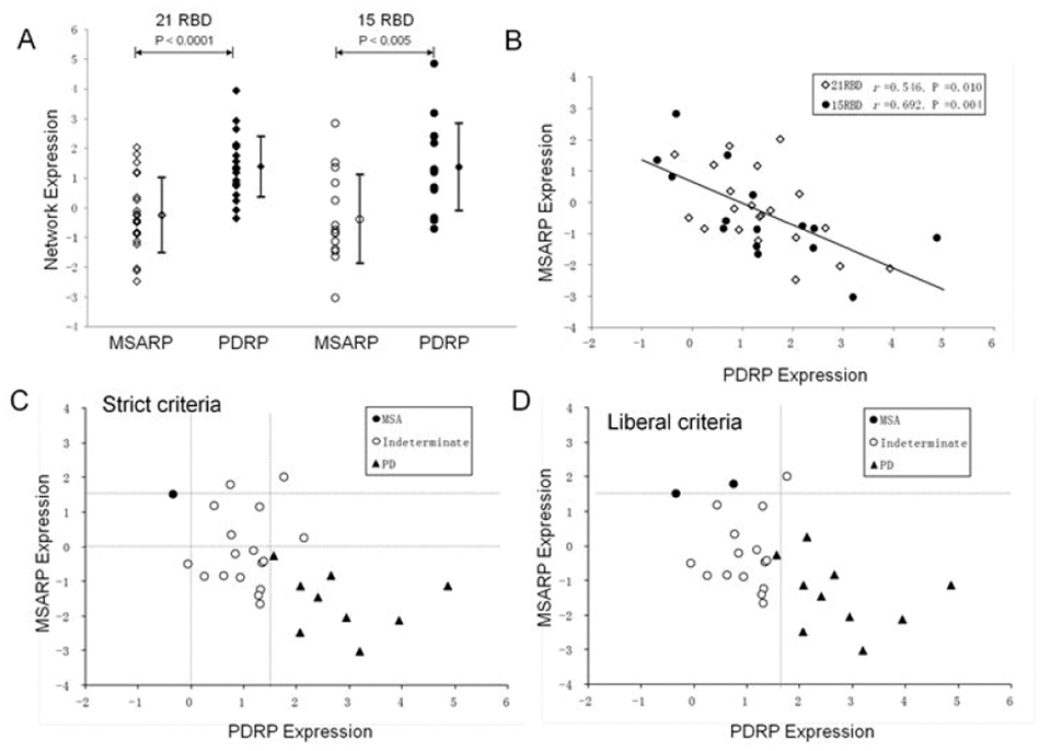
References:
802
BRAIN-0650
Poster Session
PARKINSON’S DISEASE-RELATED SPATIAL COVARIANCE PATTERNS FROM A RESTING STATE FDG AND O15-WATER PET: COMPARISONS OF NETWORK ACTIVITY MEASURED WITH FDG PET AND ASL MRI
BrainPET: Data Acquisition and Analysis
1Center for Neurosciences, Feinstein Institute for Medical Research, Manhasset, USA
Abstract
Objectives
Previous studies with FDG PET and multivariate network analysis have consistently demonstrated that Parkinson’s disease (PD) is associated with a specific disease-related spatial covariance pattern (PDRP) of regional glucose metabolism [1]. PDRP network activity correlates with disease severity ratings and is modulated by experimental therapies. The expression of PDRP can be assessed by imaging regional cerebral blood flow (rCBF) with PET/SPECT and perfusion MRI (2-3). Analogous PDRP network has also been generated by using resting-state H215O PET [4]. In this work we evaluated the comparability in network activity of these PDRPs using dual-modality FDG PET and arterial spin label (ASL) MRI.
Imaging studies were performed concurrently in 15 PD patients (58.2 ± 7.3 y; Hoehn and Yahr stage I-II) on a PET camera and a separate 3T MRI scanner. Maps of relative metabolism and rCBF were analyzed as described previously [1, 4]. Network scores of the two PDRPs were computed and z-transformed with respect to the same set of healthy controls. The scores from FDG and ASL MRI were compared in the healthy controls and in the PD patients to evaluate their reproducibility.
The PD-related spatial covariance pattern (PDRP-H215O) was characterized by increased activity in the sensorimotor cortex, thalamus, pons and cerebellum, and decreased activity in the premotor areas and occipito-parietal cortices. PDRP-H215O expression was elevated (P215O and PDRP-FDG.
We have established that rCBF images can be used for identifying disease-related covariance patterns to discriminate PD patients from controls at an individual-case basis. Subject scores from ASL MRI and FDG PET correlate equally for PDRP-FDG and PDRP-H215O, further demonstrating the similarity of both topographies that are comparable to those from ASL MRI [5]. More study is needed to test the reproducibility of PDRP with rCBF in larger samples, and assess its clinical correlation and therapeutic responses. This method supports the development of a biomarker for PD with the increasing availability of simpler and non-invasive ASL MRI.


References:
803
BRAIN-0289
Poster Session
IMPACT OF NEW SCATTER CORRECTION STRATEGIES ON HIGH-RESOLUTION RESEARCH TOMOGRAPH PET STUDIES
BrainPET: Data Acquisition and Analysis
1Radiology and Nuclear Medicine, VU University Medical Center, Amsterdam, Netherlands
Abstract
Misalignment between transmission and emission data as a result of patient motion may cause scatter scaling artefacts, which in turn can lead to inaccurate quantification, especially for high-resolution scanners such as the High-Resolution Research Tomograph (HRRT) (1). This study was performed to evaluate the impact of μ-map dilation and 3D versus 2D scatter correction on quantification of HRRT data for three tracers covering a wide range in uptake and kinetic profiles.
A total of fifteen healthy subjects received dynamic HRRT scans with arterial sampling using either (R)-[11C]verapamil (n=5), [11C]raclopride (n=5) or [11C]flumazenil (n=5). All data were corrected for attenuation, decay, scatter and random events, and reconstructed using resolution-modelled ordinary-Poisson ordered-subsets expectation maximization (12 iterations, 16 subsets). In order to reduce the effect of patient motion on scatter scaling factors, μ-maps were dilated (up to 17 mm) prior to scatter correction. Scatter correction was performed using either 2D or 3D single scatter simulations (SSS). Volumes of interest (VOI) were generated using PVElab (2). For several VOI, including those that were prominently affected by artefacts, volumes of distribution (VT) were obtained.
Three out of five (R)-[11C]verapamil studies showed prominent artefacts in the lower part of the brain with a reduction of VT up to 50%. With a μ-map dilation of 9.6 mm or more, artefacts were no longer visible (Figure 1). Use of 3D SSS showed a statistically significantly increase in VT of, on average, 6% compared with 2D SSS (p<0.05, Wilcoxon Signed Rank test). For [11C]raclopride and [11C]flumazenil studies, μ-map dilations did not result in a statistically significant changes in VT (p>0.05). In addition, [11C]flumazenil and [11C]raclopride studies also did not show a significant change in VT with an average increase of 2% when using 3D versus 2D SSS.
The effect of μ-map dilation seems to be tracer dependent. For tracers such as (R)-[11C]verapamil, where uptake is prominent in skin tissue (near the outer contour of the patient), it resulted in more reliable scatter scaling factors, but did not change (and/or deteriorate) quantification for the other tracers. 3D SSS resulted in significant changes for (R)-[11C]verapamil only. Application of both μ-map dilation and 3D SSS results in improved quantitative accuracy of HRRT studies. This method might be applicable to other PET systems that use the same scatter scaling method.
Coronal (A and C) and sagittal (B and D) images, summed over the last 58 minutes frames, of an (R)-[11C]verapamil scan that was affected by subject motion. A and B are images reconstructed without dilation of the μ-map, C and D those with a 9.6-mm dilation of the μ-map. For all the images, 3D SSS was applied. Red arrows show artefacts, while white arrows show the prominent reduction of artefacts when μ-map dilation was applied.
References:
804
BRAIN-0525
Poster Session
REFERENCE TISSUE METHODS FOR PET NUROIMAGING STUDIES REVISITED
BrainPET: Data Acquisition and Analysis
1Radiology and Nuclear Medicine, Johns Hopkins University, Baltimore, USA
2Psychiatry and Behavioral Sciences, Johns Hopkins University, Baltimore, USA
3Department of Family and Community Health, University of Maryland School of Nursing, Baltimore, USA
4Department of Medicine, Johns Hopkins University, Baltimore, USA
Abstract

805
BRAIN-0347
Poster Session
INDIVIDUALIZED MAP OF 18F-FDG METABOLISM VARIATION: COMPARISON OF ON AND OFF DEEP BRAIN STIMULATION CONDITIONS IN SEVERE BRAIN INJURED PATIENTS.
BrainPET: Data Acquisition and Analysis
1Image Guided Clinical Neurosciences and Connectomics (EA 7282), Université Clermont Auvergne, Clermont-Ferrand, France
2Neurochirurgie A, CHU de Clermont-Ferrand - Hôpital Gabriel Montpied, Clermont-Ferrand, France
3Centre Jean Perrin - Service de Médecine Nucléaire, Université Clermont Auvergne, Clermont-Ferrand, France
Abstract
Abstract
Figure: Individualized map of metabolism variation (Patient 3) overlaid on horizontal (axial) slice (inverted T1-weighted MRI) going through basal ganglia and thalamus: note the severe brain atrophy and ventricular enlargement*; left (L) hemisphere metabolism increased in DBS On (vs Off).
References:
806
BRAIN-0334
Poster Session
TOWARDS LESS-INVASIVE QUANTIFICATION OF [11C]PBR28: IMAGE-DERIVED AND POPULATION-BASED INPUT FUNCTIONS
BrainPET: Data Acquisition and Analysis
1Department of clinical neuroscience, Karolinska Institutet, Stockholm, Sweden
Abstract
Objective
[11C]PBR28 is a radioligand for imaging the 18kDa Translocator Protein (TSPO), a protein expressed in microglia. Since microglia is present throughout the brain, it is not possible to designate a reference region free from specific binding. Therefore, quantification of [11C]PBR28 PET data currently requires an arterial input function (AIF) obtained from arterial cannulation. The goal of this study was to evaluate population-based and image-derived input functions (IDIFs, PBIFs) obtained using only a few arterial or venous samples, for the analysis of [11C]PBR28 data.
Method
Fifteen control subjects (8 High affinity binders (HABs) and 7 Mixed affinity binders (MABs)) were examined using [11C]PBR28 and the HRRT PET system. AIFs were acquired for each subject. For 4 of the subjects (2 HABs, 2 MABs), whole blood radioactivity, blood-to-plasma radioactivity ratio (B/P), and radioactive metabolites were also measured in venous blood. Three different less-invasive techniques to estimate the input functions were evaluated; PBIFs (1), IDIFs obtained with arterial samples (2), and IDIFs obtained with venous samples (3). For objectives (2) and (3), a population-based blood-to-plasma ratio (B/P) was used.
A PBIF was generated for each subject by averaging the AIFs for the remaining subjects (HABs and MABs pooled), and scaled with the measured AIF 20 and 50 min post injection. For the IDIFs, the whole blood radioactivity was estimated using the pairwise correlation approach (pwc)1, and B/Ps were estimated as the population average. Here, HABs and MABs were regarded as different populations, and each subject was omitted when calculating the corresponding average. Both whole blood radioactivity and the population-based B/P were scaled for each individual, using arterial samples for objective (2), and venous samples for objective (3). For objective (2), parent fractions were estimated from arterial samples, whereas venous samples were used for (3).
Using these three different types of input functions, the distribution volumes (VT) in temporal cortex, thalamus, putamen, cerebellum and white matter were calculated using the Logan Plot (t*=30 min), and compared to corresponding values obtained using AIF.
Results
VT values obtained using IDIF and PBIF were in good agreement to those obtained with AIF (figure 1). For the temporal cortex (representative ROI), the average percent deviation from VT obtained with AIF were -0.2±7.3% for PBIF, 2.7±10.4% for IDIF with arterial samples, and -0.4±8.2% for IDIF with venous samples.
Conclusion
Overall, this study shows that IDIFs obtained with population-based B/P, as well as PBIFs, can successfully replace AIFs when estimating VT of [11C]PBR28. Further, although the sample size is small (N=4), this study suggests that venous samples can, in combination with image-derived blood curves, be used to substitute arterial sampling for the analysis of [11C]PBR28.
This study was supported by the European Union's Seventh Framework Programme (FP7/2007-2013) under grant agreement n° HEALTH-F2-2011-278850 (INMiND). The calculations for the pwc method were performed on resources provided by the Swedish National Infrastructure for Computing (SNIC) at PDC Center for High Performance Computing (PDC-HPC). Drs Kanegawa and Schain provided equal contribution.
Abstract

References
807
BRAIN-0335
Poster Session
VALIDATION OF PARTIAL VOLUME EFFECT CORRECTION BASED ON FREESURFER AND AAL SEGMENTATION
BrainPET: Data Acquisition and Analysis
1Department of clinical neuroscience, Karolinska Institutet, Stockholm, Sweden
Abstract
Objectives
A consequence of the moderate spatial resolution of PET is loss and contamination of signals obtained from adjacent brain regions. This phenomenon, called partial volume effect (PVE), can be corrected for if the anatomical boundaries and the resolution of the PET system are known. The objective of the present study was to validate a recently implemented PVE-correction (PVEc) scheme based on MR segmentation provided by either FreeSurfer or the Automatic Anatomic Labeling (AAL) template1.
Methods
To estimate the effective resolutions of two PET systems (ECAT EXACT HR and HRRT), a NEMA phantom was filled with solutions providing a radioactivity ratio of 4:1 between spheres and background. In addition, a digital version of the NEMA phantom was created and repeatedly smoothed using varying FWHMs until the recovery coefficient for the spheres matched those obtained from the phantom measurements. The PVEc scheme is based on the geometric transfer matrix approach2, and uses the effective resolution of the PET system and the segmentation of the MRI to estimate the extent of signal contamination occurring between each ROI pair, and correct the measured radioactivity values accordingly.
The PVEc scheme was applied to data from six control subjects examined twice with [11C]raclopride on the same day, using the HRRT (resolution in air ∼2mm FWHM) and the HR (∼5mm FWHM). ROIs were defined using FreeSurfer and AAL. Time-activity curves were extracted for the caudate, putamen, pallidum, thalamus, and cerebellum, and binding potentials (BPND) were calculated using SRTM with cerebellum as reference region. Following PVEc, the resolution-dependent measurement errors should be reduced, and the corrected BPND values expected to be similar for the two PET systems.
Results
The effective resolutions were estimated to 7.2mm in plane and 8.9mm transaxially for the HR system, and 4.4mm and 4.8mm for the HRRT. Using FreeSurfer ROIs for striatum, PVEc reduced the difference between the BPND estimated for the two systems (from 26.9±8.7% (ppppBPND-values, but not increased agreement. The overall improved agreement between the systems was illustrated by regression analysis (figure 1).
Conclusion
The improved agreement observed in the high density regions (from where spill-out of activity is expected) indicate that PVEc compensates for signal loss. For thalamus, PVEc had little effect, likely due to the low density of D2 receptors. In pallidum, the reduction of BPND caused by PVEc reflects that a proportion of the measured signal originates from nearby structures, such as putamen. The findings support that the proposed PVEc scheme provides detailed correction across brain regions, and may be particularly useful when studying neurodegenerative disorders where volumetric changes are expected.
This work was partially supported by CHDI Foundation Inc.
Abstract

References
808
BRAIN-0338
Poster Session
IMPROVED ACCURACY IN THE ESTIMATION OF THE DISTRIBUTION VOLUME OF [18F]MNI-659 IN CEREBELLUM
BrainPET: Data Acquisition and Analysis
1Department of clinical neuroscience, Karolinska Institutet, Stockholm, Sweden
2CHDI Foundation Inc, Princeton, USA
3Karolinska Trial Alliance, Karolinska University Hospital, Stockholm, Sweden
Abstract
Objectives
Our previous analysis of PET data acquired using the phosphodiesterase 10A radioligand [18F]MNI-659 has revealed that the cerebellar time-activity curve (TAC) cannot be adequately explained by the two-tissue compartment model (2TCM). More precisely, 2TCM consistently underestimates the TAC in the late part of the measurement. In addition, poor agreement between cerebellar distribution volumes (VT) estimated with 2TCM and those obtained with Logan plot has been observed. A potential reason for this is underestimation of signal contribution from vasculature, which has been observed to be high from parametric images. The objective of this study was to establish a reliable procedure to calculate VT in cerebellum.
Methods
15 control subjects were measured with [18F]MNI-659 and the HRRT. Regions of interests were defined using FreeSurfer processing of T1-weighted MR images, and arterial input functions were obtained. The cerebellar TACs were fitted with the 2TCM using blood volume factions (vB) estimated from fitting the whole brain (WB) TAC, and with vB fitted together with the other model parameters. χ2-test, model selection criteria test (MSC), and an F-test were performed to investigate which of these approaches resulted in a better model fit. Cerebellar VT values were also estimated using Logan plot (t*=33 min), and their correlation with VT derived with 2TCM was investigated.
The slope of the Logan plot is given by (K1/k2)(1+k3/k4+vB)1, whereas VT from the 2TCM is given by (K1/k2)(1+k3/k4)2. To obtain Logan plot-derived VT values that were comparable to those obtained with 2TCM, the slopes of the Logan plot were corrected for blood contribution by subtracting vB(K1/k2), estimated with 2TCM.
Results
Using a cerebellar specific vB resulted in a significantly better model fit as compared to the fit obtained using WB vB, as reflected by 61±27% lower χ2 values (p<10−4) and 31±19% higher MSC scores (p<10−4), and confirmed by the F-test (p<10−8). This procedure increased the estimates of VT from 0.37±0.12 to 0.56±0.11ml/cm3 (p<10−6) and vB from 0.05±0.00 to 0.08±0.01 (p<10−8), and reduced the difference between VT obtained with 2TCM and Logan plot from 71±36% to 8±18%. In both cases, the correlation between VT obtained with 2TCM and Logan plot was low (r2=0.21). Correcting the VT estimated with Logan plot for vB resulted in an improved correlation with VT obtained with 2TCM (r2=0.82), and in an average difference of 11±9%.
Conclusions
Significantly improved fitting of the cerebellar TAC is obtained if vB is estimated using the 2TCM model curve, compared with the use of vB estimated from the WB TAC. This procedure results in higher VT, which is likely closer to the true values considering the improved fit. Correcting VT obtained from Logan plot for blood volume improved the correlation with VT obtained from 2TCM (using vB from cerebellum). Therefore, Logan plot can be used to reliably quantify radioligand binding in cerebellum if the outcome measures are corrected accordingly.
This study has been supported by CHDI Foundation Inc.
Abstract

References
809
BRAIN-0615
Poster Session
EFFECT OF IMAGE RECONSTRUCTION ALGORITHMS IN PET ON THE EXPRESSION OF CHARACTERISTIC METABOLIC BRAIN NETWORK FOR PARKINSON DISEASE
BrainPET: Data Acquisition and Analysis
1Department of Nuclear Medicine, University Medical Centre Ljubljana, Ljubljana, Slovenia
2Department of Neurology, University Medical Centre Ljubljana, Ljubljana, Slovenia
3Center for Neurosciences, Feinstein Institute for Medical Research, Manhasset NY, USA
Abstract
Objective:
Characteristic metabolic brain network associated with Parkinson Disease (PD) – Parkinson Disease Related Pattern (PDRP) has been consistently reported (Refs 1-3). Our objective was to identify an analogous pattern in a Slovenian cohort of PD patients (PDRP-SLOV) at University Medical Center Ljubljana (UMCL), and to study the effect of various positron emission tomography (PET) reconstruction algorithms on its expression in PD and normal control (NC) images.
Methods:
18F-flourodeoxyglucose (FDG) PET brain images were acquired using Siemens Biograph mCT in two groups of participants. Group A comprised of 27 PD patients whose antiparkinsonian medications had been discontinued for 12 - 24 hours (age 70 ± 7 yrs, disease duration 4 ± 4 yrs, MDS-UPDRS-III 39 ± 15) and 20 age matched normal controls (NC). Group B comprised of 56 PD patients (age 71 ± 9 yrs, disease duration 5 ± 3 yrs) and 12 NC. Images were spatially normalized using SPM5.
To identify PDRP-SLOV network, Scaled Subprofile Model/Principle Component Analysis (SSM/PCA) was performed using customized neuroimaging software scanvp [Ref 4], on Group A images, reconstructed with TRUEX-TOF algorithm which incorporates Point-Spread-Function correction and Time-Of-Flight information. PDRP-SLOV was tested by ability to discriminate between PD and NC participants, and by correlating its expression in PD images with corresponding MDS-UPDRS-III score.
Group B images were reconstructed using Filtered Back Projection (FBP), Ordered Subsets Expectation Maximization (OSEM) (6 iterations, 24 subsets), TRUEX (4 iterations, 21 subsets); FBP-TOF, OSEM-TOF and TRUEX-TOF algorithms. The expressions of PDRP-SLOV were calculated for each image using Topographic Profile Rating (TPR) in scanvp and compared among different reconstruction algorithms.
Results:
The SSM/PCA on images in Group A revealed one principal component, accounting for 16% of subject-voxel variance. This was selected as PDRP-SLOV metabolic brain network (Figure 1), showing hypermetabolism in pallidum, thalamus, brain stem and cerebellum associated with hypometabolism in posterior parietal cortex. PDRP-SLOV was topographically identical to that published in American patient cohorts (Refs 1, 3). The expression of PDRP-SLOV in individual subjects discriminated PD patients from NC (p=2,99E-07) and correlated with MDS-UPDRS-III score (p<0.05) (Figure 2).
PDRP-SLOV subject scores correlated between the reference TRUEX-TOF and FBP, FBP-TOF, OSEM, OSEM-TOF and TRUEX reconstruction algorithms for PD and NC images in Group B (r≥0.999, p<0.00001 for all). Differences in individual subject scores from the reference TRUEX-TOF were 0.03 ± 0.11, 0.02 ± 0.09, 0.01 ± 0.08, 0.04 ± 0.10 and -0.05 ± 0.04 for FBP, FBP-TOF, OSEM, OSEM-TOF and TRUEX respectively (Figure 3).
Conclusion:
We have identified an analogous PDRP-SLOV using FDG/PET brain images acquired on Biograph mCT in UMCL.
Expression of PDRP-SLOV accurately discriminates between PD patients and NC and correlates with MDS-UPDRS-III score.
Different types of PET reconstruction algorithms have no significant effect on the expression of PDRP-SLOV. This study further proves that PDRP provides a reproducible imaging biomarker independent of patient population and PET scanners.


References
810
BRAIN-0664
Poster Session
PREDICTION OF ALZHEIMER'S DISEASE FROM AMYLOID-ß PROFILE IN BRAIN AND CEREBROSPINAL FLUID WITH COMBINED MULTIVARIATE ANALYSIS AND SUPPORT VECTOR MACHINE
BrainPET: Data Acquisition and Analysis
1McGill University, McConnell Brain Imaging Centre Montreal Neurological Institute, Montreal, Canada
Abstract
Accurate measures of amyloid-ß (Aß) burden in the brain are essential to develop disease modifying drugs aimed at delaying or stopping the progression of Alzheimer's disease (AD). At present, a number of measures of Aß pathology are being investigated, and there is great interest in determining which are best related to cognitive assessments and which can optimally predict future cognitive decline and AD progression [1]. Aß protein metabolism in CSF has been proposed as the most promising predictor of AD progression as it provides an early indication of impairment (or failure) in Aß deposition and/or clearance [2]. Nevertheless, this sensitive single point biomarker does not predict the full amount of Aß deposition, nor does it reflect the distribution pattern of Aß plaques that is characteristic of the disease susceptible brain and that is observed with PET measurements of Aß or post-mortem investigation. CSF and PET measures of Aß are complementary but in general only PET is available in centres studying AD, as lumbar puncture for sampling CSF is an invasive and often painful procedure that requires a skilled professional. In this work, we investigate the relationship between CSF and PET Aß to predict the CSF measure from the PET Aß concentration and deposition patterns, in a quest for a minimally invasive and complete Aß profile for early detection of AD.
A subset population (N = 299) of the Alzheimer's Disease Neuroimaging Initiative (ADNI) database (www.adni-info.org) comprising subjects with both Aß measured in CSF samples and determined by 18F-AV-45 PET at baseline was used. A canonical regression analysis (CCA) was performed to describe the relationships between CSF metabolites (Aß concentration, and levels of total and phosphorylated tau proteins) and PET regional Aß values in 78 predefined regions, with one or more co-variables amongst age, gender, Mini-Mental State Examination (MMSE) score, Apolipoprotein E (ApoE) alleles, and years of education. The most influential variables were then used as features in a support vector classification (SVC) scheme.
Tests of dimensionality for the CCA indicated that only the first canonical dimension was statistically significant at the 0.05 level (p < 1e-7), with a canonical correlation of 0.20 between the sets of variables when all were considered. This dimension was mostly influenced by PET Aß concentrations in the left inferior, superior and transverse temporal gyri, as well as nucleus accumbens bilaterally (standardised canonical coefficient (SCC) > 0.70). These regions are known sites of amyloid deposition in degenerative diseases [3]. For CSF metabolites, the dominating influence within the first dimension was exerted by Aß (SCC > 0.70). Using these variables as features for SVC we could differentiate subjects at later stages of mild cognitive impairment from AD patients with 79% accuracy. Our approach gives a simple means to reliably predict CSF Aß from PET Aß concentrations and could be used for early detection of impairment in Aß metabolism.
References
811
BRAIN-0668
Poster Session
A COMPARISON OF [18F]PBR111 BINDING POTENTIAL ESTIMATION METHODS IN THE RAT BRAIN FOLLOWING ACUTE ORGANOPHOSPHATE INTOXICATION
BrainPET: Data Acquisition and Analysis
1Dept. of Biomedical Engineering, University of California, Davis, USA
2Center for Molecular & Genomic Imaging, University of California, Davis, USA
3Dept. of Molecular Biosciences, University of California, Davis, USA
4Dept. of Pathology?#143; Microbiology?#143; and Immunology, University of California, Davis, USA
5Dept. of Pediatrics?#143; UCDMC Center for Healthcare Policy and Research, University of California, Sacramento, USA
6Dept. of Biomedical Engineering?#143; CMGI?#143; and Dept. of Radiology, University of California, Davis, USA
Abstract
References: [1] Liu, G.-J. et al., Brain Pathology (2014). [2] Callaghan P.D. et al., EJNMMI (2015). [3] Martín A. et al., JCBFM (2009). [4] Flannery B.M. et al., Tox Sci (2015) – in preparation. [5] Lammertsma A.A. et al., Neuroimage (1996). [6] Turkheimer F.E. et al., JNM (2007).

812
BRAIN-0736
Poster Session
EFFECT OF NORMOBARIC HYPEROXIA (NBO) ON HYPOXIC VOLUME AND INFARCT VOLUME FOLLOWING MIDDLE CEREBRAL ARTERY OCCLUSION (MCAO): AN 18F-FLUORO-MISONIDAZOLE (F-MISO) PET, MR AND TTC STUDY
BrainPET: Preclinical Imaging
1Wolfson Brain Imaging Centre, University of Cambridge, Cambridge, United Kingdom
2Stroke Research Group Dept of Clinical Neurosciences, University of Cambridge, Cambridge, United Kingdom
Abstract
Objectives
Whether NBO is effective in reducing infarct volume in ischemic stroke remains inconclusive. NBO modestly increases arterial oxygen content, which in turn may not significantly alleviate tissue hypoxia. In vivo hypoxia markers such as FMISO now make it possible to directly address this issue. We determined the impact of NBO in two rat strains with different leptomeningeal collaterals: well-developed affording natural partial protection (Wistar), and poorly-developed causing marked vulnerability (spontaneously hypertensive rats; SHRs).
Methods
Under isoflurane, 6 Wistars and 7 SHRs underwent distal permanent MCAo (pMCAo) under either normoxic (30% O2/70% N2O; n=3 or 4/strain) or hyperoxic (100% O2; n=3/strain) conditions. Both FMISO PET and NBO commenced ∼5min after occlusion. Dynamic PET data were acquired with a microPET P4 for 3hrs. Hypoxic volumes were defined on SUV2-3hrs maps based on a p1]. A two-compartment irreversible kinetic model was applied to the mean hypoxic volume signal and a mirror ROI [2]. Lesion volume was defined on T2w-MR acquired immediately after PET. Immediately after MR, the infarct was mapped by TTC. Additional rats were used to measure arterial PO2 (PaO2) and MCA cortex PO2 (PtO2) using an O2 probe during 2x30min cycles of normoxia/NBO.
Results
In SHRs, NBO increased PaO2 ∼4-fold from normoxia; non-ischemic cortex PtO2 rose from mean 24 to 50mmHg in SHRs (n=5; p=0.04), and from 46 to 78mmHg in Wistars (n=4; p=0.001). During MCAo, in SHRs compared to Wistars PtO2 was lower in normoxia (2.5 vs 15mmHg) and increased less with NBO: 6mmHg (p=0.08) vs 26mmHg (p=0.09), respectively. FMISO hypoxic volume was much larger in SHRs than Wistars, but was not significantly different between normoxia and NBO in either strain (mean 207 vs 349mm3 in SHRs, and 11 vs 9mm3 in Wistars). FMISO influx rate (Ki) was significantly increased (p3) also increased in SHRs (pi and k3 in the hypoxic volume were not significantly different between normoxia and NBO in either strain. A similar pattern was found for TTC (mean 136 vs 179mm3, and 18 vs 11mm3, respectively) and MR (mean 91 vs 103mm3, and 7 vs 3mm3, respectively) lesion volumes. There were strong (p
Conclusions
Regardless of strain, there was no protective effect of NBO against tissue hypoxia and infarction. The latter is consistent with previous pMCAo studies, all in Sprague-Dawleys. However, here we directly show no effect on FMISO lesion volume and only mild effects on tissue PtO2. Early reperfusion is probably necessary for NBO to show benefit, even with good collaterals. The worse tissue hypoxia during MCAo in SHRs is consistent with their intrinsically poorer vascular architecture, apparent already from their lower non-ischemic hemisphere PtO2 under normoxia.
References
813
BRAIN-0741
Poster Session
AMYLOID AND TAU DEPOSITION IN ALZHEIMER’S DISEASE PROGRESSION USING [C-11]PIB AND [F-18]THK-5117
BrainPET: Preclinical Imaging
1Medical Physics, University of Wisconsin-Madison, Madison, USA
2Pharmacology, Tohoku University, Sendai, Japan
3Medicine, University of Wisconsin-Madison, Madison, USA
4Alzheimer's Disease Research Center, University of Wisconsin-Madison, Madison, USA
Abstract
Deposition of beta-amyloid (Aβ42) and tau proteins (τ) are hallmarks of Alzheimer’s Disease (AD). Ongoing investigations have shown that amyloid deposition is evident prior to cognitive symptoms, and tau deposition correlates with cognitive decline. The goal of this work is to observe the regional distribution of amyloid and tau deposition during various stages of AD progression via [11C]PIB and [18F]THK-5117 PET, respectively.
Three subjects from the Wisconsin Registry for Alzheimer's Prevention and the ADRC with varying levels of AD risk and cognitive decline underwent T1-weighted MRI, [11C]PIB and [18F]THK-5117 PET scans. MRI scans were performed on a GE SIGNA 750, and PET scans on a Siemens ECAT EXACT HR+. The PET time series were motion corrected, denoised, and coregistered to MRI. Parametric distribution volume ratio (DVR) images were created using the Logan Reference Tissue Model with a t* of 35 min and 40 min, and a k2bar of 0.15/min and with the k2bar term removed for PIB and THK, respectively. DVR images were coregistered to MRI by applying the transformation matrix from the PET coregistration. Mean DVR values for PIB were obtained from DVR images using FreeSurfer generated volumes of interest (VOIs). A global VOI composed of the anterior cingulate, precuneus, striatum, and frontal, parietal, and temporal cortical regions was used as an index of amyloid load. Due to highly specific regional distributions of THK, DVR values were obtained using VOIs in the lateral temporal cortex (LTC) and high focal binding regions of the temporal cortex and mesial temporal lobe (MTL) using the DVR PET images. To suppress the levels of THK uptake in the white matter, all VOIs were restricted to gray matter voxels using a SPM12 generated gray matter probability mask with a threshold of 0.3. Cerebellar gray matter was used as the reference region for both PIB and THK scans.
The subjects were characterized as a stable normal (SN), a cognitive decliner (CD) and AD. The SN (DVR=1.36) and AD subjects (DVR=1.44) were classified as PiB positive (assuming a threshold of 1.25) and the CD subject revealed a slightly elevated value (DVR=1.14). For tau, all subjects revealed regions of elevated focal THK binding in the LTC (DVR>1.3), with patterns of AD > CD > SN in spatial extent. Specific THK binding could be detected in the MTL for CD (DVR=1.28) but not in SN, and slightly elevated in AD (DVR=1.07) although atrophy was evident.
These preliminary data reveal THK and PIB have distinct distributions in cortical regions for subjects with varying degrees of presymptomatic and symptomatic AD progression, with tau (THK) binding in the perirhinal cortex only present with symptoms of cognitive decline. Additional studies of paired amyloid/tau PET imaging will be needed to better characterize a potential relationship of pathology with AD progression.
Regional distribution of PIB and THK in SN, CD, and AD subjects. DVR ranges from 0.9 to 1.5 for PIB and 0.9 to 1.4 for THK.
814
BRAIN-0354
Poster Session
CHANGES IN CEREBRAL METABOLISM IN A PRECLINICAL MODEL OF BLAST-INDUCED TRAUMATIC BRAIN INJURY
BrainPET: Preclinical Imaging
1Neurology, University of Minnesota, Minneapolis, USA
2Radiology, Johns Hopkins University, Baltimore, USA
3Neurology, University of Minnesota, Minneaplis, USA
4Radiology, University of Minnesota, Minneaplis, USA
Abstract
Our study suggests that bTBI might cause hypo-metabolism on the impact side of the rat brains. Different bTBI impact location and severity may result in differing affected brain regions and degree/duration of hypo-metabolism in these regions.

References:
815
BRAIN-0835
Poster Session
THE STRIATAL BINDING OF YOHIMBINE REFLECTS A PROPORTION OF ALPHA 2C ADRENERGIC RECEPTORS
BrainPET: Preclinical Imaging
1Medicine/Neurology, University of British Columbia, Vancouver, Canada
2Physics and Astronomy, University of British Columbia, Vancouver, Canada
3Nuclear Medicine, PET Center, Aarhus C, Denmark
Abstract
Objectives:
To determine thedistribution of alpha 2C adrenoceptors versus other subtypes in the cortical,thalamic and striatal regions of the rhesus monkey brain. The striatum (caudate nucleus and putamen) receives limited noradrenergic innervation as shown by in vivo PET studies ofthe noradrenergic transporter. Yet,significant striatal binding of alpha 2 adrenoceptors ligands has been shown in vivo by PET and in vitro by post-mortem autoradiography in both human,non-human primates and rodents. A recent study 1 demonstrated the binding of a selective alpha 2C antagonist ligand in striatum and its displacement by pharmacological challenges. We have validated the use of [11C]-yohimbine as a tracer of the alpha 2 receptors2, 3. In the current pilot study, we evaluated the bindingof yohimbine in cortical, striatal and thalamic regions of the non human primate brain and the contribution of alpha 2C binding to the measuredsignal.
Four rhesus monkeys were scanned under isofluraneanesthesia in a HRRT scanner. Each animal received a 90 min baseline scanfollowed by a second scan 10 min after a pharmacological dose of ORM-13070(gift of Orion Pharmaceuticals), a selective alpha 2C adrenoceptor antagonist: to date, three doses were tested, 0.1 (N=1), 0.5 (N=2) and 1 (N=1) mg/kg. The PET images were registered to a rhesusmonkey brain atlas and the volume of distribution VT was measured incortex (frontal, motor, sensory, temporal and occipital cortices), striatum (caudateand putamen) and thalamus using an arterial metabolite corrected plasma curve asinput. The VT was then corrected for the tracer plasma free fraction fp and the data were subsequently analyzed as VT/fp
The plasma freefraction fp increased in ORM challenge studies. The low doses of ORM13070 (0.1 and 0.5 mg/kg) induceddecreases in yohimbine binding, more pronounced in striatum (10% and 37%) compared to the equal decline in cortex and thalamus (4% and 30% at the same doses). The dose of 1 mg/kg ofalpha 2C antagonist induced a similar decrease in all regions (65%), similar to pharmacological doses of yohimbine (0.02mg/kg).
This small study confirms that the striatal binding of yohimbine is due in part to binding to alpha 2C receptors. Several studies suggest that the preferred ligand of the alpha 2C receptors is dopamine. Our earlier studies with amphetamine challenge and microdialysis strongly support that yohimbine may be used as a surrogate marker of NA release in cortex and thalamus. This initial study suggests that the changes in the striatal binding of yohimbine during challenge studies however, mayalso be affected by DA.
816
BRAIN-0853
Poster Session
INTERPRETING DIHYDROTETRABENAZINE BINDING DATA IN RODENT MODELS OF PARKINSONISM
BrainPET: Preclinical Imaging
1Medicine/Neurology, University of British Columbia, Vancouver, Canada
2Physics and Astronomy, University of British Columbia, Vancouver, Canada
Abstract
In vivo evaluation of dopaminergic (DA) function in rats and mice is often performed with the use of microPET imaging and the vesicular monoamine transporter ligand, 11C-dihydro-tetrabenazine(DTBZ). Unilateral models of nigro-striatal lesion have been extensively used to study the behavioural and pharmacological consequencesof DA loss. DTBZ is routinely used to evaluate longitudinally the effects of DAlesions, genetic manipulations, resulting compensatory mechanisms and therapeutic responses. Yet, despite the use of well characterized, purposely bred animals,there remains large variability in data. In this study, we evaluated causes in the variability of the published binding data in rodent striatum: role of the animal’s origin on baseline binding values and importance of baseline values in interpretation.
Sprague Dawley (SD) rats (N=6; UBC South Campus: SC) receiveda unilateral lesion of the nigro-striatal pathway with 6-OHDA. Another group of SD rats (N=8; Charles River: CR) received a unilateral lesion of nigro-striatal pathway with lactacystin, a UPS inhibitor. All the animals were scanned at baseline, before lesion and 2 and 8 weeks post lesion. DTBZ scan were performed as previously described 1 2 in a Siemens microPET Focus120. The striatal binding potentials BPND were calculated from a Logan graphical analysis using the cerebellum as the non-specific input. The effect of unilateral lesion on DTBZ binding in the contralateral, non–lesioned striatum was evaluated by comparing baseline and post lesion data in each lesion group independently. The effect of animal’s origin was measured by comparing the baseline binding data in the SCand CR groups.
Two-tailed paired t-tests were used for all comparisons. There was a significant difference (P= 0.006) in baseline DTBZ binding in SC (3.7±0.24) vs CR rats(4.22±0.32). There was a significant difference (P<0.04) in the DTBZ binding in the unlesioned side of the 6OHDA group (4.21±0.32 versus 4.7±0.3 and 4.6±0.2 at 2 and 8 weeks after lesion) and in the DTBZ binding in the unlesioned striatum of the UPS lesioned group (3.77±0.3 versus 4.37±0.3 and 4.32±0.25 at 2 and 8weeks post lesion). In both lesion models, there was a 11-15% increase in DTBZbinding post lesion compared to respective baseline.
Adequate interpretation of in vivo PET data requires careful attention to study design. While the influence of animal origin on behavioural testing is well established, it is to the best of our knowledge the first time that significant differences in DA innervation has been shown in vivo. Furthermore, our study demonstrates clearly that the use of the non-lesioned hemisphere as control (as opposed to pre-lesionbaseline data) may lead to mis-estimation of the effects of a unilateralintervention.
817
BRAIN-0235
Poster Session
EVALUATION OF [18F]PBR111 BINDING IN YOUNG AND AGED NON-HUMAN PRIMATE BRAINS WITH AND WITHOUT PHARMACOLOGICAL MODULATION
BrainPET: Preclinical Imaging
1iSAT, AbbVie Inc., North Chicago, USA
Abstract
Objective: One of the hallmarks of neuroinflammation is increased microglia activation, characterized by overexpression of mitochondrial 18kDa Translocator Protein (TSPO). Increased microglial activation and TSPO expression have been reported with aging and wide range of neurological and psychiatric disorders [1,2]. Further, previous reports of treatment with minocycline in rodent models of brain injury have demonstrated reduction in microglia activation and binding of TSPO radioligands in pathological tissue [3] which were attributed to the brain-penetrant and anti-inflammatory properties of minocycline. This study examined whether the level of TSPO expression (a) is increased in aged relative to young non-human primates and (b) is reduced following minocycline treatment in aged non-human primates using [18F]PBR111 a second generation radioligand for TSPO.
Methods: [18F]PBR111 dynamic data were collected in young (7-8yrs) and aged (19-21yrs) cynomologus monkeys (n=4 each) with arterial sampling. Whole blood activity, plasma activity and parent fraction were determined at multiple time points over 120 minutes. All animals received test-retest scans separated by 1-3 weeks. Following the test-retest baseline scans, the aged cohort received minocycline treatment (8mg/kg daily dose) over a period of 10-14 days followed by post-treatment PET scans. Subject-space atlas based on anatomical MRI was coregistered to PET images of each animal to derive regional time activity curves. Volumes of distribution (VT) in predefined regions of the brain (thalamus, midbrain and cortex) were used as the outcome measures and were quantified using Logan and 2TCM+Vb methods. The test-retest variability of VT in each cohort and the changes in VT pre and post minocycline treatment in the aged cohort will be determined.
Results: [18F]PBR111 exhibited deflourination demonstrated by relatively high magnitude of activity in the skull. The Logan and 2TCM+Vb models were able to fit the data well. The VT ranged between 8-30 ml/cm3 across all the young animals. However, the inter-regional variation of VT within each animal was small (%CoV<17%). The test-retest variability of VT in predefined regions of interest was modest ranging between 10-40% across the young animals. Analysis of dynamic PET data from the aged animals with and without minocycline treatment is ongoing.
Conclusion: Logan and 2TCM+Vb models performed well with [18F]-PBR111 data in primates. Analysis of the test-retest variability of VT in the aged cohort and the effect of minocycline treatment is ongoing and will test whether [18F]PBR111 VT is reduced following treatment with minocycline.
Disclosures: All authors are employees of AbbVie. The design, study conduct, and financial support for this research were provided by AbbVie. AbbVie participated in the interpretation of data, review, and approval of the publication.
References:
818
BRAIN-0528
Poster Session
PET-FDG EVALUATION OF THE BRAIN GLUCOSE METABOLISM IN QUINPIROLE SENSITISED RATS AS A MODEL FOR OBSESSIVE-COMPULSIVE DISORDER
BrainPET: Preclinical Imaging
1Department of Nuclear Medicine, Molecular Imaging Center Antwerp, Antwerp, Belgium
2Department of Nuclear Medicine, University Hospital Antwerp, Antwerp, Belgium
Abstract
VOI-based analysis revealed that, when compared to the saline group, average decreases were most notable in the striatum (11.77% ± 7.36%), nucleus accumbens (NAcc; 11.10% ± 8.94%), cingulate gyrus (Cg; 12.47% ± 6.28%), orbitofrontal cortex (OFC; 8.27% ± 5.44%) and medial prefrontal cortex (mPFC; 14.11% ± 8.27%).
Coronal sections from the posterior to the anterior part of the brain, comparing the quinpirole group to the saline group, displaying T-maps (cluster size > 50 voxels) with significant hypometabolism (p < 0.01) overlaid on an MR template.
References:
819
BRAIN-0296
Poster Session
CUMI-101 BINDS TO A1 ADRENOCEPTORS IN HUMAN CEREBELLUM AND IS NOT A SUITABLE REFERENCE REGION.
BrainPET: Preclinical Imaging
1Molecular Imaging Branch, National Institute of Mental Health, Bethesda, USA
CUMI-101 binds to α1 adrenoceptors in human cerebellum and is not a suitable reference region.
Molecular Imaging Branch, National Institute of Mental Health, Bethesda, Maryland, USA
Abstract
Reference:
820
BRAIN-0807
Poster Session
EVALUATION OF [11C]PBR28 PET IMAGING TO DETECT CHANGES IN MICROGLIAL ACTIVATION IN MOUSE MODELS OF ALZHEIMER’S DISEASE
BrainPET: Preclinical Imaging
1Imanova, Imanova Limited, London, United Kingdom
2Division of brain sciences, Imperial College, London, United Kingdom
Abstract


References:
821
BRAIN-0438
Poster Session
LONGITUDINAL PET IMAGING OF ZQ175, A MOUSE MODEL OF HUNTINGTOŃS DISEASE
BrainPET: Preclinical Imaging
1Department of Clinical Neuroscience, Karolinska Institutet, Stockholm, Sweden
2Inc. Los Angeles, CHDI Foundation, Los Angeles, USA
3Inc. Princeton, CHDI Foundation, Princeton, USA
Abstract
Objectives:
Huntington’s disease is a neurodegenerative disorder, characterized by progressive loss of medium spiny neurons in the striatum. The loss of neurons leads to development of motor dysfunction, emotional disturbances, psychiatric symptoms and cognitive deficits. Huntingtońs disease is caused by expansion of the CAG-repeat within the 5′end of the IT15 coding gene for the protein Huntingtin (Htt) [1]. Several animal models have been developed to study the progression of disease as well as to evaluate potential new therapeutics. The zQ175 knock-in mouse model is considered to show high face validity for the human condition [2]. In the present study, we have used small animal positron emission tomography (PET) in order to examine the molecular phenotype of the zQ175 mouse model. We focused on subcortical and cortical markers, including the dopamine D2 and D1 receptors, the phosphodiesterase 10A (PDE10A) enzyme and the serotonin (5-HT2A) receptor.
Male heterozygous zQ175 and wild-type (WT) animals were imaged with the dopamine D2-receptor radioligand [11C]raclopride, the PDE10A radioligand [18F]MNI-659, the dopamine D1-receptor radioligand [11C]NNC 112 and the 5-HT2A radioligand [11C]MDL 100907 at 6 and 9 months of age using the nanoScan® PET/MRI and nanoScan® PET/CT scanners (Mediso Ltd, Hungary). The main outcome measure was binding potential (BPND), estimated using the cerebellum as reference region. We have selected the striatum for all radioligands and the rostral cortex, dorsal cortex and hippocampus for [11C]NNC 112 and [11C]MDL 100907 as regions of interests.
At 6 months of age, the BPND in the striatum was lower in zQ175 animals compared with WT animals by 40% in the case of [11C]raclopride (p<0.0001), by 52% in the case of [18F]MNI-659 (p<0.001), by 29% in the case of [11C]NNC 112 (p<0.001) and 12% in the case of [11C]MDL 100907 (p<0.01). In the rostral cortex, D1-receptor binding was 24% lower in zQ175 compared to WT. In the hippocampus, the BPND of [11C]MDL 100907 in zQ175 was 12% lower compared to WT. At 9 months there was a slight reduction of D1, D2 and 5-HT2A in the striatum (with a 4-7% further decrease in zQ175 compared to WT), whereas PDE10A reached a plateau. Cortical markers were also slightly further decreased at 9 months in zQ175 animals.
The present study shows a marked loss of D1- and D2- receptors as well as a loss of PDE10A enzyme in the striatum of zQ175 mice, which is in agreement with data obtained in clinical PET studies. The 5-HT2A receptors seemed to be less affected. zQ175 mice represent a suitable model for understanding the pathophysiology of Huntington’s disease and for evaluating new therapeutic strategies aimed to interfere with the progression of the disease.
References:
822
BRAIN-0474
Poster Session
PET IMAGE QUANTIFICATION IN RODENTS: UP TO NON-INVASIVE, STANDARDIZED IMAGING PROTOCOLS ALLOWING LONGITUDINAL STUDIES.
BrainPET: Preclinical Imaging
1Commissariat à l’Energie Atomique et aux Energies Alternatives (CEA) Département des Sciences du Vivant (DSV), Institut d’Imagerie Biomédicale (I2BM) Molecular Research Center (MIRCen) Centre National de la Recherche Scientifique (CNRS) Université Paris-Sud Université Paris-Saclay UMR 9199 Neurodegenerative Diseases Laboratory, Fontenay aux Roses, France
2Institut de Recherches Servier, DRD-RDNPS, Croissy sur Seine, France
Abstract
823
BRAIN-0500
Poster Session
IN VIVO COMPARISON OF TAU-SPECIFIC RADIOLIGANDS IN THE NORMAL RAT BRAIN AND IN VARIOUS RAT MODELS OF SPORADIC AND FAMILIAL TAUOPATHY USING PET IMAGING
BrainPET: Preclinical Imaging
1Commissariat à l’Energie Atomique et aux Energies Alternatives (CEA) Département des Sciences du Vivant (DSV), Institut d’Imagerie Biomédicale (I2BM) MIRCen Centre National de la Recherche Scientifique (CNRS) Université Paris-Sud Université Paris-Saclay UMR 9199 Neurodegenerative Diseases Laboratory, Fontenay-aux-Roses, France
2Institut de Recherches Servier, DRD-RDNPS, Croissy sur Seine, France
3Inserm UMR-S 1172, Université Lille 2 Faculté de Médecine IMPRT JPARC CMRR CHR, Lille, France
Abstract
824
BRAIN-0790
Poster Session
CHRONIC ETHANOL EXPOSURE AND WITHDRAWAL UPREGULATES TSPO, A MARKER OF NEUROINFLAMMATION
BrainPET: Preclinical Imaging
1Department of Biochemistry & Physiology, University of Surrey, Surrey, United Kingdom
2Radiochemical Sciences, Imanova Centre for Imaging Sciences, London, United Kingdom
3Department of Medicine, Imperial College London, London, United Kingdom
Abstract
Objectives
Recent evidence suggests a role for neuroinflammation underlining alcohol addiction, although the mechanism remains unclear. 18kDa translocator protein (TSPO) is sparsely expressed in the brain parenchyma, and resting glia, but richly expressed in activated glial cells. TSPO PET has been used to detect neuroinflammation in vivo in humans in inflammatory conditions. The aim of this study was to investigate the change in TSPO binding sites, using [3H]PBR28, during a model of chronic alcohol consumption and withdrawal to help elucidate the role of neuroinflammation in alcohol addiction.
Mice (C57Bl/6J) were treated with an ethanol-containing diet or isocaloric control diet up to 10 days, with the following escalating paradigm: 2.3%v/v for 2days, 4.7%v/v for 2days and 7%v/v for 6days. After 10 days of treatment, mice were withdrawn from ethanol. Five cohorts of ethanol-treated groups together with their respective controls (n=5-6) underwent behavioural assessments of memory and motor coordination, (novel object recognition (NOR) and Rota-Rod respectively) on either days 4 (D4), 10 (D10) or post-withdrawal days 1 (WD1), 4 (WD4) and 7 (WD7). At the end of the behavioural tests, brains were removed and coronal brain sections (20um) were taken from the level of the olfactory tubercle to the cerebellum. Total binding was determined by incubating sections with 6nM [3H]PBR28; non-specific binding was determined by the addition of 10uM PK11195. Slides were apposed to film for 8weeks and the specific binding (fmol/mg) determined in the regions of interest.
A decrease in the latency to fall was observed following the 10-day alcohol treatment which persisted 1 day post-withdrawal suggesting impaired motor coordination; no differences were observed 4 and 7 days post-withdrawal. Chronic ethanol treatment did not have a significant effect on memory as evaluated by NOR; however, ethanol-withdrawal resulted in memory impairment on WD1 and WD7.
Chronic ethanol treatment (D10) led to a significant increase in [3H]PBR28 binding in the motor cortex regions compared with control tissue (Bmax, fmol/mg tissue: M1region,147±11 vs 95±14; M2region 162±14 vs 106±16 respectively, P<0.05). This increase in TSPO binding sites was transient and no significant differences in [3H]PBR28 binding were observed at treatment D4 or withdrawal days 4 and 10. Chronic ethanol treatment did not lead to an increase in TSPO binding sites in other brain regions of interest. A significant increase in [3H]PBR28 binding was observed one day post-ethanol withdrawal in the motor cortex, caudate putamen, nucleus accumbens, hippocampus, amgydala, and cerebellum compared with control tissue (
Chronic ethanol treatment and withdrawal leads to regionally specific and time dependent increases in central TSPO binding sites. Ethanol-induced neuroinflammation may be a contributing factor to motor and cognitive impairments. This data supports the use of TSPO PET to monitor these changes in vivo.

825
BRAIN-0793
Poster Session
IMAGING DOPAMINE SYNTHESIS CAPACITY IN THE MALE RAT- A [18F]FDOPA POSITRON EMISSION TOMOGRAPHY STUDY
BrainPET: Preclinical Imaging
Psychosis Studies, Institute of Psychiatry Kings College London, London, United Kingdom
Radiochemical Sciences, Imanova Centre for Imaging Sciences, London, United Kingdom
Abstract

826
BRAIN-0088
Poster Session
IN VIVO POSITRON EMISSION TOMOGRAPHY (PET) EVALUATION OF TARGET OCCUPANCY OF ABT-419, A GLYCINE TRANSPORTER-1 (GLYT1) INHIBITOR, USING [18F]CFPYPB IN NON-HUMAN PRIMATES (NHP) AND HUMAN VOLUNTEERS
BrainPET: Neurotransmitter System Evaluation
1Translational Sciences-Imaging, AbbVie, North Chicago, USA
2Neuroscience Clinical Development, AbbVie, North Chicago, USA
3Clinical Pharmacology and Pharmacometrics, AbbVie, North Chicago, USA
4DMPK-BA, AbbVie, North Chicago, USA
5Neuroscience Discovery, AbbVie, Ludwigshafen, Germany
Abstract
Hypofunction of the NMDA receptor-mediated neurotransmission is thought to be involved in the pathology of schizophrenia. GlyT1 regulates extracellular levels of glycine, a co-agonist at the NMDA receptor. GlyT1 inhibition increases synaptic glycine, and is proposed as potential treatment to reverse NMDA hypofunction in schizophrenia. ABT-419 is a novel potent, selective and competitive GlyT1 inhibitor.
Objective: To evaluate the in vivo relationship between ABT-419 plasma concentrations and the brain GlyT1 occupancy in baboons, cynomolgus macaques and healthy human volunteers.
Methods: GlyT1 occupancy was assessed by dynamic PET with arterial sampling in NHP and healthy individuals using [18F]CFPyPB (1, 2, 3). PET imaging was conducted at baseline and following a blockade protocol with ABT-419. In NHP, ABT-419 doses were infused intravenously over a 3-h infusion protocol. [18F]CFPyPB was administered by intravenous bolus 1-h after the start of ABT-419 infusion, followed by 2-h dynamic scans. The human PET study was conducted following single-oral doses of ABT-419. [18F]-CFPyPB was administered intravenously and followed by 90 min of dynamic PET scans. The temporal relationship between ABT-419 plasma concentrations and GlyT1 occupancy was investigated by injecting [18F]CFPyPB at different times relative to ABT-419 administration up to 22-h (NHP) or 30-h post dose (human). PET images were coregistered with NHP/human MRI to apply a volume of interest template and generate regional time activity curves. Tracer kinetic analysis at baseline and post-dosing, showed that a two-tissue compartment model (2TCM) with a blood volume term is the appropriate plasma input model to calculate the regional equilibrium partition coefficient (VT). VT and occupancy plot analysis (4), indicated there was no reference region and SRTM would not be appropriate.
Results: The highest uptake of [18F]CFPyPB and GlyT1 occupancy by ABT-419 was observed in brainstem, pons, midbrain, cerebellum and thalamus. Dose-dependent target occupancy was achieved in NHPs reaching full occupancy (>90%) at high doses. In humans, a dose-dependent target occupancy was observed, but, full occupancy was not achieved due to dose-limiting tolerability issues. There was a disconnect between ABT-419 plasma pharmacokinetic profile and the brain GlyT1 target occupancy, with time dependent EC50 values and lower EC50 at the later scan time points; suggesting prolonged GlyT1 occupancy and indirect kinetics. Results are discussed in terms of kinetic modeling, PK-target occupancy relationship and translatability value among different species.
Conclusion: ABT-419 penetrated the brain and engaged the GlyT1 target achieving dose dependent occupancy in the brain. Use of a direct relationship between ABT-419 plasma levels and target occupancy is not appropriate for the occupancy analysis and further indirect kinetic modeling or biophase model is needed. Characterization of GlyT1 occupancy and ABT-419 plasma concentrations in NHP supported preliminary prediction of pharmacologically active doses in humans and initial planning for first-in-human study. The human PET data were critical for decisions on further clinical development of ABT-419. These data confirm strongly support the use of brain PET target occupancy studies for translational purpose.
References:
827
BRAIN-0606
Poster Session
CHARACTERIZATION OF MGLUR5 AVAILABILITY MEASURED BY HIGH-RESOLUTION [11C]ABP688 PET IN HEALTHY CONTROLS
BrainPET: Neurotransmitter System Evaluation
1Department of Neurology and Neurosurgery, Montreal Neurological Institute McGill University, Montreal, Canada
2The Russell H. Morgan Department of Radiology and Radiological Science, Johns Hopkins University, Baltimore, USA
3McGill Center for Studies in Aging, Douglas Mental Health University Institute McGill University, Montral, Canada
4PET Unit McConnell Brain Imaging Center, Montreal Neurological Institute McGill University, Montral, Canada
5Department of Epidemiology Biostatistics and Occupational health, McGill University, Montral, Canada
Abstract
References
828
BRAIN-0548
Poster Session
PET EVALUATION OF GABA SENSITIVITY OF BENZODIAZEPINE (BZD) SITE AGONIST [11C]RO6899880 IN THE MONKEY BRAIN
BrainPET: Neurotransmitter System Evaluation
1Department of Clinical Neuroscience, Karolinska Institutet, Stockholm, Sweden
2Research and Early Development Roche Innovation Center, Roche Pharma, Basel, Switzerland
3Neuroscience Research Unit, Pfizer Inc., Cambridge, USA
Abstract
References:
Figure: (Left) Cortical VT values after intravenous injection of [11C]RO6899880 in cynomolgus monkeys during baseline conditions or after pretreatment with tiagabine (Tia) or vehicle (Veh). (Right) Relative change in cortical VT after pretreatment with tiagabine (Tia) or vehicle (Veh).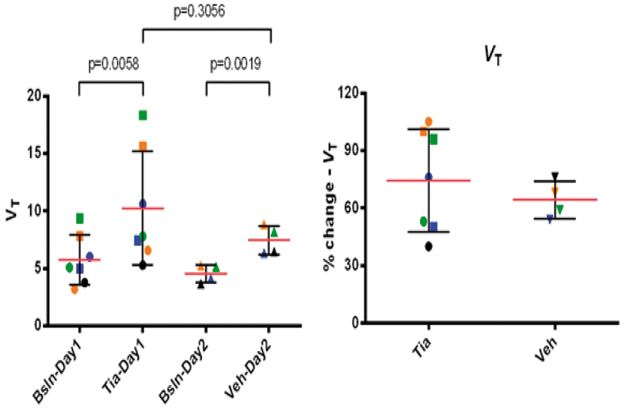
829
BRAIN-0767
Poster Session
BRAIN SEROTONIN TRANSPORTER BINDING IN A MINIPIG MODEL OF PARKINSON’S DISEASE
BrainPET: Neurotransmitter System Evaluation
1Department of Nuclear Medicine and PET-Centre, Aarhus University and Hospital, Aarhus C, Denmark
2Department of Neurosurgery, Aarhus University Hospital, Aarhus C, Denmark
3Department of Medicine/Neurology, University of British Columbia, Vancouver, Denmark
Abstract
Objectives:
Some of the debilitating non-motor aspects of Parkinson’s disease (PD) are related to the serotonin system1. To investigate the involvement of the brain serotonergic system in a PD animal model, we measured the in vivo binding of [11C]-DASB to the serotonin transporter (SERT) as a marker of serotonergic neurons. In this study, we use the in vivo capabilities of positron emission tomography (PET) imaging to study serotonin neurotransmission in a minipig model of PD induced by the intracerebroventricular injection of lactacystin, an inhibitor of the ubiquitin proteasome system.
This study was done in accordance with a protocol approved by the Danish Animal Inspectorate. Five female Göttingen minipigs were implanted in the cisterna magna with a catheter connected to a subcutaneous titanium injection port under sterile conditions. Six-eight weeks after recovery from the catheter implant, and after injections of sterile saline alone to verify patency, minipigs were scanned at baseline with [11C]-3-amino-4-(2-dimethylaminomethyl-phenylsulfanyl)-benzonitrile (11C-DASB), a label of SERT availability. Four pigs then received eight weekly injections of lactacystin dissolved in sterile saline, and one pig received saline alone, directly into the cerebrospinal fluid through the access port. They were scanned with DASB again after a cumulative dose of 200mg lactacystin. PET data were registered to an average minipig MRI atlas and processed using PMOD software. The binding potential (BPND) of DASB was obtained with the Logan graphical analysis and cerebellum activity as a region of non-displaceable binding.
Lactacystin administration induced behavioural symptoms including weakness of hindlimbs and decreased motor activity. SERT binding potential was decreased by 35-40% in striatal brain regions and by 20% in thalamic regions compared to the baseline scans.
Our imaging data suggests a loss of brain serotonergic innervation in response to protein aggregation. The decreased striatal binding of DASB observed in this minipig model of PD is to some extend consistent with previous studies done in PD patients2. The proteasome inhibition model may therefore be useful in the investigation of non-motor deficits in PD and LDOPA-induced dyskinesia.
References:
830
BRAIN-0234
Poster Session
CHARACTERIZING THE RELATIONSHIP BETWEEN DOPAMINE D2 RECEPTOR OCCUPANCY AND PLASMA LEVELS OF HALOPERIDOL IN NON-HUMAN PRIMATES USING [18F]-FALLYPRIDE
BrainPET: Neurotransmitter System Evaluation
1iSAT, AbbVie Inc., North Chicago, USA
Abstract
Objective: [18F]-Fallypride has been used to evaluate the receptor occupancy of antipsychotic drugs and the endogenous dopamine release in both preclinical species and humans. Prior studies [1,2] have evaluated the occupancy of haloperidol using [11C]-Raclopride in humans and determined EC50 to be 320 and 510ug/ml respectively. While prior studies have evaluated the relationship between dopamine D2 receptor occupancy and plasma drug levels in humans, similar work in non-human primates is lacking. The purpose of this study was to determine the relationship between dopamine D2 receptor occupancy and plasma haloperidol levels in non-human primates and compare it to that in humans.
Methods: [18F]-Fallypride dynamic positron emission tomography (PET) data were collected in 6 cynomologus monkeys without arterial sampling. 3 animals received test-retest scans separated by 4-6 weeks and 3 other animals received scans prior to and post haloperidol (3-55ug/kg) administration. The dynamic scans were of 150-180 mins in duration. Subject-space atlas based on anatomical MRI was coregistered to PET images of each animal to derive regional time activity curves. Binding potential (BPND) was used as the outcome measure using Cerebellum as the reference region and was quantified using both simplified reference tissue model (SRTM) and Logan reference method. Sensitivity analysis for t* and the choice of k2 for Logan reference method and time stability analysis for both methods were also performed. The changes in BPND pre and post pharmacological challenge were estimated and the ED50 and EC50 of haloperidol were calculated.
Results: Logan reference method was able to fit the data well and generate reproducible binding potentials in striatal regions. SRTM was only able to fit 30% of the total scans well. The highest binding potential was found in caudate-putamen (BPND=20.51±2.82) followed by globus pallidus (BPND=7.96±2.35), insula (BPND=4.06±1.45), substantia nigra (BPND=1.72±0.44) and the lowest binding potential was found in thalamus (BPND=0.68±0.22) and cortical regions (BPND=0.55±0.19) consistent with prior reports [3]. Test-retest variability of BPND estimated from Logan method was relatively low in striatal regions (%VAR25%), which might be due to the low target density in these regions. Dose dependent receptor occupancy was observed following different doses of haloperidol, however, at the lowest doses the occupancy measured in the extrastriatal region were variable and differed from the measurement in striatal regions. Therefore, when calculating the ED50 and EC50, only occupancy estimated in caudate-putamen was used. ED50 and EC50 were estimated to be 8.3 (95% CI: 5.5-12.5) ug/kg and 287.4 (95% CI: 131.1-629.8) ug/ml respectively and were comparable to estimates in human [2].
Conclusion: Dose dependent receptor occupancy was successfully measured in striatal regions following differing doses of haloperidol. When arterial sampling is not available, the Logan reference method can be used to estimate binding potential of [18F]-Fallypride in nonhuman primate brain.

Disclosures: All authors are employees of AbbVie. The design, study conduct, and financial support for this research were provided by AbbVie. AbbVie participated in the interpretation of data, review, and approval of the publication.
References:
831
BRAIN-0667
Poster Session
IN VIVO GLUTAMATE FLUCTUATIONS: A GLT-1 CHALLENGE WITH CEFTRIAXONE AND [11C]ABP688
BrainPET: Neurotransmitter System Evaluation
1Neurology and Neurosurgery, Douglas Mental Health University institute, Montreal, Canada
Abstract
Molecular imaging of glutamate neurotransmission offers unprecedented opportunities for investigating mechanisms underlying neuropsychiatric conditions. Here, we evaluated whether [11C]ABP688, a PET ligand that binds to the allosteric site of the metabotropic glutamate 5 receptor (mGlur5), is sensitive to glutamate fluctuations after a pharmacological challenge. For this, we used ceftriaxone (CEF) administration in rats, an activator of the GLT-1 transporter (EAAT2), which is known to decrease extracellular levels of glutamate. MicroPET [11C]ABP688 dynamic acquisitions were conducted in rats following a venous injection of saline (baseline) and after CEF 200 mg/kg. Binding potentials (BPND) were obtained using the simplified reference tissue method. Between-conditions statistical parametric maps indicating brain regions showing the highest CEF effects (Figure 1) guided placement of microdialysis probes, for subsequent assessment of extracellular levels of glutamate. CEF administration increased [11C]ABP688 BPND in the thalamic ventral nucleus (VA). Subsequent microdialysis assessment in VA revealed declines in extracellular glutamate concentrations. The present results support the concept that mGluR5 allosteric binding sites availability is sensitive to extracellular concentrations of glutamate. This interesting property of mGluR5 allosteric binding sites has potential applications for interrogating the role of glutamate in the diathesis of neuropsychiatric conditions, which may ultimately boost the development of glutamatergic imaging signatures and allow for the evaluation of glutamatergic-focused therapeutic approaches.
Statistical images projected on the rat brain structural imaging showing t-stat contrast mGluR5 BPND [baseline < ceftriaxone]
832
BRAIN-0332
Poster Session
MAPPING HUMAN BRAIN FATTY ACID AMIDE HYDROLASE ACTIVITY WITH PET – EFFECT OF C385A GENETIC POLYMORPHISM ON [11C]CURB BINDING
BrainPET: Neurotransmitter System Evaluation
1Research Imaging Centre, Centre for Addiction and Mental Health, Toronto, Canada
2Pharmacology and Toxicology, University of Toronto, Toronto, Canada
3Psychiatry, University of Toronto, Toronto, Canada
4Nutrition, University of Toronto, Toronto, Canada
5Research Imaging Centre, Centre for Addiction and Mental Health, Toronto, Canada
Abstract
833
BRAIN-0771
Poster Session
CHANGES OF METABOTROPIC GLUTAMATE RECEPTOR TYPE 1 BINDING OF [11C]ITMM BY AGING
BrainPET: Neurotransmitter System Evaluation
1Research Team for Neuroimaging, Tokyo Metropolitan Institute of Gerontology, Tokyo, Japan
2Molecular Imaging Center, National Institute of Radiological Sciences, Chiba, Japan
Abstract
Figure: Total distribution volume (VT) and distribution volume ratio (DVR) of the [11C]ITMM in young and aged human subjects.
References:
834
BRAIN-0391
Poster Session
PRELIMINARY RESULTS SUGGEST POSITIVE CORRELATION ACROSS SUBJECTS BETWEEN PEAK HEIGHT OF DA RESPONSE TO CIGARETTE SMOKING AND SATISFACTION OF CRAVING IN FEMALE SMOKERS
BrainPET: Neurotransmitter System Evaluation
1Biomedical Engineering, Yale University, New Haven, USA
2Psychiatry, Yale University, New Haven, USA
3Diagnostic Radiology, Yale University, New Haven, USA
Abstract
References:
Positive correlations across subjects were observed in the striatum between peak height of DA release induced by cigarette smoking and subjective rating of satisfaction of craving in female smokers (N=8).
835
BRAIN-0726
Poster Session
DOPAMINE D1 LIGANDS SCH23390 AND NNC112 EXHIBIT PARTIAL AGONISTIC PROPERTIES AS MEASURED IN VIVO
BrainPET: Neurotransmitter System Evaluation
1CAMIS/Radiology, Massachusetts General Hospital Harvard Medical School, Boston, USA
2Martinos Center, Massachusetts General Hospital Harvard Medical School, Boston, USA
Abstract
Abstract
CBV curves for h-DA conditions illustrating decrease in CBV following either NNC (top) or SCH (bottom) injection. Comparison of b-DA and h-DA CBV curves (h-DA CBV curves were found by subtracting AMP only curves from AMP + antagonist curves).

836
BRAIN-0027
Poster Session
GESTATIONAL AND POSTNATAL ETHANOL CONSUMPTION MODIFIES CART PEPTIDE SYSTEM IN RAT BRAIN: CORRELATES WITH ANXIETY, DEPRESSION AND MEMORY DEFICITS IN OFFSPRING
Psychiatric Disorders & Addictions
1Pharmacology, Department of Pharmaceutical Sciences R.T.M. Nagpur University Nagpur India, NAGPUR, India
2Neuroscience, Indian Institute of Science Education and Research Pune India, Pune, India
Abstract
Objective: Cocaine- and amphetamine-regulated transcript peptide (CART) plays a vital role in brain development and ethanol-mediated behavioral abnormalities. Herein, we attempted to evaluate the role of CART in the brain of offspring obtained form alcoholic female rats and its correlation with anxiety, depression and memory phenotype.
Methods: Female rats were fed with ethanol-containing, or ethanol-free nutritionally balanced liquid diet from 8 days prior of conception and continued till 25 days post parturition to coincide with weaning. We examined the adolescent (day 30) and adult (day 90) offspring for anxiety, depression and memory by using elevated plus maze (EPM), forced swim test (FST) and novel object recognition test (NORT), respectively. Furthermore, half of the pups from each litter were killed at day 30 and others at 90, for profiling of CART using immunocytochemistry.
Results: Offspring of alcoholic mothers at both age groups spent significantly more time in closed arms of EPM, showed more immobility time in FST and failed to recognize novel object versus familiar object in NORT. This indicates presence of anxiety, depression and amnesia. Thirty days old pups showed significant augmentation in the CART-immunoreactivity in the cells of dorsal hypothalamus (DA), while a dramatic reduction was noticed in the perifornical area (Pef). We found no changes in the lateral hypothalamus (LH) and paraventricular nucleus (PVN) at this age of pups compared with control. In adult offspring, the CART-immunoreactivity was significantly increased in the cells of PeF and LH and decreased in the DA and PVN.
Conclusions: We suggest that, the endogenous CART system in discrete brain areas, in a temporally specific manner, may be involved in the behavioral abnormalities in the offspring following ethanol ingestion by the mother.
837
BRAIN-0819
Poster Session
DYNAMIC MAGNETISATION TRANSFER MRI IN MAJOR DEPRESSION DISORDER: DELAYED TISSUE RESPONSE TO CARDIAC PULSATION IN THE BASAL GANGLIA.
Psychiatric Disorders & Addictions
1Lloyds Institute of Neuroscience, Trinity College Dublin, Dublin, Ireland
Abstract
Objectives:
In Major Depressive Disorder (MDD), several brain regions, such as the prefrontal cortex or the basal ganglia, are affected by a loss of glial cells, and in particular of astrocytes [1, 2]. Astrocytes are known to mediate between the brain parenchyma and its vasculature. Therefore, patients suffering from depression possess an abnormal blood circulation and metabolism in those regions [2]. Here, we want to study how those structural changes may affect the tissue pulsation which is related to blood circulation. Using a new method that we have developed, it is possible to measure the sudden change of intracellular water concentration evoked by the cardiac pressure wave [3].
The MRI sequence consists of a magnetisation transfer (MT) preparation phase followed by single-slice echo-planar imaging sequence across the basal ganglia. MT effect was introduced by a negative (-5236.8 ± 894.18 Hz) and a positive off frequency (6982.5 ± 894.18 Hz) RF pulse. Imaging parameters were voxel size of 3.5 x 3.5 x 3 mm, TR = 60 ms, TE = 12 ms, FA = 35° and 3000 repetitions.
4 depressed patients (between 25 and 42 years old) and 4 control subjects (between 27 and 44 years old) were scanned with the protocols approved by the local ethics committee.
For each subject, ROI's from the basal ganglia and the visual cortex were averaged to showcase the difference between both brain regions and groups. For the control group, only strong cardiac frequencies are resolved in the basal ganglia as well as the cortex. However, the spectra for the depressed patients show lower cardiac response in general but also different behavior in the basal ganglia, where we found a direct cardiac response from the tissue as well as a second response at around half of the cardiac circle. This results in an apparent shorting by the detected frequency by a factor 2, which can be seen in frequency spectrum in the figure.

To our knowledge, the observation of a cardiac related MT change and its variation in MDD patients has not yet been reported. The delayed response is unexpected. Most likely, the suppression of astrocytes in the depressed brain hinders the response of the tissue or the brain tissue uses another mechanism to compensate water dislocation due to the pressure wave. This could be explained with the decrease in astrocytes, which is linked to a decrease in aquaporin-4 (AQP4) water channels, which work as a regulator of blood flow, glucose and metabolism among other functions [1]. At present, we can only hypothesize about the mechanisms, but we strongly believe that we have found a new imaging biomarker for MDD.
References:
838
BRAIN-0225
Poster Session
SELF-TRANSCENDENCE TRAIT AND ITS RELATIONSHIP WITH IN VIVO SEROTONIN TRANSPORTER AVAILABILITY: A HIGH-RESOLUTION PET-MRI STUDY
Psychiatric Disorders & Addictions
1Psychiatry/Neuroscience Research Institute, Gachon University Gil Medical center, Incheon, Korea
2Neuroscience Research Institute, Gachon University Gil Medical center, Incheon, Korea
Abstract
Objectives: Self-transcendence is an inherent personality trait relating to the experience of spiritual aspects of the self. We examined the relationship between self-transcendence and serotonin transporter (SERT) availability in brainstem and thalamus, which are known to contain high SERT density, using high-resolution magnetic resonance imaging (MRI) and positron emission tomography (PET) with [11C]DASB to elucidate potential roles of serotonergic innervation in this trait.
Methods: Sixteen healthy subjects completed 7.0-Tesla MRI and high-resolution PET. The regions of interest included midline raphe nucleus groups and thalamic subdivisions. For the estimation of the SERT availability, the binding potential (BPND) was derived using the simplified reference tissue model (SRTM2). The Temperament and Character Inventory was used to measure self-transcendence.
Results: The self-transcendence total score had significant negative correlations with the [11C]DASB BPND in the left pulvinar and the caudal raphe. The subscale score for self-forgetful experiences correlated negatively with the [11C]DASB BPND in both pulvinar nuclei. The spiritual acceptance subscale score correlated negatively with the [11C]DASB BPND in the median raphe.
Conclusions: These results indicate that self-transcendence trait is associated with SERT availability in specific subregions of raphe nuclei and thalamus, suggesting that the serotonin system may serve as an important biological basis for human self-transcendence and spiritual experiences. The functional activity of these nuclei and their related neural circuitry may have a crucial role in the manifestation of self-transcendence.
References:
839
BRAIN-0394
Poster Session
STUDIES ON THE ANIMAL MODEL OF POST-STROKE DEPRESSION WITH HIGH VALIDITY AND APPLICATION OF ATYPICAL ANTIPSYCHOTIC ARIPIPRAZOLE
Psychiatric Disorders & Addictions
1School of Korean Medicine, Pusan National University, Yangsan-si Gyeongnam, Korea
Abstract
Objectives: The existence of a causal relationship between depression and vascular deficits has been hypothesized clinically as a post-stroke depression. We investigated whether cerebrovascular abnormalities such as ischemic stroke develop to ideal animal model of post-stroke depression by additional chronic mild stress (CMS) procedure. And atypical antipsychotic aripiprazole (ARZ) applied to this model.
Methods: Behavioral, histopathological and immunohistochemical analysis were performed for examination of the depressive disorders and therapeutic effect of aripiprazole in CMS, left middle cerebral artery occlusion (MCAO) and CMS after MCAO (MCAO+CMS) mice.
Results: In all
Conclusions: Our results suggest that chronic mild stress after ischemic stroke leads to severe depressive-like behavior than each CMS or MCAO treated mice via extrafocal neurodegeneration and degradation of neurogenesis, and these behavioral and histopathological changes are reversed by treatment of atypical antipsychotic aripiprazole.
840
BRAIN-0207
Poster Session
MECP2 SUMOYLATION AT LYS-412 RESCUES MECP2 MUTANT-INDUCED BEHAVIORAL DEFICITS IN A MOUSE MODEL OF RETT SYNDROME
Psychiatric Disorders & Addictions
1Neuroscience, Academia Sinica, Taipei, Taiwan
Abstract
Objectives:
The methyl-CpG-binding protein 2 (MeCP2) gene, Mecp2, is an X-linked gene encoding the MeCP2 protein and mutations of Mecp2 cause Rett syndrome (RTT). But the molecular mechanism of Mecp2 mutation-caused RTT is less well known. The aim of the present study was to investigate the molecular mechanism of Mecp2 mutant-induced behavioral deficits using a mouse model of RTT.
The major methods adopted for the present study include: in vitro SUMOylation assay for MeCP2, oligo pull-down assay for MeCP2 methyl-DNA binding, co-immunoprecipitation, western blot, CREB DNA binding in brain tissue, quantitative real-time PCR for Bdnf mRNA expression, Mecp2 plasmid transfection and Lentiviral vector transduction to animal’s brain, recombinase Cre injection to mouse basolateral amygdala, immunohistochemistry, adoption of loxP-Mecp2-loxP conditional knockout mice for behavioral assays including social interaction and cued fear conditioning learning.
We have found that MeCP2 could be SUMO-modified by the E3 ligase PIAS1 at Lys-412 and MeCP2 SUMOylation is MeCP2 phosphorylation (at Ser-421 and Thr-308)-dependent. MeCP2 SUMOylation is increased by N-methyl-D-aspartate and IGF-1 treatments. Further, most of the Mecp2 mutations identified in RTT patients showed decreased level of MeCP2 SUMOylation. Moreover, MeCP2K412R sumo-mutant transfection did not alter MeCP2 methyl-DNA binding, but it increased its association with CREB. On the other hand, MeCP2-SUMO1 fusion plasmid transfection decreased its association with CREB and HDAC1 in a dose-dependent manner, suggesting release of CREB from the repressor complex. Enhanced MeCP2 SUMOylation also increased CREB DNA binding to gene promoter and brain-derived neurotrophic factor mRNA expression in the rat brain. Meanwhile, it rescues the behavioral deficits, including social interaction and cued fear conditioning memory, in Mecp2 conditional knockout mice.
MeCP2 could be SUMO-modified by the SUMO E3 ligase PIAS1 both in vitro and in rat brain and MeCP2 SUMOylation is MeCP2 phosphorylation (at Ser-421 and Thr-308)-dependent. Decreased MeCP2 SUMOylation is observed in most of the Mecp2 mutations identified in RTT patients. Transduction of Lenti-MeCP2-SUMO1 fusion vector to basolateral amygdala rescues the behavioral deficits in Mecp2 conditional knockout mice. Drugs that enhance MeCP2 SUMOylation without altering the expression level of MeCP2 may have therapeutic potential against RTT and other autism-like syndrome. Here, we have identified a novel molecular mechanism for RTT.
This work was supported by a Grant (MOST 103-2320-B-001-004-MY3) from the Minister of Science and Technology in Taiwan.
References (selected):
841
BRAIN-0466
Poster Session
ASSESSING THE DOPAMINERGIC BASIS OF DELUSIONAL IDEATION: THE RELATIONSHIP BETWEEN D1 AND D2 RECEPTOR AVAILABILITY AND DELUSIONAL BELIEFS IN HEALTHY SUBJECTS
Psychiatric Disorders & Addictions
1Department of Clinical Neuroscience, Karolinska Institutet, Stockholm, Sweden
Abstract

References
842
BRAIN-0471
Poster Session
DIURNAL AND SEASONAL VARIATION OF THE BRAIN SEROTONIN SYSTEM IN HEALTHY MALE SUBJECTS
Psychiatric Disorders & Addictions
1Department of Clinical Neuroscience, Karolinska Institutet, Stockholm, Sweden
2Department of Neuroscience, Karolinska Institutet, Stockholm, Sweden
Abstract

References
843
BRAIN-0337
Poster Session
PET STUDY OF THALAMIC DOPAMINE D2-RECEPTORS IN DRUG-NAIVE PATIENTS WITH SCHIZOPHRENIA
Psychiatric Disorders & Addictions
1Clinical Neuroscience, Karolinska Institutet, Stockholm, Sweden
Abstract
844
BRAIN-0518
Poster Session
ELEVATED LEVELS OF ORBITOFRONTAL DOPAMINE D2-RECEPTORS IN PATIENTS WITH SOCIAL ANXIETY DISORDER
Psychiatric Disorders & Addictions
1Department of Clinical Neuroscience, Karolinska Institutet, Stockholm, Sweden
Abstract

References
845
BRAIN-0617
Poster Session
NEURO-ANATOMY PERSPECTIVE AND FUNCTION IN CLASSIC HYSTERICAL PSYCHOSIS
Psychiatric Disorders & Addictions
1Psychiatry, UMF Carol Davila, Bucharest, Romania
2Psychiatry, Sapunari Psychiatric Hospital, Calarasi, Romania
3Psychology, Sapunari Psychiatric Hospital, Calarasi, Romania
Abstract
The actuality of the research topic: Hysterical psychosis classic issue is known since Charcot without being existed then explanations of functional neuro-sensory area.
Objective: The present paper is a presentation of the case of a patient aged 61 years, admitted to the Sapunari Psychiatric Hospital for major depressive symptoms accompanied by psychotic phenomena (auditory hallucinations).
*** Current hospitalization: Early treatment instituted, but regressive evolution type, with disorganization in the thinking and behavior, delusional ideation negative type and guilt.
**** Presentation of neurological investigations, the differential diagnoses neurological and metabolic (meningo-encephalitis, dementia Jacobs Kreutzfeld).
*** EEG: presence of theta, post psychiatric treatment, without any other markers epileptic disorders. During the entire period of hospitalization, patient has not had a fever and leukocytes and leukocytes were not changed. Viral markers,
*** CT a moderate degree of cortical atrophy. MRI Cerebral: areas of demyelination/gliosis supratentorial unspecified, probably micro-vascular substrate. Gaps sequelae Ponto right midbrain minimal (less than 0.5 cm). We asked two checkups during hospitalization neurological and advice infective diseases, all refuting need for lumbar puncture.
***
Psychiatric evolution went to disorganization of thought and behavior, not SK type, but dissociative: dietary and verbal negativity, sketch of catalepsy/catatonia, waxy flexibility, stereotypes, posts. The patient presented Answers “alongside”, “The will of not knowing”, suggestibility, regression going to urinary incontinence. Auditory hallucinations denies, but asserts complex visual hallucinations, type scenes and presents delusional ideation cotardoid negative type, with immortality, enormous guilt and shame for concern.
Conclusions: Evolutions of symptoms (particularity evolution of this psychiatric case) raises modern explanations on neural lesion anatomic substrate.
846
BRAIN-0251
Poster Session
A NEW METHOD FOR EVALUATION OF REGIONAL PERFUSION ABNORMALITIES OF MILD TRAUMATIC BRAIN INJURY WITH NEUROPSYCHOLOGICAL IMPAIRMENT USING STATISTICAL IMAGING ANALYSIS FOR TC-ECD SPECT
Psychiatric Disorders & Addictions
1Department of Rehabilitation Medicine, The Jikei University School of Medicine, Tokyo, Japan
2Division of Rehabilitation Medicine, National Center for Child Health and Development, Tokyo, Japan
Abstract
Objective: The objective of this study was to identify specific brain lesions with regional perfusion abnormalities possibly associated with neuropsychological impairments (NPI), as sequela after mild traumatic brain injury (MTBI), using Tc-ECD SPECT and its novel analytic software.
Methods: We studied 23 patients with diffuse axonal injury with NPI group (Impaired-DAI), 26 with MTBI with NPI group (Impaired-MTBI) and 24 with MTBI without NPI group (Healthy-MTBI). In each subject, Tc-ECD SPECT images were analyzed by easy Z-score imaging system (eZIS) and voxel-based stereotactic extraction estimation (vbSEE). Segmented into lobule levels, ROIs were set in 140 areas in whole brain, and relative regional low Tc-ECD uptake was computed as ‘‘extent’’ (rate of coordinates with Z score >2.0 in the ROI). ROC analysis was performed using” extent’’ to discriminate the three groups.
Results: The highest area under the curve (AUC) value for data of Impaired-DAI and Healthy-MTBI groups was obtained in ROI on the left anterior cingulate gyrus (LtACG), with AUC of 0.93, optimal ‘‘extent’’ cutoff value of 10.9%, sensitivity 87.0%, specificity 83.3%. The highest AUC value for data of Impaired-MTBI and Healthy-MTBI groups was also in the LtACG, with AUC of 0.87, optimal ‘‘extent’’ cutoff value of 9.2%, sensitivity 73.1%, specificity 83.3%.
Conclusions: Using two analytic software packages, eZIS and vbSEE, we were able to identify specific areas with low regional Tc-ECD uptake, which were possibly associated with NPIs after MTBI. This trend was most marked in the left anterior cingulated gyrus in MTBI patients with NPIs and those with DAI. The optimal ‘‘extent’’ cutoff value, as a criterion for SPECT abnormality, might help the diagnosis of NPIs after MTBI.
847
BRAIN-0037
Poster Session
CHARACTERIZATION OF CEREBROVASCULAR FUNCTION IN MICE WITH CEREBRAL SMALL VESSEL DISEASE
Dementia and Neurological Disorders
1Institute for stroke and dementia (ISD), LMU, Munich, Germany
Abstract
848
BRAIN-0098
Poster Session
MTRNR2L12: A CANDIDATE BLOOD MARKER OF EARLY ALZHEIMER’S DISEASE-LIKE DEMENTIA
Dementia and Neurological Disorders
1Department of Medical Genetics, Jagiellonian University, Krakow, Poland
Abstract
Objectives:
Histological changes typical for Alzheimer’s disease (AD) are constantly reported in autopsied brain tissue of deceased individuals with Down syndrome (trisomy 21) aged above 30-40 years. Interestingly, significant variability of cognitive profiles can be observed in adult persons with Down syndrome. On the one hand symptoms of AD-like dementia develop in them frequently in the fourth or fifth decade of life, but on the other hand several seniors remain not affected. This phenomenon suggests presence of genetic variability influencing metabolic pathways crucial for development of AD-like dementia in Down syndrome.
Recent studies suggest that several genes playing central role in metabolic pathways follow similar expression patterns in brain and in blood. Consequently, alteration of genome expression in brain in case of AD development might be partially detectable in blood.
Considering the above facts we aimed at seeking of potential genomic blood markers of AD-like dementia development in adults with Down syndrome.
A cohort of 48 adults with Down syndrome aged >35 years who were institutionalized or attended day-care centers was recruited for the study.
To allow for detection of potential markers of early dementia, the participants were divided into two age groups: younger persons (<55 years) and older persons (>55 years).
The cognitive function of each study participant was assessed with use of the Prudhoe Cognitive Function Test (PCFT). The cognitive status was defined as good/acceptable (PCFT score >25%) or as severe cognitive disability (PCFT score up to 25%).
Whole genome expression in blood mononuclear cells was compared between subgroups of younger and of older persons with various cognitive statuses. SurePrint Human Gene Expression 8x60K v2 Microarrays (Agilent) were used for this purpose.
The study was accepted by the Jagiellonian University Ethics Committee.
The PCFT scores in the subgroups of persons with good/acceptable cognitive function ranged 50-100% in the younger and 57-97% in the older patients. In patients with severe cognitive disability the PCFT could be administered only in three cases (administration of the test in other patients was not possible due to poor contact).
Comparison of the groups of younger and of older persons by means of microarrays revealed significant differences with regard to three transcripts (ESPNL, XLOC_007536 and USP27X-AS1). However, these transcripts do not seem to be involved in pathogenesis of dementia. No significant differences were detected when comparing the group with good/acceptable cognitive status against the group with severe cognitive disability. However, subgroups of younger participants with severe cognitive disability differed significantly when compared with older patients with good/acceptable cognitive status (corrected p=0.00026; Fold Change=17.76) with regard to expression of a single gene: MTRNR2L12, an isoform of humanin, considered to be a protective factor in familial AD.
Expression of MTRNR2L12 might be an easy to measure blood marker of severe cognitive disability and, possibly, of early dementia in patients with Down syndrome. Further studies should be performed to evaluate potential usefulness of measurement of MTRNR2L12 transcript in patients with Alzheimer’s disease.
The study was sponsored by the Polish National Science Centre (DEC-2011/03/B/NZ5/01328).
849
BRAIN-0423
Poster Session
GLOBAL GENE EXPRESSION PROFILING OF NEW ANIMAL MODEL FOR COGNITIVE DYSFUNCTION
Dementia and Neurological Disorders
1Biomedical Research Institute, Chung Ang University College of Medicine, Seoul, Korea
2National Primate Research Center, Korea Research Institute of Bioscience and Biotechnology, Cheongju, Korea
3Department of Radiology, Chungbuk National University College of Medicine, Cheongju, Korea
Abstract
Alzheimer’s disease (AD) is a neurodegenerative disorder which observed amyloid plaques and neurofibrillary tangles. Appropriated medicine is not available so far. There are many animal models to imitate AD using mutant genes of presenilin and APP. However, these animal models which used mutant genes are based to familial dementia to developed young age. We proposed new animal model for AD using ibotenic acid (Ibo) and amyloid beta (Aβ) to induce neuronal cell loss. We injected ibotenic acid and Aβ into bilateral hippocampus of mouse at 6 week age. Animal behavioral test and histological analysis were shown to decline of learning, memory, neuronal cell loss and deposits of amyloid plaque in hippocampus regions. Total RNAs were used for global gene network. These results suggested that new animal model may be a useful tool to study as AD-like disease.
850
BRAIN-0092
Poster Session
NO RELATIONSHIP BETWEEN CORTICAL BETA-AMYLOID DEPOSITION AND CURRENT DEPRESSIVE SYMPTOMS IN ALZHEIMER’S DISEASE AND MILD COGNITIVE IMPAIRMENT
Dementia and Neurological Disorders
1Institute of Medical Science, University of Toronto, Toronto, Canada
2Multimodal Imaging group, Centre for Addiction and Mental Health, Toronto, Canada
3Cerebral Imaging Centre, Douglas Mental Health Institute, Montreal, Canada
Abstract
Depressive symptoms are frequently seen in patients with dementia and mild cognitive impairment (MCI). Evidence suggests that there may be a link between depressive symptoms and Alzheimer’s disease (AD)-associated pathological changes, such as an increase in cortical beta-amyloid (Aβ). However, limited in-vivo studies have explored the relationship between current depressive symptoms and cortical Aβ accumulation in patients with MCI and AD. Our study, using a large sample consisting of 455 patients with MCI and 153 patients with AD from the Alzheimer’s disease Neuroimaging Initiatives databases, investigated whether current depressive symptoms predict cortical Aβ deposition. Depressive symptoms were assessed using Geriatric Depression Scale and Neuropsychiatric Inventory-Depression/Dysphoria. Cortical Aβ deposition was quantified using Positron Emission Tomography with the Aβ probe 18F-Florbetapir. Standardized uptake value ratio (AV-45 SUVR) from the frontal, cingulate, parietal and temporal regions were estimated. In addition, a global AV-45 SUVR defined as the average of frontal, cingulate, precuneus, and parietal cortex regions, was also used. Through correlation analyses, we observed that current depressive symptoms were not related to cortical Aβ deposition, after controlling for potential confounds that may affect Aβ accumulation, including history of previous major depression. We also observed that there was no difference in cortical Aβ deposition between matched subjects with high and low depressive symptoms, as well as no difference between matched subjects with presence and absence of depressive symptoms. In conclusion, we found no relationship between current depressive symptoms and cortical Aβ deposition in patients with MCI and AD. This finding is consistent with the Aβ hypothesis of AD, which suggests that cortical Aβ accumulation occurs early in the illness, prior to the emergence of clinical symptoms.
851
BRAIN-0183
Poster Session
PROTECTIVE POTENTIAL OF METFORMIN ON MEMBRANE LINKED FUNCTIONS IN DIABETIC AGING FEMALE RATS.
Dementia and Neurological Disorders
1School of Life Sciences, Jawaharlal Nehru University, New Delhi, India
Abstract
Objective: The objective of this study was to investigate protective potential of metformin on membrane linked functions and glucose transporter in diabetic aging female rats.
Background: The emerging view is that diabetic brain features many symptoms that are best described as accelerated brain aging. Diabetes is considered to be a kind of 'accelerated aging” by increasing susceptibility to degenerative condition, including kidney disease, retinopathy, hypertension, coronary artery disease, stroke and atherosclerosis. Diabetes mellitus leads to functional and structural changes in the brain which appear to be most pronounced in the elderly.
Methods: Young (3 months) adult (12 months) and aged (24 months) rats will be diabetic by using alloxan monohydrate. After metformin was given i.p dose 200mg/Kg for one months to both control and diabetic aging rats. Learning was tested in a Morris water maze. A detailed study was carried on membrane linked enzymes, membrane fluidity, lipofuscin, antioxidant enzymes, glucose transporter, bcl-2 and DNA degradation to identify the antidiabetic and antiaging role of metformin using biochemical, molecular and histiochemical study.
Results: Present study shows that there was a similar pattern of increased lipid peroxidation, neurolipofuscin, DNA degradation and monoamine oxidase activity and a decrease in membrane fluidity, Na+ K+ ATPse, Ca2+ ATPase, sueroxidase dismutase and glutathione S-transferases activities, glucose transporter-4 (GLUT4) in both aging and diabetes. Metformin was found to be an effective treatment in stabilizing and normalizing the membrane functions; therefore this therapy can be considered an alternative to be explored further as a means of diabetic and aged related disorders control. Metformin treatment also helped to reverse the age related changes studied, to normal levels, elucidating an anti-aging, antidiabetic and neuroprotective action.
Conclusions: The cumulative deficits in learning and membrane functions in aged diabetic rats indicate that the effects of diabetes and ageing on the brain could interact. The results of this study will be useful for pharmacological modification of the aging process and applying new strategies for control of age related disorders including metabolic syndrome.
852
BRAIN-0302
Poster Session
ASTROCYTE-DERIVED LIPOCALIN-2 MEDIATES HIPPOCAMPAL NEURONAL LOSS IN THE GLOBAL ISCHEMIC MODEL OF VASCULAR DEMENTIA
Dementia and Neurological Disorders
1Dept of Pharmacology, Kyungpook National Univsersity School of Medicine, Daegu, Korea
Abstract
853
BRAIN-0054
Poster Session
CORRELATION OF DOPAMINERGIC AND SEROTONERGIC DYSFUNCTION IN A RAT MODEL OF PARKINSON'S DISEASE
Dementia and Neurological Disorders
1Department of Nuclear Medicine, Gangnam Severance Hospital, Seoul, Korea
2Department of Nuclear Medicine, Inha University Hospital, Incheon, Korea
3Department of Anatomy, Yonsei University Wonju College of Medicine, Wonju, Korea
4Department of Neurology, Gangnam Severance Hospital, Seoul, Korea
5Department of Molecular Imaging, Korea Institute of Radiological & Medical Sciences, Seoul, Korea
Abstract
References:
854
BRAIN-0422
Poster Session
ASSOCIATION OF WHITE MATTER HYPERINTENSITIES AND COGNITION WITH AMBULATORY BLOOD PRESSURE MONITORING (ABPM).
Dementia and Neurological Disorders
1Department of Neurology and Gerontology, Iwate Medical University, Morioka Iwate, Japan
2Division of Ultrahigh Field MRI Institute for Biomedical Sciences, Iwate Medical University, Morioka Iwate, Japan
Abstract
Objectives
White matter hyperintensities (WMH) are a common finding on MRI. Associations of WMH with aging, hypertension and cognitive impairment have been reported. The circadian blood pressure abnormality is reported as a risk factor of cerebrovascular disease. We assessed the circadian blood pressure and associations with the severity of WMH and cognitive impairment in hypertensive patients.
Methods
From March 2013 to November 2014, 101 consecutive hypertensive patients (69+/-8 years of age, 65% men) were recruited from our outpatient department. Ambulatory blood pressures monitoring (ABPM) parameter included BP level, variability during waking and sleep hours, and circadian blood pressure type. We assessed severity of WMH by Fazekas score and volumetric of WMH using 3T MRI and cognitive impairment by Mini-Mental State Examination (MMSE max score 30, cut off <20). Statistical analyses were performed to identify the association between WMH, cognitive impairment, and ABPM parameters.
Results
There was significant association between WMH Fazekas score total, WMH volume, high age, 24h systolic blood pressure (SBP), 24h daytime SBP, nighttime SBP, and MMSE score (27+/-3) (p<0.05). No significant relationship was found between circadian blood pressure type, WMH severity, and MMSE score.
Conclusions
Elevated blood pressure by ABPM is a risk factor for WMH independent of the circadian variability. WMH is associated with low cognitive scores.
References
855
BRAIN-0870
Poster Session
Brain hypometabolism coincides with or precedes cortical thinning in patients with ALS-FTD
Dementia and Neurological Disorders
1Department of Electrical and Electronics Engineering, Institute of Technology and Science, Pilani, Hyderabad Campus, Hyderabad, India
2Department of Biomedical Engineering, Lerner Research Institute, Cleveland Clinic, Cleveland, Ohio, USA
3Department of Neurology, Neuromuscular Center, Neurological Institute, Department of Neurosciences, Lerner Research Institute, Cleveland Clinic, Cleveland, Ohio, USA
Abstract
Objectives:
We previously used voxel based morphometry (VBM) to show decreased motor and extramotor grey matter (GM) volume in brains of patients with amyotrophic lateral sclerosis (ALS) and frontotemporal dementia (ALS-FTD) when compared to neurologic controls1. However, VBM analysis can potentially result in erroneously high GM values because it includes both cortical thickness and cortical foldings (gyri and sulci) as a single GM region. The relationship between structural and functional changes is also unknown. Therefore, we examined whether GM volumetric changes in ALS-FTD patients resulted from changes in cortical thickness, area or both, and compared these structural changes with metabolic changes as revealed by positron emission tomography (PET).
Methods:
High-resolution 3D T1-weighted MRIs were obtained at 1.5T in 18 patients with ALS-FTD and in 15 unaffected neurologic controls during routine clinical scanning. ALS-FTD patients also underwent fluorine-18-2-fluoro-2-deoxy-D-glucose (18F-FDG) PET imaging on a hybrid PET/CT Siemens Biograph TruePoint scanner within 0 to 14 days of MRI. Brain GM structural changes were assessed using VBM and cortical thickness, whereas metabolic changes were assessed using cerebral glucose metabolic rate (CMRglc) obtained from PET images.
Results:
GM volume and cortical thickness were significantly decreased (p<0.05) in motor and extramotor regions of ALS-FTD patients compared to controls. Cortical area showed no significant difference in any brain region. In addition, cerebral glucose metabolism rate was significantly reduced (p<0.05) in brain regions where structural changes were also observed but sometimes not.
Conclusions:
Significant reductions mostly in cortical thickness seemed to account for GM volume decreases detected by VBM in ALS-FTD. Metabolic changes corresponded well with structural changes in most motor and extramotor areas and occurred even in the absence of GM volume decrease in at least one region. Concurrent changes in GM structure and function suggest that neurodegeneration in ALS-FTD patients may occur as a “neuronopathy”.
References:
856
BRAIN-0108
Poster Session
STRONG IMPROVEMENT OF APOE/LDL-R SIGNALS AND AMYLOIDGENESIS BY TELMISARTAN IN POST-STROKE SHR-SR
Dementia and Neurological Disorders
1Department of Neurology, Okayama University, Okayama, Japan
Abstract
Recent studiessuggested that stroke and a factor of metabolic syndrome such as hypertensionand dyslipidemia is an important risk factor of Alzheimer’s disease (AD) viapromoting inflammatory responses. Telmisartan, an angiotensin receptor blocker(ARB), is expected to reduce not only the level of blood pressure (BP) but alsoneuroinflammation and neurotoxicity, then we examined the effects oftelmisartan on both cholesterol transport-related proteins (ApoE/LDL-R) and AD pathologyin spontaneously hypertensive rat stroke resistant (SHR-SR) after transientmiddle cerebral artery occlusion (tMCAO).
SHR-SR received tMCAO for 90 min at 12 weeksof age, and then were divided into 3 experiment groups including a vehicle,low-dose telmisartan (0.3 mg/kg/day), and high-dose telmisartan (3 mg/kg/day).The low dose served to improve the metabolic syndrome of SHR-SR withoutlowering the BP while the high dose was used to improve metabolic syndromewhile lowering BP.
Immunohistologicalanalysis showed that ApoE expression of cortical neurons was strong in thevehicle group at 6, 12 and 18 months of age, and that this ApoE expression patternwas very similar between the ipsilateral and contralateral sides of cerebral ischemia.LDL-R expression of cortical neurons was transiently increased at 6 months ofage only on the ipsilateral side. However, telmisartan dramatically suppressedthe expression of ApoE/LDL-R at both doses. Meanwhile, the numbers of amyloid β(Aβ)-positive neurons and senile plaque (SP) in the ipsilateral cerebral cortexprogressively increased with age until 18 M in the SHR-SR after tMCAO. To our surprise,low-dose/high-dose telmisartan significantly reduced the number of Aβ-positiveneuron as well as SP at 6, 12, and 18 M.
These findings suggest that both low and highdoses of telmisartan prevented the activation of ApoE/LDL-R in SHR-SR aftertMCAO, reducing both intracellular Aβ and extracellular SP accumulations aftertMCAO in SHR-SR, with a further improvement by combined BP lowering. Such astrong effect of telmisartan could provide a preventative approach for AD inpost-stroke patients with hypertension.
857
BRAIN-0121
Poster Session
PROTECTIVE EFFECT OF TELMISARTAN AGAINST PROGRESSIVE OXIDATIVE BRAIN DAMAGE AND SYNUCLEIN PHOSPHORYLATION IN STROKE-RESISTANT SPONTANEOUSLY HYPERTENSIVE RATS
Dementia and Neurological Disorders
1Department of Neurology, Okayama University, Okayama, Japan
Abstract
858
BRAIN-0419
Poster Session
CLINICAL FEATURES OF PATIENTS WITH SUSPECTED NON-ALZHEIMER PATHOLOGY (SNAP)
Dementia and Neurological Disorders
1Neurology, Hokuriku National Hospital, Nanto, Japan
2Nuclear Medicine, Public Central Hospital of Matto Ishikawa, Hakusan, Japan
3Psychiatry, Hokuriku National Hospital, Nanto, Japan
Abstract
(hippocampal volume on MRI and cortical metabolism on [18F]-fluorodeoxyglucose (FDG)–PET) in 189 consecutive patients who visit our memory clinic. We categorized patients as SNAP based on absence of amyloid pathology and presence neurodegeneration pattern of Alzheimer disease.
859
BRAIN-0336
Poster Session
INFLUENCE OF TSPO AFFINITY-BINDING ON [18F]DPA-714 WHOLEBODY BIODISTRIBUTION: A PET STUDY IN HEALTHY SUBJECTS AND ALZHEIMER PATIENTS
BrainPET: Other
1SHFJ I2BM DSV, CEA, Orsay, France
2Sainte Anne Hospital, Neurology department, Paris, France
Abstract
Neuroinflammation is thought to play a crucial role in the early stages of AD. 18 kDa-Translocator Protein (TSPO) is expressed at a low level in healthy brain and is up-regulated during inflammatory processes that may occur in neurodegenerative diseases. It is considered as a promising target for microglial activation early imaging and can be measured by PET using [18F]DPA-714 radioligand. TSPO ligand uptake was reported to be highly dependent on radioligand binding affinity status based on a genetic polymorphism.
We aimed to quantify the uptake of [18F]DPA-714 in several organs depending on the genetic status of the subject, in healthy volunteers and AD patients.
55 subjects (27 males and 28 females) including 30 healthy subjects and 25 Alzheimer patients (AD; 17 at early, 8 at dementia stage) biologically confirmed (episodic memory deficits associated with an AD-CSF profile and/or positive amyloid PET) were analyzed. All subjects underwent a [18F]DPA-714 static wholebody PET imaging starting 100 min after radioligand injection (102±8 min; five minutes acquisition per bed position). The unchanged [18F]DPA-714 concentration was determined in venous blood samples. [18F]DPA-714 uptake was determined in spleen, myocardium, liver, gastric wall, kidneys, salivary glands, vertebra bodies, and lungs. All subjects were genotyped for the TSPO polymorphism defining three populations: high (HAB; n=28); mixed (MAB; n=24) and low (LAB; n=3) affinity-binders.
No statistical difference was observed in the uptake of [18F]DPA-714 (SUV), between healthy subjects and patients, whatever the binding-affinity group. The distribution pattern of [18F]DPA-714 in organs was similar in each group with slightly lower values in MAB (-18% mean) and significant lower values in LAB subjects (-40%) compared to HAB subjects, excluding the liver where the uptake was significantly higher in LAB (+66% compared to HAB). The highest uptake was found in the gastric wall (6.5±1.8 in HAB; 5.9±1.9 in MAB and 2.7±0.2, in LAB), intermediate in the myocardium, vertebral bodies and parotids (4.2±1.0; 3.7±1.0 and 2.2±0.3) followed by the spleen and the kidney cortical (3.0±0.5 and 2.5±0.7 and 1.6±0.3). The lowest uptake was observed in lungs (1.0±0.3; 0.7±0.4 and 0.5±0.2). Liver activity was 3.3±0.9; 3.3±0.9 and 4.9±0.6.[18F]DPA-714 (unchanged) concentrations in venous plasma exhibited a four time increase in LAB subjects compared to HAB and MAB (SUV= 0.60±0.09 vs 0.13±0.06 and 0.18±0.11).
When correcting the activity measured in the organs by the plasma concentration, the differences between the affinity-binding groups appeared higher: compared to HAB, the uptake in MAB was 30% lower and was dramatically decreased in LAB subjects (-70%; but only -45% in liver). These results reflected the specific uptake of the radiotracer in the organs depending on the affinity status, and showed that liver uptake is partly due to the metabolism.
The biodistribution of [18F]DPA-714 was similar in healthy controls and in AD patients. Plasma concentrations were highly increased in LAB compared to HAB and MAB probably due to a lower binding in the TSPO containing organs. The [18F]DPA-714 activity in the organs have to be corrected by the unchanged radioligand concentrations in plasma for an accurate estimation of the specific uptake
860
BRAIN-0610
Poster Session
IMAGING NEUROINFLAMMATION IN THE EARLY STAGE OF ALZHEIMER'S DISEASE: A PET STUDY USING [18F]DPA-714
BrainPET: Other
1Laboratoire Imagerie Moléculaire In Vivo (IMIV) UMR 1023 Inserm/CEA/Université Paris Sud - ERL 9218 CNRS CEA/I2BM/Service Hospitalier Frédéric Joliot, CEA, Orsay, France
2Sainte Anne Hospital Paris, SHFJ I2BM DSV CEA, Orsay, France
3Langage and Memory Neurology Unit, Sainte Anne Hospital, Paris, France
4Neurospin I2BM DSV CATI multicenter Neuroimaging, CEA, Gif sur Yvette, France
5NeuroRadiology, Sainte Anne Hospital, Paris, France
Abstract
Neuroinflammation is thought to play a crucial role in the early stages of Alzheimer’s Disease (AD). 18 kDa Translocator Protein (TSPO) is up-regulated during inflammatory processes that may occur in neurodegenerative diseases.
Our goal was to compare the [18F]DPA-714 binding in AD patients and healthy controls.
Twenty AD patients, as defined by a typical progressive amnesic deficit of the hippocampal type and a positive amyloid PET imaging ([11C]-PiB PET) (13 at an early stage and 7 at dementia stages) were compared to 19 healthy controls with negative amyloid PiB-PET imaging. All subjects underwent a [18F]DPA-714 PET imaging. PET scans were acquired on a HRRT scanner (Siemens) for 90 minutes. Dynamic scans were corrected for head motion and co-registered with 3D T1-weighted MRI. Relative [18F]DPA-714 brain uptake was measured using cerebellum as pseudo reference region, in 76 anatomical regions previously segmented on MRI.
Analyses of the TSPO polymorphism showed that 34 subjects were high (HAB) or mixed (MAB) binders, forming the TSPO+ group (HAB: 6 controls, 8 AD; MAB: 9 controls, 11 AD) while 5 were low binders (TSPO-: 4 controls, 1 AD). Compared to normal controls, TSPO+ AD patients showed increased [18F]DPA-714 uptake: the global cortical uptake was 1.44±0.20 in HAB-AD patients versus 1.24±0.15 in HAB-controls (p<0.0001) and 1.41±0.25 in MAB-AD versus 1.35±0.28 in MAB-controls. The highest increase was found in the parietal, precuneus and posterior cingular regions (mean increase of +20 and +8% in both HAB and MAB). The [18F]DPA-714 uptake of the AD patients at the early stage was intermediate between controls and dementia-AD groups, reaching significant difference between the three groups for the HAB-subjects. No differences were found in the AD and controls TSPO low binders.
Neuroinflammation changes associated with AD could be detected using [18F]DPA-714 PET, even at the early stage of the disease. Using a simple quantification approach, the detection sensitivity depended on the affinity status of the subjects. [18F]DPA-714 in PET imaging might become a relevant biomarkers for monitoring AD.
861
BRAIN-0076
Poster Session
EXPLORING THE REWARD VALUE OF SOCIAL RELATIONSHIPS AND DOPAMINE D2/3 RECEPTOR AVAILABILITY IN THE VENTRAL STRIATUM OF HUMANS WITH [11C]-(+)-PHNO
BrainPET: Other
1Research Imaging Centre, Centre for Addiction and Mental Health, Toronto, Canada
Abstract

References:
862
BRAIN-0055
Poster Session
PARTIAL VOLUME CORRECTION OF BRAIN PET STUDIES USING ITERATIVE DECONVOLUTION IN COMBINATION WITH HYPR DENOISING
BrainPET: Other
1Department of Radiology and Nuclear Medicine, VU University Medical Center, Amsterdam, Netherlands
2Department of Radiology Oncology and Radiation science, Uppsala University, Uppsala, Sweden
Abstract
Recovery of measured activity concentrations in grey matter regions of the Hoffman phantom for different implementations of IDM with and without HYPR.
Reference List
863
BRAIN-0805
Poster Session
DECREASED REGIONAL CEREBRAL BLOOD FLOW EVOKED BY DYNAMIC EXERCISE: A POSITRON EMISSION TOMOGRAPHY STUDY
BrainPET: Other
1Faculty of Sports and Health Studies, Hosei University, Tokyo, Japan
2Department of Neurosurgery, Tokyo Medical and Dental University, Tokyo, Japan
3Research Team for Neuroimaging, Tokyo Metropolitan Institute of Gerontology, Tokyo, Japan
4Department of Radiological Technology, Hokkaido University of Science, Sapporo, Japan
Abstract
.
864
BRAIN-0683
Poster Session
[18F]FCWAY, A SEROTONIN 1A RECEPTOR RADIOLIGAND, IS A WEAK SUBSTRATE FOR EFFLUX TRANSPORTERS AT THE HUMAN BLOOD-BRAIN BARRIER
BrainPET: Other
1Molecular Imaging Branch, National Institute of Mental Health, Bethesda, USA
2Nuclear Medicine, XiangYa Hospital/Central South University, Changsha, China
3Laboratory of Cell Biology, National Cancer Institute, Bethesda, USA
4Psychiatry, University of Texas Health Sciences Center San Antonio, San Antonio, USA
5Neurology, Columbia University, New York, USA
6Clinical Center PET, National Institute of Health, Bethesda, USA
7Clinical Epilepsy Section, National Institute of Neurological Disorders and Stroke, Bethesda, USA
Abstract
Intravenous tariquidar infusion increased (A) total distribution volume (VT) and (B) VT with correction for plasma free fraction (VT/fP) of [18F]FCWAY in five centrally-located brain regions of three healthy subjects (mean ± SD).
Reference:
865
BRAIN-0614
Poster Session
[18F]T807 PET AND DIFFUSION TENSOR IMAGING REVEAL ASSOCIATION BETWEEN TAU PATHOLOGY AND NEURONAL FIBER INTEGRITY IN TRAUMATIC BRAIN INJURY
BrainPET: Other
1Center for Advanced Medical Imaging Sciences & Department of Radiology, Massachusetts General Hospital & Harvard Medical School, Boston, USA
2Department of Neurology, Massachusetts General Hospital & Harvard Medical School, Boston, USA
3Department of Radiology, Massachusetts General Hospital & Harvard Medical School, Boston, USA
4Department of Physical Medicine and Rehabilitation, Spaulding Rehabilitation Hospital & Harvard Medical School, Boston, USA
Abstract
[18F]T807 (A,B) and tractography (C-F) compared between a subject with severe TBI (B,D,F) and matched control (A,C,E). Tau pathology (B) corresponded with reduced neuronal bundles in callosum (D) and dorsal projections of corona radiata (F). Cortical [18F]T807 (A) in a former professional athlete corresponds with loss of white matter tracks which were preserved contralaterally (B,C).

866
BRAIN-0102
Poster Session
[18F]-RADIOLABELLING OF BASE SENSITIVE O-ARYLCARBAMATES USING A MOM PROTECTING GROUP; APPLICATION TO FAAH RADIOTRACERS
BrainPET: Other
1Research Imaging Centre, Centre for Addiction and Mental Health, Toronto, Canada
Abstract
Objectives.
Direct [18F]-radiolabelling of O-arylcarbamates has proven intractable because of their inherent sensitivity to even the mildest of bases. Thus multi-step synthetic strategies have been required to prepare [18F]-radiotracers which incorporate this functionality. Such radiotracers have become important in the area of targeting the extensive family of serine hydrolases such as FAAH and MAGL. While carbamates are often used as protecting groups for amines methods of protection of carbamate functionalities themselves are scarce. We report here a novel method using the MOM group to protect the O-arylcarbamate moeity during the [18F]-radiolabelling procedure. This approach greatly simplifies the preparation of [18F]-radiolabelled O-arylcarbamates.
N-Alkyl-O-arylcarbamates, containing a tosylate ester suitable for introduction of the [18F]-fluoride were treated with trimethylsilyl chloride and paraformaldehyde under anhydrous conditions, then quenched with methanol, to give MOM-protected carbamates. Conditions for radiolabelling of the protected precursor and deprotection were then explored. The optimised method was applied to the radiosynthesis of a potential radotracer suitable for PET imaging of the serine hydrolase, FAAH.
The MOM-group was readily incorporated into a variety of O-arylcarbamates. Anhydrous solvents were required for reproducible yields. Model reactions demonstrated that the parent unprotected O-arylcarbamate was generated upon warming with aqueous acid. Standard [18F]-radiolabelling conditions (e.g. K2CO3, 2.2.2.K, CH3CN, 80°C) gave high (>85%) incorporation of [18F]-fluoride. The MOM-group was easily removed (>95%) by quenching the reaction mixture in either aqueous hydrochloric or sulphuric acid at 80°C for 5 mins. The synthetic sequences were automated on a commercial module (Synthra), including HPLC purifications and formulations. Using this method a radiolabelled FAAH inhibitor was prepared in 49% radiochemical yield after formulation (uncorrected) and used successfully in rodent experiments as a potential FAAH radiotracer.


MOM-protection of O-arylcarbamates is a viable technique to enable facile radiolabelling of these base sensitive substrates. The method greatly simplifies existing methods and provides higher yields and reliability. A practical example of an [18F]-FAAH radiotracer is demonstrated.
867
BRAIN-0886
Poster Session
MODELING THE HYPOXIC/ISCHEMIC BLOOD-BRAIN BARRIER USING INDUCED PLURIPOTENT STEM CELL SOURCE
Late Breaking Abstracts
1Pharmaceutical Sciences, Texas Tech University Health Sciences Center, Amarillo, USA
Abstract
Objectives: Stroke constitutes the fourth cause of death worldwide and a high morbidity rate in surviving patients. Despite the important effort to find novel therapeutical targets and treatment to alleviate stroke injury, we are still lacking a clinically relevant therapy capable to restore neuronal function and promote brain recovery in patients. The objective of this study is to assess and validate a novel in vitro model of cerebral hypoxia/ischemia using human brain microvascular endothelial cells (BMECs) and neurons derived from induced pluripotent stem cells (iPSCs).
Methods: In this study, we used the human iPSC IMR90-c4 cell line 1 and induced the differentiation of these cells into brain microvascular endothelial cells (BMECs) and neurons following established differentiation protocols 2, 3. In addition, immortalized human brain microvascular endothelial cells (hCMEC/D3) were used to compare our BMECs to an established human in vitro model. Hypoxia was obtained either by chemical activation (CoCl2) or by incubation into an hypoxic chamber (1%O2). Ischemic condition was obtained by exposing BMECs monolayers to oxygen-glucose deprivation (OGD). Changes in the barrier function were assessed by measurement of transendothelial electrical resistance (TEER) and permeability to sodium fluorescein (NaF), whereas changes in cell junctions’ complexes were measured by immunocytochemistry. In addition, changes in cell viability were assessed by Trypan Blue exclusion and by MTT assays.
Results: We firstly investigated the ability of our BMECs monolayers to respond to CoCl2, a treatment known to disruption rat BMECs monolayers 4, 5. Interestingly, after 24h of treatment, we noted a significant decrease in barrier function at 30uM and 100uM. In contrast, hCMEC/D3 monolayers failed to respond to such hypoxic stimulus, as we noted no changes in barrier function even at 100uM. Furthermore, we noted a significant decrease in barrier function during either hypoxic stress after 24 hours; such effect was accentuated by OGD stress. Notably, such decrease in barrier function was occurring earlier than previous studies suggesting a high susceptibility to OGD than established models. Our data also the ability to model an “ischemic/reperfusion” injury, as BMECs showed a decrease at 6 hours OGD stress followed by a recovery at 24 hours of “reperfusion”.
Conclusions: In this study, we demonstrate the ability of iPSC-derived BMECs monolayers to respond to hypoxic/ischemic stress in a more susceptible fashion than previous in vitro models. Furthermore such model response to OGD showed a temporal pattern similar to cerebral ischemia in vivo, as we showed the ability of our model to mimic ischemic/reperfusion injury. Our next goal is to assess the response of iPSC-derived neurons to OGD stress and to better understand how hypoxia inducible factor (HIF-1) activation correlates with the onset of barrier disruption in our model.
References:
868
BRAIN-0935
Poster Session
USE IT OR LOSE IT: THE EFFECTS OF DETRAINING ON CEREBRAL BLOOD FLOW IN MASTER ATHLETES.
Late Breaking Abstracts
1Kinesiology Exercise for Brain Health, University of Maryland, College Park, USA
2Neuroscience and Cognitive Science, University of Maryland, College Park, USA
3Kinesiology, University of Maryland, College Park, USA
Abstract
869
BRAIN-0887
Poster Session
INCREASED PHASIC RELEASE OF DOPAMINE IN THE RIGHT CAUDATE OF ADHD VOLUNTEERS
Late Breaking Abstracts
1Psychiatry, University of Minnesota, Minneapolis, USA
2Nuclear Medicine, University at Buffalo, Buffalo, USA
Abstract

References:
870
BRAIN-0888
Poster Session
REDUCED TONIC RELEASE OF DOPAMINE IN THE RIGHT CAUDATE OF ADHD VOLUNTEERS
Late Breaking Abstracts
1Psychiatry, University of Minnesota, Minneapolis, USA
2Nuclear Medicine, University at Buffalo, Buffalo, USA
Abstract

Objectives: Dopamine neurotransmission is dysregulated in most psychiatric conditions but the nature of dysregulation remains unclear because of the use of indirect methods to study dopamine neurotransmission in the human brain. Thus, different sets of data suggest that dopamine is either hyperactive or hypoactive (1) in attention deficit hyperactivity disorder (ADHD). To reconcile contradictory data we used dynamic molecular imaging technique (2-9) to study phasic and tonic release of dopamine in ADHD. In a recent experiment we observed increased phasic release in the right caudate (10) and in this experiment we estimated the tonic release. Methods: We studied the tonic release in adult ADHD (n=11) and healthy control (n=11) volunteers of either sex. In this experiment after volunteers were positioned in the positron emission tomography (PET) camera, an intravenous bolus of a radiolabeled dopamine receptor ligand 11C-raclopride was administered intravenously. Immediately after the ligand administration PET data were acquired dynamically in list mode and volunteers were asked to stay still in the scanner for 45 min. The PET data were acquired for 45 min and analyzed using simplified reference tissue model (SRTM) (11) to estimate values of a number of receptor kinetic parameters in each voxel and also in the regions of interest. These parameters included the ligand binding potential (BP). Results: Since ligand BP changes in proportion to the amount of endogenously released dopamine (11, 12), we compared the ligand BP in ADHD and healthy control volunteers. The mean BP in ADHD volunteers was 3.21±1.30, which was 27% higher than the mean BP measured in the healthy control volunteers (2.53±0.85). The difference however was not significant statistically. Comparison of the BP in different striatal regions revealed significant difference in the right caudate (Figure 1) where the mean ligand BP in ADHD volunteers (3.19±0.23) was higher (p=0.003) than that in the healthy control volunteers (2.86±0.26). Conclusions: Results suggest that the reduced tonic release in the right could be the primary deficit of dopamine neurotransmission. Because the tonic and phasic release have reciprocal relationship, the reduced tonic release leads to an increase in the phasic release we observed in another experiment (10). The data indicate that the deficit of dopamine neurotransmission in ADHD is limited to the right caudate and is characterized by reduced tonic and increased phasic release (10) of dopamine.
References
871
BRAIN-0893
Poster Session
D-CYCLOSERINE TREATMENT IN A RAT STROKE MODEL INCREASES NMDA RECEPTOR AND BDMF LEVELS IN HIPPOCAMPUS
Late Breaking Abstracts
1Neurology, Stony Brook Medicine, Stony Brook, USA
Abstract
Objectives: Ischemic stroke triggers a massive but transient glutamate efflux and activation of NMDA receptors, followed by long lasting loss of NMDA receptor function. We have recently reported that D-cycloserine (DCS), a partial NMDA agonist improves neurological and cognitive outcome in transient focal ischemia; although the mechanisms mediating these effects are not entirely clear. The present studies were designed to examine the effects of DCS on neuroinflammation, NMDA receptor density, BDNF and aromatase levels following transient focal ischemia in rats.
Methods: Rats subjected to a transient (90 min) Middle Cerebral Artery Occlusion (MCAO) were administered DCS (10 mg/kg, N=14) or vehicle (PBS, N=14) 24 hours post reperfusion. Eight rats were included as non-ischemic controls. Animals were killed more than 4 weeks after MCAO. Consecutive coronal cryosections were processed for quantitative autoradiography with the neuroinflammation marker [3H]PK11195 and the NMDAR antagonist [3H]MK801; and quantitative immunohistochemistry was used to assess BDNF and aromatase expression in the same brains.
Results: As expected, MCAO resulted in a long lasting decrease in NMDA receptor density and a concomitant increase in the neuroinflammation marker TSPO in the hippocampus relative to non-ischemic controls. DCS treatment reversed the decrease in NMDA receptor density and increased hippocampal BDNF, with no effect on neuroinflammation. Aromatase expression in cortical and striatal regions was increased by MCAO and reduced by DCS.
Conclusions
872
BRAIN-0924
Poster Session
AROMATASE AVAILABILITY IN AMYGDALA LINKED TO OBESITY AND SELF-CONTROL: PET STUDIES IN HEALTHY MEN AND WOMEN
Late Breaking Abstracts
1Neurology, Brook MedicineStony, BrookStony, USA
2Psychiatry, Mount Sinai School of Medicne, New York, USA
3Imaging, NIAAA, Bethesda, USA
4Imaging, NYU Med Ctr, New York, USA
5Neurlogy, Universitat Autonoma de Barcelona, Barcelona, Spain
6Medicine, Brookhaven National Lab, Upton, Spain
7Imaging, NIAAA, Behthesda, USA
8Medicine, Brookhaven National Lab, Upton, USA
Abstract
Objectives: Aromatase, the Cyp19A gene product, is the last enzyme in estrogen biosynthesis from androgenic precursors. The aromatase gene is locally expressed and differentially regulated in many organs and tissues, including fat cells and brain. Aromatase expression is reportedly increased in fat tissue of subjects with a high body mass index (BMI), but the relationship between BMI and brain aromatase availability has not been investigated to date. Here we use [11C]vorozole, an aromatase inhibitor radiotracer to assess aromatase availability in brains of normal weight, overweight and obese individuals with PET to test the hypothesis that brain aromatase, specifically in amygdala, is inversely correlated with BMI and positively correlated with personality traits related to inhibitory control of behavior.
Methods: Forty eight otherwise-healthy individuals, divided among normal weight (BMI 18-25, N=14), overweight (25<BMI≤30, N=16) and obese (BMI>30, N=12) were scanned following administration of [11C]vorozole. PET data were acquired over a 90 min session and regions of interest placed bilaterally over the amygdala. Brain and plasma time activity data were used to calculate the total distribution volume (VT) from a two-compartment model as well as model free graphical analysis (Logan plot). A subgroup of the subjects (N=29) were administered the multidimensional personality questionnaire (MPQ). The results were analyzed by ANOVA and Pearson’s regression analysis.
Results: High BMI was associated with a significant decrease in VT in the amygdala. Thus, ANOVA revealed highly significant main effect of BMI (F=9.68, p=0.0004); with both the overweight (p=0.005) and obese (p=0.0001) groups significantly lower than the normal weight group (Fisher’s PLSD posthoc test). In addition, There was a highly significant, inverse correlation (R2=0.38, p<0.0001), between BMI and VT in amygdala, which was evident in both men (R2=0.32, p=0.001) and women (R2=0.42, p=0.0006). Analysis of the MPQ scores revealed a significant positive correlation between scores on trait constraint (composed of harm avoidance, control and traditionalism) and BMI (R2=0.25, p=0.01).
Conclusions
873
BRAIN-0920
Poster Session
APPL2 ATTENUATES ADULT NEUROGENESIS IN OLFACTORY BULB
Late Breaking Abstracts
1School of Chinese Medicine, The University of Hong Kong, Hong Kong, Hong Kong China
2Department of Medicine, The University of Hong Kong, Hong Kong, Hong Kong China
Abstract
References:
874
BRAIN-0941
Poster Session
CHARACTERIZATION OF A MELANOTRANSFERRIN DERIVED PEPTIDE CAPABLE OF DELIVERING THERAPEUTICS ACROSS THE BLOOD-BRAIN BARRIER.
Late Breaking Abstracts
1Michael Smith Laboratories, University of British Columbia, Vancouver, Canada
2R&D, biOasis Technologies Inc, Richmond, Canada
3Dept of Chemistry, University of British Columbia, Vancouver, Canada
4R&D, biO, Richmond, Canada
Abstract
The Blood-Brain Barrier (BBB) restricts the passive entrance of most molecules. However, mechanisms must exist to allow entry of desirable compounds (e.g. vitamins and minerals). The well known iron transport protein, transferrin (Tf), does not efficiency cross the BBB but another transferrin family member, melanotransferrin (MTf), is readily able to transcytose the BBB1. We also wished to explore the differences between Tf and MTf that allow that later to cross the BBB. To this end, a twelve amino acid peptide derived from MTf (MTfpep) has been identified which is able to cross the BBB, via receptor mediated transcytosis, as well as or better than the entire protein. Studies of MTfpep, through bioinformatics guided NMR experiments have suggested this peptide has the propensity to adopt a conformation, in solution, similar to the shape it adopts as part of the parent protein. This structural characteristic, coupled with key primary sequence differences suggests why MTf is able to cross the BBB but Tf is not. Furthermore, identification of MTfpep and the motif required for BBB transcytosis has lead to the identification of a novel candidate transcytosis receptor found on the surface of the BBB. The MTf and MTfpep platform is also being explored as a vector for the delivery of therapeutic compounds across the BBB for treatment of central nervous system disease. Most recently, MTfpep conjugated to siRNA was shown to prevent or ameliorate damage associated with stroke in a mouse model.
1 Moroo, I. et al. Identification of a novel route of iron transcytosis across the mammalian blood-brain barrier. Microcirculation
875
BRAIN-0883
Poster Session
CAUDATIN INHIBITS HUMAN GLIOMA CELLS GROWTH THROUGH TRIGGERING DNA DAMAGE-MEDIATED CELL CYCLE ARREST
Late Breaking Abstracts
1Key Lab of Cerebral Microcirculation in Universities of Shandong Taishan Medical University Taian Shandong 271000 China, Taishan Medical University, Taian, China
2Taishan Vocational College of Nursing Taian Shandong Province 271000 China, Taishan Vocational College of Nursing, Taian, China
Abstract
References:
876
BRAIN-0884
Poster Session
ENHANCED NEUROPROTECTION OF MINIMALLY INVASIVE SURGERY JOINT LOCAL COOLING LAVAGE AGAINST ICH-INDUCED INFLAMMATION INJURY AND APOPTOSIS IN RATS
Late Breaking Abstracts
1Taishan Vocational College of Nursing Taian Shandong Province 271000 China, Taishan Vocational College of Nursing, Taian, China
2Key Lab of Cerebral Microcirculation in Universities of Shandong Taishan Medical University Taian Shandong 271000 China, Taishan Medical University, Taian, China
Abstract
*Corresponding author: Cun-dong Fan, Xiao-yan Fu, Bao-liang Sun, Tel: +86-538-6230027, E-mail: tsmc_nks@tsmc.edu.cn; xyfu@tsmc.edu.cn; blsun@tsmc.edu.cn
References:
877
BRAIN-0925
Poster Session
SELENOCYSTINE INDUCES S-PHASE ARREST IN HUMAN GLIOMA CELLS BY TRIGGERING ROS-MEDIATED DNA DAMAGE AND MODULATING ERK AND AKT PHOSPHORYLATION
Late Breaking Abstracts
1Neurology, Affiliated Hospital of Taishan Medical University Taian Shandong 271000 China, Taian, China
2Neurology, Taishan Vocational College of Nursing Taian Shandong Province 271000 China, Taian, China
3Neurology, Key Lab of Cerebral Microcirculation in Universities of Shandong Taishan Medical University Taian Shandong 271000 China, Taian, China
Abstract
878
BRAIN-0907
Poster Session
NARINGIN, A NATURAL COMPOUND PROTECTS AGAINST CEREBRAL ISCHEMIA-REPERFUSION INJURY THROUGH REDUCING PEROXYNITRITE
Late Breaking Abstracts
1School of Chinese Medicine, The University of Hong Kong, Hong Kong, Hong Kong China
Abstract
Objectives: Thrombolytic therapy is essential therapeutic strategy for ischemic stroke to quickly restore blood and oxygen supply[1]. However, recanalization causes cerebral ischemia-reperfusion (I/R) injury by producing free radicals including superoxide, nitric oxide (NO), peroxynitrite (ONOO-), etc. ONOO- is a representative reactive oxygen species (RNS) triggering a series of molecules cascades to further induce brain damage[2]. In this study, we tested the hypothesis that naringin, a non-toxic plant bioflavonoid, could reduce ONOO- and attenuate neuronal damage during cerebral I/R injury in vivo and in vitro.
Methods: In the in vivo study, male Sprague-Dawley rats weighed 260-280g were subjected to middle cerebral artery occlusion (MCAO) for 2 hours followed by reperfusion for 22 hours to induce cerebral I/R injury. Naringin was intravenously administrated at the onset of reperfusion respectively at 80 mg/kg, 120mg/kg, 160mg/kg. FeTMPyP, a peroxynitrite decomposition catalyst, was used as positive control at 3mg/kg. Neurological deficits score was calculated by mNSS scale and infarct volume was evaluated by TTC staining. Pathological morphology was detected by Hematoxylin-Eosin staining and TUNEL staining for apoptotic cell death. Western blot analysis was used to detect iNOS, NADPH oxidase subunits and cleaved caspase 3 expressions. In the in vitro study, SH-SY5Y cells were subjected to oxygen-glucose-deprivation for 10hours and reoxygenation for 14hours (OGD/R). Cell viability was evaluated by MTT. Superoxide anion radical (O2.-) and NO were detected by HEt and DAF-2DA staining respectively with fluorescent microscopy and 3-nitrotyrosine expression, a footprint of ONOO–, was detected by western blot.
Results: (1) Naringin dose-dependently reduced the neurological deficit score, decreased the infarct volume and attenuated pathological morphological changes by compared with MCAO model group in vivo; (2) Naringin reduced apoptotic cell death by inhibiting cleaved caspase 3 activation in ischemia-reperfused brains in vivo; (3) Naringin inhibited the expressions of iNOS and NADPH oxidase subunits-p47phox and p67phox and 3-nitrotyrosine level in the ischemic brains and decreased NO level in serum in the rats after cerebral I/R injury in vivo; (4) Naringin reduced the level of O2.- and NO and down-regulated the expression of 3-nitrotyrosine and attenuated apoptotic cell death in SH-SY5Y cells under OGD/R condition in vitro. Naringin revealed similar effects to FeTMPyP.
Conclusions: Naringin might be a promising neuroprotective agent against cerebral I/R injury and its mechanisms could be attributed to its capacity of reducing NADPH oxidase subunits and iNOS expression level and inhibiting peroxynitrite-mediated cell death.
References:
879
BRAIN-0895
Poster Session
NEUROPROTECTIVE ROLE OF TRANSTHYRETIN THROUGH MEGALIN IN ISCHEMIC BRAIN INJURY
Late Breaking Abstracts
1Molecular Neurobiology, Instituto de Biologia Molecular e Celular (IBMC), Porto, Portugal
Abstract
Transthyretin (TTR) is a protein involved in the transport of thyroxin and retinol, also associated to nerve regeneration in the peripheral nervous system. Recently in our lab, TTR was shown to have a neuroprotective role in focal cerebral ischemia (J. Neurochemistry, (2010) 115, 1434), using a permanent MCAO stroke model. Several studies pointed, in the last years, to the neuroprotective role of TTR in the central nervous system: (i) individuals heterozygous for TTR T119M, a non-pathological mutation of TTR associated with increased levels of the protein in the plasma, show a reduced risk of cerebrovascular disease, and increased life expectancy compared with non-carriers; (ii) TTR can be a good predictor for young strokes, since patients have a worse clinical outcome if they have decreased levels of serum TTR at the time of stroke; (iii) smaller incidence of stroke in women, due to the neuroprotective role of sex steroids (which upregulate TTR in CSF); (iv); in C.elegans model, a transthyretin-like protein was determinant in the recognition of apoptotic cells by phagocytes. These effects triggered by TTR might involve transducing receptors, such as megalin, that binds TTR. The goal of this work is to unveil the molecular mechanisms involved in TTR-induced neuroprotection in cerebral ischemia, namely through its interaction with megalin, using in vitro ischemia models.
Cultured hippocampal neurons from Wt, TTR KO and Megalin TTR KO mice were subjected to glutamate excitotoxic insults. Protein levels were determined by Immunoblot, and gene expression was analyzed by qPCR. Protein distribution in neurons was also assessed by immunocytochemistry, including neurite outgrowth quantification. Cell death was assessed by nuclear condensation with Hoechst 33342.
We found that cultured hippocampal neurons from TTR KO mice are more sensitive to an glutamate excitotoxic insult than neurons from Wt mice, as neurite proteins MAP2 and Tau are significantly more affected in TTR KO mice neurons. Moreover, if TTR is administered in a therapeutic way, after the excitotoxic stimulus, a clear neuroprotection in the number and lenght of neurites in neurons from TTR KO cultures is observed. This neuroprotective effect of TTR seems to occur mainly in dendrites rather than in axons. More important when TTR is added to Megalin(+/-) TTR KO cultures after the excitotoxic insult, the neuroprotective effect is lost. Administration of TTR post glutamate stimulation does not prevent the nuclear condensation of apoptotic neurons. However, if TTR is given to cultured hippocampal neurons from TTR KO mice 6h before the glutamate stimulus, as a preventive strategy, a decrease in apoptotic neurons is observed. By opposition in megalin deficient cultures (Megalin(+/-) TTR KO) this soma neuroprotection is lost, indicating that TTR neurite and soma neuroprotection is Megalin dependent.
Taken together, our results show that TTR should be explored as a potential therapeutic/neuroprotective protein or even used as an indicator of risk factor in stroke outcome. Further studies to characterize the signaling pathways in in-vitro/in-vivo ischemia models behind this neuroprotection are underway.
880
BRAIN-0936
Poster Session
COMPLEX CEREBRAL ANGIOARCHITURE AND NEURONAL METABOLISM LEAD TO WIDE VARIATIONS OF HEMATOCRIT, RBC SATURATION IN THE CAPILLARY BED
Late Breaking Abstracts
1Bioengineering, University at Illinois at Chicago, Chicago, USA
2Physics, University of California San Diego, La Jolla, USA
Abstract
Objective
A quantitative understanding of themulti-scale neurovascular coupling between microcirculatory blood flow andneuronal metabolism would improve the treatment of neurodegenerative diseases. Previousattempts to computationally model cerebral metabolic regulation at the cellularlevel are hampered by the complex microcirculatory structure of the brain,limiting studies to thin subsections of cortical gray matter. To overcome thisobstacle, a 3D computational model was constructed from high-resolutiontwo-photon images which encompass both the cerebral angioarchitecture and relativepositions of neuronal and non-neuronal cells in the primary somatosensorycortex of four mouse brains. This mechanistic computer model predicts thehemodynamic states of blood pressure, hematocrit, and RBC velocity, as well asred blood saturation, the oxygen transport across the blood brain barrier, andthe oxygen tension within individual brain cells.
The cerebral angioarchitecture of theprimary somatosensory cortex was obtained from high-resolution two-photon microscopyimages. Vessels were assigned as either surface pial arterioles, penetratingarterioles capillaries, draining venules or surface pial venules based on theirsize, orientation, positon, and connectivity. The non-linear biphasickinematics of plasma skimming was computed throughout the blood vessel network.Metabolic activity was assigned to each brain cell, and oxygen tension wascomputed for each brain cell and throughout the extracellular space. Oxygentension in subcellular organelles was also computed for each cell.Additionally, the oxygen saturation of the red blood cells (RBCs) and theplasma oxygen tension in each vessel was determined, as well as the oxygenextraction of each vessel through the microvessel angioarchitecture.
Hemodynamic parameters of bloodflow, pressure, hematocrit, RBC velocity, RBC saturation and intracellularoxygen tension are validated against mouse measurements using in vivo models. The effects of oxygenperfusion in response to vasodilation of different arterial group is computed.The change in oxygen perfusion to brain cells in each cortical layer and thevenous oxygen saturation is measured following vasodilation of the surfacearterioles and penetrating arteriole groups. The effects of pial andpenetrating arteriole occlusions on brain tissue oxygen tension is measuredusing the computational model. We found that the occlusion of smaller diameter6.4um vessel cut off the supply of oxygen to cells more severely than theocclusion of a larger 10um vessel.
The complex hierarchy of thecerebral microcirculatory angioarchitecture leads to non-uniform distributionsin hemodynamic parameters, including hematocrit, blood pressure and RBC saturation.Our 3D computational model of the mouse primary somatosensory cortex revealsthat there are wide variations in these hemodynamic parameters, especially inthe capillary bed. Our model quantitates the increase in oxygen perfusion tobrain cells in response to vasodilatory events. Additionally, we can identifyvessel hierarchies that supply large section of the primary somatosensorycortex by simulating the decrease in blood flow following a micro-occlusionevent. This work at steady state gives a baseline for future large-scaledynamical models of oxygen n supply to the brain tissue.

881
BRAIN-0914
Poster Session
CORRELATION ANALYSIS OF MOLECULAR BLOOD BRAIN BARRIER DISRUPTION MARKERS AND STROKE SEVERITY
Late Breaking Abstracts
1School of Chinese medicine, The University of Hong Kong, hong kong, Hong Kong China
Abstract
Early prediction of stroke severity is important. During stroke, the blood–brain barrier (BBB) is compromised by endothelial cell death, and cytosolic contents released from injured brain tissues have the potential to cross the BBB. This suggests that the measurement of brain-derived proteins in plasma could be used to monitor stroke onset and severity. In our clinical observational study, we recruited 120 ischemic stroke patients and detect Caveolin-1, AQP-4, MMP-9, HMGB1, ONOO concentrations in ischemic stroke patients’ plasma, cerebral spinal fluid and analyze the correlations with BBB permeability and edema degree by MRI method. Caveolin-1 level in ischemic stroke patients plasma had significantly decreased compared to th healthy control group (p<0.05); CSF caveolin-1 level showed an increase comparing to the control group (p<0.05). ONOO level in plasma of ischemic stroke patients was higher than that of healthy control group (p<0.05). There was no significant ONOO change in CSF.
882
BRAIN-0898
Poster Session
THE DOUBLE MODELS STUDY OF REGION BLOOD PERFUSION AND GLUCOSE METABOLISM OF THE PREFRONTAL LOBES IN DEPRESSED PATIENTS WITH FIRST-EPISODE
Late Breaking Abstracts
1Radiology, Henan Provincial People's Hospital, Zhengzhou, China
Abstract
883
BRAIN-0899
Poster Session
PRELIMINARY STUDY OF BRAIN GLUCOSE METABOLISM CHANGES IN PATIENTS WITH LUNG CANCER OF DIFFERENT HISTOLOGICAL TYPES
Late Breaking Abstracts
1Radiology, Henan Provincial People's Hospital, Zhengzhou, China
Abstract
Background Cerebral glucose metabolism changes are always observed in patients suffering from malignant tumors .This preliminary study is aimed at investigating the brain glucose metabolism changes in patients with lung cancer of different histological types.
Methods One hundred and twenty patients with primary untreated lung cancer who visited Zhengzhou University People’s Hospital from February 2012 to July 2013 were divided into three groups based on histological types confirmed by biopsy or surgical pathology, which included adenocarcinoma (52 cases), squamous cell carcinoma (43 cases) and small-cell carcinoma (25 cases). The whole body 18F-fluorodeoxyglucose (18F-FDG) positron emission tomography (PET)/computed tomography (CT) of these cases were retrospectively studied. The brain PET data of the three groups was analyzed individually using statistical parametric maps (SPM) software, with 50 age and gender-matched healthy controls for comparison.
Results The brain resting glucose metabolism in all three lung cancer groups showed regional cerebral metabolic reduction. The hypo-metabolic cerebral regions were mainly distributed at the left superior and middle frontal, bilateral superior and middle temporal and inferior and middle temporal gyrus. Besides, the hypo-metabolic regions were also found in the right inferior parietal lobule and hippocampus in the small-cell carcinoma group. The area of the total hypo-metabolic cerebral regions in the small-cell carcinoma group (total voxel value 3255) was larger than those in the adenocarcinoma group (total voxel value 1217) and squamous cell carcinoma group (total voxel value 1292).
Conclusions The brain resting glucose metabolism in patients with lung cancer shows regional cerebral metabolic reduction and the brain hypo-metabolic changes are related to the histological types of lung cancer.
884
BRAIN-0937
Poster Session
BRAIN T2-WEIGHTED SIGNAL INTENSITY RATIO IN CHILDREN WITH SICKLE CELL DISEASE WITH AND WITHOUT STROKE
Late Breaking Abstracts
1Neurosciences Unit, UCL Institute of Child Health, London, United Kingdom
Abstract
BACKGROUND
Iron is paramagnetic, accumulates with age, is higher in the deep gray matter in neurodegeneration and if increased in tissues, shortens MRI T1 and T2. There are few data in children with sickle cell disease (SCD). Our aims were to measure T2-weighted signal intensity ratio (T2SIR), a ratio comparing regional T2 with CSF T2, as a proxy for brain iron status in children with SCD and controls and to compare T2SIR with serum Ferritin, with transcranial Doppler as a measure of cerebral blood flow and with overnight oximetry studies as a measure of exposure to hypoxia.
METHODS
Using ImageJ, 2 masked observers (coefficient variation <5%) measured T2 in regions of interest, excluding visible infarction where necessary, from archived 1.5T MRI data (Siemens Vision; TR=3,458 ms, TE=96 ms) in controls and children with haemoglobin (Hb) SS and SC and generated T2SIR and Pearson correlations with serum ferritin. Controls (n=21) and children with HbSC (HbSC; n=7) or HbSS with no (HbSS-NL; n=23), covert (HbSS-CL, n=19) or overt lesions (HbSS-OL; n=7) and children with overt stroke without SCD (HbAA; n=8) were compared (one way analysis of variance; post hoc Dunnett’s test). In the HbSS patients, correlations were computed for T2SIR in the various regions and velocities in the right and left middle, anterior and posterior cerebral and the basilar arteries as well as degree of hypoxic exposure computed as mean and minimum overnight hemoglobin oxygen saturation and the percentage of the night spent with hemoglobin oxygen saturation <90%. T2SIR in the same regions was also obtained in 8 HbSS-CL children randomised to 3 years of blood transfusion or standard care in the Silent Infarct Transfusion Trial.
RESULTS
Serum ferritin significantly correlated (p<.05) with T2SIR in right (R) and left (L) caudate and putamen. Compared with controls and children with overt stroke without SCD of similar age, T2SIR for R/L caudate, R putamen, R/L globus pallidus, and R/L red nucleus was lower in HbSS-OL (p<.01**) and in R/L globus pallidus and R/L red nucleus in HbSS-CL (p<.05). Left posterior cerebral artery velocity was positively correlated with T2SIR in the right caudate, right globus pallidus, left putamen and left red nucleus. For the left red nucleus, there was also a significant negative correlation with the percentage of the night spent with an oxygen saturation <90% during the overnight oximetry study. Compared to those randomised to standard care, there was a reduction in T2SIR in most brain areas in those who were chronically transfused for 3 years in the Silent Infarct Transfusion Trial
DISCUSSION
The significant correlations with serum Ferritin suggest that T2SIR is a proxy measure of brain iron. The most obvious reason for brain iron accumulation in SCD is regular blood transfusions. However, neurodegeneration may play a role, perhaps related in part to ischemic and hypoxic exposure. Studies quantitating and comparing brain T2 measurements with measures of CBF and exposure to hypoxia are warranted.
885
BRAIN-0930
Poster Session
PERICYTE REGULATION OF NEUROVASCULAR COUPLING AND OXYGEN SUPPLY TO THE BRAIN
Late Breaking Abstracts
1Department of Physiology and Biophysics and the Zilkha Neurogenetic Insitute, University of Southern California, Los Angeles, USA
2Department of Physics, University of California San Diego, La Jolla, USA
3Optics Division Athinoula A. Martinos Center for Biomedical Imaging Department of Radiology, Massachusetts General Hospital and Harvard Medical School, Charlestown, USA
Abstract
References:
886
BRAIN-0926
Poster Session
ACUTE KETAMINE INFUSION IN RAT DOES NOT AFFECT IN VIVO [11C]ABP688 BINDING TO METABOTROPIC GLUTAMATE RECEPTOR SUBTYPE 5
Late Breaking Abstracts
1Molecular Imaging Center Antwerp, University of Antwerp, Antwerp, Belgium
2University hospital Antwerp Department of nuclear medicine, Molecular Imaging Center Antwerp University of Antwerp, Antwerp, Belgium
Abstract

887
BRAIN-0927
Poster Session
A NEW METHOD TO CALCULATE BLOOD-BRAIN BARRIER PERMEABILITY ON A SINGLE CAPILLARY LEVEL IN LIVING MICE USING TWO-PHOTON MICROSCOPY
Late Breaking Abstracts
1Neuroscience and Pharmacology, University of Copenhagen, Copenhagen, Denmark
Abstract
Objectives:
Our aim was to develop a robust procedure to measure BBB permeability of a single capillary using the two-photon microscopy.
Measurements were performed on anesthetized C57bl6/j mice. Sodium Fluorescein, a low molecular weight fluorescent dye, was injected into the bloodstream intravenously and visualized using an Olympus two-photon microscope FVMPE-RS FLUOVIEW. Fast scanning mode was employed to collect images yielding a high temporal resolution.
To be able to calculate capillary permeability it is necessary to know the concentration of a diffusive compound inside and outside a capillary. This issue comprise one of the main challenges and usually leads to the excessively simplificated or complicated experimental setups.
Here we established an image acquisition protocol as well as data analysis algorithm that allows to obtain different types of images, including those which correspond to either extravascular or overall dye signal. Filtering them from other images (reflecting the average distribution of a dye) it is further possible to extract the fluorescent intensity values (proportional to dye concentration) inside and outside a capillary which can be directly used to calculate permeability.
The method requires the use of only one fluorescent tracer and doesn’t require comprehensive image processing tools, which makes it handy and robust for the current task.
We provide an example of a two-photon microscopy based approach to study single brain capillaries permeability of living mice in real time.
The method will potentially allow to study BBB pathological conditions as well as test fluorescently labeled drugs or nanocarriers on their ability to cross BBB.
References:
888
BRAIN-0890
Poster Session
DOES BRAIN ISCHAEMIA INDUCE CHANGES IN THE VASCULAR EFFECTS OF PROTEASE ACTIVATED RECEPTORS?
Late Breaking Abstracts
1School of Biomedical Sciences, Chinese University of Hong Kong, Shatin, Hong Kong China
Abstract
Objectives
Vascular responses and brain diseases such as ischaemic stroke are inter-related. Many studies have demonstrated altered vascular responses following brain injury (Hansen-Schwarts et al., 2003, Ansar et al., 2010). Four types of protease activated receptors are known; PAR1 to PAR4. They are suggested to have both neuroprotective and neurodegenerative effects in the brain under pathological conditions (Luo et al., 2007). The impact of brain ischaemia on the vascular effects of PARs has not been investigated. Using activating peptides of the four PARs, this study aims to determine whether their vascular effects could be different in middle cerebral arteries (MCA) isolated from normal and ischaemic rat brains.
MCA were isolated from normal rats and from rats subjected to 45min occlusion of MCA 24h previously. Two 2mm vessel rings were obtained from each MCA and mounted to a myograph system for tension recording. Activating peptides of the four PARs (PAR APs) were tested on the precontracted tone produced by 100nM U46619. Involvement of nitric oxide (NO) in their vascular effects was tested by addition of 100µM of the nitric oxide synthase inhibitor L-NAME prior to cumulative additions of PAR APs.
In MCA isolated from normal rat brains, PAR-1 AP (SFLLRN-NH2) and PAR-4AP (AYPGKF-NH2) had no effect, whereas, PAR-2 AP (SLIGRL-NH2) produced 38% relaxation and PAR-3 AP (SFNGGP-NH2) produced 46% contraction on the U46619-precontracted tone. In MCA isolated from ischaemic rat brains, PAR-4 AP remained ineffective, but PAR-1 AP produced a significant 17% relaxation, and PAR-2 AP produced 47% relaxation, whereas, PAR-3 AP produced 102% contraction on the U46619-precontracted tone. Pretreatment with L-NAME completely inhibited the vasodilator effects of PAR-1 AP and PAR-2 AP in both types of MCA.
The present findings have demonstrated that PAR-1 AP possesses vasodilator effect only in MCA isolated from ischaemic rat brains. On the other hand, PAR-2 AP produced vasodilatation in MCA isolated from both normal and ischaemic rat brains. All the vasodilator effects of these two peptides were abolished by L-NAME; indicating involvement of NO. In contrast, PAR-3 AP is a vasoconstrictor, and PAR-4 AP is not vasoactive in both types of blood vessels. Compared to MCA from normal rats, MCA from ischaemic rats showed slightly better vasodilator responses to PAR-1 AP and PAR-2 AP, but greatly augmented vasoconstriction to PAR-3 AP. The latter exaggerated vasoconstrictor effect could potentially worsen ischaemia-induced injuries in the brain.
References
889
BRAIN-0877
Poster Session
HIGH DOSE OF ATORVASTATIN INDUCES CEREBRAL HEMORRHAGE BY DAMAGING BLOOD VESSEL STABILITY VIA SMALL GTPASES MEDIATED SRC/VE-CADHERIN/CATENINS PATHWAYS IN HUMAN ENDOTHELIAL CELL
Late Breaking Abstracts
1State Key Laboratory of Quality Research in Chinese Medicine, Institute of Chinese Medical Sciences, Macau, Macau
Abstract
Abstract
Objectives:
Statins are a class of drugs used to lower high cholesterol levels and to prevent its complications such as cardiovascular diseases by inhibiting the enzyme HMG-CoA reductase. However, clinical trials showed that statins appear to increase the risk of intracerebral hemorrhage (ICH). Previously studies indicated that statins induced-rupture vessel integrity mediated bleeding could be reproduced in zebrafish larvae. Loss of junctions of the vascular endothelium can result in the rupture of vessels and bleeding into interstitial spaces.
Methods:
In view of this, we employed human endothelial cells in vitro and zebrafish in vivo to study the mechanisms of change of vascular integrity and endothelium permeability underlying the atorvastatin-induced cerebral bleeding process.
Results:
Our present study showed that atorvastatin-induced cerebral hemorrhage in zebrafish could be completely rescued by exposure of embryos to mevalonic acid (MVA) and isoprenoids (i.e. IPP, FPP, GGPP) but not cholesterol. Moreover, atorvastatin induced loss of cell-cell junctions by derangement of the actin cytoskeleton and impairment of VE-cadherin (VEC) distribution. Nevertheless, MVA and isoprenoids completely recovered the actin cytoskeleton derangement and VEC loss. Further studies showed that atorvastatin inhibited the post-translation isoprenylation of small GTPases and disrupted the regulation on Src/VEC/catenins associated with cell junction and actin cytoskeleton. In addition, inhibition of Src kinase prevents the VEC loss and disruption of cell-cell junctions caused by atorvastatin.
Conclusions:
Taken together, we have mapped the mechanism of this disruption to the regulation of small GTPases on VEC/catenins mediated adheren junctions, as well as the actin cytoskeleton. More importantly, we have presented results highlighting the inhibition of Src signaling might be beneficial for the management of cerebral hemorrhage.
References:
890
BRAIN-0932
Poster Session
NEURAL STEM CELL (NSC)-ENCODED HYPOXIA-INDUCIBLE FACTOR-1ALPHA(HIF-1ALPHA) PROMOTES THE ENDOGENOUS REGENERATIVE RESPONSE TO STROKE
Late Breaking Abstracts
1Neurosciences, University of New Mexico, Albuquerque, USA
Abstract
Focal cerebral ischemia induced by middle cerebral artery occlusion (MCAO) stimulates a multilineage cytogenic response from NSCs within adult subventricular zone (SVZ), which includes production of both glial and neuronal lineages (Li et al., 2010). HIF-1a is a key mediator of the adaptive cellular responses to hypoxia through direct transcriptional regulation of cell and molecular processes, including those involved in angiogenesis, stem cell maintenance and differentiation. Our previous studies have shown that stabilized HIF-1a protein is constitutively expressed within NSCs of the adult SVZ (Roitbak et al., 2011). Using conditional, tamoxifen-inducible HIF-1a knockout (HIF-1a iKO) mice in which tamoxifen administration resulted in concomitant expression of yellow fluorescent protein (YFP) and bi-allelic Hif1a exon 2 gene deletion in nestin+ NSCs and their progeny (nestin-CreERT2:R26R-YFP:Hif1afl/fl), we have demonstrated that induced Hif1α gene deletion in adult NSCs results in their gradual loss, which is preceded by regression of the SVZ vasculature under non-pathologic condition(Li et al., 2014). To investigate whether NSC-encoded HIF-1a regulates the regenerative response to stroke and vascular recovery in the area of stroke damage, tamoxifen was administered to adult male HIF-1a iKO and wild type mice 14 days prior to 60 minutes transient occlusion of the middle cerebral artery (MCAO), and the mice were sacrificed at two weeks after stroke. YFP positive cells were observed throughout the injured striatal parenchyma in stroke mice. Hif1α gene deletion resulted in fewer YFP+ cells within the injured striatum 2 weeks post-MCAO, but caused a 1.8-fold increase in the percentage of doublecortin+/YFP+ cells within the ischemic striatum (21.9 ± 1.7% vs 12.3 ± 0.8%, HIF-1a KO vs. WT, p<0.01, n=5/group), indicating a switch toward the neuronal lineage fate. Blood vessel density was decreased by 29% in the injured striatum in HIF-1a iKO mice compared with that of wild type mice (1.2 x 10⁻⁵ ± 1.7 x 10⁻⁶ vs. 1.7 x 10⁻⁵ ± 5.4 x 10⁻⁷ blood vessel number/micron3, respectively, p<0.05 n=5/group). These studies suggest that NSC-encoded HIF-1a promotes the SVZ regenerative response, vascular recovery, and NSC cell fate following stroke injury.
Li et al., 2010 Glia 58:1610.
Li et al., 2014 Journal of Neuroscience 34(50): 16713
Roitbak et al., 2011 Cell Mol Neurobiol. Jan; 31(1): 119
Supported by the American Heart Association Grant-in-Aid 09GRNT2290178
891
BRAIN-0940
Poster Session
FEEDING SUPPORT ALLOWS C57BL/6 MICE TO SURVIVE FILAMENT MCA OCCLUSION LONG-TERM
Late Breaking Abstracts
1Laboratory of Experimental Stroke Research, Institute for Stroke and Dementia Research, München, Germany
2Stroke-Immunology Group, Institute for Stroke and Dementia Research, München, Germany
Abstract
Acknowledgements: A. Lourbopoulos is supported by an Intra European (IEF) Marie-Curie Fellowship.
892
BRAIN-0928
Poster Session
DERAILMENT IN NITRIC OXIDE-DEPENDENT NEUROVASCULAR AND NEUROMETABOLIC COUPLING IN AGING
Late Breaking Abstracts
1Faculty of Medicine Polo I Piso I, Center for Neuroscience and Cell Biology, Coimbra, Portugal
Abstract
The functional and structural integrity of the brain relies on its ability to locally adjust blood flow and the delivery of metabolic substrates to meet the metabolic demands imposed by neuronal activation. This process – neurovascular coupling – and ensued alterations on glucose and O2 metabolism - neurometabolic coupling - are accomplished by complex and concerted communication between neural and vascular cells[1]. Recently, direct evidences critically supported the key role of nitric oxide (·NO) in the neurovascular coupling, acting in hippocampus as the direct mediator of the process[2]. In addition, ·NO is be involved in the regulation of ensued processes, including energy metabolism/cellular respiration by interfering with mitochondrial respiratory complexes, as well as bioenergetic signaling pathways[3,4]. While some evidences support the notion that neurovascular and neurometabolic coupling are perturbed during brain aging, the topic is still controversial [5] Supported by the simultaneous measures of ·NO, O2 and CBF dynamics in hippocampus in vivo we aimed to address the neurovascular and neurometabolic coupling mediated by neuronal-•NO in aging.
The experiments were conducted in Fischer F344 inbred rats at two different ages 3-6 months of age (young group) and 23 months of age (old aged group). The animals were evaluated in terms of behavioral and cognitive performance using several behavior tests. The simultaneous measurements of •NO, O2 and CBF were achieved by home-made arrays consisting of •NO and O2 microelectrodes, micropipette and a laser Doppler probe positioned in the hippocampus of isoflurane-anesthetized animals essentially as previously described [2]. Neuronal activation was elicited by a localized stimulation with glutamate solution.
Aging was accompanied by a gradual and significant decrease in locomotion and exploratory behavior, as well as by a decreased cognitive performance. Glutamatergic activation in hippocampus promoted transient increases in ·NO concentration levels, followed by transient CBF elevation from a background and biphasic fluctuation of O2 tension. The latter reflects both, the early increase in tissue O2 consumption and the blood-dependent O2 delivery. Quantitatively, it was observed that while •NO production was not significantly affected during aging in this model, the coupled CBF change and the variations of O2 tension were impaired. In brief, the CBF changes were diminished in terms of amplitude and increased onset delay. Also, although CBF changes were decreased along the age, the O2 component associated with the hyperemic response was increased in older animals, while no significant alterations occurred in the component associated to O2 consumption.
Overall, the results points towards an imbalance in the regulation of both ·NO-mediated neurovascular and neurometabolic coupling during normal aging, in close correlation with a compromised cognitive function.
Work supported by Fundação para a Ciência e a Tecnologia (PTDC/BBB-BQB/3217/2012 and Pest-C/SAU/LA0001/2013-2014). CFL acknowledges fellowship SFRH/BPD/82436/2011
References
893
BRAIN-0934
Poster Session
OXIDASE-BASED MICROBIOSENSOR FOR REAL TIME MONITORING OF GLUCOSE AND NEURONAL ACTIVITY IN AWAKE BEHAVING ANIMALS
Late Breaking Abstracts
1Faculty of Medicine Polo I Piso I, Center for Neuroscience and Cell Biology, Coimbra, Portugal
2Department of Anatomy and Neurobiology, Center for Microelectrode Technology University of Kentucky, Lexington, USA
Abstract
The brain is a high energy demanding organ: despite accounting for only 2% of the total body mass, it consumes up to 25% of the total glucose budget. It´s adequate function is critically dependent on the tight coupling between neuronal activity and the continuous delivery of energy substrates from blood stream, which implies that changes in blood supply and glucose and oxygen metabolism must be attuned, with temporal and regional precision, to the physiological demands imposed by neural activation. Both phenomena - neurovascular and neurometabolic coupling - underpin modern neuroimaging techniques, and yet are not fully understood. Our understanding of such complex mechanisms is critically dependent on the availability of suitable tools that allow the dynamical probing, with high temporal and spatial resolutions, of the functional and coordinated interaction between neurons, astrocytes and the vasculature in situ. Enzyme-based amperometric microbiosensors are attractive tools for real time monitoring in vivo the dynamics of neurotransmitters and metabolism-related biomarkers, such as glucose. In this work we describe the development and characterization of a glucose oxidase based microbiosensor for in vivo real-time continuous monitoring of glucose in awake-behaving rodents using mass-fabricated multisite microelectrode array (MEAs).
Glucose oxidase (GOx)-modified MEAs were used as microelectrode platforms for in vivo measurements of glucose. The MEA-based GOx biosensors were tested in vitro for their general analytical properties towards glucose measurement in brain extracellular space. In vivo measurements were performed both in anesthetized and in awake rats using the FAST-16 electrochemical system (Quanteon, LLC, USA). The self-referencing technique was used to address basal levels of glucose and fluctuations along 24-h periods in behaving animals in different brain regions, and in response to arousal status. The data recorded at high frequency was analyzed off-line to extract local field potential (LFP)-related currents allowing to measure changes metabolic markers in together with ongoing neuronal network activity (inferred from LFP features).
MEA-based GOx microbiosensor presented suitable analytical toward glucose measurement in vivo, as evidenced by the good sensitivity, response time, linear range and selectivity against major interferents. While some dependency on oxygen was observed, the Km was low, allowing the use of these biosensors for studies involving physiological fluctuations of oxygen. Preliminary data support the feasibility of glucose measurements in both anesthetized and freely moving animals with the MEA-based GOx biosensors. Spontaneous fluctuations in glucose levels were detected as a function of animal activity, awareness and sleep-awake cycle. In hippocampus, the basal levels were estimated to range from 0.5 to 1.5 mM.
This work shows the successful use of glucose oxidase_based microbiosensor to direct study tonic and phasic glucose dynamic fluctuations in awake-behaving rodents.
This work was supported by Fundação para a Ciência e a Tecnologia (PTDC/BBB-BQB/3217/2012 and Pest-C/SAU/LA0001/2013-2014). CFL acknowledges FCT post-doctoral fellowship SFRH/BPD/82436/2011
894
BRAIN-0911
Poster Session
NEUROVASCULAR COUPLING AND HEMODYNAMIC RESPONSES IN RAT: A SIMULTANEOUS ECOG-FNIRS STUDY
Late Breaking Abstracts
1Medicine, Centre Hospitalier Universitaire, Amiens, France
2CEA, Neurospin, Gif/Yvette, France
Abstract
The nature of the coupling between the hemodynamic signal and electrical activity of the brain is still under debate. However, optical imaging does not measure neural activity per se; rather, it measures the changes in concentration of blood oxygen in the tissue which is associated with changes in brain neural activity. This complementary method with higher temporal resolution (rather than fMRI) has been used to investigate the hemodynamic basis of neuronal activity in human and animals in more detail. We used a multichannel frequency-domain-based optical spectroscopy imaging system (Imagent®, ISS Inc.) and electrocorticogram (ECoG, A.N.T®) to acquire oxygenated hemoglobin (HbO) and deoxygenated hemoglobin (Hb) concentration changes and electrophysiological responses to somatosensory and auditory stimuli in fourteen adult male Sprague–Dawley rats (mean body weight: 350–450 g). We used electrical forepaw and auditory stimuli which is two robust paradigms for studying neurovascular coupling. For probing the somatosensory and auditory cortex, a special electroptode® was designed to hold the emitter/detector fibers and electrodes to fit to the small skull of the rat (n=14). The hemodynamic responses are characterized by an initial dip followed by an increase in total hemoglobin (tHb), an increase in oxyhemoglobin (HbO) concentration with a peak latency of 5 to 6.5 sec after stimulus onset. A decrease in deoxyhemoglobin (Hb) occurred simultaneously. Studying the neurovascular coupling in humans by EEG and optical imaging faced some limitation due to the vascularization of the skin, the presence of the bone and the artifacts due to movements. To overcome these problems, in the present animal study we developed a multimodal electrical and optical approach to investigate directly on the cortex the neurovascular coupling in response to electrical somatosensory and auditory syllabic stimuli. Such multimodal analysis allows characterizing the temporo-spatial specificity of the auditory and somatosensory cerebral responses of both the neuronal and vascular functional systems in rats.
895
BRAIN-0909
Poster Session
DECREASED MU-OPIOD RECEPTOR BINDING IN PATHOLOGICAL GAMBLING: A PET STUDY WITH [11C]CARFENTANIL
Late Breaking Abstracts
1Turku PET Centre, University of Turku, Turku, Finland
2Department of Radiology, Turku University Hospital, Turku, Finland
3Department of Mental Health and Substance Abuse services, National Institute for Health and Welfare, Helsinki, Finland
4Division of Clinical Neurosciences, Turku University Hospital and University of Turku, Turku, Finland
Abstract

896
BRAIN-0879
Poster Session
ON THE RELATIONSHIP BETWEEN INTRACRANIAL PRESSURE, BLOOD FLOW VELOCITY AND ARTERIAL BLOOD PRESSURE FOR PATIENTS WITH CLOSED TBI
Late Breaking Abstracts
1Statistics, University of Washington, Seattle, USA
2Engineering and Mathematics, University of Washington, Bothell, USA
Abstract
897
BRAIN-0885
Poster Session
ECM HYDROGEL INJECTION FOR THE TREATMENT OF STROKE: CHARACTERIZATION OF ACUTE HOST CELL INVASION.
Late Breaking Abstracts
1Radiology, University of Pittsburgh, Pittsburgh, USA
2Bioengineering, University of Pittsburgh, Pittsburgh, USA
3Surgery, University of Pittsburgh, Pittsburgh, USA
Abstract
Stroke is a severe cerebrovascular accident that results in a localized brain tissue loss, affecting nearly 800,000 Americans each year. Extracellular matrix (ECM) derived from urinary bladder lamina propria and basement membrane has shown therapeutic potential in rats following intracerebral injection for experimentally produced traumatic brain injury in rats. ECM can be prepared in liquid form at room temperature and dependent on concentration creates a hydrogel under physiologic conditions providing ideal material properties for intracerebral injection through a thin needle. To determine an appropriate concentration of ECM (0, 1, 2, 3, 4, 8 mg/ml) for injection and retention of material in the stroke cavity, ECM was injected in liquid form with injection parameters (coordinates and volume) determined by MRI 12 days post-middle cerebral artery occlusion, while simultaneously draining necrotic liquefied brain tissue. Retention of ECM, as well as host cell invasion and their phenotypes were assessed 24 hours post-injection using immunohistochemistry. ECM concentrations <3mg/ml did not gel and were not retained in the cavity. A significant host cell invasion was observed with 20-30% of these being putative microglia/macrophages (Iba1+) across all concentration groups. The number of astrocytes (GFAP+) cells invading the ECM acutely was negligible and there was no evidence of neuronal progenitor invasion. This characterization demonstrates that an ECM hydrogel can be readily injected and retained within a lesion cavity, while attracting host cells into the damaged region. Further time points and behavioral studies will be required to further evaluate the therapeutic potential of this approach.
898
BRAIN-0921
Poster Session
IS SUSCEPTIBILITY TO SILENT CEREBRAL INFARCTION IN RELATIVES, AKIN TO “SODIUM SENSITIVITY” IN NORMOTENSIVE SUBJECTS, MEDIATED THROUGH CAPILLARY ENDOTHELIAL DYSFUNCTION AND CELLULAR MECHANISMS?
Late Breaking Abstracts
1Clinical Research, Wellspring's Universal Environment P/L, Melbourne, Australia
Abstract
Introduction: Risk stratification using the Framingham Stroke Risk Profile (FSRP) is enhanced when markers of endothelial dysfunction, plasma homocysteine and urinary albumin:creatinine ratio are added. Carotid and internal carotid intima:wall ratio added while serum cholesterol did not add to this. The authors indicated that silent infarcts do not necessarily correlate with side of intimal plaque, which raises questions as to the pathogenesis of silent infarcts and strokes, unrelated to large vessel findings.
Aim: To understand the pathophysiological basis of subclinical infarcts in asymptomatic relatives by comparing results in normotensive “sodium-sensitive” subjects.
Methods: 117/172 subjects aged 3-72y were studied(1) using a two week period cross over design, NaCl tablets or placebo.
Results: Sodium intake resulted in weight gain and rise in hematocrit in 43/117 subjects associated with a rise in diastolic blood pressure (DBP), in whom fluid distribution altered. Systolic blood pressure (SBP) also rose (corr. DBP, 0.44, p<0.01) but different factors determined SBP response, which was related to age (or initial systolic BP). Creatinine clearance on the reduced sodium diet was inversely related to change in SBP and to change in DBP response, indicating “sodium sensitivity” of SBP, which is age related, as well as DBP response, are associated with capillary involvement.
Conclusion: Implicating capillary dysfunction in asymptomatic relatives with silent infarcts and stroke, as in normotensive subjects who are “sodium sensitive”, helps to unravel the pathophysiological mechanisms that relate to the increased stratification of risk of silent infarcts and strokes that are not explained by larger vessel involvement.
References:
899
BRAIN-0943
Poster Session
REGULATION OF NEUROPROTECTION-ASSOCIATED TRANSCRIPTION FACTOR NPAS4 IN NEUROINFLAMMATION
Late Breaking Abstracts
1Graduate Program in Neuroscience, University of British Columbia, Vancouver, Canada
2Pathology and Laboratory Medicine, University of British Columbia, Vancouver, Canada
Abstract
Background/Objectives:
Multiple Sclerosis (MS) is the most common central nervous system (CNS) neurodegenerative disorder affecting young Canadian adults. The cause of MS remains unknown; however, demyelination and axonal loss associated with disability are believed to be driven by inflammation and infiltration of various immune cells and their mediators (1). Thus, developing therapies that reduce inflammation or provide neuroprotection are most promising to limit disease progression. Neuronal Per Arnt Sim 4 (NPAS4) is a brain-specific, activity-dependent, basic helix-loop-helix (bHLH)-PAS transcription factor that regulates genes important for neuronal and axonal survival, inhibitory synapse development and synaptic plasticity (2). NPAS4 is not constitutively expressed in the CNS, and its upregulation is associated with neuroprotection against disease and neurodegenerative cell-stress inducers (3). We have recently documented its dysregulation during autoimmune demyelination in a model of MS. The objective of this study is to examine the functional relevance of NPAS4 expression patterns in MS disease development and under the influence of inflammatory cells and mediators. We hypothesize that changes in NPAS4 expression levels influence MS disease development and progression.
Embryonic primary cortical-enriched neuronal cultures were exposed to excitatory stimuli, oxidative stress mediators, glutamate or co-cultured with splenocytes. NPAS4 expression levels were determined via western blotting and immunocytochemistry, while cell viability was assessed through morphological cellular characteristics and lactate dehydrogenase assay.
As previously reported, primary neuronal cultures demonstrate negligible levels of NPAS4, and upregulation with excitatory stimuli such as 4-aminopyridine (4AP), potassium chloride (KCl) and bicuculline was confirmed (2). Protein expression is typically quite rapid, peaking within 2-4hrs following exposure to 4AP and bicuculline before dropping off, whereas KCl maintains high NPAS4 levels for up to 8 hours following exposure. Consistent with immune cell involvement, we observed an early upregulation of NPAS4 expression in primary neuronal cultures following co-culture with splenocytes, which was significantly reduced by a cocktail of inhibitors targeting NMDA and glutamate receptors. In view of these results, immune cell mediators generated by splenocytes, such as glutamate and oxidative stressors were further investigated. Exposure to low levels of glutamate and an oxidative stress mediator, hydrogen peroxide, were found to rapidly increase NPAS4 expression, in a dose and time dependent manner. The direct contributions of NPAS4 to survival in these settings are under investigation.
Understanding the role of processes that govern CNS inflammation and neurodegeneration is an important first step in developing more efficient therapies for MS that focus on limiting disease progression through neuroprotection. Our study describes novel findings related to NPAS4 regulation under inflammatory settings, and suggests an important role for NPAS4 in protecting neurons through initial disease development and progression.
References:
900
BRAIN-0891
Poster Session
IDENTIFICATION OF DYSFUNCTIONAL STRESS RESPONSE PATHWAYS IN POST-ISCHEMIC BRAINS OF OLD MICE: IMPLICATIONS FOR IMPAIRED FUNCTIONAL RECOVERY FROM ISCHEMIC STRESS
Late Breaking Abstracts
1Department of Anesthesiology, Duke University Medical Center, Durham, USA
Abstract


901
BRAIN-0931
Poster Session
ROLE OF THE TYROSINE PHOSPHATASE STEP IN REGULATING POST-ISCHEMIC NEUROINFLAMMATION
Late Breaking Abstracts
1Neurology, University of New Mexico, Albuquerque, USA
2Neurosciences, University of Florida, Gainesville, USA
Abstract
Objective - Cerebral ischemia induced brain damage begins as oxygen-glucose deprivation from the loss of blood supply result in the excessive release of the excitatory neurotransmitter glutamate, over-activation of N-methyl-D-aspartate-subtype of glutamate receptors and intracellular Ca2+ overload. Emerging evidence indicates that an ischemic insult not only activates multiple cytotoxic pathways, but also triggers some endogenous protective responses capable of limiting injury. The neuron-specific tyrosine phosphatase, STEP is one such protein whose rapid activation provides initial neuroprotection during an ischemic insult. Degradation of active STEP over time leads to loss of its neuroprotective effects, thus allowing the activation of deleterious processes. Maintaining STEP availability with intravenous administration of a degradation resistant STEP-derived peptide effectively reduces ischemic brain damage. Conversely, genetic deletion of STEP in mice exacerbates brain damage following ischemia (Deb et al., 2013). The objective of this study is to elucidate the molecular basis of exacerbated ischemic brain damage caused by the loss of endogenous STEP, and to further determine whether restoring STEP signaling can target multiple components of the ischemic cascade to confer neuroprotection.
Methods – Mild ischemic insult was induced by MCAO (30 min) in both wild-type and STEP KO mice followed by reperfusion (0, 3, 6, 12, 24h). The temporal profile of p38-MAPK phosphorylation, expression of cyclo-oxygenase-2, cleavage of tight-junction proteins and extravasation of IgG in the ipsilateral hemisphere were assessed by immunoblot analysis. Microglial phenotype and activation were assessed using immunohistochemistry and enzyme assay. Fluorojade staining was performed to assess the progression of brain damage. In a subset of STEP KO mice, the STEP-derived peptide was administered intravenously to determine whether restoration of STEP signaling could attenuate these deleterious pathways. (Animal care and protocols approved by IACUC, UNM, following NIH guidelines).
Results – Our findings show that both in the striatum and cortex, the phosphorylation of the stress-activated kinase p38-MAPK and level of the pro-inflammatory enzyme cyclo-oxygenase-2 are elevated in the STEP KO mice within 30 min of the ischemic insult. Both p38-MAPK activity and cyclo-oxygenase-2 level remain elevated in the STEP KO mice even 24h after the onset of the insult. Additional mechanistic studies show transformation of microglial morphology to the reactive amoeboid form, activation of MMP-9 and -3, cleavage of tight junction protein zona occludens-1 and extravasation of IgG in the STEP KO mice brain. Consistent with these findings, accelerated ischemic brain damage is also observed in the STEP KO mice. Intravenous administration of the STEP-derived peptide attenuates p38-MAPK activation, reduces cyclo-oxygenase-2 expression, MMP-9 and -3 activity as well as prevents hemorrhagic transformation.
Conclusions – These findings show that in the absence of STEP, enhanced inflammatory alteration involving microglial activation leads to the exacerbation of ischemic brain injury. The study provides the first evidence for the role of a tyrosine phosphatase in the modulation of neuroinflammatory pathways and raises the possibility that a STEP-based approach could be of significant benefit for stroke treatment.
References –
902
BRAIN-0892
Poster Session
REVERSIBLE INACTIVATION MAPPING OF CORTICAL SITES REQUIRED FOR VOLUNTARY FORELIMB MOVEMENTS IN VGAT-CHR2 TRANSGENIC MICE.
Late Breaking Abstracts
1Psychiatry, University of British Columbia, Vancouver, Canada
2Center for neuroscience studies, Queens Univeristy, Kingston, Canada
Abstract
Objectives: The aim of the study is to understand the sensory-motor interactions of the cortical regions that are required to execute a voluntary forelimb movement. Methods: Awake water restricted VGAT-ChR2 mice with transcranial windows were head-fixed and trained to pull a robotic lever to get water rewards. A blue laser was targeted to contralateral and ipsilateral points of the cortex to cause reversible local inhibition (by activation of the GABAergic neurons) while the mouse pulls the lever repeatedly over a 1 min sampling epoch. The total number of pulls done by the mouse at each cortical point was compared with the control point which was out of the cranial windows to probe their role in directed movements. Results: Different frequencies (10, 50, 100Hz) of the blue laser light at different laser power (1, 3, 6mW) were targeted to the regions of interest. Our preliminary results show a 6mW train of 5ms pulses delivered at 100Hz to the contralateral primary motor cortex(M1) reduced the number of rewarded lever pulls by 90±16.8% relative to a control site, while 50 and 10 Hz were less effective. This extent of inhibition was not seen while photo-activating other contralateral points such as M2, visual, retrosplenial cotex, parietal association area and even ipsilateral M1. Moreover, M1 inhibition was fully reversible as the mouse started pulling the lever within seconds once the laser was off the contralateral M1 site. Conclusions: As expected contralateral M1 is required for the execution of voluntary movement and M2, putative hubs such as the retrosplenial cortex, parietal association area were not required to initiate pulling. Future experiments will be done to look at the behavioral recovery of lever pulling after making focal strokes at the forelimb motor cortex in these transgenic mice using photo thrombotic lesion model.
903
BRAIN-0894
Poster Session
A 4 DAY-CLINICAL MICRODIALYSIS MONITORING IN A PATIENT WITH TRAUMATIC BRAIN INJURY (TBI) SUGGESTS A POSSIBLE LINK BETWEEN BRAIN SEROTONIN AND CEREBRAL ENERGY METABOLISM
Late Breaking Abstracts
1Neurobiology, Hospices Civils de Lyon, Bron, France
2NeuroDialytics, Centre de Recherche en Neuroscience de Lyon, Lyon, France
3Neuro Intensive Care Unit, Hospices Civils de Lyon, Bron, France
Abstract
While the continuous microdialysis monitoring of cerebral biomarkers of energy metabolism is frequently used in neurocritical care, very few data on monoamine neurotransmitters, such as serotonin (5-HT), have been reported using clinical microdialysis. Consequently, taking the opportunity of a feasibility study for a project on TBI, we sought to determine 5-HT levels in the brain microdialysis samples obtained from a TBI patient, concomitantly with biomarkers of energy metabolism.
Methods
The probes i) for cerebral microdialysis (CMA 70, flow rate, 0.3 μL/min), for PtO2 (LICOX) and ii) for intracranial pressure were placed into the cortical region containing the traumatic injury of a 23 year-old male patient. The dialysates were collected every hour and the brain extracellular concentrations of glucose, lactate and pyruvate were bedside analyzed on an ISCUSFlex microdialysis analyzer. In addition, the remaining volumes of dialysates were used for 5-HT analysis using a recently developed highly sensitive UHPLC-ED method.1
Results
The novelty of this study is the continuous monitoring of extracellular 5-HT in the human cerebral cortex using UHPLC-ED. Microdialysis samples collected between the 5th and the 9th day after TBI exhibited 5-HT concentrations varying between 0.13 and 6.12 nmol/L. Such a high variability in 5-HT concentration led us to determine whether the changes in cerebral 5-HT could be associated with variations in other biomarkers or events related to the patient’s management. For instance, on the second day of monitoring, a marked fall in averaged 5-HT concentration (from 2.5 to 1.1 nmole/L, p< 0.07) occurred when insulin was administered to control hyperglycemia. This fall was associated with a bigger drop in microdialysate glucose concentration (from 3.7 to 1.1 mmol/L, p<0.001) concomitant with a fall in blood glucose concentration (from 13.7 to 7.2 mmol/L). As a consequence, brain/blood glucose ratio varied from 0.27 to 0.15, a decrease which may be linked to a reduction in brain glucose uptake. Brain lactate exhibited a decrease at the time of insulin therapy (from 6.4 to 5.7 mmol/L; p<0.05) as pyruvate did (from 248 to 211 µmol/L), leaving the lactate to pyruvate ratio unaltered. However, the decrease in lactate level was modest as compared to the fall in glucose (-11% vs –64%), leading to a 2.6 fold increase in lactate to glucose ratio. Finally, a series of correlation analyses showed that brain 5-HT concentrations were correlated to brain lactate concentrations during the 10-hour hyperglycemic episode (r2 = 0.4798, p<0.05), but that correlation was totally lost when glycemia was normalized after insulin administration.
Conclusions
These data obtained while performing the probably first monitoring of extracellular 5-HT concentration in the human cerebral cortex allow hypothesizing a possible link between 5-HT and energy metabolism in the human brain, as previously suggested by animal studies. However, further studies performed on cohorts of patients are needed to investigate this possible link, particularly in extreme conditions, i.e. in TBI or subarachnoid hemorrhage.
References
904
BRAIN-0901
Poster Session
CLINICAL NEUROCHEMISTRY OF SUBARACHNOID HEMORRHAGE (SAH)- OUTCOME PREDICTION BY MONITORING ENERGY BIOMARKERS: BRAIN ARTERIO-VENOUS VERSUS MICRODIALYSIS INVESTIGATIONS OF 18 PATIENTS WITH SEVERE SAH
Late Breaking Abstracts
1Neurobiology, Hospices Civils de Lyon, Bron, France
2Centre Hospitalier Universitaire de Limoges Biochemistry and Molecular Genetics, University of Limoges Medical School, Limoges, France
3Pharmacology and Intensive care, Geneva University Hospitals, Geneva, Switzerland
4Neuro-intensive Care Unit Neurological Hospital, Hospices Civils de Lyon, Lyon, France
5Neurobiology, Hospices Civils de Lyon, Lyon, France
Abstract
Objectives
In order to improve outcome prediction with biomarkers, in subarachnoid hemorrhage (SAH), we sought to evaluate the respective interest of the two main biochemical approaches available, which are either local (cerebral microdialysis, cMD), or global (retrograde jugular vein catheterization, RJVC).
Methods
In this observational prospective study of 18 patients with severe aneurysmal subarachnoid hemorrhage (aSAH), The probes i) for cerebral microdialysis (CMA 70, flow rate, 0.3 μL/min), for PtO2 (LICOX) and ii) for intracranial pressure, were placed into the cortical region containing the aneurysm. The dialysates were collected every hour and the brain extracellular concentrations of glucose, lactate and pyruvate were bedside analyzed on an ISCUSFlex microdialysis analyzer.
Results
According to their neurological outcome, evaluated at 12 months with the Glasgow Outcome Scale (GOS), the patients were divided into two groups of outcome, either favorable (GOS 4- 5), or unfavorable (GOS 1 - 3). This study included 400 sets of paired arterial and jugular venous samples and 8138 brain microdialysates. The microdialysis lactate/pyruvate ratio, exhibited the best sensitivity (90%) for predicting an unfavorable outcome, although its specificity was much less satisfactory. On the other hand, the frequency of increased lactate microdialysis hypoxic events as well as the frequency of reduced RJVC metabolic ratio events were found to predict an unfavorable outcome with an 86% specificity.
Conclusions
This observational prospective study of 18 severe aSAH patients shows that these two methods of investigation, either local (cMD) or global (RJVC) are complementary and that their combination, taking into account the above mentioned three biomarkers, allows to increases the accuracy of outcome prediction, to reach a 90% sensitivity and a 71% specificity.
Reference
905
BRAIN-0876
Poster Session
PLANT NATURAL PRODUCT PUERARIN SUPPRESSED 6-HYDROXYDOPAMINE (6-OHDA)-INDUCED NEUROTOXICITY VIA INDUCING MITOCHONDRIAL ENZYME ARGINASE-2
Late Breaking Abstracts
1School of Chinese Medicine, The University of Hong Kong, Hong Kong, Hong Kong China
Abstract
Aberrant production of nitric oxide (NO) isimplicated in the progression of neurodegenerative diseases. The aim of the presentstudy was to explore whether plant natural product puerarin could attenuatenitric oxide (NO)-mediated neurotoxicity via modulating the enzymes in theL-arginine-NO pathway. Neurotoxin 6-hydroxydopamine (6-OHDA) is well known toinduce neurodegeneration via a NO-dependent mechanism. We first validated thatpuerarin protected rat dopamingeric PC12 cells against 6-OHDA-inducedneurotoxicity in a concentration-dependent manner. We subsequently profiled thecellular responses to puerarin by a proteomic response fingerprinting approach.A total of sixteen protein spots with > 1.5-fold change of intensity wereselected and identified by mass spectrometry. As one of puerarin-upregulatedproteins, mitochondrial arginase-2 hydrolyzes L-arginine to L-ornithine,thereby competing with neuronal NOS for substrate L-arginine in mitochondria.Thus, we hypothesize that puerain may attenuate nitric oxide (NO)-mediatedmitochondrial injury via increasing arginase-2 expression. Western blot andRT-PCR analyses confirmed that puerarin increased arginase-2 expression in aconcentration- and time-dependent manner. Accordingly, puerarin suppressed6-OHDA-induced NO production and neurotoxicity in PC12 cells and primary ratmidbrain neurons. Arginase inhibitor BEC diminished the effect of puerarin on6-OHDA-induced NO production and neurotoxicity. The activation of arginase-2 bypuerarin represents an endogenous mechanism for specific control of NO-mediatedmitochondrial damage. Thus, puerarin is useful lead for suppressing NO-mediatedneurotoxicity in neurodegenerative diseases.
906
BRAIN-0875
Poster Session
FACILITATING THE ADOPTION OF OXYGEN PARTIAL PRESSURE IMAGING WITH TWO-PHOTON MICROSCOPY
Late Breaking Abstracts
1Optics Division, Athinoula A. Martinos Center for Biomedical Imaging, Massachusetts General Hospital and Harvard Medical School, Charlestown, Massachusetts 02129, USA
2Department of Biochemistry and Biophysics, University of Pennsylvania, Philadelphia, Pennsylvania 19104
Abstract
2PLM of oxygen is a combination of state-of-the-art two-photon enhanced phosphorescent probes and a unique variant of two-photon laser scanning microscopy - both of which are not presently available commercially. The transformative power of 2PLM of oxygen has been demonstrated and described in several high-impact publications (Sakadžić et al., 2010; Devor et al., 2011; Lecoq et al., 2011; Parpaleix et al., 2013; Spencer et al., 2014; Sakadžić et al., 2014; Gagnon et al., 2015), producing great interest in the neuroscience community. Consequently, we have been receiving requests from laboratories in the US and across the world to provide the probes and to assist in implementing this new technology. We are aiming to set up a self-sustaining resource that will promote widespread use of the two-photon oxygen imaging technology, making this new powerful method available to a broad group of neuroscience researchers.
907
BRAIN-0896
Poster Session
CONTRIBUTION OF THE K63-DEUBIQUITINASE CYLINDROMATOSIS (CYLD) TO NEURONAL CELL DEATH AFTER FOCAL CEREBRAL ISCHEMIA
Late Breaking Abstracts
1Experimental Stroke Research, Institut für Schlaganfall- und Demenzforschung (ISD), München, Germany
Objectives
Abstract
After ischemic stroke, the affected tissue splits up into two different zones: the ischemic core, in which cells die quickly and are not accessible to treatment and the surrounding penumbra in which cells die with a delay of several hours. The precise molecular mechanisms which mediate delayed neuronal cell death are, however, not fully understood yet. The K63-deubiquitinating enzyme Cylindromatosis (CYLD) has been recently identified as key regulator of necroptosis, a form of regulated necrotic cell death. Necroptosis depends on the receptor interacting protein kinase 3 (RIP3) and seems to be responsible for cell death under physiological and several pathological conditions including stroke (Linkermann and Green 2014). Since the role of CYLD for delayed neuronal cell death after stroke is not known, we aimed to evaluate its contribution to ischemic brain damage.
Methods
Focal cerebral ischemia was induced by 1 hour occlusion of the Middle Cerebral Artery in CYLD-knock-out mice and their wild-type littermates. 24 hours after reperfusion the brains were harvested and processed for immunohistochemistry and Western blot to characterize the spatial and temporal expression of CYLD, respectively. Infarct volumes were evaluated by cresyl violet staining, neuronal cell death by TUNEL and NeuN co-labeling. Brain edema was quantified using the wet-dry method and neurological deficits were evaluated at 1h and 24h after reperfusion by the modified Bederson score.
Results
CYLD was mainly expressed in cortical and hippocampal neurons. Cerebral ischemia induced a decrease of CYLD expression in the affected hemisphere. In CYLD-deficient animals the ischemic lesion was significantly smaller than in WT littermates (27.3 ± 17.1% vs. 44.4 ± 13.7%, respectively; P=0.02) 24 hours after stroke. Co-labeling of TUNEL/NeuN revealed that deletion of CYLD increased survival of cortical neurons by more than 30% (74 ± 13% in CYLD-/- vs. 40 ± 13% in WT, P = 0.01) and CYLD-/- animals had a significantly better functional outcome (1.2 ± 0.7, 1.7 ± 0.5 in CYLD-/- vs WT, P=0.05). Brain edema formation was not affected by deletion of CYLD.
Conclusion
Our study shows that CYLD is expressed in the brain, that it is present in cortical and hippocampal neurons, and that it is down-regulated after cerebral ischemia. Most importantly deletion of CYLD reduces lesion size and the number of damaged neurons and improves functional outcome after experimental stroke. These findings suggest that CYLD may represent a novel molecular switch responsible for neuronal cell death after ischemic stroke.
Reference
908
BRAIN-0944
Poster Session
FEASIBILITY OF DIFFUSE CORRELATION SPECTROSCOPY FOR PROLONGED MONITORING OF CEREBRAL AUTOREGULATION DURING NEUROCRITICAL CARE
Late Breaking Abstracts
1Radiology, Massachusetts General Hospital, Charlestown, USA
2Neurology, Massachusetts General Hospital, Charlestown, USA
Abstract
OBJECTIVES. Cerebral autoregulation (CA) is a protective mechanism that maintains adequate cerebral perfusion in response to varying cerebral perfusion pressure (CPP) [1]. CA can be impaired in a number of brain injuries [2], increasing the risk of secondary ischemic insults. Monitoring of cerebral blood flow (CBF) and assessment of CA are therefore essential components of neurocritical care. Continuous assessment of CA can be derived from ‘reactivity indices’, computed as moving correlation coefficients between CBF and the slow waves of CPP, or alternatively arterial blood pressure (ABP) [4]. Impaired autoregulation is characterized by CBF passively following CPP fluctuations, with high reactivity indices (>0.2-0.4). The most common CBF surrogate is CBF velocity in the large cerebral arteries assessed by transcranial Doppler [4], but TCD may be unstable over long monitoring epochs. Cerebral oximeters afford non-invasive monitoring of cerebral oxygenation (SO2). The correlation between CPP and SO2 [6] has shown clinical promise as a continuous index of autoregulation during surgery [7]. However, SO2 is difficult to interpret without a measure of cerebral perfusion [8]. Diffuse correlation spectroscopy (DCS) is an emerging non-invasive optics modality that measures a CBF index (CBFi) by monitoring the rapid fluctuations of near-infrared light diffusing through moving red blood cells [9]. Here, we present initial data demonstrating the potential of DCS in cerebral perfusion and autoregulation monitoring among patients in the Neurosciences Intensive Care Unit (NeuroICU).
METHODS. Three patients with aneurysmal SAH were recruited from the Massachusetts General Hospital NeuroICU. After the patient’s legal representative provided informed consent, DCS emitters and detectors were attached on the patient’s scalp with a clinical adhesive. Data were acquired continuously for up to 20 hours, synchronously with invasive ABP and CPP. A static autoregulation curve was obtained by plotting the DCS-derived absolute CBFi vs. CPP over the whole duration of the recording. By analogy with the ‘reactivity indices’ previously defined [3], a continuous index of blood flow autoregulation BFAx was computed as the Pearson correlation coefficient, over a moving 5 min window, between the CBFi and the CPP time series down-sampled at 0.5Hz.
RESULTS. Figure 1 presents the dependence of CBFi and BFAx as a function of CPP for one patient over 2 hours. The static autoregulation curve on Fig.1a displays evidence of impaired autoregulation, as the autoregulatory plateau of CBF is only maintained for high CPP values above approximately 70 mmHg. At lower values, CBFi drops linearly with CPP. Using a threshold on BFAx of 0.3, Fig.1b presents indication of impaired autoregulation for CPP<57 mmHg. The large error bars reflect the fact that instances of impaired autoregulation occurred at varying CPP over the course of the monitoring, suggesting the dynamic nature of the autoregulation disruption and the importance of continuous real-time monitoring.

CONCLUSIONS. DCS is a promising modality for direct, non-invasive monitoring of CBF and continuous assessment of autoregulation in the NeuroICU.
REFERENCES.
909
BRAIN-0945
Poster Session
MODULATION OF CEREBRAL ANGIOGENESIS: A THERAPEUTIC INTERVENTION TOWARDS ALZHEIMER'S DISEASE
Late Breaking Abstracts
1Medical Genetics, University of British Columbia, Vancouver, Canada
Abstract
The widely studied mechanism of pathogenesis of Alzheimer's Disease (AD), the Amyloid cascade hypothesis, states that the accumulation of Amyloid β (Aβ) in vasculature, caused by its impaired clearing, leads to the cognitive decline. The therapeutics based on this hypothesis have proven to be unsuccessful. Though Aβ accumulation is central for this hypothesis, it has been observed to be accumulated in the brain without AD symptoms. Vascular dysfunction is shown as a crucial pathological hallmark of AD, giving way to the alternative Angiogenesis hypothesis according to which the defective neurovasculature and impaired cerebral blood flow compromise the clearance of Aβ.
Our objectives are to establish hypervascularization via neoangiogenesis as a mechanistic explanation for amyloid associated Tight Junction pathology and to look for therapeutic interventions that modulate angiogenesis to restore the Blood Brain Barrier (BBB). We assumed that the BBB leakiness is due to amyloid triggered angiogenesis and not by vascular deterioration and apoptosis induced by hypoxia and neuroinflammation. We have validated this by using techniques like immunostaining and confocal imaging of brain tissues, quantification by western-blot analysis and microvessel density quantification with stereological analysis. Expression of tight junction proteins along with markers of apoptosis and angiogenesis demonstrated that the BBB integrity was compromised in aged Tg2576 mice when compared to age matched wild types. A significant increase in the incidence of disrupted tight junction proteins was seen directly linked to the increased microvessel density but not to apoptosis1. This suggests that Aβ itself is vasculotropic and agrees with our assumption. We have shown that immunization with Aβ peptide not only resolved amyloid triggered neoangiogenesis but also reduced hypervascularity by 50% from diseased to normal state2. This provides scope for clinically approved anti-cancer drugs with anti angiogenic properties to modulate cerebral angiogenesis in order to ameliorate Aβ load, vasculature damage, hypervasularization and neuronal loss. Hence the facilitation of new therapeutic intervention for not only AD but also vascular diseases like Parkinson's disease, macular degeneration, hypotension and hypertension.
910
BRAIN-0923
Poster Session
ASSESSMENT OF REORGANIZATION OF CEREBRAL ACTIVATION DURING WALKING ALONG A COMPLEX PATHWAY AFTER A SUB-CORTICAL STROKE USING DYNAMIC UPTAKE OF 18F-FDG
Late Breaking Abstracts
1McConnell Brain Imaging Centre, Montreal Neurological Institute, Montreal, Canada
2Centre for Interdisciplinary Research in Rehabilitation of Greater Montreal, McGill University, Montreal, Canada
Abstract
There is a higher incidence of falls in stroke victims than in the general population. Such events especially occur when patients are turning or performing wait transfer, and are a major source of injury in a generally older and frailer population. Brain imaging during prolonged, large-scale motion in humans is technically difficult, as it cannot be performed inside a scanner. There is therefore limited information of cerebral activity patterns during ambulation both in normal subjects and in stroke patients. Here, we have assessed whether 18F-FDG PET imaging was capable of showing changes in cerebral activity linked to different ambulatory conditions in post-stroke patients as compared to normal controls.
In this preliminary study, four post-stroke subjects (all with lower limb involvement on the Chedoke-McMaster Scale; average time post-stroke: 25,8 months) and four matched controls were submitted on different days to 2 different locomotion tasks, one involving long segments of walking in a straight line with occasional 180° turns in-between segments, and the other with an irregular pattern involving multiple turns around circulation cones. Immediately before they started walking, subjects received an IV injection of on average 185 MBq of 18F-FDG . They then walked for 40 minutes (allowing full uptake of the tracer). Within 10 minutes, they were brought to the imaging suite and imaged (20 minute emission scan followed by a 10 minutes transmission scan) on a Siemens HR+ PET system. Straight-walking activity was subtracted from that in the multiple-turns task to generate Z-score maps. This project was approved by the MNI REB.
Patterns of activation were much more asymmetrical in stroke subjects than in controls in the superior parietal lobules and somato-motor cortices, with more activity changes between the 2 conditions on the side of the stroke in those with a better functional outcome, and contralateral to the lesion in those with more functional limitations. Also, whereas controls showed increased activity in the vermis during the turning task, stroke subjects showed increased activity in cerebellar hemispheres.
It is therefore possible to study cerebral activation during sustained and complex motor sequences using 18F-FDG PET imaging, and to use this approach to evaluate normal and impaired gait. Our results show differences in the way stroke patients handle complex ambulatory patterns as compared to controls, with significant reorganization of activation patterns not only at the cerebral but also at the cerebellar level. We are continuing acquisition of data to confirm the present findings, qhich could help better understand the mechanisms leading to falls in such patients.
911
BRAIN-0942
Poster Session
INFLUENCE OF APOE ε4 ON CSF BIOMARKERS OF THE NEUROVASCULAR UNIT DURING MILD DEMENTIA AND ALZHEIMER’S DISEASE
Late Breaking Abstracts
1Department of Physiology and Biophysics, Zilkha Neurogenetic Insitute Keck School of Medicine University of Southern California, Los Angeles, USA
2Department of Psychology, University of Southern California, Los Angeles, USA
3Department of Neurology, The Hope Center for Neurodegenerative Disorders The Knight Alzheimer's Disease Research Center Washington University School of Medicine, St. Louis, USA
4Department of Neurology, The Hope Center for Neurodegenerative Disorders Washington University School of Medicine, St. Louis, USA
Abstract
912
BRAIN-0900
Poster Session
IMAGING GLUCOSE ASSIMILATION IN MICE BRAIN WITH SUBCELLULAR RESOLUTION
Late Breaking Abstracts
1Laboratory for Biological Geochemistry, École polytechnique fédérale de Lausanne, Lausanne, Switzerland
2Centre Interdisciplinaire de Microscopie Electronique, École polytechnique fédérale de Lausanne, Lausanne, Switzerland
3Electron Microscopy Facility, Université de Lausanne, Lausanne, Switzerland
4Nestlé Institute of Health Science, École polytechnique fédérale de Lausanne, Lausanne, Switzerland
5Institute of Physics of Biological Systems, École polytechnique fédérale de Lausanne, Lausanne, Switzerland
Abstract
References:
913
BRAIN-0908
Poster Session
NEONATAL DOMOIC ACID DECREASES IN VIVO BINDING OF [11C]YOHIMBINE TO ALPHA-2 ADRENOCEPTORS IN ADULT RAT BRAIN
Late Breaking Abstracts
1Department of Nuclear Medicine and PET Center, Aarhus University and Hospital, Aarhus C, Denmark
2Translational Neuropsychiatry Unit, Aarhus University, Risskov, Denmark
3Department of Biomedical Sciences, University of PEI, Charlottetown, Canada
Abstract
References:
914
BRAIN-0902
Poster Session
LONGITUDINAL [11C]PBR28 IMAGING OF ACUTE NEUROINFLAMMATION IN A CLINICALLY RELEVANT ISCHEMIC STROKE RAT MODEL
Late Breaking Abstracts
1Department of Clinical Neuroscience, Karolinska Institutet, Stockholm, Sweden
2Department of Neuroscience, Karolinska Institutet, Stockholm, Sweden
Abstract
Objectives
Adequate estimation of neuroinflammatory processes following ischemic stroke is essential for better understanding of disease mechanisms, and for the development of treatment strategies. With [11C]PBR28 (radioligand for the 18 kDa Translocator Protein(TSPO)), we monitored longitudinally the inflammatory response after transient cerebral ischemia in rats, using a recently developed rat stroke model which produces isolated focal cortical infarcts with clinical relevance in size and pathophysiology [1].
Six Sprague-Dawley rats (300-400g) were anaesthetized with isoflurane in 100% O2. Occlusion of the M2 segment of the middle cerebral artery (M2CAO) was maintained for 90 min, and animals were imaged at 1, 4, 7 and 14 days after reperfusion (one animal missing day 14) with a bolus injection of [11C]PBR28 through tail vein cannulation in nanoScan® PET/MRI and PET/CT systems (Mediso Ltd, Budapest Hungary). PET scans were co-registered to individual T1-weighted MRI images in PMOD 3.3 (Zurich, Switzerland). As a primary outcome BPND was estimated with SRTM, using the contralateral cortex as reference region.
[11C]PBR28 showed high uptake in the Infarct region from day 4 with gradual decrease at later time points. At day 4, 7 and 14 there was a significantly increased uptake of [11C]PBR28 in the Infarct region compared to the Contralateral Cortex on the same day and the Infarct region at day 1. No significant increase was detected in the Contralateral Cortex during the 14 days of imaging. BPND in the Infarct region was 0.22 ± 0.19 (n = 6) on day 1 and there was a significant increase in BPND on day 4: 1.73 ± 0.78 (n = 6), day 7: 1.59 ± 0.96 (n = 6) and day 14: 0.86 ± 0.64 (n = 5) compared to day 1 (p < 0.05).
The longitudinal follow up of inflammatory response with the TSPO radioligand [11C]PBR28 in rats after cerebral ischemia showed significantly up-regulated TSPO binding in the Infarct region from day 4, indicating activation of microglia. This activation gradually decreased between day 4 and day 14. The present M2CAO model appears to be well suited for studies on neuroinflammation with a more rapid response compared to the MCAO model [2].
The research leading to these results has received funding from the European Union's Seventh Framework Programme (FP7/2007-2013, GA 278850, INMiND), Uppdrag Besegra Stroke (supported by the Swedish Heart–Lung Foundation, Karolinska Institutet, Friends of Karolinska Institutet USA and the Swedish order of St John), Söderbergska Stiftelsen and COST Action TD1007 ('Bimodal PET-MRI molecular imaging technologies and applications for in vivo monitoring of disease and biological processes”).
References
915
BRAIN-0903
Poster Session
CEREBRAL DYSGEMIA OF CHILDREN AND TEENAGERS
Late Breaking Abstracts
1Federal State Agency, «1409 Naval hospital of the Baltic Sea fleet», Kaliningrad, Russia
Abstract
The problem of venousbrain circulation disorder in childhood is of high priority at present.
Research objective: Assessment of cerebral venous hemodynamics inchildren and teenagers having cranialgia with updating cause-effect relationsof venous discirculation.
Vasoconstriction inthe vein of Galen is accompanied by ipsilateral hypersthenia of vertebralartery, internal carotid artery and middle cerebral artery (effect ofreflectory changes), and is also interrelated with flexures, sigmoid coiling ofinternal carotid artery.
The link of “headachesyndrome” with accelerated venous blood flow along the veins of Galen turnedout to be quite low (r = +0,22, p<0,05).
916
BRAIN-0933
Poster Session
AMPHETAMINE INDUCED PSYCHOMOTOR IMPROVEMENT IN RELATION TO STRIATAL DOPAMINE RELEASE IN HEALTHY SUBJECTS
Late Breaking Abstracts
1Centre for Human Drug Research, Centre for Human Drug Research, Leiden, Netherlands
2Radiology & Nuclear Medicine, VU University Medical Centre, Amsterdam, Netherlands
3Janssen Pharmaceutica N.V, Janssen Research and Development, Beerse, Belgium
Abstract

917
BRAIN-0919
Poster Session
A COST EFFICIENT APPROACH TO CLARITY
Late Breaking Abstracts
1Chemistry and Biochemistry, University of Alaska Fairbanks, Fairbanks, USA
2Institute of Arctic Biology, University of Alaska Fairbanks, Fairbanks, USA
Abstract
Reference:
918
BRAIN-0915
Poster Session
PRELIMINARY FINDINGS OF DIFFUSION KURTOSIS IMAGING IN COMATOSE CARDIAC ARREST PATIENTS
Late Breaking Abstracts
1Athinoula A. Martinos Center for Biomedical Imaging, Massachusetts General Hospital, Charlestown, USA
2Dept of Radiology, Massachusetts General Hospital, Boston, USA
3Dept of Neurology, Massachusetts General Hospital, Boston, USA
4Dept of Cardiology, Massachusetts General Hospital, Boston, USA
5Dept of Psychiatry, Spaulding Rehabilitation Hospital, Charlestown, USA
6Dept of Neurology, Yale School of Medicine, New Haven, USA
Abstract
Objectives:
Once circulation has been reestablished in cardiac arrest (CA) patients, the likelihood of neurologic recovery is the key determinant in decisions regarding withdrawal vs. continuation of life-sustaining treatment. Diffusion-weighted MRI (DWI) has proven to be sensitive to brain injury after CA; however, DWI’s specificity in predicting neurologic outcome is questioned. Changes in diffusional heterogeneity measured by diffusion kurtosis MRI (DKI1) have been proposed as a more sensitive and specific marker for microstructural injury than fractional anisotropy (FA). We examined whether DKI may provide further insight into tissue integrity and potential for recovery of consciousness in comatose cardiac arrest patients.
DKI from 6 CA patients and one 26-year-old female healthy control were analyzed. The CA patients were comatose at admission and were treated with therapeutic hypothermia. All subjects were imaged on a 3T scanner. DKI was acquired using 30 diffusion-encoding directions with b-values=1000 s/mm2, and 2000 s/mm2 (3 × 3×3 mm3) and 10 b-value=0 s/mm2 images. Mean kurtosis (MK), axial kurtosis (AK), radial kurtosis (RK), mean diffusivity (MD), axial diffusivity (AD), radial diffusivity (RD) and FA maps were calculated using the Diffusion Kurtosis Estimator.2 Median whole-brain values in patients with poor outcomes (failure to regain consciousness before discharge) were compared with those from patients who regained consciousness (two-tailed t-test). Average values from the healthy control were calculated for comparison.
Patients’ mean±SD age was 39±19, and 83% were male. Duration of arrest was 45±19 min, with one patient’s onset time unclear. 50% were conscious by discharge and 50% were dead due to withdrawal of life-sustaining treatment. Figure 1 shows examples of FA, MD, and AK in the healthy control and two coma patients, one with good and one with poor outcome. There was a significant difference in time-to-MRI between good and poor outcome groups (10±1.7 vs 3±0 days, P=0.02). Significant differences across all metrics were found between good and poor outcome groups except for FA (Figure 2). Greatest differences were found axially, with a 46% increase in AK and a 28% decrease in AD.
DKI metrics are more sensitive to brain injury post-cardiac arrest than FA. Patients who failed to regain consciousness demonstrated higher values for all kurtosis metrics, particularly axially, consistent with findings from stroke patients.3 Differences in timing of MRI acquisition and potential bias from self-fulfilling prophecy are limitations of these preliminary findings. Nonetheless, this is the first report investigating acute diffusional heterogeneity changes in post-cardiac arrest patients. Whether kurtosis metrics add further prognostic power to diffusivity metrics remains to be determined.
Example FA, MD and AK images from a healthy control and two CA patients. Differences between whole-brain diffusivity and kurtosis metrics in a healthy control and in patients with good and poor outcome. *P=0.01; †P =0.02 good vs poor outcome.
