
Abstract
Minipigs are commonly utilized in dermal toxicology studies, necessitating documentation of background findings, including neoplastic lesions. We describe a case of a Yucatan minipig with a squamous cell carcinoma at the mucocutaneous junction of the lip. The neoplasm appeared grossly as a slightly raised area of skin with ulceration. Histologically, there were nests and trabeculae of neoplastic squamous epithelial cells undergoing keratinization. Multifocally, rafts of these cells were within lymphatic vessels. Squamous cell carcinoma has not previously been reported in Yucatan or other laboratory minipigs, however, has been reported uncommonly in nonlaboratory pot-bellied pigs. Although squamous cell carcinoma has been associated with ultraviolet exposure or papillomavirus in various species, this was unable to be confirmed in this case.
Due to the anatomic and physiologic similarities between human and porcine skin, swine are commonly utilized in dermal studies including dermal toxicology studies, dermal absorption studies, and phototoxicity studies. 1,2 Minipigs are commonly used instead of full-size farm breeds due to ease of handling, reduced amounts of test article in dosing, earlier maturation, and relative ease of training and socialization. 2 A complete understanding of background pathology of skin in this species is important, given the growing importance of the minipig in dermal preclinical studies. Background lesions that have been reported in Yucatan minipigs include epidermal hyperplasia, exudate, and inflammation; however, to the authors’ knowledge, squamous cell carcinoma of the skin or oral cavity has not been reported in Yucatan or other purpose-bred laboratory swine strains. 2 -4 Here, we describe a case of squamous cell carcinoma in a control Yucatan minipig at the mucocutaneous junction of the lip.
An adult female Yucatan minipig was used for a surgical method development study and did not receive test article. There was no dermal exposure component to the study. The animal was sourced from a commercial supplier (Sinclair Research) and housed indoors at a contract research facility. Shortly after arrival at the facility, the animal had decreased appetite. This was resolved after offering food enrichment. One month after arrival at the facility, an approximately 1.5 × 1.2 cm raised ovoid area on the rostral aspect of the upper lip at the junction of the haired skin and mucosa was noted by surgical staff during endotracheal intubation. The lesion was raised 0.5 cm from the surrounding skin and pink around the edges with central red areas of ulceration. No other lesions were noted during the veterinary assessment of the oral cavity. The animal was euthanized in accordance with current American Veterinary Medical Association and Institutional Animal Care and Use Committee guidelines at the termination of the study, and a sample of this lesion was collected and preserved in 10% neutral-buffered formalin. No other superficial gross findings were noted at the time of euthanasia. The sample was processed routinely for histologic examination and stained with hematoxylin and eosin prior to examination by a board-certified pathologist.
Histologically, at the junction of the haired and nonhaired skin of the lip, the epidermis was segmentally hyperplastic, progressing to areas of disorderly maturation, which then progressed to areas where neoplastic squamous cells breach the basement membrane. These epidermal neoplastic cells were arranged in broad trabeculae and nests supported by variably dense fibrovascular stroma (Figure 1A). The superficial surface of the neoplasm was lined by a thick band of parakeratotic hyperkeratosis and was colonized by a mixed population of bacterial rods and cocci (Figure 1B). Multifocally, within the epidermis, there was full-thickness loss of cellular architecture with replacement by karyorrhectic debris (necrosis). The cells were polygonal, had a moderate to a large amount of brightly eosinophilic cytoplasm, and had distinct cell borders and prominent intercellular bridges. The centers of the neoplastic trabeculae and nests multifocally underwent keratinization (Figure 1C), and neoplastic cells whorled around small, central, brightly eosinophilic, round accumulations of keratin (keratin pearls). The nuclei were round to ovoid, finely stippled, and had 1 to 3 nucleoli. Anisokaryosis and anisocytosis were mild, with 4 mitoses in 10 high-power fields. Rafts of neoplastic cells were present within lymphatic vessels multifocally (Figure 1D). Surrounding nests of neoplastic cells were moderate numbers of epithelioid macrophages and scattered multinucleated giant cells, which occasionally contained a small amount of phagocytized necrotic debris. The superficial to mid dermis surrounding the neoplastic population contains small to moderate numbers of lymphocytes, plasma cells, and histiocytes.
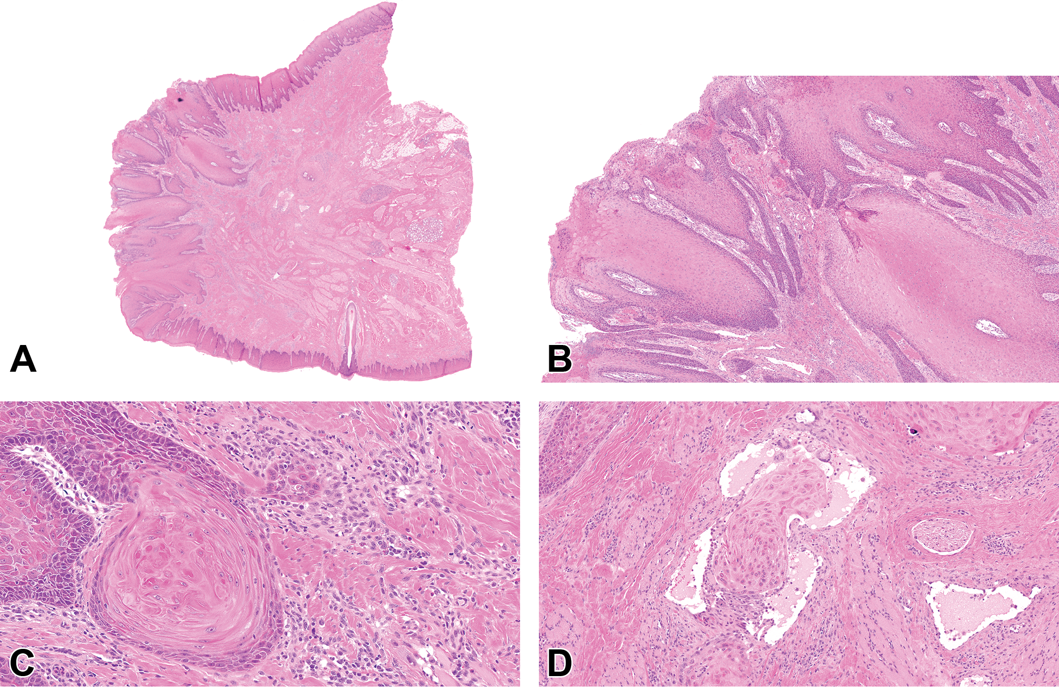
Cutaneous squamous cell carcinoma in a Yucatan minipig. (A) Arising from the epidermis is a neoplastic population of squamous epithelial cells arranged in broad trabeculae and nests (hematoxylin and eosin [H&E]). (B) The surface is lined by a thick band of keratin and is multifocally eroded (H&E). (C) The neoplastic cells undergo central keratinization and multifocally breach the basement membrane (H&E). (D) Rafts of neoplastic cells are present in lymphatic vessels and there are scattered multinucleated giant cells (H&E).
Cutaneous neoplasia in laboratory minipigs is rare, though melanoma has been reported. 5 To the authors’ knowledge, this is the first report of a cutaneous squamous cell carcinoma in Yucatan or other laboratory minipigs. Given that minipigs are commonly utilized for dermal studies, it is important to document this finding as a spontaneous lesion to aid in future study interpretation. Epidermal hyperplasia has been reported as a background lesion in Yucatan and other minipigs breeds used in toxicology studies; however, progression to squamous cell carcinoma is not noted, and this finding is often considered to be associated with treatment or handling, including dose site preparation. 2 -4 Oral and cutaneous squamous cell carcinomas have been infrequently reported in pot-bellied pigs kept as pets and may occur in farm pigs as well. 6- 11
In animals and humans, including swine, there is an association between squamous cell carcinoma and ultraviolet (UV) light exposure. 9,12- 14 This was considered unlikely in this case, as this animal was housed indoors at both the breeding facility and the research facility and thus not exposed to UV light. Additionally, solar elastosis and actinic keratosis, which are commonly observed in UV-associated neoplasms, were not observed in this case. 12,14 Squamous cell carcinomas are typically thought of as slow-growing and locally invasive, usually lacking metastasis except for poorly differentiated neoplasms. 14 This case was unusual in that there was the presence of lymphatic vascular invasion in a relatively well-differentiated lesion; however, nodal or distant metastasis was not evaluated due to a limited macroscopic and histologic evaluation.
Squamous cell carcinoma has also been associated with papillomavirus infection in various species, but not in swine. 15- 17 There was no evidence of papillomavirus infection detected histologically; however, no additional testing was performed to completely rule out this possibility. Typically, swine papillomavirus causes a sexually transmitted genital papilloma usually present on the vulva or preputial diverticulum which undergoes spontaneous regression. 17 In this case, there was no macroscopic evidence of genital lesions and the husbandry of the animals would preclude sexual transmission, as males and females are housed separately. We consider it most likely that the neoplasm in this case was spontaneously arising and this represents a rare finding of a spontaneous dermal neoplasm in the Yucatan minipig.
Footnotes
Acknowledgments
The authors would like to acknowledge the Charles River Laboratories Histology team for their technical support and imaging for this report.
Declaration of Conflicting Interests
The author(s) declared no potential, real, or perceived conflicts of interest with respect to the research, authorship, and/or publication of this article
Funding
The author(s) received no financial support for the research, authorship, and/or publication of this article.