
Abstract

In routine microscopic review of sections of normal tissues and experimental studies of animals, pathologists often come across interesting variations of normal structures (Figures 1-2). These variations of normal can invoke visions of plants, animals, people, machines, and other interesting formations. Mouse HistoArt was first reported in 2012. 1 Today, there is one major pathology art web site (Facebook group of over 11,000 members, #pathart: Art in Pathology 2 ) which showcases pathology art and includes 2 basic types of art: Natural, with no drawings on the histology image (Figure 1A, C, D-F) and Illustrated, with drawings depicted on the structures (Figure 2B).
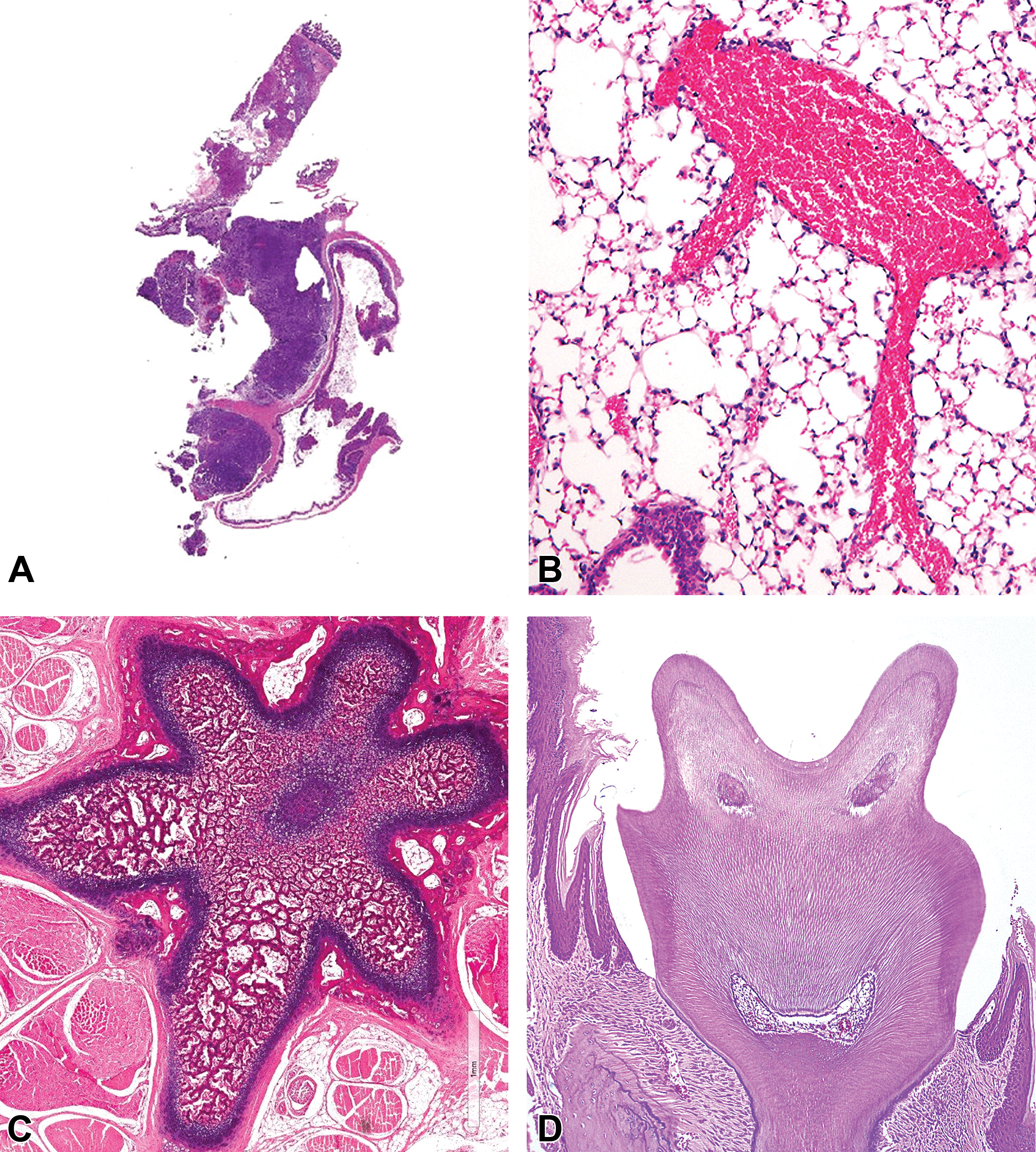
A, Microscope man—what a pathologist experiences after reviewing too many slides (mouse tumor). B, Long-legged hemorrhagic mouse (mouse lung). C, Starfish (rat tail vertebra). D, Smile for the camera (mouse tooth).
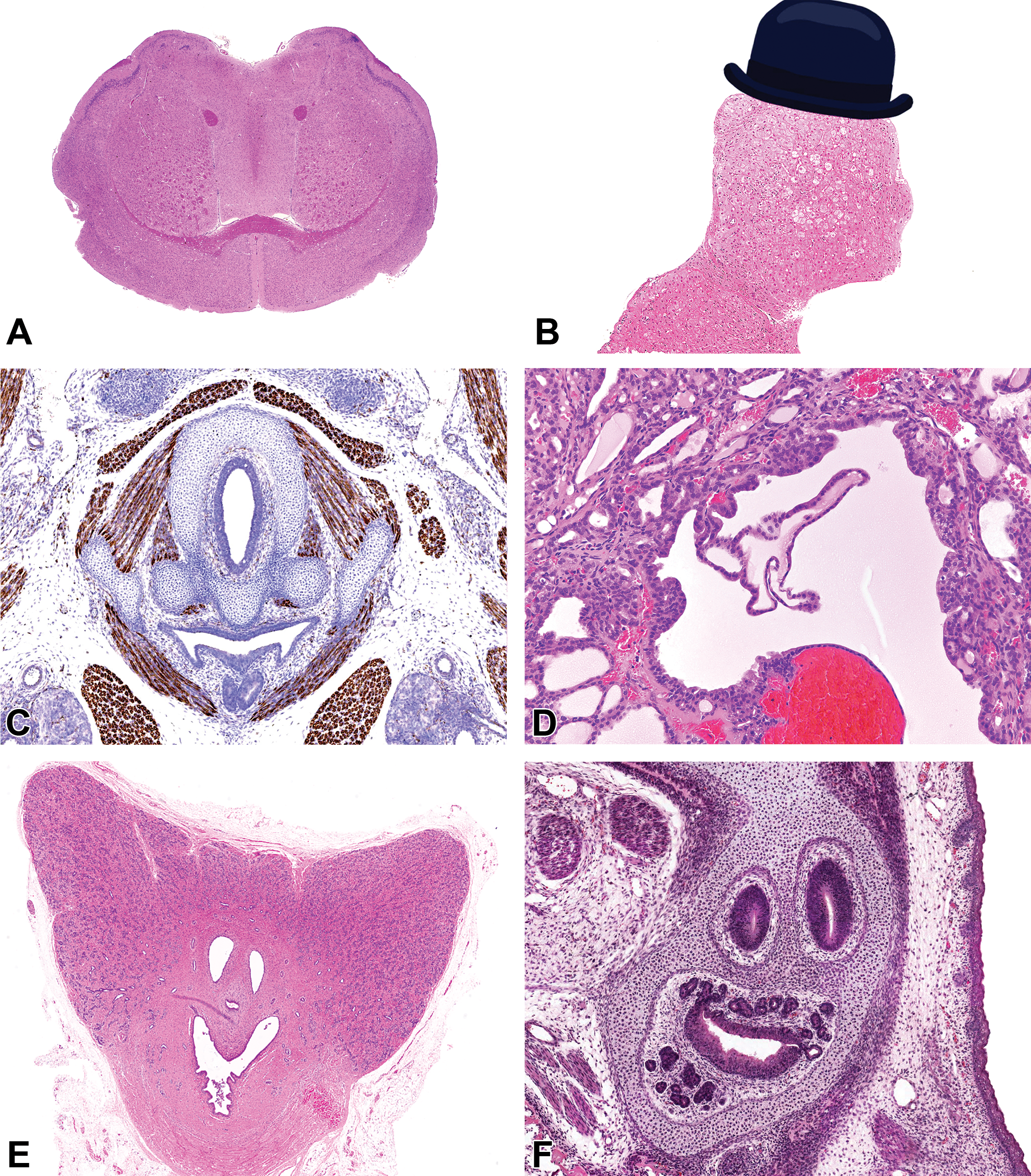
A, Happy toad face (mouse forebrain). B, Liver man (nonhuman primate). C, Dracula’s realization: “I need to get my fangs cleaned like I need a hole in the head” (mouse embryo laryngeal region, anti-desmin immunohistochemistry). D, Dinosaur hanging by a tail (mouse thyroid). E, Happy bat (monkey prostate). F, Casper, the friendly ghost with a 5 o’clock shadow (mouse embryo).
In regular times and periods of crisis, we should always remember to ask: What is life without whimsy?
Footnotes
Declaration of Conflicting Interests
The author(s) declared no potential conflicts of interest with respect to the research, authorship, and/or publication of this article.
Funding
The author(s) received no financial support for the research, authorship, and/or publication of this article.