
Abstract
Smoking is a major risk factor for heart attack, stroke, and lung cancer. Tobacco smoke (TS) causes bronchitis, emphysema, persistent cough, and dyspnea. Smoking cessation minimizes risks of TS-related disease. To determine whether smoking cessation could reverse TS-induced pulmonary changes, 10-week-old male spontaneously hypertensive rats were exposed to TS or filtered air (FA) for 39 weeks and allowed to live out their normal lifespan. Significantly (P ≤ .05) decreased survival was noted by 21 months in TS versus FA rats. In TS rats, persistent peribronchiolar, perivascular, alveolar, and subpleural inflammation were observed with pervasive infiltration of pigmented foamy macrophages and plausible intra-alveolar fibrosis and osseous metaplasia. Alveolar airspace was significantly (P ≤ .05) increased in TS versus FA rats as was the volume of stored epithelial mucosubstances in the left central axial airway. Increased mucin contributes to airflow obstruction and increased lung infection risks. Findings suggest TS-induced changes do not attenuate with smoking cessation but result in irreversible damage similar to chronic obstructive pulmonary disease. The observed persistent pulmonary changes mirror common TS effects such as chest congestion, sputum production, and shortness of breath long after smoking cessation and represent important targets for treatment of former smokers.
Introduction
Tobacco use is one of the most common causes of avoidable disease and death. It is associated with numerous bothersome and harmful effects in humans including cough, chronic phlegm production (“smoker’s cough”), fatigue, and increased severity of lung infections. 1,2 Smoking is recognized as the most important risk factor for chronic obstructive pulmonary disease (COPD) and is the third leading cause of death in the world today. 3 Smoking is also associated with a high incidence of cardiovascular disease, which represents the most common cause of illness and death worldwide. 4
Currently, there are more than 37 million adult smokers in the United States, 5 where each year, more than 16 million people have smoking-related symptoms, and more than 480 000 people die due to smoking-related disease. 2 Smoking is also a global issue. Countries with the highest incidence of smoking today include China, Korea, Russia, Indonesia, and India. 6
Past studies have used animal models such as dogs, mice, rats, and guinea pigs to observe the effects of tobacco smoke (TS) on lung pathology. Findings in these animal models have shown similar results with exposure to TS, such as goblet cell proliferation and airspace enlargement. 7 –11 Published studies involving active human smokers also show cellular infiltration and morphological changes in airway epithelia. 12,13 In the present study, the spontaneously hypertensive (SH) rat model was used to examine the effects of chronic TS exposure. Based on previous work, 14 –17 and in contrast to other rodent models, 18 the SH rat appears to be an optimal model for TS-induced lung inflammation with significant cellular, inflammatory, and structural changes in the lung consistent with human smokers. A striking finding in SH rats is the rapidity with which these pathologic changes develop following limited periods of smoking at fairly low concentrations of TS. 14,16 However, no studies have examined the senescence-associated effects of long-term smoking cessation in the lungs of SH rats. Given that human studies are highly variable and other animal models do not exhibit the range of adverse pulmonary effects known to affect long-term smokers, this study is significant in its presentation of new data that suggest the SH rat model is not just ideal for studying concurrent effects of TS exposure but also for observing structural and cellular effects after cessation. Since smoking cessation is thought to be the key to reducing the risk of disease progression, this study focused on the long-term pulmonary sequelae of TS exposure after a prolonged period of smoking cessation.
The purpose of this study was to (1) examine the long-term pulmonary effects of smoking in rats following a chronic (9 months) TS exposure and a subsequent smoking cessation period culminating at the end of their normal lifespan (average = 19 months) and (2) determine whether prolonged smoking cessation would allow the lungs to recover, repair, and return to a normal but natural state of aging. Morphological and cellular structures of the main axial airway and lung parenchyma were examined up to 10 months post-TS exposure. Inflammation in the airways, perivascular regions, pleura, and general lung parenchyma was qualitatively and semiquantitatively assessed. Alveolar size was measured using morphometry of the mean alveolar linear intercept length (average free distance in the air spaces) to examine potential differences within the gas exchange regions of former smoker (TS) rats relative to their never-smoker filtered air (FA) counterparts. Specific central airway generations of the bronchial tree were also examined using morphometric methods to determine airway epithelial volume and abundance of mucosubstance.
In the present study, the age of the rats over the TS exposure period would be approximately 20 to 70 years of age in humans. 19 Previous studies of human former smokers with COPD and a history of smoking cessation for 1 month to 14.5 years (mean 4.8 years) showed no difference in macrophage and neutrophil numbers in airways compared to current smokers, suggesting ongoing airway inflammation persists after cessation. 20 Based upon this finding, we hypothesized TS-induced changes in the lungs of SH rats, including inflammatory changes present in the airways and other regions within the lung, would not be completely repaired following long-term smoking cessation. Structural changes in alveolar space size and airway epithelial thickness were also anticipated in this model of long-term smoking cessation. Through the use of the SH rat model, this study may provide possible insight for further research on mechanisms of smoking-related disease progression and post-smoking treatment in former human smokers.
Materials and Methods
Animal Treatment and Exposure
Eight-week-old male SH rats were purchased from Charles River Laboratories (Raleigh, North Carolina) and allowed to acclimate for 2 weeks prior to exposure. A total of 74 rats were exposed to either TS (n = 36; experimental group) or FA only (n = 38; control group). All procedures performed on the rats were reviewed and approved by the University of California, Davis Institutional Animal Care and Use Committees. Inhalation of TS from 2R4F research cigarettes (University of Kentucky, Lexington, Kentucky) started at 10 weeks of age. Exposure of TS was performed in a smoking system built in our laboratory. 21 Rats were housed by threes in cages with narrow, straight-wire stainless-steel lids. All chambers were kept on a 12-hour light–dark cycle and controlled for temperature and humidity at 23°C to 24°C and 70%, respectively.
All TS-exposed rats were rotated, on a weekly basis, through 1 of 3 glass and stainless-steel chambers (Figure 1), with 4 cages and 12 rats per chamber. All TS animals were exposed for 6 hours/d, 3 days/wk, for 39 weeks (9 months). The total suspended particulate (TSP) concentrations in the 3 chambers ranged from 30 to 80 mg/m3 due to particulate loss as the air passed from consecutive chambers in series. As TS passed serially from the first to the third chamber, particulates deposited on the chamber walls and connecting lines between each chamber. To ensure equal smoke distribution to all rats during the entire experiment, cage positions were rotated weekly in a systematic way within the chambers. 22 Control rats were kept in an adjacent animal room, in a similar inhalation chamber ventilated with high-efficiency particulate air (HEPA)-FA. 21
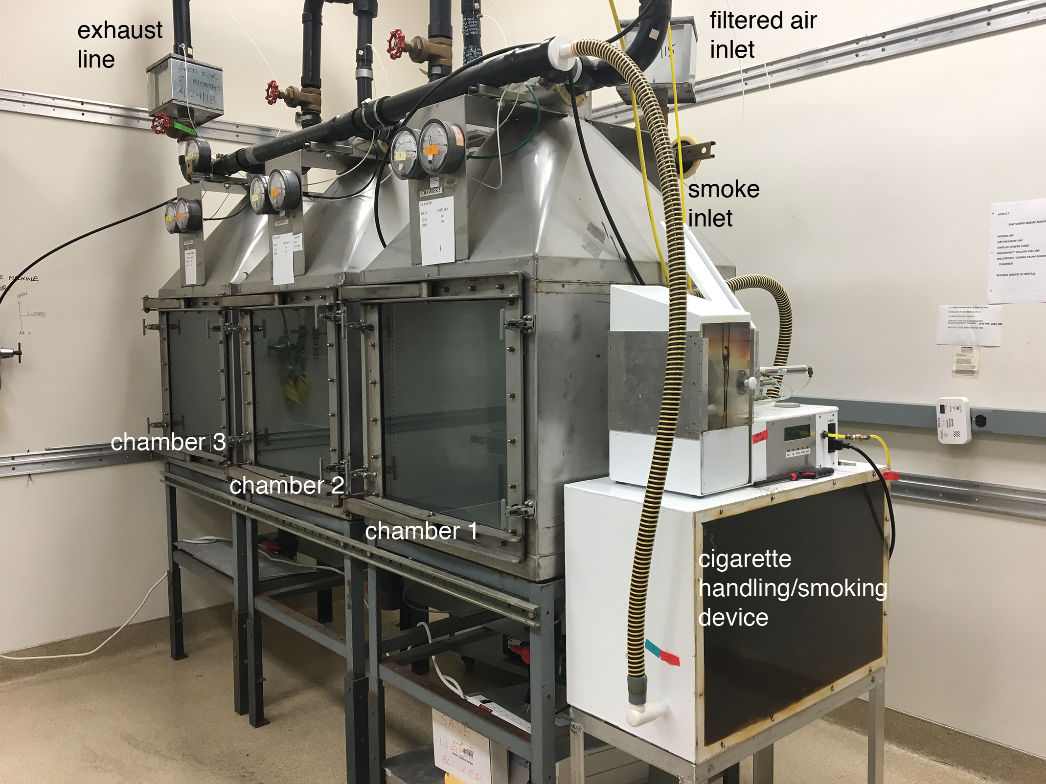
Tobacco smoke exposure chambers. From right to left, in chambers 1 to 3, the averaged chamber total suspended particulate (TSP) concentrations for the entire exposure period were 87 mg/m3, 57 mg/m3, and 44 mg/m3, respectively. The average TSP concentration over the 39-week exposure period was approximately 63 mg/m3.
To determine the level of TS exposure, measurements were taken for the following constituents in smoke: carbon monoxide (CO) every 30 minutes, TSP every 2 hours, and nicotine once per week, as reported previously. 21 These measurements were taken over the entire exposure period.
Necropsy and Lung Fixation
After the 39-week exposure period, rats were subsequently maintained in FA and permitted to live out their normal lifespan (average = 87 weeks [19 months] of age). At all times during the course of the experiment, the rats had access to food and water ad libitum. The rats were observed daily and weighed weekly. Once any rat was observed to be moribund or lethargic, it was deeply anesthetized with 1 mL of a 65 mg/mL pentobarbital solution by intraperitoneal injection, and lung tissue samples were collected. The left lung lobe was inflation-fixed at 30-cm water pressure with 4% paraformaldehyde for 1 hour and embedded in paraffin wax for subsequent histological analysis. 18,23
Histological Analysis
A total of 22 rats were randomly selected from the control and experimental groups (n = 10 and 12, respectively) for histological examination. Five-micron-thick paraffin-embedded lung tissue sections from these rats were transversely cut with a microtome (HM 355; Microm, Walldorf, Germany), placed on glass slides (Fisher Scientific, Pittsburgh, Pennsylvania), and stained with hematoxylin and eosin (H&E) or Alcian blue periodic acid Schiff (AB-PAS) stains from American MasterTech Scientific (Lodi, California). One section from each rat was stained with H&E to determine cellular infiltrates and airspace size. A serial section stained with AB-PAS was used to examine intracellular stored mucosubstances of the central airways. All histological assessments were performed blind.
Hematoxylin and eosin staining was performed by deparaffinizing mounted tissue paraffin-embedded sections in 3 changes of toluene, hydrating in decreasing concentrations of ethanol (100%, 95%, and 70%) and staining by Harris hematoxylin (American MasterTech) with a final wash in distilled water. Tissue sections were treated with differentiating solution (American MasterTech) to remove excess hematoxylin and bluing solution (American MasterTech) to enhance the contrast of the staining. Tissue sections were stained with eosin Y (American MasterTech), dehydrated in increasing concentrations of ethanol (70%, 95%, and 100%), cleared through 3 changes of toluene, and coverslipped. The AB-PAS staining was performed by deparaffinizing and hydrating tissue sections with a wash in running tap water. Tissue sections were treated with 3% acetic acid (American MasterTech) and stained by Alcian blue (American MasterTech). Tissue sections were placed in 0.5% periodic acid (American MasterTech, Lodi) and Schiff reagent (American MasterTech) followed by dehydrating, clearing, and coverslipping.
Semiquantitative Histological Assessment by H&E
Histopathological scoring was completed to determine the morphological changes and degree of lung inflammation in and/or around the airways, blood vessels, and lung parenchyma using an ordinal, semiquantitative rubric for extent and severity described previously 24 and shown in the Supplemental Table 1 and Supplemental Figures 1 to 4. Severity scores ranged from 0 to 3. Scores of 0 were given for sections with little to no inflammation, while scores of 3 were given for sections with marked inflammation. Extent was defined as the proportion of the tissue section involved in the specified pathological change and extent scores including 0 (no lung involvement), 1 (≤1/3 of the tissue section involved), 2 (1/2 of the tissue section involved), and 3 (≥ 2/3 of the tissue section involved) were used. The overall score was a combined assessment of severity and extent wherein overall score = severity × extent.
Assessment of Alveolar Airspace Size by H&E
Emphysema, a long-term lung disease that leads to shortness of breath and hyperventilation, is defined as the development of lung airspace enlargement. 25 Previous research suggests analysis of airspace enlargement is an appropriate method for identifying emphysema because of its ability to discriminate significant variations, even in aging mice that have not been exposed to TS. 26 For these reasons, H&E-stained tissue sections were used for morphometric assessment of the alveolar airspace. The mean linear intercept (MLI; the mean free distance in the air spaces) was calculated with Image J software (version 2.0.0; National Institutes of Health, Bethesda, Maryland). 27 For each tissue section, a total of 10 parenchymal fields of view were captured in a systematic, uniform, random but nonoverlapping manner using a 20× objective lens on an Axiolab light microscope (Zeiss, Jena, Germany). Fields containing large airways, blood vessels, or artifacts were not photographed and thus excluded from the analysis. 28 Resulting images were projected onto a monitor using an Axiocam 105 color camera (Zeiss, Jena, Germany) and overlaid with a test grid generated by Stereology Toolbox software (Morphometrix, Davis, California) to assess MLI by counting the number of points intercepting alveolar tissue. 18,29 The airspace size was expressed as the average length of the line between intercepts in µm and was compared between TS and FA groups.
Analysis of Mucin Content and Epithelial Thickness by AB-PAS
The AB-PAS-stained sections were used to determine intraepithelial mucosubstance abundance. To this end, central airway mucin cells were quantified by point counting in Image J. For each section, images of 4 regions (12, 3, 6, and 9 o’clock positions) around the main axial airway of the left lung were captured using a 40× objective lens on a Zeiss Axiolab light microscope with a Zeiss Axiocam 105 color camera. Measurements were performed using a Mertz rolling cycloid arc grid generated by Image J and overlaid with 190 points per image. Points that intercepted mucin cells, basal lamina, and epithelia were counted to calculate the volume of mucin in the airway epithelium per surface area of basal lamina in µm3/µm2. 30 All points falling on the epithelium were also used to calculate the epithelial volume per surface area of basal lamina to determine airway epithelial thickness. 31 The areas of mucin content were expressed as a percentage of the total epithelial area.
Statistical Analysis
Student t tests were performed at a significance level of P < .05 to determine statistical differences between the FA and the TS exposure groups. All results were reported as the arithmetic mean ± standard error of the mean. Statistical analyses were performed using R software (version 3.5.0; R Development Core Team, Vienna, Austria).
Results
All animal results are reported for the smaller subset of 22 rats unless otherwise stated.
Concentrations of TS Constituents
Average concentrations were 210 ± 19 ppm of CO, 63 ± 13 mg/m3 of TSP (estimated as the equivalent of about 1 pack per day simulating human smoking) and 5 ± 2 mg/m3 of nicotine. 32 The range of TSP concentrations in TS to which the SH rats were exposed in the present study, 30 to 80 mg/m3, would be relevant to concentrations encountered by a moderate smoker. 14 These concentrations were within the levels used in a number of other TS inhalation studies with rats and mice. 33 –37 In a previous study, a high TSP concentration (176 mg/m3), which acclimated mice can tolerate well, was used, and no treatment-related deaths were observed. 28 Another past study, using a different exposure system, subjected mice to up to 300 mg/m3 of TSP. 38
Body Weight
The average near end-of-life body weight for FA- and TS-exposed rats was 383 ± 18 g and 363 ± 13 g, respectively. No significant (P < .05) difference in body weight was found between the 2 treatment groups.
Age and Survival
The average age at the time of necropsy was significantly (P = .004) different between FA- and TS-exposed rats (22 ± 0.4 months vs 20 ± 0.4 months, respectively), with the TS group having a shorter lifespan of approximately 2 months compared to that of the FA group. This difference is the equivalent of approximately 3.5 years in humans. 19 Survival curves plotted for the TS and FA groups (Figure 2) were found to be significantly (P = .002) different. The survival curves for the complete set of 74 animals has been included in the Supplemental Material. These survival curves (Supplemental Figure 5) are similar to that of the 22 animals randomly selected for this study.
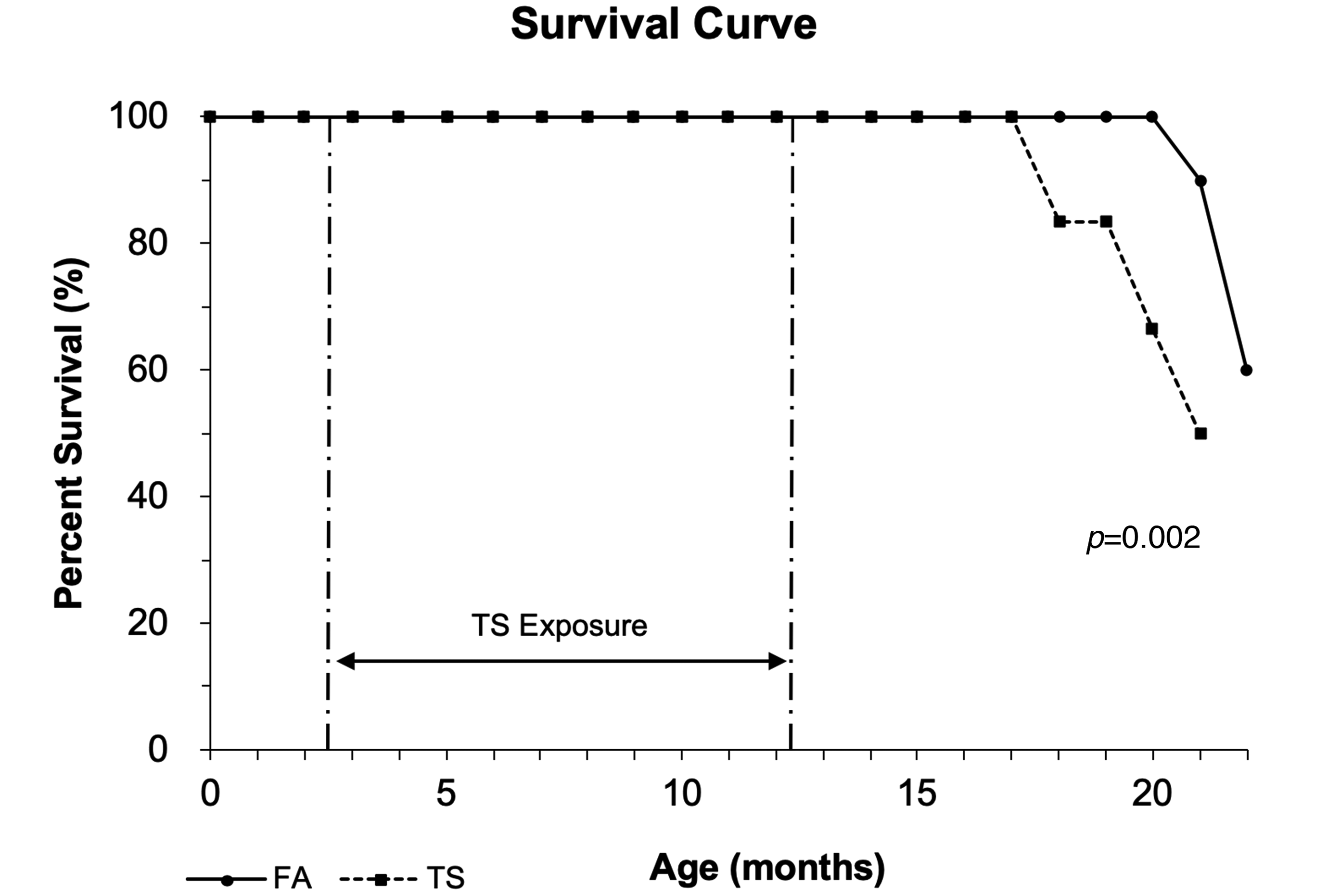
Rats chronically exposed to tobacco smoke (TS) have shorter lifespans than those breathing filtered air (FA). Rats were exposed to TS (square markers with dashed lines; n = 12) or FA (round markers with solid lines; n = 10) for 9 months (6 hours/d, 3 days/wk, for 39 weeks), from ages 10 to 49 weeks, and subsequently maintained in an FA environment until death. The resulting percentage of survival is shown for each group over time. Survival curves were compared using a Student t test at a significance level of P < .05.
Inflammatory Changes Observed by H&E
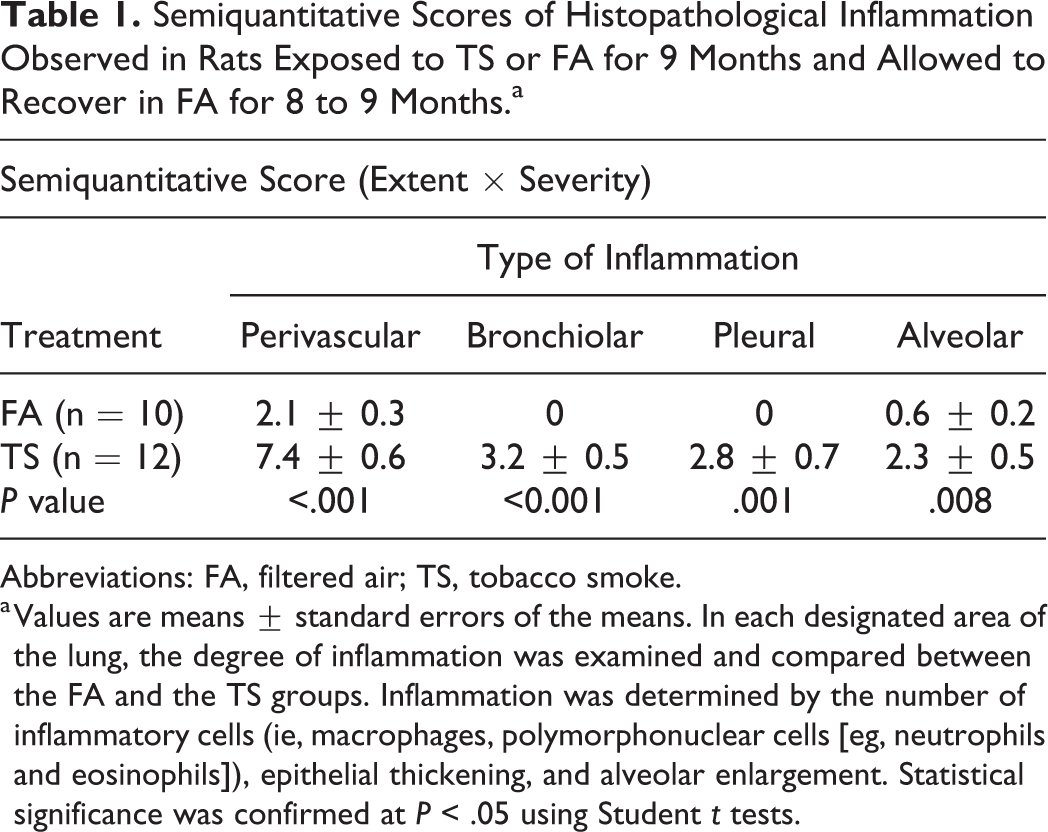
Semiquantitative analysis showed significantly (P < .01) greater scores for bronchiolar, perivascular, alveolar, and subpleural inflammation in lungs of TS versus FA rats (Table 1). In the former versus the latter, a number of inflammatory markers, such as macrophage influx (Figures 3C and D), pseudogland formation (Figure 4C and D), and goblet cell metaplasia (Figure 4C and D), were found to be increased in the lung. Changes suggesting osseous metaplasia (Figures 5A and B) and focal or multifocal alveolar fibrosis (Figure 5C) were observed in the most severe cases as well as numerous irregular, enlarged, multinucleated luminal macrophages (Figure 5D), cellular debris (Figure 5D), and what appeared to be occasional mast cells (not shown) in the alveoli. Some rats exhibited brown and foamy macrophages that appeared to be hemosiderin-filled; these cells seemed more abundant in TS versus FA rats (Figure 3). Hemosiderin is an iron-storage complex that, when freely present in lung tissues as observed, may be associated with macrophage phagocytosis of abundant red blood cells and hemoglobin resulting from pulmonary hemorrhage or heart failure. 39,40 The presence of pigmented macrophages in the lungs of TS-exposed rats may also be associated with the ingestion of tars from TS. 41 However, brown foamy macrophages were not quantified. Airway epithelial thickening also appeared to be greater in TS versus FA rats (Figure 6). Quantitative analysis of this end point is discussed below (see Mucosubstance and Airway Epithelium Analysis by AB-PAS section).
Semiquantitative Scores of Histopathological Inflammation Observed in Rats Exposed to TS or FA for 9 Months and Allowed to Recover in FA for 8 to 9 Months.a
Abbreviations: FA, filtered air; TS, tobacco smoke.
a Values are means ± standard errors of the means. In each designated area of the lung, the degree of inflammation was examined and compared between the FA and the TS groups. Inflammation was determined by the number of inflammatory cells (ie, macrophages, polymorphonuclear cells [eg, neutrophils and eosinophils]), epithelial thickening, and alveolar enlargement. Statistical significance was confirmed at P < .05 using Student t tests.
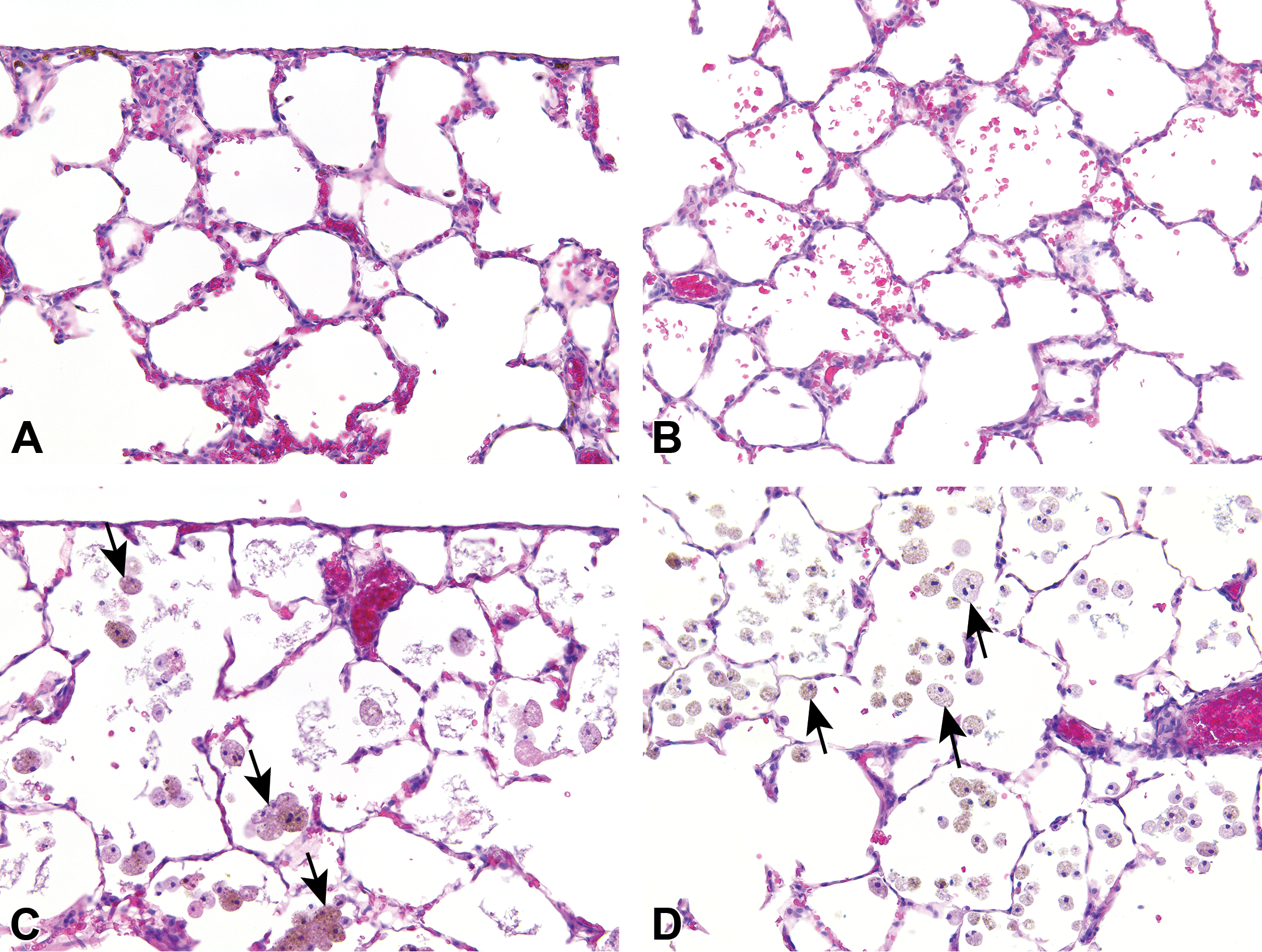
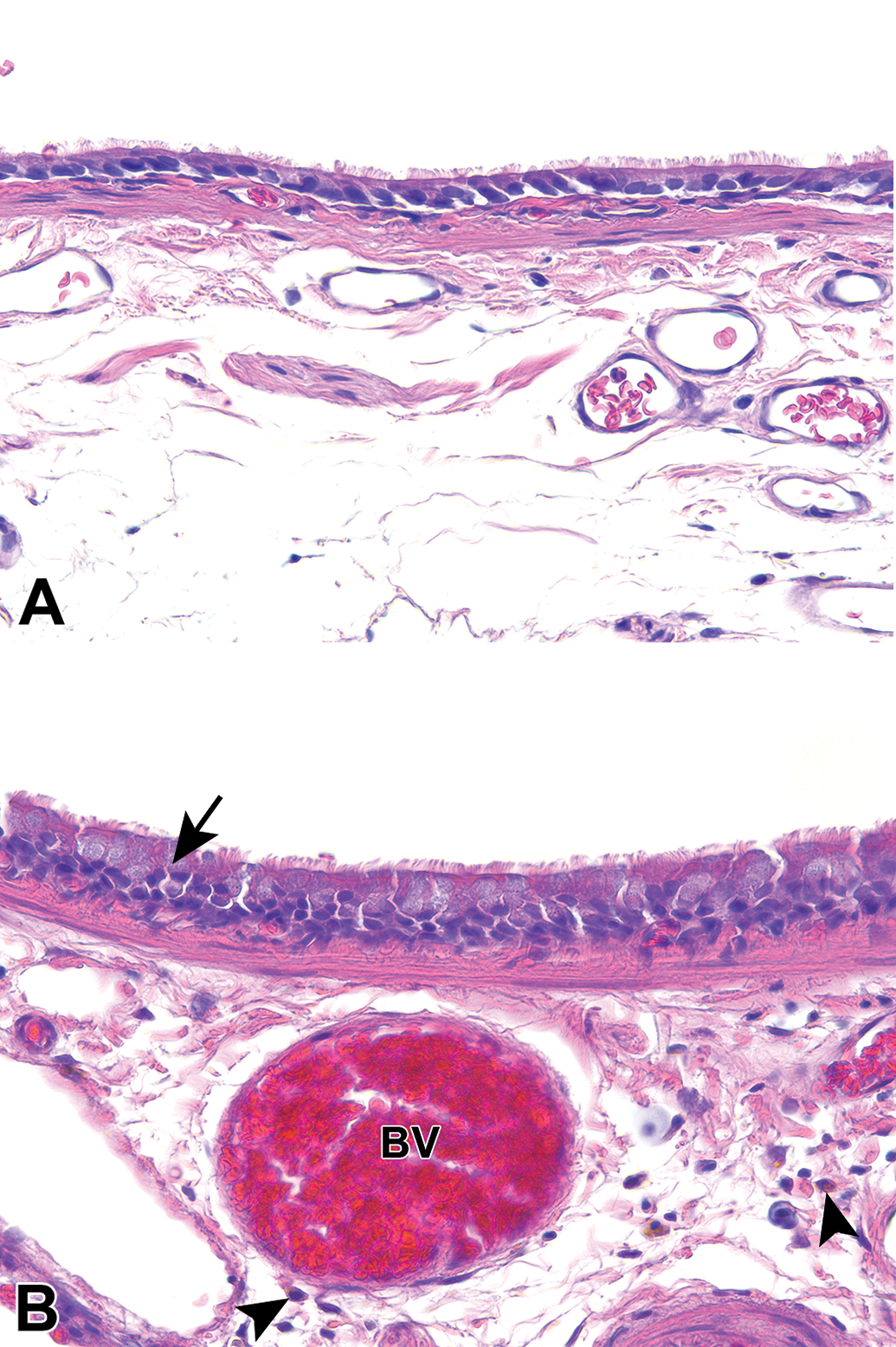
Marked inflammatory changes present in the lungs of tobacco smoke (TS)- versus filtered air (FA)- exposed rats. Panels are bright-field microscopy images of lung tissues from rats exposed to FA (panels A and B) or TS (panels C and D) for 9 months and subsequently maintained in FA until the end of their lifespan (approximately 20 or 22 months, respectively). Tissue sections were stained with hematoxylin and eosin. Arrows indicate infiltrating foamy, brown-pigmented macrophages in the subpleural and parenchymal regions of the lung (panels C and D, respectively). Panels C and D are representative of the most severe inflammation in the treatment group. All panel images are shown at the same magnification.
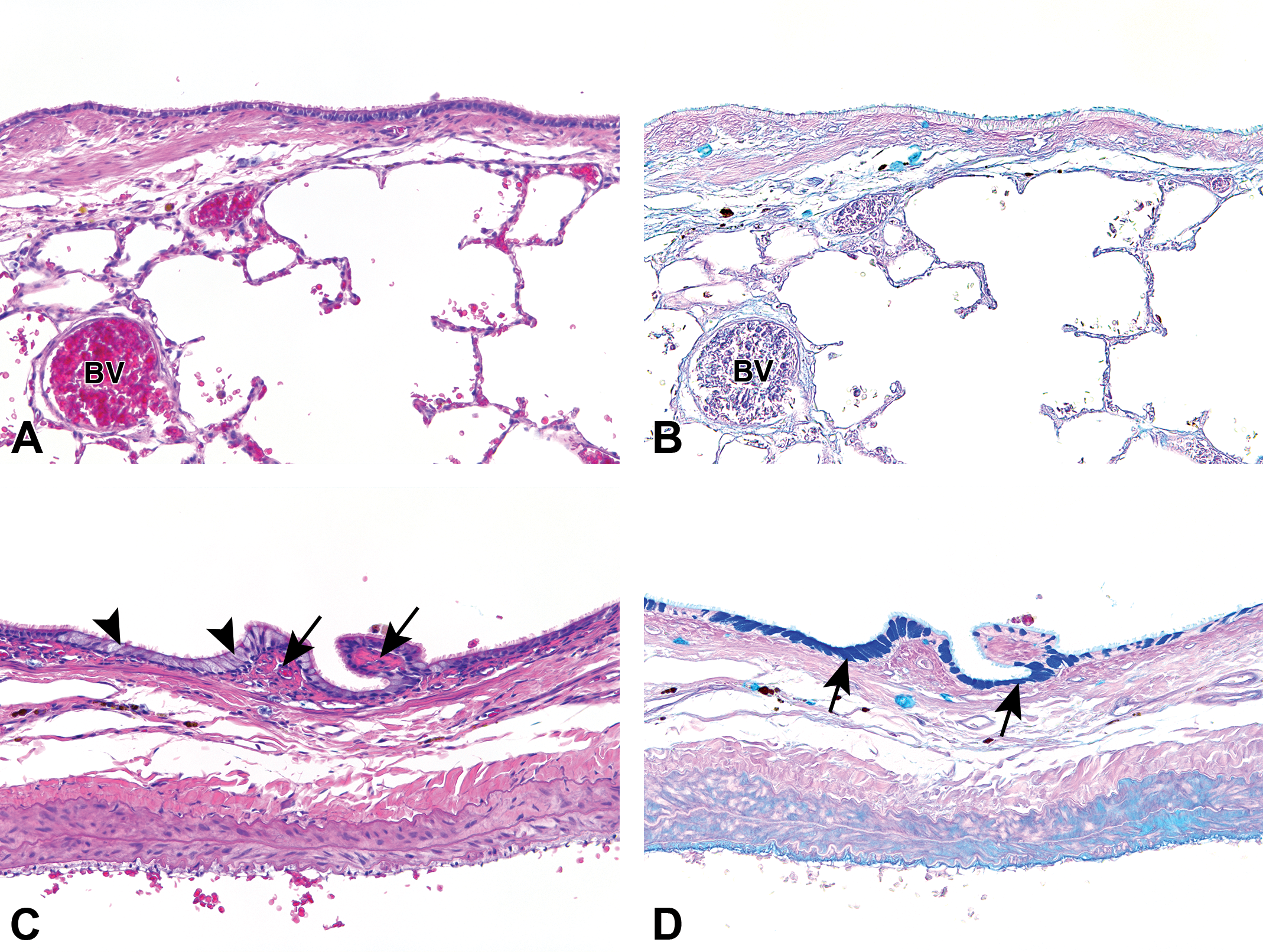
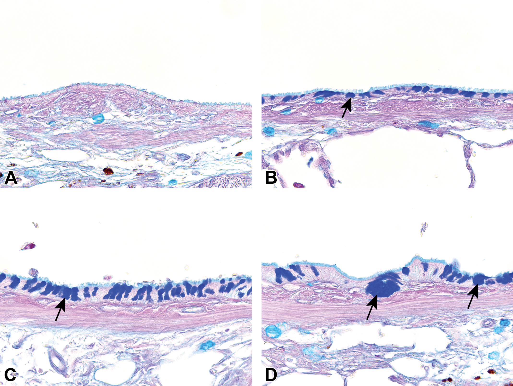
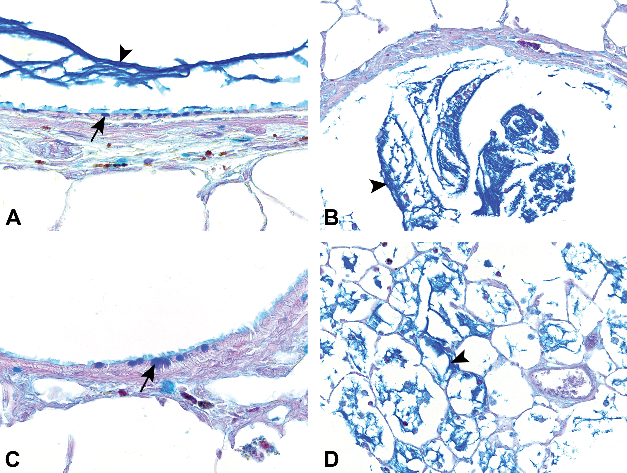
Morphological changes in the central airway epithelium of tobacco smoke (TS)-exposed rats. Bright-field images are from serial tissue sections recovered after a 9-month exposure to filtered air (FA; panels A and B) or TS (panels C and D) followed by an 8- to 9-month smoking cessation period with exposure to FA only. Sections were stained with hematoxylin and eosin (panels A and C) or Alcian blue and periodic acid-Schiff (panels B and D) stains. Arrows indicate pseudoglands in the central airway (panel C) and mucus metaplasia in the airway epithelium (panel D). The arrowheads in panel C indicate goblet cells. All panel images are shown at the same magnification. BV indicates blood vessel.
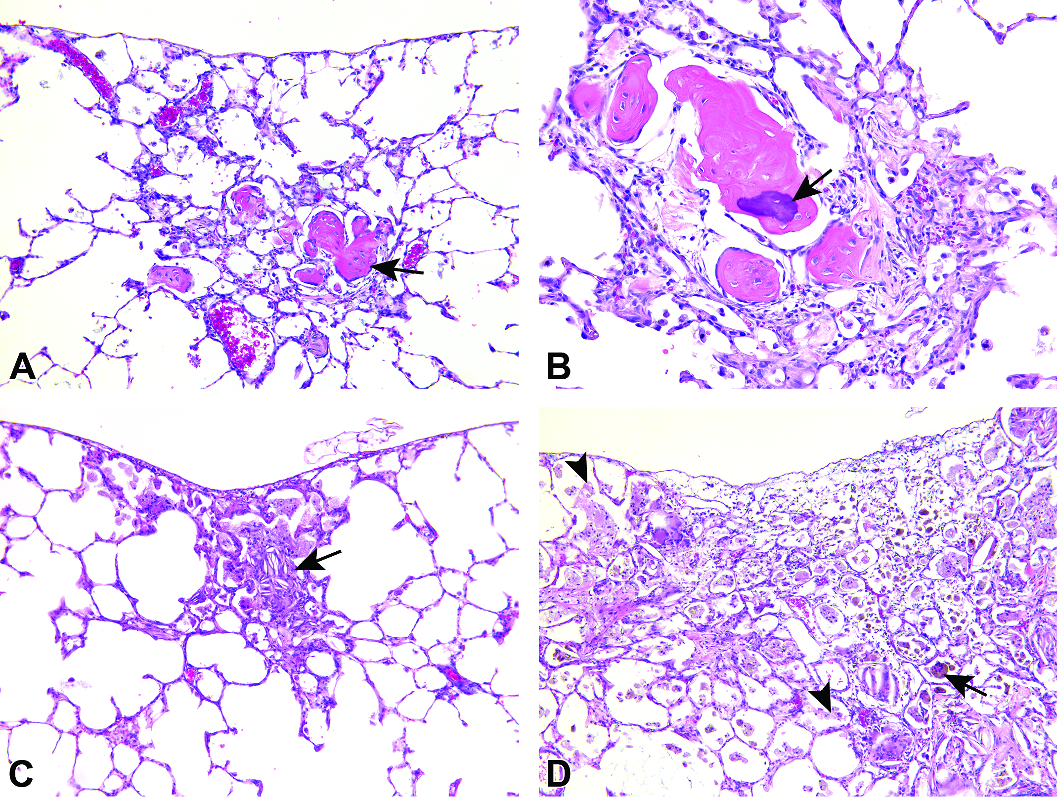
Tobacco smoke (TS)-induced alterations in the lungs of rats. Bright-field images are from hematoxylin and eosin-stained tissue sections recovered after 9 months of TS exposure followed 8- to 9-months of smoking cessation in FA alone. Arrows indicate subpleural parenchymal osseous metaplasia (panels A and B), subpleural fibrosis (panel C), and alveolitis with numerous luminal alveolar macrophages (panel D). Cellular debris (arrowheads) is seen in the lumen of some alveoli (panel D). Panel A, C, and D were captured using original objective ×10 and panel B using original objective ×20.
Tobacco smoke (TS)-exposed rats show morphological changes in the central airway epithelial cells and peribronchiolar inflammation in the lungs. Panels are bright-field microscopy images of lung tissues stained with hematoxylin and eosin. Arrow indicates the epithelium of the central airway, which seems thicker in the lungs of rats exposed for 9 months to TS (panel B) versus filtered air (panel A) and subsequently allowed to live out the rest of their normal lifespan (20- vs 22-months, respectively) in FA. The arrowheads in panel B show peribronchiolar cellular infiltration. All panel images are shown at the same magnification. BV indicates blood vessel.
Structural Changes Within Lung Parenchyma by H&E
The MLI analysis (Figure 7) quantitatively confirmed that differences in alveolar size observed by H&E (Figure 8) were statistically significant (P = .002), with greater alveolar size in TS versus FA rats (Figures 7 and 8). However, varying degrees of alveolar wall thickness were observed in both the groups with no distinct exposure-related patterns; some walls were thickened, while other alveolar structures appeared to be quite thin (not shown). Both groups showed thinned alveolar walls, which can be associated with emphysema, in some focal areas within the lungs. Alveolar thinning was especially noticeable in the ventral regions of the left lung lobes of FA and TS rats. These emphysematous changes may be due to aging. 42
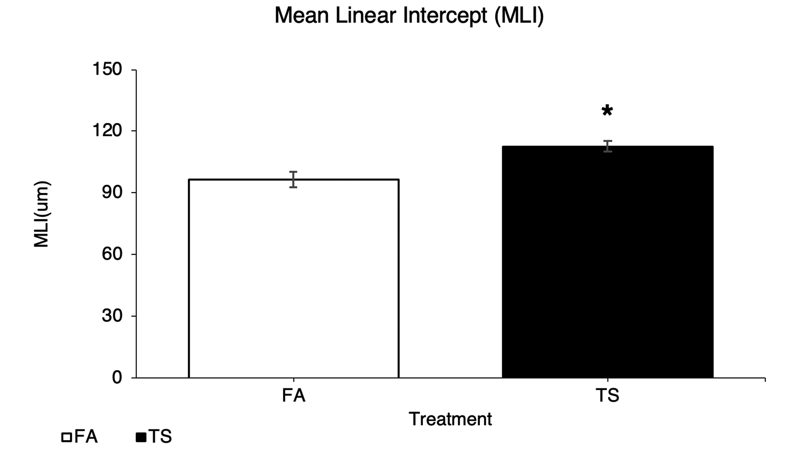
Tobacco smoke (TS)-exposed rats have significantly (P = .002) greater areas of air space enlargement relative to filtered air (FA) controls. Graph shows results of mean linear intercept measurements, which were taken in both groups after consecutive periods of TS or FA exposure (9 months) and smoking cessation (8-9 months exposure to FA).
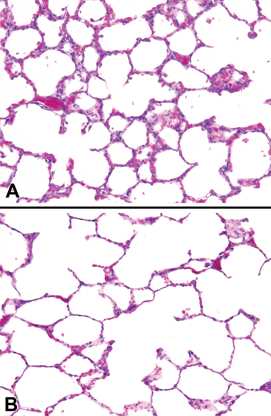
Tobacco smoke (TS)-induced morphological changes in the lung parenchyma of SH rats. Hematoxylin and eosin stain was used for pathological assessment. Tobacco smoke-exposed (9 months) rats showed alveolar airspace enlargement and thinner alveolar septal walls (panel B) compared to filtered air-treated rats (panel A). Both panel images are shown at the same magnification.
Mucosubstance and Airway Epithelium Analysis by AB-PAS
Airway mucus hypersecretion is an important characteristic of COPD, which can be associated with a continuous decline in pulmonary function among patients with COPD. 43 The average volume fraction of stored mucosubstances in the central airways was 4.97% ± 1.48% of the total epithelial area in rats treated with FA and 34.26% ± 3.97% in rats exposed to TS (Figures 9 and 10). Compared to FA, TS exposure significantly (P < .001) increased mucin content in the central airway but did not significantly affect airway epithelial thickness (Table 2). These metaplastic changes may be due to TS that caused the normal bronchial epithelial cells to be replaced by mucous cells, which resulted in an increase in mucosubstances but no change in airway epithelial thickness. 44 The distribution of bronchial epithelial mucus metaplasia was noted several airway generations down the bronchial tree (Figures 11A–C), and mucin in the airway and alveolar lumina was occasionally present in TS rats (Figure 11).
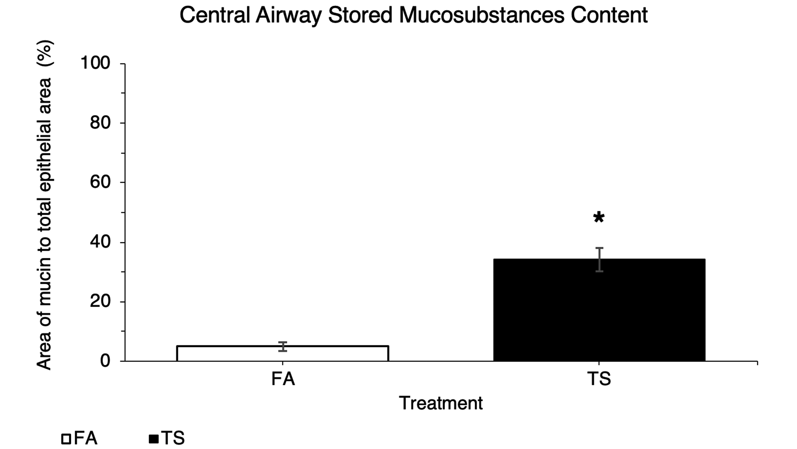
Tobacco smoke (TS)-exposed rats have significantly (P < .001) greater mucin content in the central airway relative to filtered air (FA) controls. Graph shows results of mucosubstance abundance analysis, which were taken in both groups after consecutive periods of TS or FA exposure (9 months) and smoking cessation (8-9 months exposure to FA).
Tobacco smoke (TS)-exposed rats exhibit more intraepithelial mucosubstances than filtered air (FA)-treated rats. Panels are bright-field microscopy images of lung tissues treated with Alcian blue and periodic acid-Schiff stain. The number of mucin cells was increased in TS-exposed (9 months) group (panels B-D) compared to the FA-treated group (panel A). Arrows in panels B-D indicate mucosubstances stained blue. All panel images are shown at the same magnification.
Quantitative Analysis of Airway Epithelial Thickness in Rats Exposed to TS or FA for 9 Months and Allowed to Recover in FA for 8 to 9 Months.a
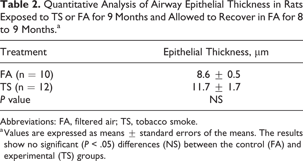
Abbreviations: FA, filtered air; TS, tobacco smoke.
a Values are expressed as means ± standard errors of the means. The results show no significant (P < .05) differences (NS) between the control (FA) and experimental (TS) groups.
The distribution of mucosubstances observed within the lungs of tobacco smoke (TS)-exposed rats. Panels are bright-field microscopy images of lung tissues treated with Alcian blue and periodic acid-Schiff stain. Arrows in panels A and C indicate the bronchial epithelial mucus metaplasia observed in distal airways. Arrowheads indicate mucin in the airway lumen (panels A and B) and alveolar airspace (panel D) occasionally present in some of the TS-exposed (9 months) rats. Panels A and C were captured using original objective ×40, and panels B and D were captured using original objective ×20.
Discussion
General Implications of Smoking Cessation
The present study, which was novel in its examination of chronic TS exposure, smoking cessation, and senescence, demonstrated persistent TS-associated inflammation and morphological changes in the lungs of SH rats following a 9-month TS exposure and a subsequent 8- to 9-month smoking cessation period in FA alone and suggested that smoking cessation was not sufficient to reverse these pathological changes. We believe our long-term SH rat model of smoking cessation is an exemplary chronic COPD model for the examination of relationships between senescence and smoking while also stressing the importance of early smoking cessation to avoid persistent lung damage.
Body Weight
Results of body weight analysis showed rats from the control and experimental groups were not statistically different, although the average weight of TS rats appeared slightly lower than the controls. The nonsignificant trend toward lower body weight in the former versus the latter is consistent with previous findings in longitudinal studies, where current smokers had a lower weight or body mass index (a measure of body fat based on height and weight) than nonsmokers. 45,46 Indeed, the possibility of weight gain sometimes deters current smokers from quitting. However, results herein do not support such claims, and many other studies show variability in weight gain when quitting. 47,48 While it is known that smoking and nicotine can increase metabolic rate while decreasing appetite and reducing body weight, 49 these effects are clearly counterbalanced by the long-term negative health effects of smoking as seen in our findings.
Lifespan
The average lifespan of the male SH rat is 22 months (range 15-26 months). 50 Our findings show former smoker (TS) rats have a reduced lifespan of approximately 2 months on average compared to that of never-smoker (FA) rats. Research on human survival in smoking populations suggests that survival increases with smoking cessation, in contrast to continued tobacco use. 51 Further evidence suggests although smoking cessation slows the accelerated rate of lung function decline, exsmokers continue to have a permanently greater risk of morbidity and mortality due to COPD, compared to that of never-smokers. 52 This may, in part, explain our findings of a statistically significant decrease in the lifespan of TS-exposed rats, compared to FA control rats (Figure 2). Senescent TS-exposed rats may also be more susceptible to cardiovascular diseases as well as other age- or TS-related pathologies (renal, liver, testicular, etc) that could also lead to a shorter lifespan.
Long-Term Histological Sequelae of Lung Morphology Following Smoking Cessation
Histopathology scoring
To survey the pulmonary inflammation of the rats exposed to TS, the histological appearance of the lung and the degree of inflammation were investigated following semiquantitative assessment and scoring in this study. Greater signs of inflammation, as indicated by increased levels of macrophages, were seen throughout the lungs of TS rats compared to those of FA rats (Table 1; Figures 3 –6). These results suggested the development of chronic pulmonary inflammation. 53 Chronic inflammation is a strong predictor of lung cancer development, 54 but lung cancer was not observed in this study.
The present study showed that TS rats exhibited airway inflammation with peribronchiolar cellular infiltration (Figure 6B) and goblet cell metaplasia (Figure 4). These effects have been demonstrated in a previous study of Sprague-Dawley rats exposed acutely to TS. 55 Other studies on former-smoker mice have reported persistent airway inflammation after smoking cessation suggesting that these TS-induced changes were irreversible. 56,57
In the present study, moderate-to-severe inflammation was found in the perivascular regions of the lungs with a few cases of arterial thickening and abnormal vascular cells in TS-exposed rats. Previous studies found similar alterations including perivascular inflammation, vascular congestion, and vascular wall thickening in rats exposed to TS and suggested that the vascular changes were associated with pulmonary hypertension which likely resulted from inflammation-induced hypertrophy of the vascular wall. 58,59
In addition to peribronchiolar and perivascular inflammation (Table 1), increased cellularity (Figures 3C-D) in alveolar and subpleural regions was also found in TS rats when compared to FA controls from the present study. Moreover, in most of the TS-exposed rats, scarring and fibrosis (Figure 5C) were observed in the subpleural and parenchymal regions of the lungs. Despite previous research findings in which TS appeared to increase the risk of developing pulmonary fibrosis, the association between pulmonary fibrosis and TS is still under debate. 60,61 Nevertheless, effects similar to ours have been reported in past studies of TS-exposed beagle dogs and F-344 rats, wherein alveolar fibrosis accompanied alveolar and subpleural inflammation. 62
Alveolar morphometric analysis of lung gas exchange regions
Other observed changes due to TS exposure in the present study included the significant (P = .002) increase in alveolar size (Figure 7) and loss of the alveolar wall structure (Figure 8B), which are characteristics of human emphysema. 63 Chronic inflammation in the lung leads to destruction of the airway walls and loss of elasticity in the lung parenchyma, and these types of lung injury reduce expiratory airflow. 64
Valenca et al 65 suggested that TS-exposed C57/BL6 mice have a pulmonary emphysema-like condition, with thickening of alveolar septa due to collagen deposits. March et al 66 indicated no significant TS-induced thickening of alveolar septa but a significant TS-induced shape change in the alveolar air spaces in both B6C3F1 mice and Fischer-344 rats. The progressive loss of alveolar septa during the formation of emphysema in current-smoker humans has been reported by Vlahovic et al 67 and suggests that gas exchange in the early stages of emphysema primarily decreases due to the loss of entire segments of alveolar septa. The same group also showed interstitial thickening in the remaining alveolar septal wall with an increase in elastin and collagen in more severe areas of emphysema suggesting a repair process after TS-induced injury to the lung of humans. Ri et al 68 claimed that the increase in alveolar space size observed in a C57BL/6 ex-smoker mouse model was more prominent than controls although alveolar septal thickness was normalized after smoking cessation, suggesting that emphysema progressed with septal remodeling during smoking cessation. These may explain the varying thickness of alveolar septal walls and irreversibly enlarged alveolar airspaces observed following smoking cessation in this study (Figure 8).
Airway epithelial cell morphometric analysis
The chronic pathologies observed in TS-exposed rats from the present study are precursors to fatal lung diseases that negatively affect lifespan and quality of life. Increased epithelial cells in airways can be linked to bronchitis and COPD, while greater amounts of mucin in the central airway can obstruct airflow and gas exchange. 69 Epithelial cells also serve as secretors of cytokines and chemokines that promote the recruitment of leukocytes leading to increased levels of inflammation. 70 Persistent mucus secreted by mucin cells increases lung susceptibility to infection and dyspnea. 71,72
The increase in bronchiolar epithelial cell thickness observed in some of the TS-exposed rats in the present study is suggestive of COPD development. The significant development of pseudoglands (Figure 4C) was one of the most prominent markers that distinguished TS-exposed rat airways from those of controls in this study (Figure 4). We believe the pseudoglands were created by abnormalities in the airway reconstruction process due to onset TS damage. Other research has shown that pseudoglands are indicative of abnormally repaired epithelia and can negatively affect activity of secretory cells leading to decreased immune defense. 73
Combined effects on overall lung health
Both emphysema and chronic bronchitis are conditions of COPD, a major cause of chronic morbidity and mortality throughout the world. 74 Both lung conditions are associated with long-term TS exposure and affect the ability of lungs to take in oxygen. Sustained low levels of oxygen in the blood place extra stress on the heart and force the heart to pump harder in order to send blood through the lungs. This contributes to the risk of heart failure by increasing heart and breathing rates that compound heart strain. 75 Chronic obstructive pulmonary disease is also characterized by chronic inflammation in the lung tissue associated with a systemic inflammatory response that affects many other organs and raises the probability of cardiovascular events. 76 Asthma is another prominent disease characterized by long-term airway inflammation and alteration in the airway wall. 77 Many of the changes seen in the airways of the SH rats of this study, including airway epithelial thickening (Figure 6) and goblet cell hyperplasia (Figure 4), are also seen in patients with asthma. 78 From the results of our recovery study, it is clear that TS exposure causes irreversible changes throughout the lungs, negatively affecting air exchange that cannot be attenuated through smoking cessation.
Normal lung aging in SH rats
Functional and structural changes are exhibited in aging lungs, 79 but detailed descriptions of age-related alterations in the respiratory system of SH rats are limited. Previous experiments in F344/N rats revealed age-related morphological changes, including extension of distal airways and enlargement of alveolar airspace size. 80 The increase in the size of the alveolar space without any inflammation or alveolar wall destruction is also observed in the aging lung of humans and defined as “senile emphysema.” 42 This supports our findings of thinner alveolar walls and larger airspace size seen in some of the senescent rats from the FA group. In addition to structural changes, alterations in muscle function and pulmonary immunologic function are correlated with age, 81 although these end points were not measured herein. Taking all of these age-associated changes into consideration, pulmonary diseases including COPD and emphysema have significant consequences in the elderly population. 82 This may explain the presence of persistent alterations induced by TS, which are irreversible even after a period of cessation, in the senescent SH rats in the current study.
Relevance of the SH rat to a model of COPD
A variety of animal models involving dogs, guinea pigs, mice, and various strains of rats have been used to study the mechanisms of COPD induced by TS exposure. The characteristic features of COPD include emphysema, mucus hypersecretion, chronic inflammation, and airway remodeling. 83 All animal models have presented slightly different degrees and severity in each of these abnormalities. Variations have been found to be strain dependent and could explain our results, which did not show complete foundation of emphysema but rather beginning signs of its development. This suggests a genetic component in the development of emphysema, a key point of importance for predicting the disease onset in humans. 84 As many animal models similarly reflect specific human lung changes onset by TS, 85 it is important to note whether they also show acute exacerbations or comorbidities of COPD such as diabetes, skeletal muscle atrophy, lung cancer, cardiovascular disease, or osteoporosis. 86 However, the present study aims to explore the use of the SH rat model for future studies into smoking cessation and treatments for long-term effect, since they develop changes similar to humans upon TS exposure and respond similarly after cessation.
In conclusion, this research provides further evidence to encourage current smokers to discontinue smoking early on to prevent life-long problems due to residual inflammation and structural lung changes. Although there is some evidence for smoking’s metabolic benefits including increased metabolism which leads to weight loss, such gains are clearly outweighed by the persistent adverse consequences of lung inflammation and structural and functional remodeling even after cessation. Furthermore, knowledge of the types of morphological changes brought about by TS can aid in research of possible treatments for the persistent symptoms in former smokers. Because the changes observed are associated with problems experienced by many smokers, drugs targeting epithelial cells, mucin content, or inflammatory cells may be effective treatments for post-smokers struggling with the aftereffects of TS. Future investigations into the molecular mechanisms and relevant biological markers that represent these persistent changes after smoking cessation are important and may determine which pathways to target for pharmacological interventions.
However, there may be some possible limitations in this study. First, only one examined timepoint was used to compare to the control group. Therefore, potential alterations in the lungs of SH rats with increasing periods of smoking cessation throughout their lifespan were not examined. Future studies should examine shorter timepoints in smoking cessation. Second, both males and females should also be examined to analyze the effects of smoking cessation in the lungs. Since the effects of long-term smoking exposure followed by cessation may be different between males and females, further studies may need to consider both genders to investigate how sex affects the lung changes in rats to better understand human COPD disease.
Supplemental Material
Supplemental Material, Ching-Wen_Wu_Supplemental_file - Long-Term Sequelae of Smoking and Cessation in Spontaneously Hypertensive Rats
Supplemental Material, Ching-Wen_Wu_Supplemental_file for Long-Term Sequelae of Smoking and Cessation in Spontaneously Hypertensive Rats by Ching-Wen Wu, Tammy Yau, Ciara C. Fulgar, Savannah M. Mack, Alina M. Revilla, Nicholas J. Kenyon and Kent E. Pinkerton in Toxicologic Pathology
Supplemental Material
Supplemental Material, Ching-Wen_Wu_supplemental_table_1 - Long-Term Sequelae of Smoking and Cessation in Spontaneously Hypertensive Rats
Supplemental Material, Ching-Wen_Wu_supplemental_table_1 for Long-Term Sequelae of Smoking and Cessation in Spontaneously Hypertensive Rats by Ching-Wen Wu, Tammy Yau, Ciara C. Fulgar, Savannah M. Mack, Alina M. Revilla, Nicholas J. Kenyon and Kent E. Pinkerton in Toxicologic Pathology
Supplemental Material
Supplemental Material, wu_supplemental_fig1_flt - Long-Term Sequelae of Smoking and Cessation in Spontaneously Hypertensive Rats
Supplemental Material, wu_supplemental_fig1_flt for Long-Term Sequelae of Smoking and Cessation in Spontaneously Hypertensive Rats by Ching-Wen Wu, Tammy Yau, Ciara C. Fulgar, Savannah M. Mack, Alina M. Revilla, Nicholas J. Kenyon and Kent E. Pinkerton in Toxicologic Pathology
Supplemental Material
Supplemental Material, wu_supplemental_fig2_flt - Long-Term Sequelae of Smoking and Cessation in Spontaneously Hypertensive Rats
Supplemental Material, wu_supplemental_fig2_flt for Long-Term Sequelae of Smoking and Cessation in Spontaneously Hypertensive Rats by Ching-Wen Wu, Tammy Yau, Ciara C. Fulgar, Savannah M. Mack, Alina M. Revilla, Nicholas J. Kenyon and Kent E. Pinkerton in Toxicologic Pathology
Supplemental Material
Supplemental Material, wu_supplemental_fig3_flt - Long-Term Sequelae of Smoking and Cessation in Spontaneously Hypertensive Rats
Supplemental Material, wu_supplemental_fig3_flt for Long-Term Sequelae of Smoking and Cessation in Spontaneously Hypertensive Rats by Ching-Wen Wu, Tammy Yau, Ciara C. Fulgar, Savannah M. Mack, Alina M. Revilla, Nicholas J. Kenyon and Kent E. Pinkerton in Toxicologic Pathology
Supplemental Material
Supplemental Material, wu_supplemental_fig4_flt - Long-Term Sequelae of Smoking and Cessation in Spontaneously Hypertensive Rats
Supplemental Material, wu_supplemental_fig4_flt for Long-Term Sequelae of Smoking and Cessation in Spontaneously Hypertensive Rats by Ching-Wen Wu, Tammy Yau, Ciara C. Fulgar, Savannah M. Mack, Alina M. Revilla, Nicholas J. Kenyon and Kent E. Pinkerton in Toxicologic Pathology
Supplemental Material
Supplemental Material, wu_supplemental_fig5 - Long-Term Sequelae of Smoking and Cessation in Spontaneously Hypertensive Rats
Supplemental Material, wu_supplemental_fig5 for Long-Term Sequelae of Smoking and Cessation in Spontaneously Hypertensive Rats by Ching-Wen Wu, Tammy Yau, Ciara C. Fulgar, Savannah M. Mack, Alina M. Revilla, Nicholas J. Kenyon and Kent E. Pinkerton in Toxicologic Pathology
Footnotes
Acknowledgments
The authors thank Dale Uyeminami and Janice Peake for technical laboratory assistance during the course of this study. Special thanks to Dr Rona Silva for her assistance in the technical editing of this article as well as Dr. Hanspeter Witschi, Dr. Robert Maronpot, and Dr Alan Buckpitt for their valuable comments that have enhanced the quality of this research.
Declaration of Conflicting Interests
The author(s) declared no potential conflicts of interest with respect to the research, authorship, and/or publication of this article.
Funding
The author(s) disclosed receipt of the following financial support for the research, authorship, and/or publication of this article: This research was supported in part by the National Cancer Institute grant CA96217. CWW and AMR were supported through the Forensic Science Graduate Program of the University of California, Davis, and a student internship from the Center for Health and the Environment.
Supplemental Material
Supplemental material for this article is available online.
References
Supplementary Material
Please find the following supplemental material available below.
For Open Access articles published under a Creative Commons License, all supplemental material carries the same license as the article it is associated with.
For non-Open Access articles published, all supplemental material carries a non-exclusive license, and permission requests for re-use of supplemental material or any part of supplemental material shall be sent directly to the copyright owner as specified in the copyright notice associated with the article.