
Abstract
Anatomic pathology and clinical pathology end points are standard components of almost every nonclinical general toxicity study conducted during the risk assessment of novel pharmaceuticals and chemicals. On occasion, an ultrastructural pathology evaluation using transmission electron microscopy (TEM) may be included in nonclinical toxicity studies. Transmission electron microscopy is most commonly used when a light microscopic finding may require further characterization that could inform on the pathogenesis and/or mechanism of action. Regulatory guidance do not address the use of TEM in general study designs nor whether these assessments should be performed in laboratories conducted in compliance with Good Laboratory Practices. The Scientific and Regulatory Policy Committee of the Society of Toxicologic Pathology (STP) formed a Working Group to assess the current practices on the use of TEM in nonclinical toxicity studies. The Working Group constructed a survey sent to members of societies of toxicologic pathology in the United States, Europe, Britain, and Japan, and responses were collected through the STP for evaluation by the Working Group. The survey results and regulatory context are discussed, as are “points to consider” from the collective experience of the Working Group. This survey indicates that TEM remains an essential diagnostic option for complementing toxicologic pathology evaluations.
*This Points to Consider article is a product of a Society of Toxicologic Pathology (STP) Working Group commissioned by the Scientific and Regulatory Policy Committee (SRPC) of the STP. It has been reviewed and approved by the SRPC and Executive Committee of the STP but it does not represent a formal Best Practice recommendation of the Society; rather, it is intended to provide key “points to consider” in designing nonclinical studies or interpreting data from toxicity and safety studies intended to support regulatory submissions. The points expressed in this document are those of the authors and do not reflect views or policies of the employing institutions. Readers of Toxicologic Pathology are encouraged to send their thoughts on these articles or ideas for new topics to the Editor.
Introduction
Conventional wisdom among toxicologic pathologists may be that the golden years of using transmission electron microscopy (TEM) for ultrastructural pathology evaluations have been superseded by the modern techniques of molecular pathology. Especially over the past 20 years, the value of TEM use in the pharmaceutical industry has been questioned on the basis of cost and speed, particularly within the constraints of regulatory requirements. 1 However, digital photography has greatly enhanced the throughput of TEM, and ultrastructural morphology can often be the only method by which many ambiguous or atypical light microscopic findings can be accurately characterized. In an effort to understand the current regulatory context and practices regarding the use of ultrastructural pathology end points for the assessment of general toxicity studies in pharmaceutical drug development and chemical safety assessment, the Scientific and Regulatory Policy Committee (SRPC) of the Society of Toxicologic Pathology (STP) formed a Working Group. The Working Group participants included pathologists with expertise in electron microscopy who are members of one or more of the global societies of toxicologic pathology and work across multiple areas of industrial pathology including pharmaceutical drug development, biotechnology, chemical companies, and contract research organizations. The Working Group reviewed current practices for the inclusion of ultrastructural pathology end points on general toxicity studies through development of a survey distributed to members of the global societies of toxicologic pathology. The survey included up to 20 questions created by the SRPC Working Group. Responses for each participating organization were collected by the STP and tabulated for analysis by the Working Group. It was distributed to all members of the STP, as well as affiliated toxicologic pathology societies in Europe (ESTP), Britain (BSTP), and Japan (JSTP) to gain global perspective. There were 258 individual responses, representing participants from industry, contract research organizations, government, academia, and consultancies. The survey did not preclude more than one pathologist responding from the same institution. The objective of this article is to report the results of this global survey designed to assess the status of TEM as a tool in nonclinical drug discovery and safety assessment and to offer “points to consider” based on these results and the collective experience of the authors. An important aspect of this survey was to ascertain the prevalence of application of TEM for ultrastructural evaluation and under what circumstances TEM is being used to augment nonclinical safety studies in support of drug development and chemical safety assessment and whether regulators are, or should be, concerned whether ultrastructural pathology evaluations are performed in full compliance with the Good Laboratory Practices (GLP) of the US regulation (21 Code of Federal Regulations Part 58).
General Perspectives on Performing Transmission Electron Microscopy
Responses were obtained from 258 individuals. Not all respondents answered every question. The vast majority of respondents were working in an industrial setting, such as a contract research organization (24%) or a pharmaceutical/agricultural/chemical company (56%) which employs at least 5 pathologists (60%). A smaller proportion of respondents were working as consultants (7%), with one consultant identified as exclusively providing TEM consultations and 1% of respondents identified themselves as preclinical regulatory agency reviewers. Most respondents had a professional veterinary degree (DVM/VMD; 86%), ACVP/ECVP pathology board certification (52%) and/or a combined DVM/PhD (48%), and professional experience with TEM for more than 10 years (64%).
The likelihood and/or frequency of utilizing TEM on basic research/drug discovery or toxicity studies is shown in Table 1. The majority of respondents indicated that their organizations perform ultrastructural pathology evaluations using TEM as part of GLP nonclinical toxicity studies intended to support regulatory filings (65%), rather than earlier in discovery/investigative-based toxicity studies (41%). When TEM is pursued in basic research and discovery, reasons provided included early safety derisking of a light microscopy finding (in lead optimization phase), ultrastructural evaluations of light microscopy findings for animal model of disease phenotyping, experimental cellular imaging, drug formulation optimization, and drug labeling experiments. When TEM is pursued on toxicity studies for regulatory filings, the majority (70%) of respondents indicated that TEM studies are uncommonly performed, generally less than 2 studies per year, on average. Reasons provided for not performing TEM studies included absence or rarity of need, cost, and lack of expertise. A few respondents commented on the decommissioning of TEM facilities at their organizations, while others commented that TEM is used rarely so they would have to outsource and the cost and time required is an obstacle. Most (64%) respondents who perform the sample evaluation for TEM studies also perform the interpretation.
Survey Responses on the Frequency That TEM Is Used in Drug Development.a
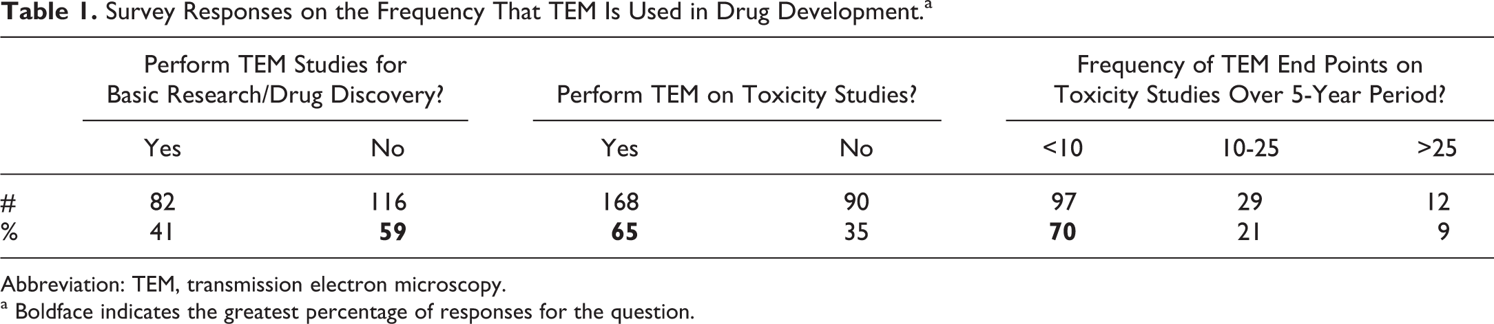
Abbreviation: TEM, transmission electron microscopy.
aBoldface indicates the greatest percentage of responses for the question.
The most common objective by far for pursuing TEM evaluations was to characterize the ultrastructural morphology of a light microscopic finding (96%; Figure 1). Multiple responses could be selected for objectives as to why TEM was performed, but only 5 of 138 responses chose another objective without also choosing “characterizing a light microscopy finding.” The second most frequent objective was to investigate pathogenesis or a hypothesis (65%), but only 4 respondents selected this in the absence of also selecting the objective of characterizing a light microscopy finding. Twenty-six (19%) respondents indicated a TEM study was necessary to fulfill a regulatory agency request or requirement, but this was selected as the sole objective by only one respondent. The most infrequent objective for pursuing TEM analysis was to establish a no observable effect level/no observable adverse effect level (NOEL/NOAEL) for a compound-related ultrastructural finding, with only 12 (9%) respondents.
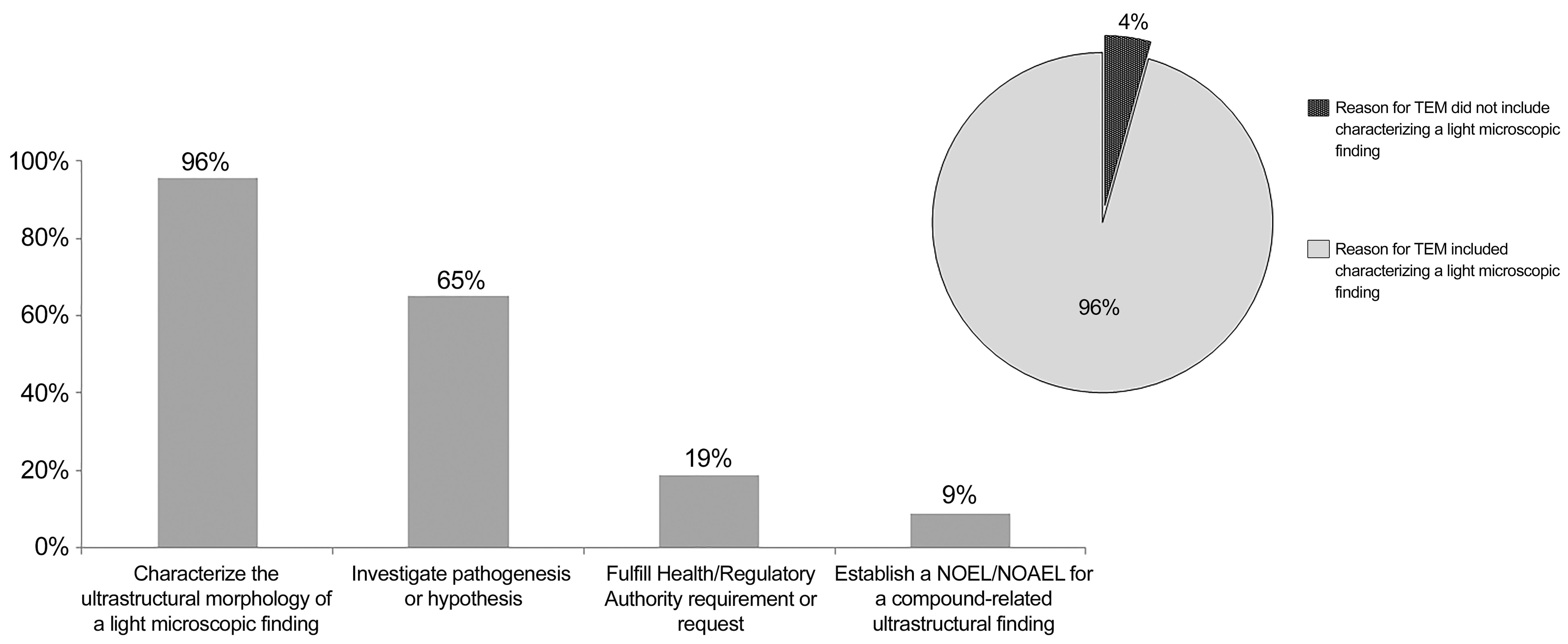
Bar chart illustrating the percentage of respondents indicating key reasons for pursuing TEM analysis on toxicity studies.
Specific Perspectives on Technical Aspects of Performing TEM Studies
Most (88%) respondents did not include routine prospective tissue collection for TEM in standard general toxicity study protocols. The respondents that did collect for possible ultrastructure evaluation as standard protocol almost exclusively collected liver (100%) and kidney (94%). Lung was mentioned twice (12%) as a tissue that was prospectively collected, while heart and spleen were both mentioned once. One respondent indicated that all tissues in toxicity studies were routinely fixed from 1 of 10 rats per treatment group using whole-body perfusion fixation, and that in 4-week dog toxicity studies, liver, lung, and kidney were fixed by immersion for possible ultrastructural evaluation as a protocol default. Another respondent specified that routine collection of liver and kidney for TEM evaluation was limited to chronic studies.
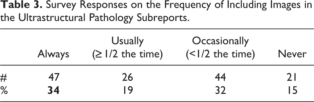
When considered independently, archived study materials would always include the signed final ultrastructural pathology report, with archived specimens primarily including blocks (86%), followed by slides with semi-thin sections (63%), grids with ultrathin sections (60%), digital images (60%), wet tissues (46%), digital or printed observation logs (41%), and printed images (37%; Table 2). When combined, the archived specimens were mainly blocks and slides (61%), followed by blocks, slides, and grids (51%). Only 6% of respondents archived all named TEM study materials. Most respondents (85%) included TEM images in their TEM reports but not always as the standard procedure, with only 34% of respondents stating that images are always included in the reports (Table 3).
Survey Responses on Archiving Practices for TEM Study Materials.a
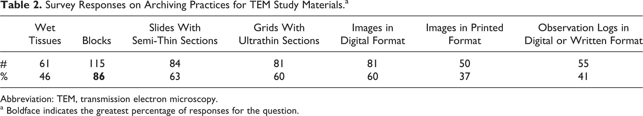
Abbreviation: TEM, transmission electron microscopy.
a Boldface indicates the greatest percentage of responses for the question.
Survey Responses on the Frequency of Including Images in the Ultrastructural Pathology Subreports.
Specific Perspectives on TEM Studies Performed in GLP-Compliant Studies
Most (79%) respondents indicated that they perform TEM studies as a part of GLP-compliant nonclinical toxicity studies (Table 4). There was variability in the responses regarding how frequently the TEM processing and evaluation were also GLP compliant, with 27% of respondents suggesting that TEM was always performed in a GLP-compliant laboratory for GLP studies, while 38% of respondents stated that less than half of TEM studies were also GLP compliant and 17% respondents stating that none of the TEM studies were GLP compliant, and were therefore considered a GLP exception on GLP-compliant studies. Only 8% of respondents were challenged by Health/Regulatory Authorities due to the TEM portion of a study not being GLP compliant; 2 respondents commented that the studies had to be repeated and performed GLP. Interestingly, GLP-compliant TEM studies were not peer reviewed for most respondents (42%) and were only consistently peer reviewed for 18% respondents (Table 5).
Survey Responses on the Incorporation of TEM in General Toxicity Studies That Will Support Regulatory Submissions.a
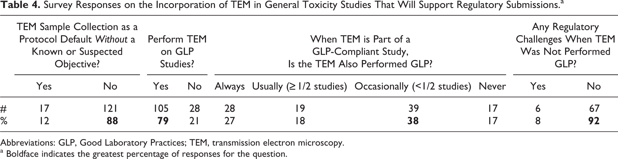
Abbreviations: GLP, Good Laboratory Practices; TEM, transmission electron microscopy.
a Boldface indicates the greatest percentage of responses for the question.
Survey Responses on the Frequency of Pathology Peer Reviews for TEM Studies.a
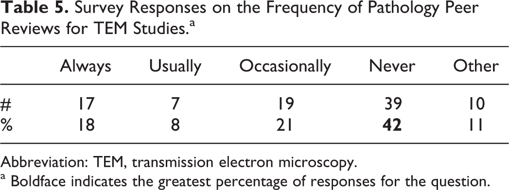
Abbreviation: TEM, transmission electron microscopy.
aBoldface indicates the greatest percentage of responses for the question.
According to 27% of respondents, the time spent performing a GLP-compliant TEM study was not any longer compared to a non GLP-compliant TEM study, while for a few (9%) respondents, the time was considered to be twice as long (Table 6). For most (78%) respondents, the opinion was that the scientific value of a non-GLP-compliant TEM study was equivalent to a GLP-compliant TEM study.
Survey Responses on Impact of Performing TEM Studies GLP or Non-GLP.a
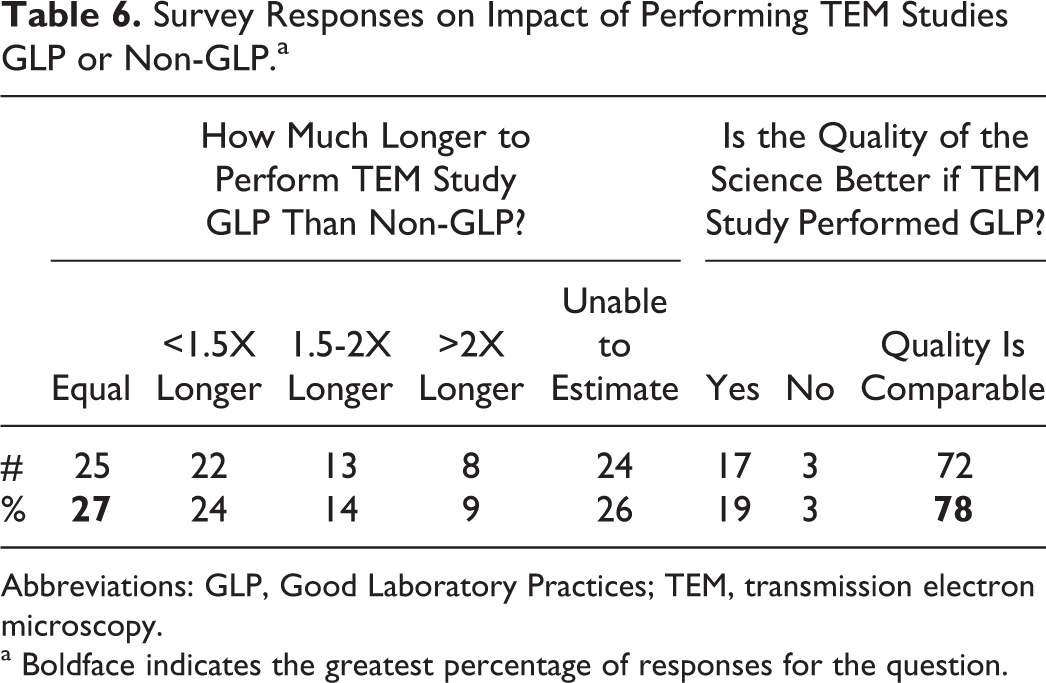
Abbreviations: GLP, Good Laboratory Practices; TEM, transmission electron microscopy.
a Boldface indicates the greatest percentage of responses for the question.
Specific Perspectives on Regulatory Aspects of Performing TEM Studies
Regardless of GLP status, the majority of respondents who have performed TEM for regulatory submissions included a written TEM subreport. These reports differ from the routine light microscopy evaluations and postmortem report as the TEM reports often include images and do not routinely include individual animal data tables since the data are not typically inputted into a laboratory information management system. An example of a TEM report that was submitted to the United States Food & Drug Administration (FDA) to support the approval of a New Drug Application is included in Supplemental Appendix A.
Working Group “Points to Consider” For Use of Ultrastructural Pathology in Nonclinical Toxicology Studies
This survey confirmed that TEM remains an essential tool for complementing toxicologic pathology evaluations in nonclinical studies but is infrequently utilized due to lack of need, access, experience, and cost and time constraints. Key points gleaned from the survey and from the perspectives of the Working Group members are: Transmission electron microscopy evaluations are rarely necessary but are certainly not obsolete or replaceable because it provides an exact diagnosis for certain subcellular pathologic changes that cannot be accurately characterized otherwise. Oftentimes, such a specific diagnosis leads to a better understanding of potential mechanism of toxicity and toxicologic assessment. Ultrastructural evaluations still provide a superior method for confirming the process underlying light microscopic findings, including but not limited to identifying the underlying cause for inclusion bodies (dilated rough endoplasmic reticulum ultrastructurally; Figure 2) and vacuolization (phospholipidosis ultrastructurally; Figure 3). Therefore, access to a quality TEM facility is beneficial and may be critical to meeting preclinical safety assessment needs of a program. Responses to this survey support that for GLP-compliant toxicity studies, it is appropriate to take a GLP-exception if TEM analysis is conducted non-GLP. Transmission electron microscopy is generally performed on only a subset of tissues/animals to qualitatively characterize pathology findings and aid interpretation of light microscopic findings, which are the gold standard to determine severity, incidence, and the NOEL/NOAEL. Therefore, working within the spirit of GLP but not full GLP compliance for the TEM portion of a GLP study is considered sufficient to maintain scientific integrity. Study materials submitted to archive include reports including protocols, blocks, and semi-thin (1 µm) slides, sample identification forms, observation logs (if any), and printed or digital images. Including TEM images in reports is a common practice but does not need to be a standard procedure. Survey results suggest that peer reviews for ultrastructural studies are not routinely performed. Pathology peer review of semi-thin sections by light microscopy or stained grids by TEM is cumbersome and not recommended. Formal or informal peer review of digital or printed images by a second pathologist is recommended by the Working Group. An alternative to peer review is to include representative images (as figures) of the ultrastructural observations which support the pathologist’s interpretation in the final report. This ensures a reviewer can always seek a “second opinion.” The infrequent practice of ultrastructural pathology peer reviews may be why a large number of survey respondents include TEM images in submitted reports, despite this not being a recommended practice for other forms of pathology images (eg, Hematoxylin & Eosin (H&E), Immunohistochemistry (IHC)). It is rare to include TEM as a protocol default on nonclinical toxicity studies if ultrastructural characterization is not a known or suspected objective. This Working Group does not recommend collection of TEM samples as a routine protocol default without an a priori data-driven rationale. Thus, post-fixing formalin-fixed tissues in a fixative appropriate for TEM processing may be required in cases where light microscopy findings on the current study suggest further characterization by TEM may be helpful. Post-fixing formalin-fixed tissue often produces adequate specimen quality, though this can be highly dependent upon the tissue, species, and type of ultrastructural change being evaluated. A notable exception is with mitochondria where delayed formalin fixation can cause artifactual changes which can hinder close evaluation of these organelles. With preassigned tissues being collected for TEM in general toxicology studies, acceptable sample collection and fixation can be obtained, with necropsy regimens altered to reach the balance of obtaining the routine specimens, data, and properly collected samples for ultrastructural evaluation (ie, rapid collection, chilled fixative, appropriate sample thickness, optimizing perfusion by fixative). The objective for conducting TEM for a large percentage of respondents was for ultrastructural characterization of light microscopic findings. Knowing the nature of the light microscopic change in advance of the proposed ultrastructural study is important in determining the proper sampling and management to optimize the opportunity of capturing the lesion and justifying the expenditure of the resources. In some instances (immunogold TEM for localization; or efficacy studies), pursuing ultrastructural evaluations in the absence of light microscopy findings may be of value, but it is not recommended by this Working Group for routine nonclinical toxicity studies. Generally, only a limited number of representative animals are evaluated from select dose groups to qualitatively characterize a change, and examination of a few concurrent study controls lacking the finding is often performed. Transmission electron microscopy is not considered a practical method to determine the incidence or severity of a finding across dose groups. Therefore, the Working Group also does not recommend establishing NOEL or NOAELs on TEM evaluations given the limited amount of tissue and the limited number of animals that can be reasonably assessed ultrastructurally, although a small percentage of respondents had used ultrastructural evaluation for this purpose. Electron microscopy findings can, however, be used to understand the potential adversity of the light microscopic findings (eg, lack of degenerative changes in organelles). Digital imaging in electron microscopy has provided great benefit, increasing ease of imaging and archiving and providing more rapid collection of an extensive subset of images from each grid. In preclinical safety, the decreased turnaround time and fewer required resources is crucial for continued investment by organizations in maintaining TEM capabilities. A pathologist should be consulted in study design when TEM evaluation for ultrastructural evaluation is intended. The majority/plurality of respondents perform TEM non-GLP and consider the most important consideration to be maintaining high-quality technical expertise and scientific accuracy. With an increased trend for companies to reduce or eliminate internal GLP TEM capabilities, access to GLP TEM is more limited. Access to high-quality non-GLP facilities with the necessary technical expertise should supersede GLP status, when not all factors can be met.
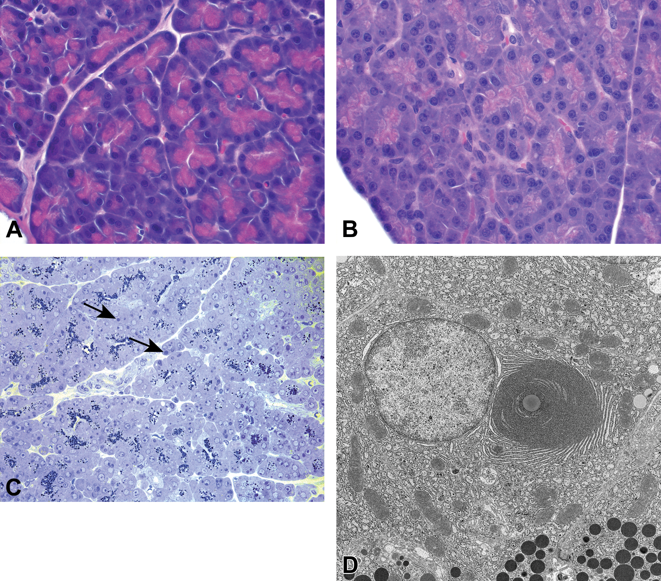
A, Normal exocrine pancreas from a vehicle-treated control rat, H&E. B, Test article–related light microscopy finding of basophilic cytoplasmic inclusions in pancreatic acinar cells, rat, H&E. C, Toluidine blue 1-µm-thick section of pancreatic acinar cells revealing zymogen granule depletion and cytoplasmic inclusion bodies (arrows), rat. D, A concentric membranous body of rough endoplasmic reticulum (RER) is encircling a lipid droplet; RER cisternae are dilated with electron-dense deposits, rat.
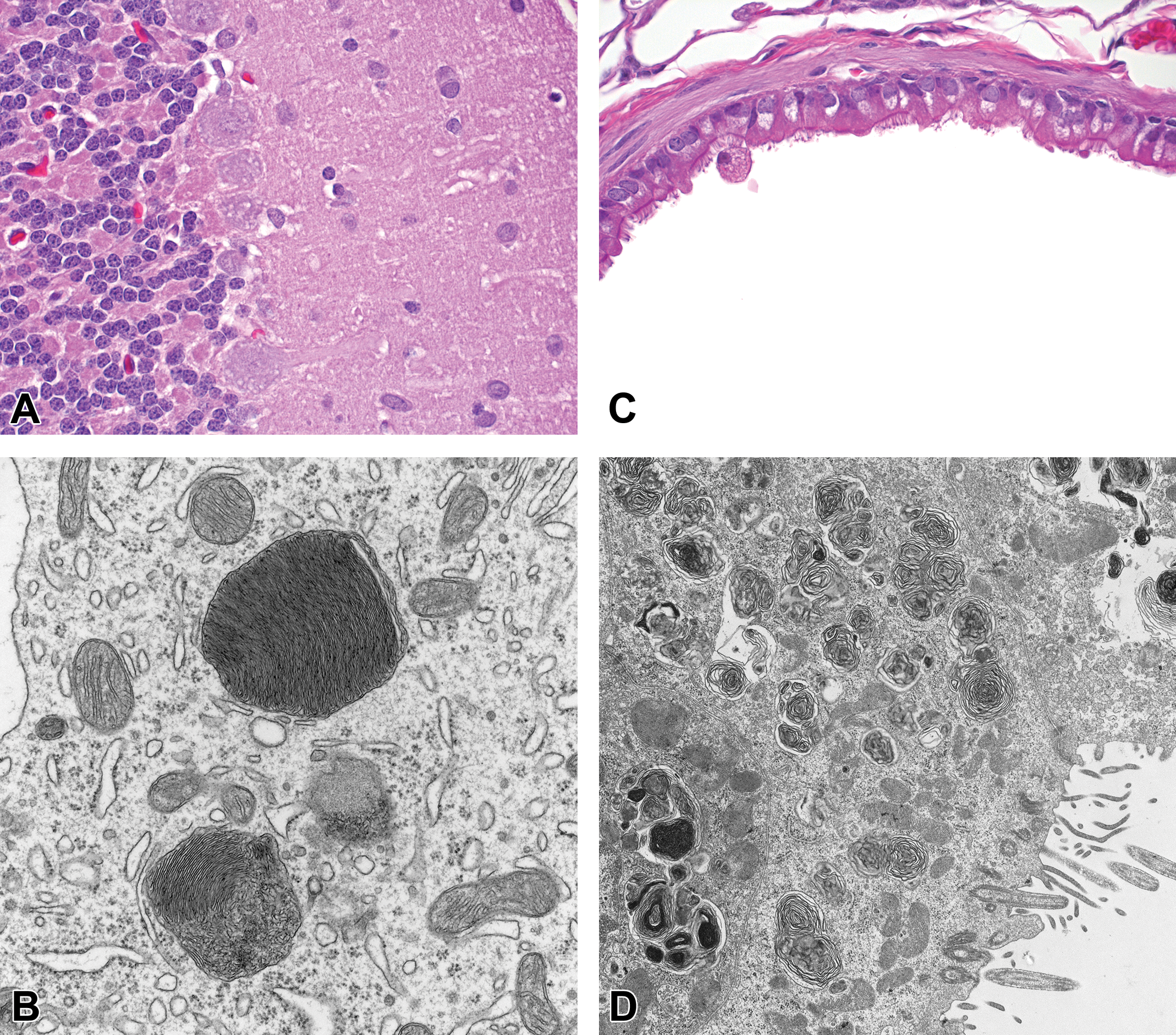
A, Cerebellum from a rat with vacuoles noted in the cytoplasm of Purkinje cells, H&E. B, Transmission electron microscopy of the Purkinje cells show profiles of lysosomal lamellar bodies (LLB) consistent with phospholipidosis. C, Light microscopic detection by H&E staining of test-article related vacuolization throughout the cytoplasm of bronchiolar epithelial cells in this mouse was also consistent with phospholipidosis when evaluated ultrastructurally (D).
Conclusions
Transmission electron microscopy evaluations are rarely necessary but can often complement early drug discovery efforts and nonclinical safety assessments on regulated toxicity studies when further characterization of a light microscopy finding is required. In some instances, TEM is still the gold standard for confirming certain processes (eg, glomerular pathology, increase smooth endoplasmic reticulum (SER) in hepatocytes/enzyme induction) and is certainly not obsolete or replaceable. However, the survey results and the Working Group’s perspective is that access to TEM facilities has reduced over the past few decades, especially GLP-compliant TEM laboratories, with many organizations closing TEM facilities in lieu of replacing old equipment. A heavier reliance on outsourcing or collaborating with academic facilities for TEM support can also influence the decision to perform the TEM processing and evaluations in a non-GLP-compliant manner. The survey did not address future innovations, but it is the opinion of this Working Group that the frequency of TEM use may increase with development of new therapeutic modalities and delivery methods (eg, nanotechnology). In chemical safety assessment, TEM was already used to evaluate the tissue response of the lung to exposure of nanoparticles (multiwalled carbon nanotubes) 2 or the tissue deposition of inhaled TiO2. 3 In addition, novel therapeutics and/or delivery may also result in increased interest by regulatory agencies for additional derisking at the subcellular level due to less understanding of mechanisms of toxicity. In addition, TEM has yet to be replaced by other techniques, including high-throughput genetic profiling, metabolomics, and immunohistochemistry, for a variety of safety issues including mitochondrial toxicity and phospholipidosis. Even with development of more predictable and detailed in vitro toxicity assessments, many toxicity findings still require TEM to confirm the association of findings to an actual subcellular impact, at least while additional tools or biomarkers are being validated. The key challenge, however, is that interpretation of ultrastructural images requires training and experience that many pathologists are no longer obtaining either formally in training programs or through practice in their careers. Transmission electron microscopy remains a valuable tool in preclinical drug safety and in some cases is still the only way to characterize a toxicity or understand the impacts of new therapeutics.
Supplemental Material
Supplemental Material, DS1_TPX_10.11770192623319835170 - Scientific and Regulatory Policy Committee Points to Consider*: Review of Scientific and Regulatory Policy Committee Points to Consider: Review of Current Practices for Ultrastructural Pathology Evaluations in Support of Nonclinical Toxicology Studies
Supplemental Material, DS1_TPX_10.11770192623319835170 for Scientific and Regulatory Policy Committee Points to Consider*: Review of Scientific and Regulatory Policy Committee Points to Consider: Review of Current Practices for Ultrastructural Pathology Evaluations in Support of Nonclinical Toxicology Studies by Natalie D. Keirstead, Evan B. Janovitz, James T. Meehan, Bruce E. LeRoy, John R. Megill, Richard A. Peterson, Regis G. Masson and Heike A. Marxfeld in Toxicologic Pathology
Supplemental Material
Supplemental Material, DS2_TPX_10.11770192623319835170 - Scientific and Regulatory Policy Committee Points to Consider*: Review of Scientific and Regulatory Policy Committee Points to Consider: Review of Current Practices for Ultrastructural Pathology Evaluations in Support of Nonclinical Toxicology Studies
Supplemental Material, DS2_TPX_10.11770192623319835170 for Scientific and Regulatory Policy Committee Points to Consider*: Review of Scientific and Regulatory Policy Committee Points to Consider: Review of Current Practices for Ultrastructural Pathology Evaluations in Support of Nonclinical Toxicology Studies by Natalie D. Keirstead, Evan B. Janovitz, James T. Meehan, Bruce E. LeRoy, John R. Megill, Richard A. Peterson, Regis G. Masson and Heike A. Marxfeld in Toxicologic Pathology
Supplemental Material
Supplemental Material, DS3_TPX_10.11770192623319835170 - Scientific and Regulatory Policy Committee Points to Consider*: Review of Scientific and Regulatory Policy Committee Points to Consider: Review of Current Practices for Ultrastructural Pathology Evaluations in Support of Nonclinical Toxicology Studies
Supplemental Material, DS3_TPX_10.11770192623319835170 for Scientific and Regulatory Policy Committee Points to Consider*: Review of Scientific and Regulatory Policy Committee Points to Consider: Review of Current Practices for Ultrastructural Pathology Evaluations in Support of Nonclinical Toxicology Studies by Natalie D. Keirstead, Evan B. Janovitz, James T. Meehan, Bruce E. LeRoy, John R. Megill, Richard A. Peterson, Regis G. Masson and Heike A. Marxfeld in Toxicologic Pathology
Supplemental Material
Supplemental Material, DS4_TPX_10.11770192623319835170 - Scientific and Regulatory Policy Committee Points to Consider*: Review of Scientific and Regulatory Policy Committee Points to Consider: Review of Current Practices for Ultrastructural Pathology Evaluations in Support of Nonclinical Toxicology Studies
Supplemental Material, DS4_TPX_10.11770192623319835170 for Scientific and Regulatory Policy Committee Points to Consider*: Review of Scientific and Regulatory Policy Committee Points to Consider: Review of Current Practices for Ultrastructural Pathology Evaluations in Support of Nonclinical Toxicology Studies by Natalie D. Keirstead, Evan B. Janovitz, James T. Meehan, Bruce E. LeRoy, John R. Megill, Richard A. Peterson, Regis G. Masson and Heike A. Marxfeld in Toxicologic Pathology
Footnotes
Acknowledgments
The authors would like to thank Takayuki Tsuchiya, Sabu Kuruvilla, and reviewers from the SRPC and Executive STP Committees for their critical comments on this article.
Declaration of Conflicting Interests
The author(s) declared no potential, real, or perceived conflicts of interest with respect to the research, authorship, and/or publication of this article.
Funding
The author(s) received no financial support for the research, authorship, and/or publication of this article.
Supplemental Material
Supplemental material for this article is available online.
References
Supplementary Material
Please find the following supplemental material available below.
For Open Access articles published under a Creative Commons License, all supplemental material carries the same license as the article it is associated with.
For non-Open Access articles published, all supplemental material carries a non-exclusive license, and permission requests for re-use of supplemental material or any part of supplemental material shall be sent directly to the copyright owner as specified in the copyright notice associated with the article.