
Abstract
Information on background changes in the ocular tissues of rabbits (Oryctolagus cuniculus), a common species employed in ophthalmic toxicology studies, is sparse. This complicates interpretation of changes in light of small sample sizes on any single study. The purpose of this publication is to document the interstudy incidence of spontaneous or iatrogenic changes occurring in eyes of control rabbits. Photomicrographs of select lesions are provided. The data set was derived from a total of 54 studies conducted over an eleven-year period at Alcon Research Ltd., a Novartis Division, which featured topical ocular and contact lens routes of administration. It includes a total of 1,222 pigmented and albino New Zealand rabbits and a total of 2,084 eyes which were either untreated or treated with innocuous control articles. There were no noteworthy differences across routes of administration. Changes in anterior segment ocular and adnexal tissues were more common than in posterior segment ocular tissues. Overall, mononuclear cell infiltration was the most common finding. The retina was the posterior tissue most commonly observed with spontaneous changes, with folds and rosettes being the most common retinal finding. Retinal changes were more common in albino as compared to pigmented rabbits. Understanding the incidence and characteristics of spontaneous ocular lesions facilitates accurate and consistent diagnosis and data interpretation.
The number of animals included on toxicology studies is necessarily limited. Reasons for this limitation range from matters of logistical or execution feasibility to emphasis on the animal welfare 3Rs, with reduction of animal use being a main thrust in this area. An understanding of the incidence of nontest article–related spontaneous or iatrogenic histopathology lesions would be an aid in accurate data interpretation in light of small group numbers. Characterization of these findings, illustration via photography, and use of standardized terminology can encourage consistent diagnoses and descriptions among toxicologists and pathologists.
While the rabbit may not be a suitable test system for some ophthalmic products such as biologics (Zuch de Zafra et al. 2017), its utility in toxicology studies of other ophthalmic pharmaceuticals and medical devices is widely accepted. The normal histologic anatomy of the rabbit eye has been the subject of reports for several decades (Björkman, Nicander, and Schantz 1960; Davis 1929; Prince 1964; Sheppard 1962; Williams 2013). Reports on background lesions in the rabbit eye which are detected primarily via in vivo techniques such as biomicroscopic and ophthalmoscopic examination are also available (Munger, Langevin, and Podval 2002; Holve, Mundwiler, and Pritt 2011; Jeong et al. 2005). However, data in the scientific literature are limited with regard to the incidence of background histopathology lesions in the laboratory rabbit and even more limited with regard to lesions occurring in the eye. Schafer and Render (2013a, 2013b) discuss the histologic preparation of ocular tissues and describe findings which can occur following administration of certain classes of chemicals as a result of certain types of procedures (iatrogenic) or as spontaneous phenomena. The extensive history of research and development in the areas of ophthalmic pharmaceuticals and medical devices within this laboratory affords the opportunity to review an abundance of interstudy histopathology data to determine the frequency at which spontaneous or iatrogenic findings may be observed. This is an aid to toxicologists and pathologists seeking to determine whether a lesion observed at a low frequency within a discrete study may be a spontaneous, background finding or a low-level signal of test article toxicity. A robust database of background lesions will help overcome the barriers of small sample size in individual toxicology studies by leveraging cumulative historical data. Having a reference point for the incidence of background lesions can limit the number of occasions where an “equivocal test article relationship” designation is assigned to a finding.
Material and Method
Retrospective analysis and tabulation of lesions occurring in the tissues of control animals from topical ocular and contact lens wear studies was performed. Data were compiled from 54 studies over an eleven-year period, 2005 to 2016. The in-life phases of the studies occurred in the laboratories of Alcon Research Ltd., a Novartis Division (Fort Worth, TX). Studies ranged from 1 day to 9 months in duration. Animals included in the tabulation are those which served as untreated and vehicle controls as well as marketed product controls. Animals which served as vehicle controls were only included if it was judged that the vehicle used would generally be accepted as innocuous (e.g., vehicles composed of ingredients listed in compendia, designated as generally recognized as safe, or widely used in marketed pharmaceuticals and/or medical devices). Included as marketed product controls were lubricating eye drops and contact lens disinfection solutions. These products have a long history of safe use in the market and may commonly be used as comparators in studies of products under development. This resulted in a total population of 1,222 rabbits. Both eyes were not evaluated in all rabbits in all studies, depending on study design. However, considering that most studies included bilateral (OU) design, the total number of eyes evaluated was 2,084.
It is important to understand not only those lesions which occur completely spontaneously but also those which may occur from minor noninvasive procedures. Therefore, separate tabulations are provided for findings in untreated, vehicle, and marketed product control–treated eyes. Results from studies featuring soft contact lens wear are tabulated separately, given the potential of these treatments to elicit different types of minor microscopic changes, as compared to topical ocular treatment. Of the 54 studies referenced (Table 1), 39 are topical ocular studies and 15 are contact lens studies. Of the topical ocular studies, 12 studies used New Zealand White (albino) rabbits (200 rabbits, 352 eyes) and 27 studies used New Zealand White × New Zealand Red (pigmented) rabbits (536 rabbits, 1,006 eyes). All 15 contact lens studies used albino rabbits (486 rabbits, 726 eyes).
Study Demographics.
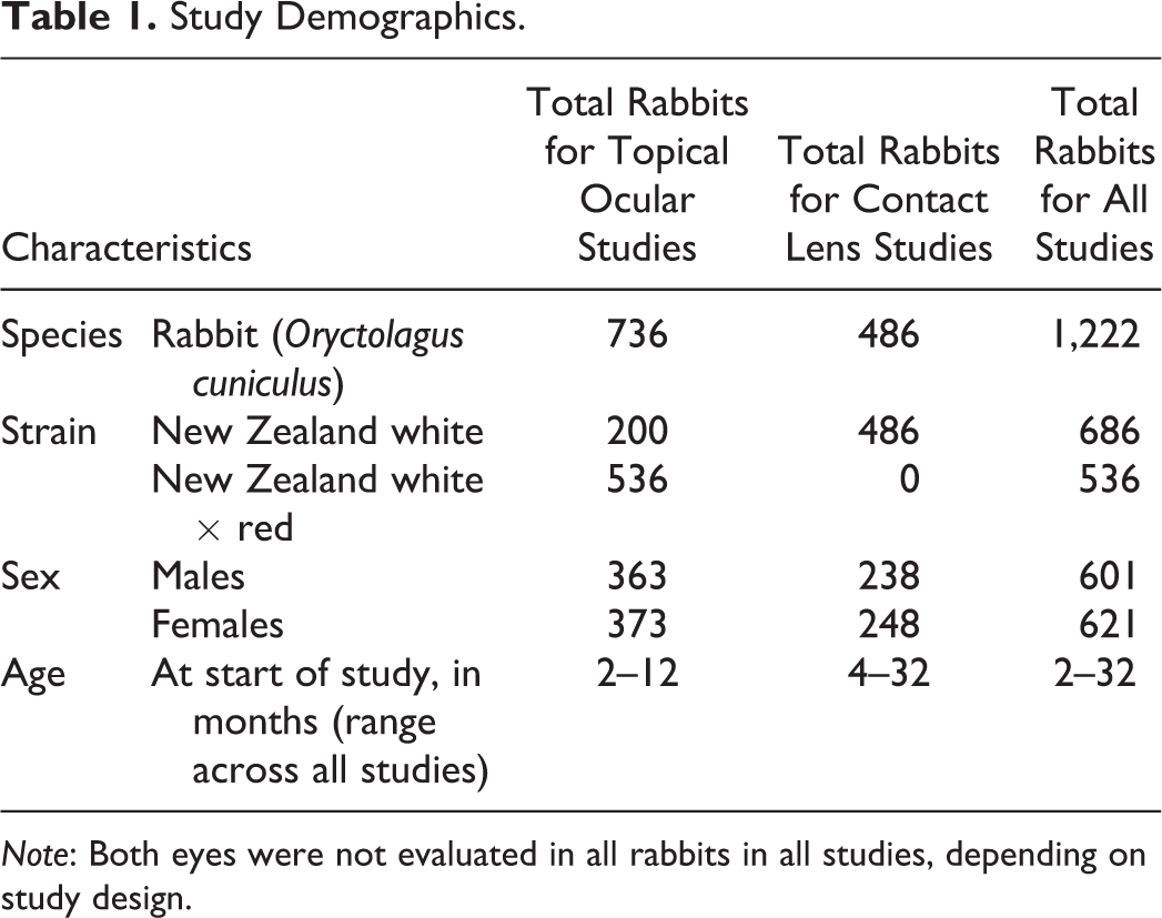
Note: Both eyes were not evaluated in all rabbits in all studies, depending on study design.
During in-life phases of the studies in this data set, animals were individually housed in standard laboratory caging. Cage pan liners were replaced three times weekly and the animals were moved into sanitized caging at least every two weeks, per facility standard operating procedures. Rabbits were maintained on PMI Nutrition International (Brentwood, MO)-certified rabbit feed 5325 and had ad libitum access to fresh potable water that was tested for contaminants on a biannual basis. The animals were maintained in a controlled environment, between 64°F and 72°F (approximately 18–22°C), with a relative humidity ≥30%. All animals in the studies were maintained on an approximate 12 hr on and 12 hr off room light cycle with the light levels set at an intensity in the range of 30 to 37 foot candles, which is standard for rabbits in the laboratory environment. It would be expected that light intensity at the cage levels varied some for individual animals depending on cage location within the racks. We would not expect there to be any influence of environmental light on spontaneous ocular lesions, given the standard light levels and cycles used within the facility.
Immediately after euthanasia, ocular tissues were harvested and placed in Davidson’s solution for fixation for up to 48 hours, rinsed with water for 15 min, and then preserved in 10% neutral buffered formalin. Tissues were transferred to the protocol-designated pathology laboratory. Pathology laboratories utilized were Experimental Pathology Laboratories Inc. (Sterling, VA), Seventh Wave Laboratories, LLC (Chesterfield, MO; Maryland Heights, MO), and Vet Path Services Inc. (Mason, OH). Tissues were embedded in paraffin, sectioned to glass slides, and stained with hematoxylin and eosin. The most common sectioning method included separation of the globe into three separate paraffin blocks: one containing the temporal portion of the globe, another including the central superior to inferior portion of the globe to include the optic nerve and most anterior curvature of the cornea, and a third section with the nasal portion of the globe. For the majority of studies (52 of 54) in this data set, one slide was generated from each of these three blocks (3 slides per eye). For the remaining two studies, three slides (at 200 µm intervals) were taken from each block (9 slides per eye).
A review of the study reports determined that histopathology data for the majority of studies were tabulated on a per-eye basis. Therefore, it was possible to determine whether a given finding occurred in both eyes (bilateral) of a given animal or in only one eye (unilateral). In a few cases, however, the histopathology data were tabulated on a per-animal basis. In these cases, it was not possible to discern whether a finding occurred unilaterally or bilaterally from the pathology report and tables, so the finding was counted as a single incidence for the purposes of this document. A description of the animal population used in this research is provided in Table 1. When severity was assigned to findings, two scales were used to grade findings in the individual study pathology reports: a 4-point scale of minimal, mild, moderate, and marked or a 5-point scale of minimal, slight/mild, moderate, moderately severe, and severe. In this review of findings, mild is considered to be equivalent to slight and marked is considered equivalent to moderately severe. This enabled harmonization of scales to minimal, mild, moderate, and marked within this review (there were no severe findings). Some findings were without severity rating and others were reported as “present.”
In the interest of harmonized nomenclature, wherever possible, terminology to describe findings was translated to that proposed in the then draft manuscript from INternational HArmonization of Nomenclature and Diagnostic Criteria (INHAND) for the rodent eye (Ramos et al. 2018). However, a few of the ocular findings in these studies are not described in the rodent INHAND list.
Research Involving Animals
The tabulation of data herein was accomplished via retrospective analysis of studies conducted in the routine support of pharmaceutical and medical device product development. Therefore, no additional animals were used to acquire these data. Study protocols were reviewed, approved, and study conduct overseen by the Institutional Animal Care and Use Committee of Alcon Research Ltd. Animal care and handling was in accordance with institutional and governmental guiding principles for the use of animals, including the Alcon and Novartis Animal Welfare Policies, the United States Department of Agriculture (USDA) Animal Welfare Act and Regulations, and the National Research Council’s Guide for the Care and Use of Laboratory Animals.
Results
Lesions occurring in studies featuring topical ocular dosing are presented in Tables 2 and 3 for albino and pigmented rabbits, respectively. Lesions occurring in studies featuring contact lens wear are presented in Table 4. Photomicrographs of select lesions are cross-referenced within Tables 2 –4.
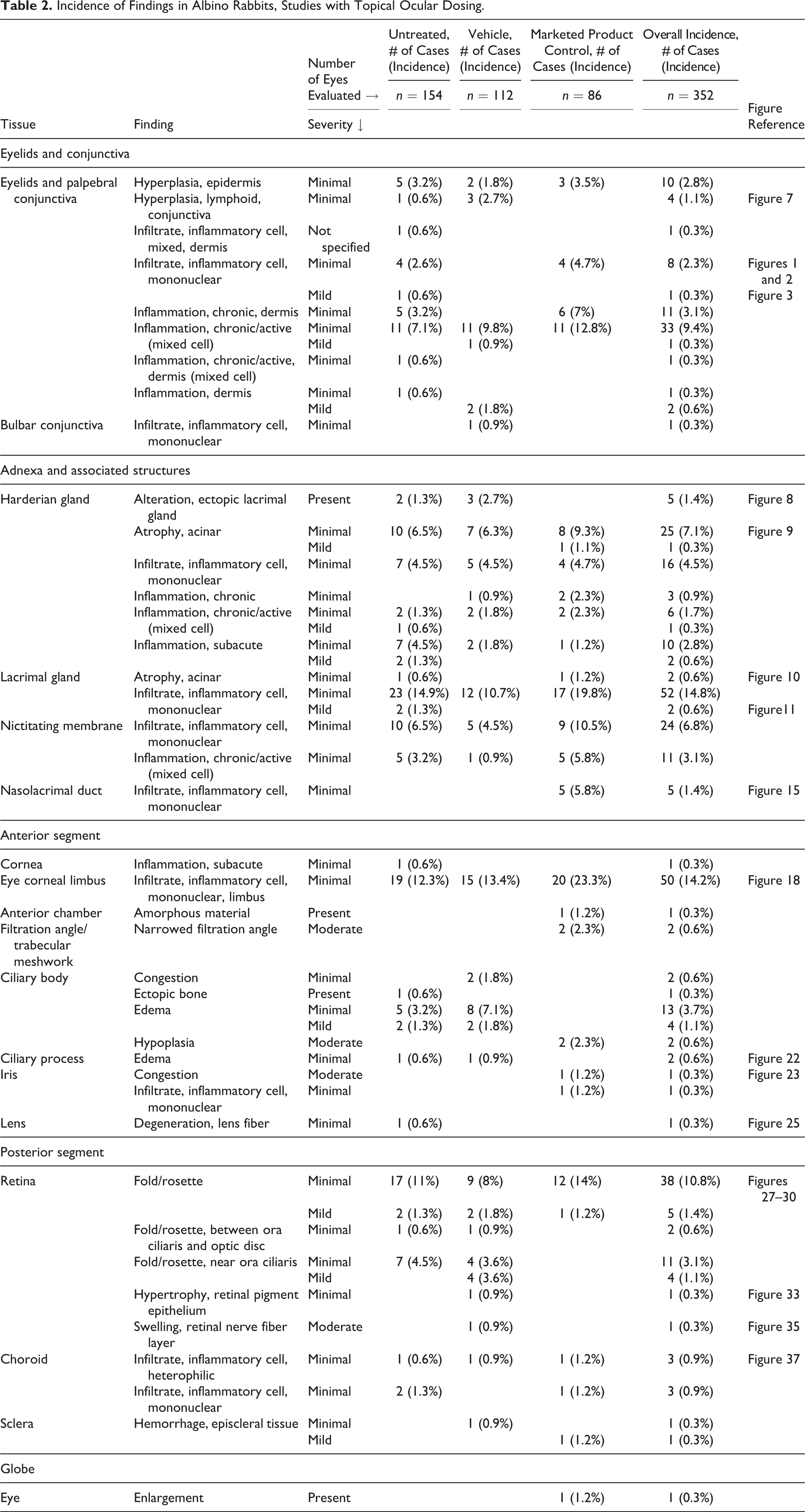
Incidence of Findings in Albino Rabbits, Studies with Topical Ocular Dosing.
Incidence of Findings in Pigmented Rabbits, Studies with Topical Ocular Dosing.

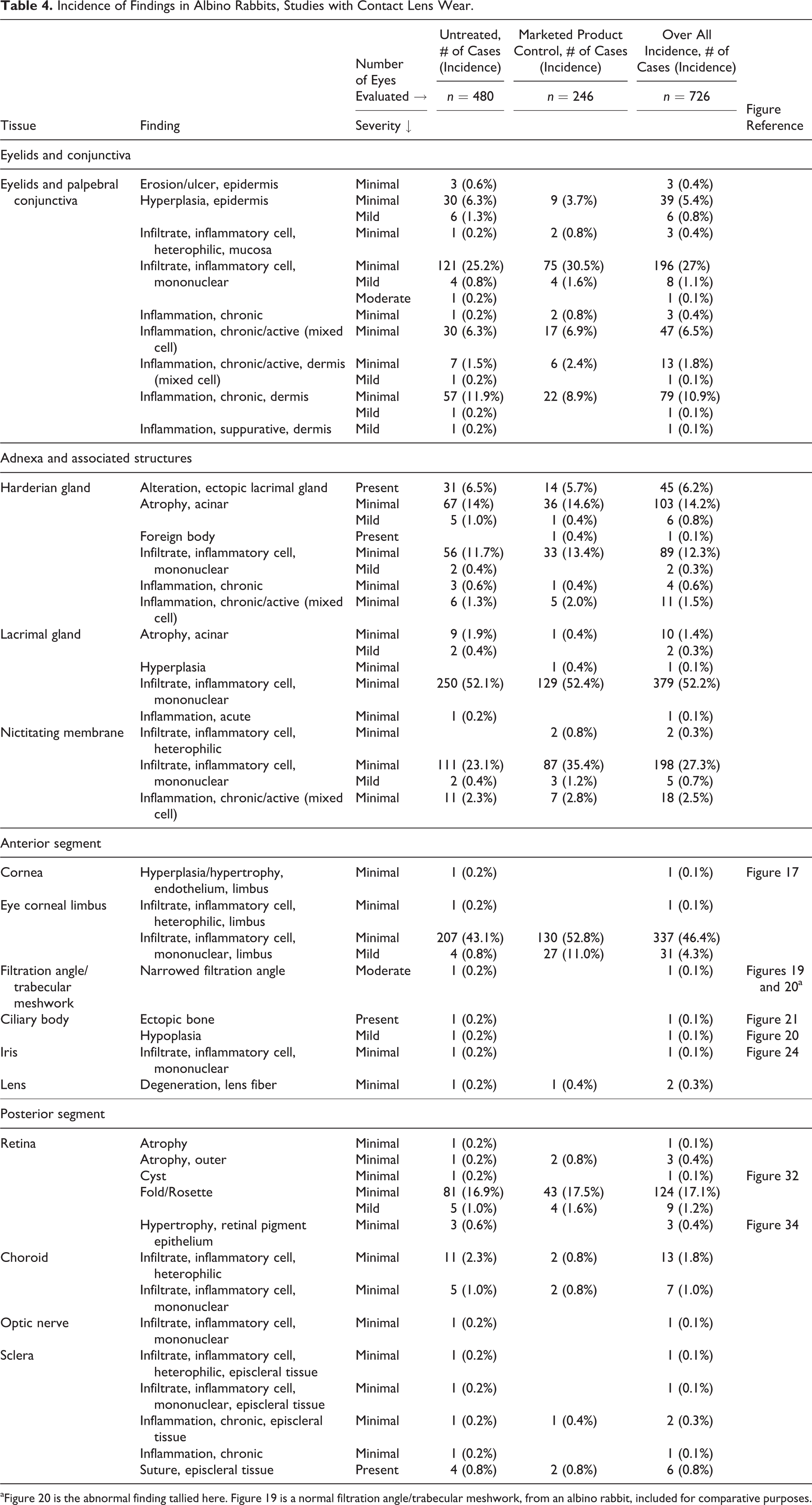
Incidence of Findings in Albino Rabbits, Studies with Contact Lens Wear.
Eyelids and Conjunctiva
Eyelids and palpebral conjunctiva
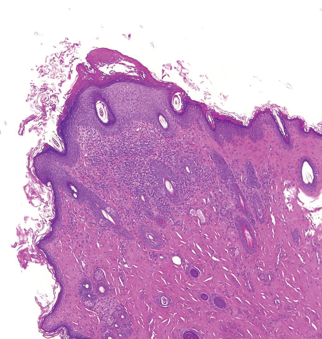
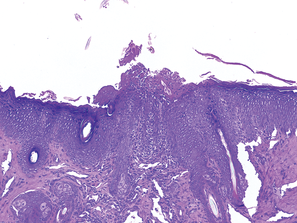
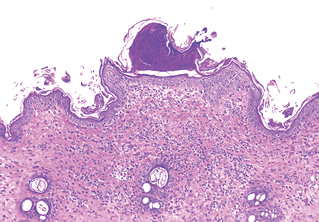
Eyelid findings were quite common, and some were seen up to an incidence of nearly 13% in albino topical ocular studies (inflammation), 14% of pigmented topical ocular studies (infiltrate), and 31% of albino contact lens studies (infiltrate). The most common findings in the eyelids across all three cohorts were inflammatory cell infiltrates and inflammation, variably characterized as minimal to mild or rarely moderate, heterophilic, mononuclear (Figures 1 –3), mixed cell (Figure 4), chronic, chronic/active, or subacute. This was localized to the palpebral conjunctiva, epidermis, the underlying dermis, or was periadnexal in the dermis. Minimal epidermal erosion and/or ulceration (Figure 5) with crusting (Figure 6), minimal to mild epidermal hyperplasia, and minimal keratinization were also occasionally recorded (<7%). Infrequent (<3%) minimal to mild lymphoid aggregates and/or lymphoid hyperplasia were also observed in the eyelids and palpebral conjunctiva. Although diagnosed as lymphoid hyperplasia, some cases are recognized as conjunctiva-associated lymphoid tissue (CALT; Figure 7).
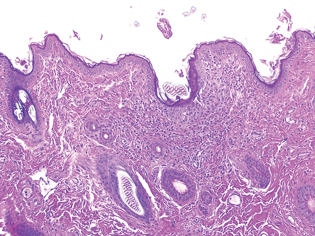
Albino rabbit topical ocular study. Right eye, eyelid, infiltrate, mononuclear cells, minimal. A focal infiltration of mononuclear inflammatory cells (lymphoid) in the superficial dermis between the basal layer of the epithelium and the follicular structures. Hematoxylin and eosin stain. Original objective 10×.
Albino rabbit topical ocular study. Right eye, eyelid, conjunctiva, infiltrate, mononuclear cells, minimal. Distinct aggregates of mononuclear inflammatory cells (lymphoid) are present subjacent to the conjunctival epithelium. Hematoxylin and eosin stain. Original objective 10×.
Albino rabbit topical ocular study. Left eye, eyelid, infiltrate, mononuclear cell, mild. A focal infiltration of mononuclear inflammatory cells (lymphoid) is in the superficial dermis, between the basal layer of the epithelium and the follicular structures, with a focal thickening of the superficial epithelium (hyperkeratosis). Hematoxylin and eosin stain. Original objective 10×.
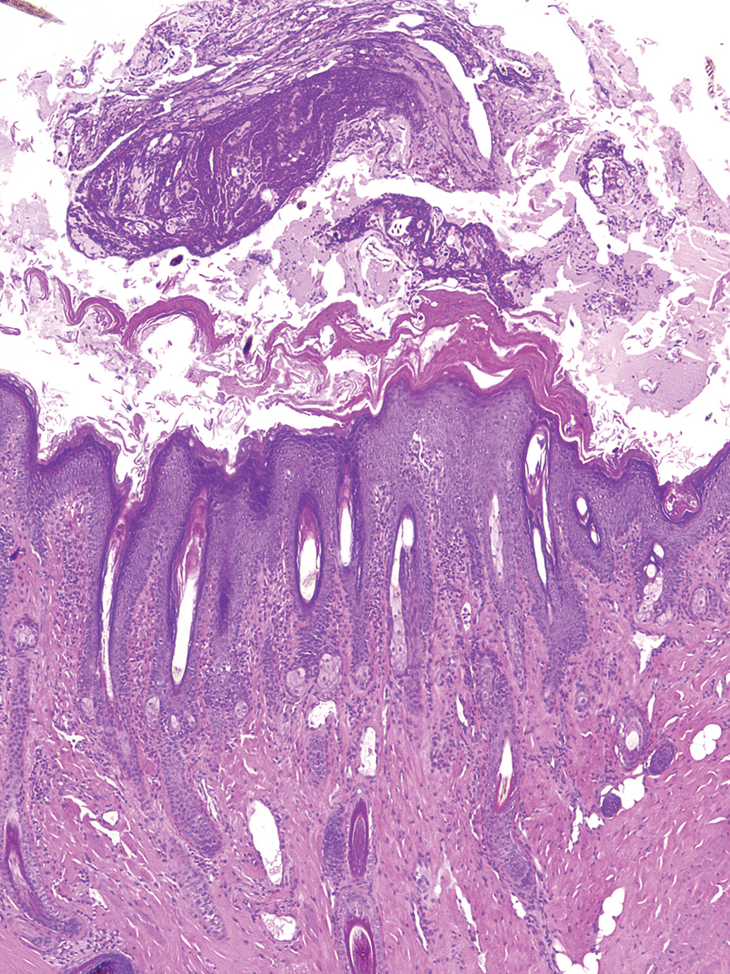
Pigmented rabbit topical ocular study. Left eye, eyelid, dermis, inflammation, mixed cell, moderate. A multifocal infiltration of mixed inflammatory cells is in the superficial dermis, between the basal layer of the epithelium and around follicular structures, with a locally extensive thickening of the superficial epithelium (hyperkeratosis) and overlying serocellular crust. Hematoxylin and eosin stain. Original objective 5×.
Pigmented rabbit topical ocular study. Left eye, eyelid, ulcer, minimal. A focal discontinuity (ulcer) in the epithelium is covered by a serocellular crust and has a mixed inflammatory cell population that extends from the crust into the contiguous superficial and deep dermis. Hematoxylin and eosin stain. Original objective 10×.
Pigmented rabbit topical ocular study. Right eye, eyelid, crust. A focal serocellular epithelial crust overlies dermis that is locally infiltrated by a mixed population of inflammatory cells, which surround the follicular structures. Hematoxylin and eosin stain. Original objective 10×.
Albino rabbit topical ocular study. Right eye, eyelid, hyperplasia, lymphoid, minimal. Although diagnosed as lymphoid hyperplasia, this is recognized as conjunctiva-associated lymphoid tissue. Distinct, roughly oval foci of lymphocytes rise above the adjacent epithelium and exhibit regions of darker (marginal) and lighter (germinal center) tinctorial qualities. Hematoxylin and eosin stain. Original objective 5×.
Bulbar conjunctiva
The only findings in the bulbar conjunctiva and/or subconjunctiva were minimal infiltration of mononuclear cells (<1%) and minimal to mild lymphoid aggregates (<2%). No bulbar conjunctival or subconjunctival lesions were reported in the albino contact lens studies.
Adnexa and Associated Structures (Harderian Gland, Lacrimal Gland, Nictitating Membrane, and Nasolacrimal Duct)
Harderian gland
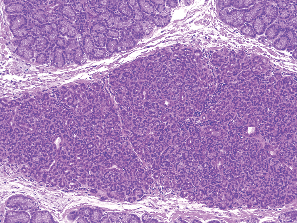
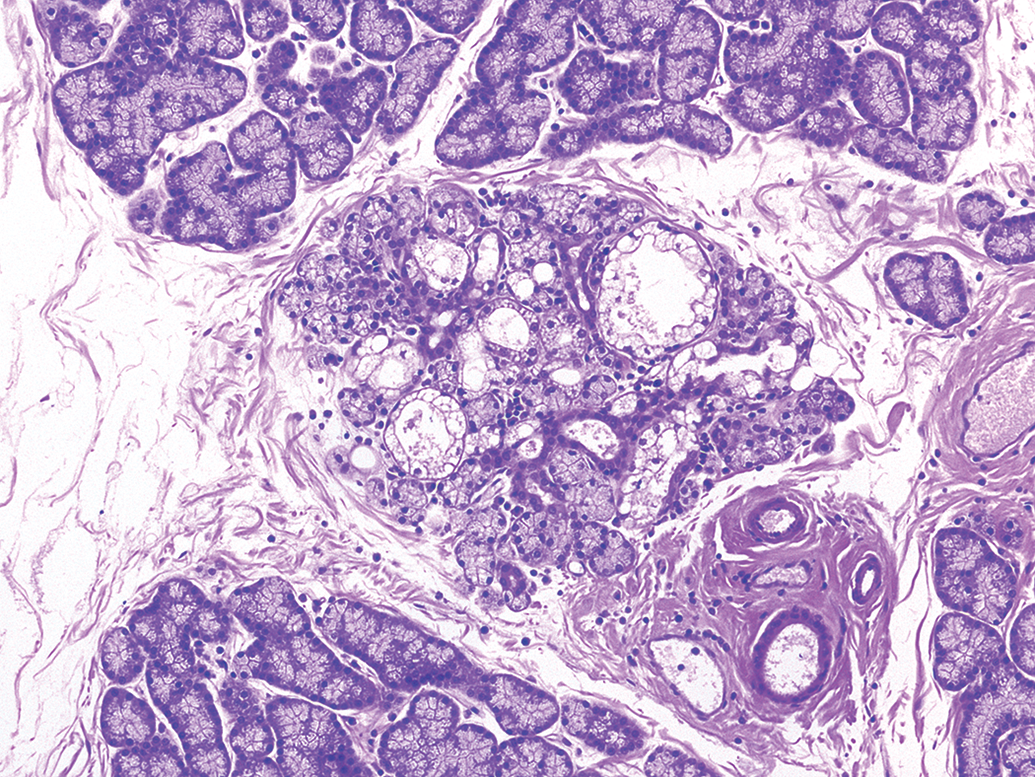
In the Harderian gland, the most common findings were ectopic lacrimal gland (up to 7%; Figure 8), minimal to mild acinar atrophy (up to 15%; Figure 9), and minimal to mild inflammatory cell infiltration/inflammation (mononuclear, subacute, chronic, chronic/active, or mixed cell, up to 13% across cohorts).
Albino rabbit topical ocular study. Left eye, Harderian gland, alteration, ectopic lacrimal gland. Small, distinct foci of lacrimal gland are present between normal Harderian gland. Hematoxylin and eosin stain. Original objective 10×.
Albino rabbit topical ocular study. Right eye, Harderian gland, atrophy with infiltrates, minimal. Normal foamy glandular epithelial cells are either markedly reduced in size or lost from the affected tissue, and the interstitium is expanded by inflammatory cells (lymphoid). Hematoxylin and eosin stain. Original objective 10×.
Lacrimal gland
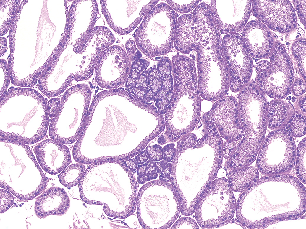
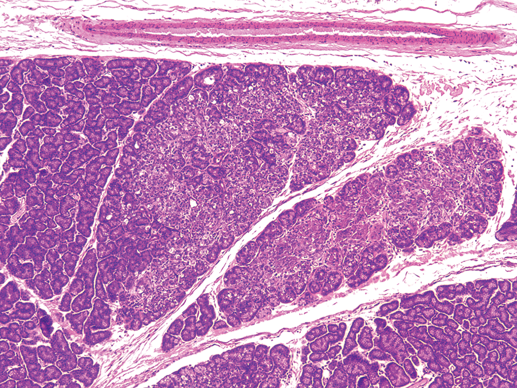
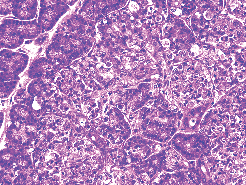
Similarly, in the lacrimal glands, minimal to mild atrophy (up to 4%; Figure 10) was observed. Mononuclear cell infiltrates (Figure 11) were a common observation (up to 52%) though almost always minimal in severity. Rare (<1%), single cases of minimal degeneration (Figure 12) and mild necrosis (Figures 13 and 14) were recorded in the pigmented rabbit topical ocular studies as were single cases of minimal hyperplasia and minimal acute inflammation in the albino contact lens studies.
Albino rabbit topical ocular study. Left eye, lacrimal gland, atrophy, minimal. A glandular lobule consists mainly of small cells with an infiltration of inflammatory cells (lymphoid) scattered throughout. Hematoxylin and eosin stain. Original objective 10×.
Albino rabbit topical ocular study. Right eye, lacrimal gland, infiltrate, mononuclear cell, mild. Aggregates of inflammatory cells (lymphoid) extend around and between vascular and ductal tissue. Hematoxylin and eosin stain. Original objective 10×.
Pigmented rabbit topical ocular study. Left eye, lacrimal gland, degeneration, minimal. A glandular lobule is mainly replaced by vacuolated and disorganized cells. An infiltration of inflammatory cells (lymphoid) is scattered throughout. Hematoxylin and eosin stain. Original objective 10×.
Pigmented rabbit topical ocular study. Right eye, lacrimal gland, necrosis, mild. Multiple glandular lobules have areas adjacent to normal tissue that have loss of normal architecture. Cells are vacuolated with pale cytoplasm and often pyknotic nuclei. Hematoxylin and eosin stain. Original objective 5× (higher magnification shown in Figure 14).
Pigmented rabbit topical ocular study. Right eye, lacrimal gland, necrosis, mild. A focus of gland on the edge of normal tissue is characterized by tissue with loss of normal architecture. Cells are vacuolated with pale cytoplasm and often have pyknotic nuclei. Hematoxylin and eosin stain. Original objective 20× (higher magnification of Figure 13).
Nictitating membrane
In the nictitating membrane, minimal mononuclear cell infiltrates (up to 11%) and chronic/active (mixed cell) inflammation (up to 6%) were recorded in the albino topical ocular cohort. Minimal to mild infiltrates were recorded at similar (up to 7%) frequency in the pigmented topical ocular cohort, along with individual, minimal severity cases (<1%) of keratinized cyst, heterophilic infiltrate, lymphoid aggregate, and lymphoid hyperplasia. Minimal or mild mononuclear infiltrate (up to 35%) was also recorded in the albino contact lens cohort. Also in the albino contact lens cohort, minimal chronic/active (mixed cell) inflammation occurred at a 2% to 3% incidence as did rare (<1%) cases of heterophilic infiltrate. A marked focal hemorrhage was observed in the nictitating membrane of a vehicle-treated eye from a pigmented rabbit. This hemorrhage was also visible during the in-life phase of the study via gross and slit lamp biomicroscopic evaluation.
Nasolacrimal duct
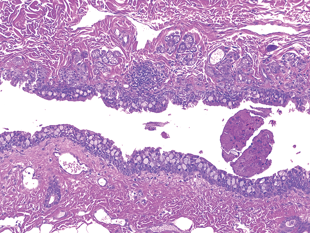
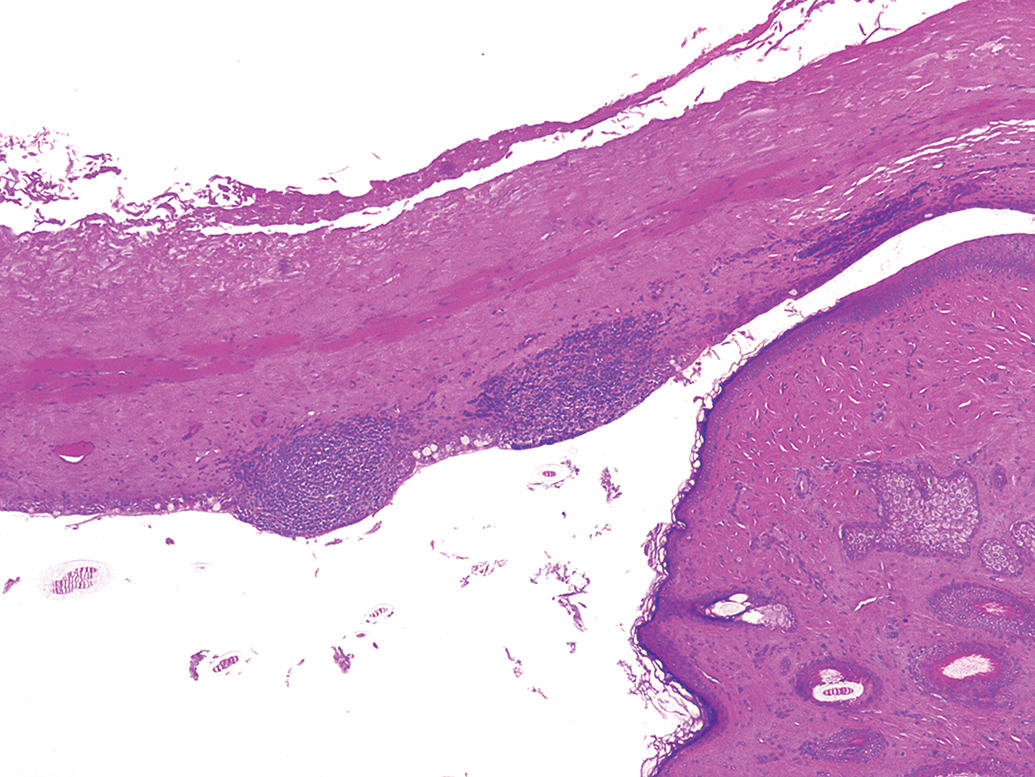
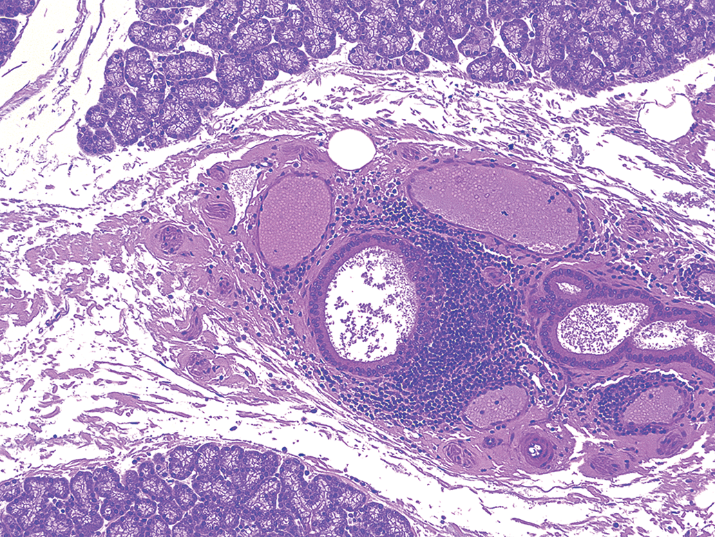
Background findings in the nasolacrimal duct were rare, with none in the contact lens cohort. In the other cohorts, cases of minimal to mild mononuclear or heterophilic inflammatory cell infiltrates were observed at up to a 6% incidence. Some cases of mononuclear inflammatory cell infiltrates were found to also have mucosa-associated lymphoid tissue (MALT) in section (Figure 15). A few cases (up to 1%) of hemorrhage were also evident (Figure 16).
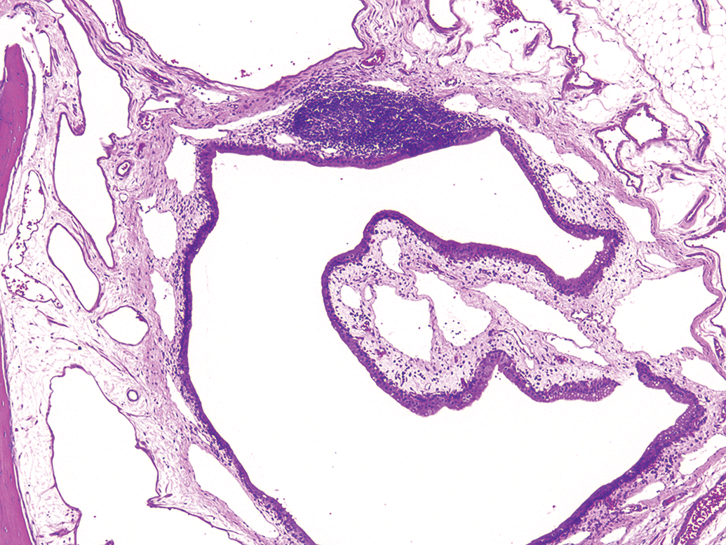
Albino rabbit topical ocular study. Regional, nasolacrimal duct, infiltrate, mononuclear cell, minimal. There are diffusely scattered mononuclear cells in the mucosa. Also, there is mucosa-associated lymphoid tissue recognized in section, characterized by a distinct, roughly oval focus of lymphocytes subjacent to the duct epithelium and exhibiting regions of darker (marginal) and lighter (germinal center) tinctorial qualities. Hematoxylin and eosin stain. Original objective 5×.
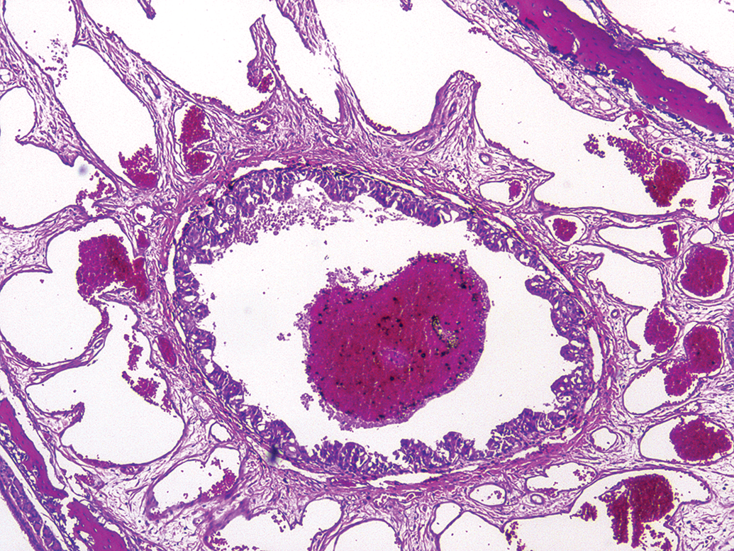
Pigmented rabbit topical ocular study. Right eye, nasolacrimal duct, hemorrhage. Large pools of erythrocytes are present in the lumen of the nasolacrimal duct and the adjacent nasal turbinate architecture. Hematoxylin and eosin stain. Original objective 5×.
Anterior Segment
Cornea
Lesions in the cornea proper were very rare (<1% in all cohorts) and consistent with previous reports (Moore, Dubielzig, and Glaza 1987) with single examples of minimal subacute inflammation (albino topical ocular), increased epithelial mitosis (pigmented topical ocular), and endothelial hyperplasia/hypertrophy (Figure 17). The most frequent lesion (three pigmented topical ocular eyes) was minimal subcorneal lymphoid aggregate.
Albino rabbit contact lens study. Left eye, cornea, endothelium, hyperplasia/hypertrophy, minimal. A small, circular focus of endothelial hyperplasia/hypertrophy is located on the endothelial surface and extends into the aqueous chamber. Hematoxylin and eosin stain. Original objective 20×.
At the corneal limbus, infiltration of mononuclear cells (Figure 18) was noted rather consistently. In the albino and pigmented topical ocular cohorts, the severity was always minimal and the incidence ranged from 7% to 23% of eyes. In albino contact lens eyes, up to 53% of eyes were affected with the majority of cases being of minimal severity but with some cases of mild severity.
Albino rabbit topical ocular study. Left eye, limbus, infiltrate, mononuclear cells, minimal. A locally extensive infiltration (aggregate) of mononuclear inflammatory cells (lymphoid) is present between the epithelium and the limbal vessels. Hematoxylin and eosin stain. Original objective 10×.
Anterior chamber
In a single eye from an albino topical ocular rabbit treated with a common marketed lubricant eye drop, pronounced accumulations of eosinophilic amorphous material were observed. This was consistent with excessive precipitated proteinaceous material. These findings were concurrent with observations of iridial congestion (discussed below), ciliary body hypoplasia, and narrowing of the filtration angle. All of these findings occurred in an animal diagnosed with congenital buphthalmos during histopathology as discussed further in the following paragraphs.
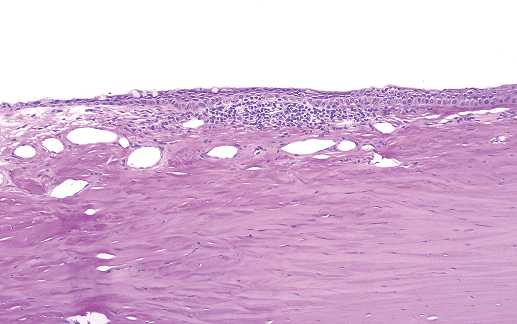
Filtration angle
Infrequent animals (one case in the contact lens cohort, two cases in the albino topical ocular cohort) had congenitally narrowed filtration angle (goniodystrophy) that manifested as a small ciliary body (ciliary body hypoplasia) with lack of any significant trabecular meshwork in the filtration angle (Figures 19 and 20). These cases correlated with in-life findings of enlarged globes which developed during the course of the study.
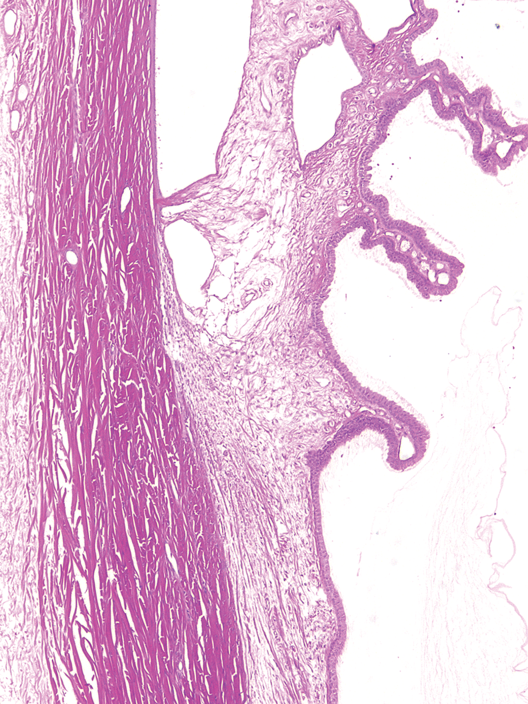
Albino rabbit topical ocular study. Right eye, ciliary body, normal filtration angle. The eye has a normal filtration meshwork (cf. with Figure 20). Hematoxylin and eosin stain. Original objective 5×.
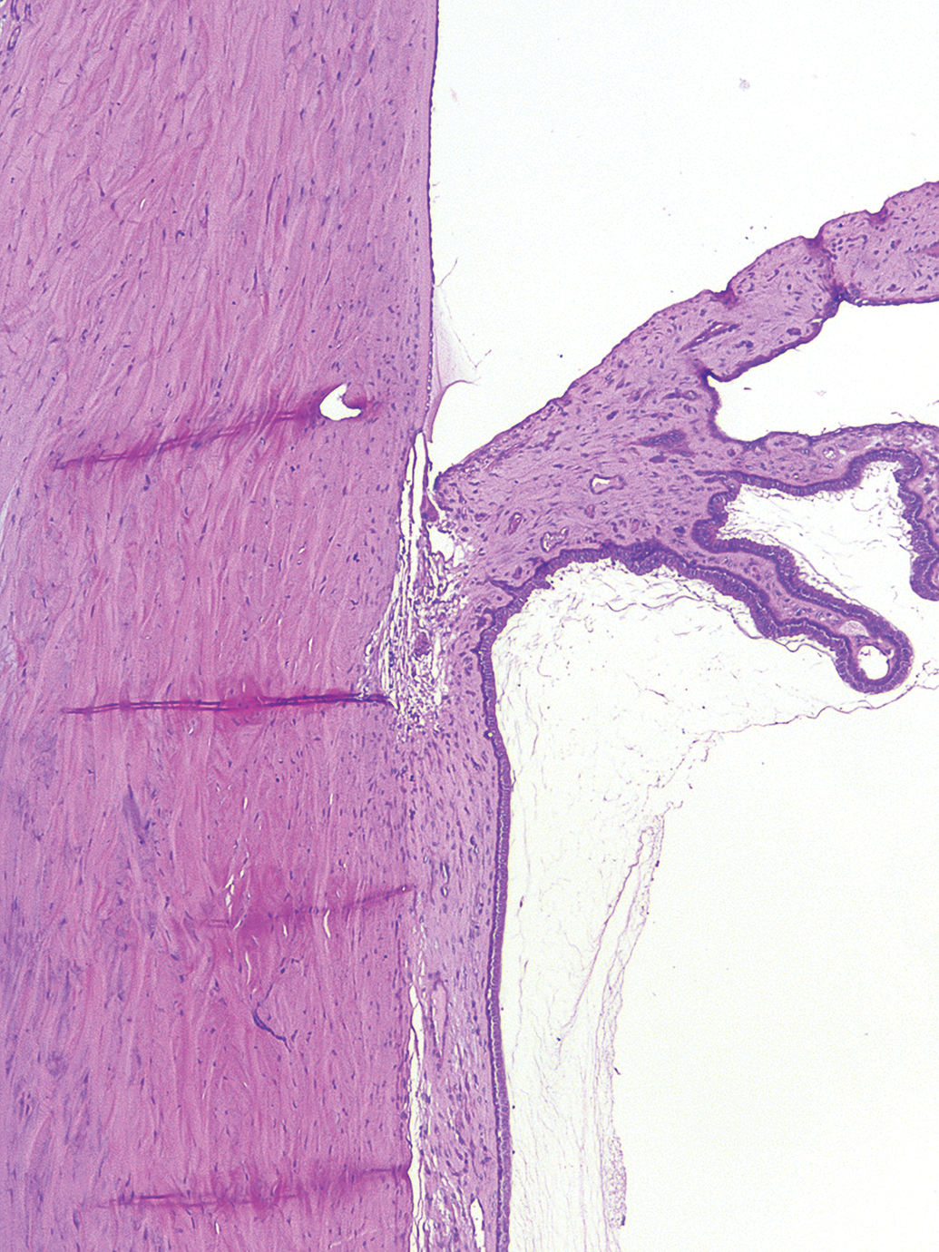
Albino rabbit contact lens study. Left eye, ciliary body, hypoplasia, mild and filtration angle, narrowed (goniodysgenesis), moderate. Narrowed filtration angle in rabbits is a recessively inherited trait and often results in slow onset of increased intraocular pressure and glaucoma. Findings consistent with congenital buphthalmos. Compare with normal architecture of Figure 19. Hematoxylin and eosin stain. Original objective 5×.
Ciliary body
Microscopic findings in the ciliary body of rabbits were relatively infrequent. Most commonly diagnosed was minimal to mild edema in up to 7% of eyes, but exclusive to the albino topical ocular cohort. Congestion of the ciliary body was likewise observed in two albino topical ocular eyes. Diagnoses of mononuclear inflammatory cell infiltrates of the ciliary body were a rare occurrence, being identified in only two pigmented eyes (<1%) from topical ocular studies.
Ectopic bone in the ciliary body was observed in only two eyes (<1%) out of all of the rabbits evaluated (Figure 21). Ectopic bone in the ciliary body may also rarely be observed in other species such as dogs (K. Schafer, pers. comm.). The findings in both instances in these rabbits consisted of a small mass of cancellous bone in the anterior ciliary body adjacent to the trabecular meshwork and at the root of the iris leaflet. Hypoplasia of the ciliary body (Figure 20) was observed in two albino topical ocular eyes and in one albino contact lens eye (0.1–2%), which were also diagnosed with narrowed filtration angles, consistent with congenital buphthalmos as discussed previously.
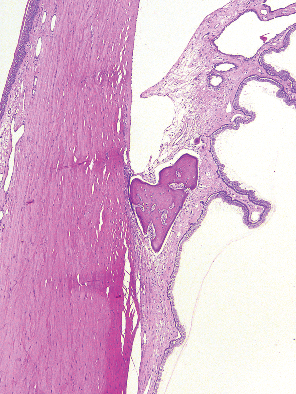
Albino rabbit contact lens study. Left eye, ciliary body, ectopic bone. Focus of ectopic bone is present at the junction of the root of the iris and ciliary body. Hematoxylin and eosin stain. Original objective 5×.
Ciliary process
The ciliary processes traverse from the anterior portion of the ciliary body to the posterior surface of the iris and are a feature of rabbit eyes and other species with little or no lens accommodation ability (Prince 1964). Minimal edema of the ciliary processes was observed in two (<1%) albino topical ocular eyes (Figure 22). Moderate congestion of the iris and associated ciliary process (Figure 23) was observed in a single albino topical ocular eye which was also diagnosed with ciliary body hypoplasia and narrowed filtration angle.
Albino rabbit topical ocular study. Left eye, ciliary body (processes), edema, minimal. The ciliary processes are expanded by proteinaceous fluid within the stroma. Hematoxylin and eosin stain. Original objective 5×.
Albino rabbit topical ocular study. Left eye, iris, congestion, moderate. The iris and associated ciliary processes are expanded by dilated venous sinuses. In this particular eye, ciliary body hypoplasia and narrowing of the filtration angle (goniodysgenesis) were also diagnosed consistent with congenital buphthalmos. Hematoxylin and eosin stain. Original objective 5×.
Iris
In the iris, very few findings were recorded. Minimal mononuclear cell infiltrates (Figure 24) were reported in a total of four animals (0.1–1%) across all studies with cases in all cohorts, albino and pigmented rabbit topical ocular studies, and albino contact lens studies. The mononuclear cell infiltrates consisted of very small foci of lymphocytes and other cells in the iridal stroma. Infiltrates were so minor that they were not visible during in vivo slit lamp examination.
Albino rabbit contact lens study. Left eye, iris, infiltrate, mononuclear cell, minimal. The iris stroma has a small cluster of mononuclear inflammatory cells (lymphoid) associated with an iridal vessel. Hematoxylin and eosin stain. Original objective 10×.
Lens
Alterations of the lens were extraordinarily infrequent, being observed in only three animals. All three of these were diagnosed as lens fiber degeneration or as cataract (Figure 25). Two of these occurred in an untreated animal and an animal in a marketed product control group in contact lens studies and the other one of these occurred in a rabbit in an albino topical ocular study for a 0.3% incidence in each cohort. One of these three cases was identified via slit lamp biomicroscopy during the in-life phase of the study.
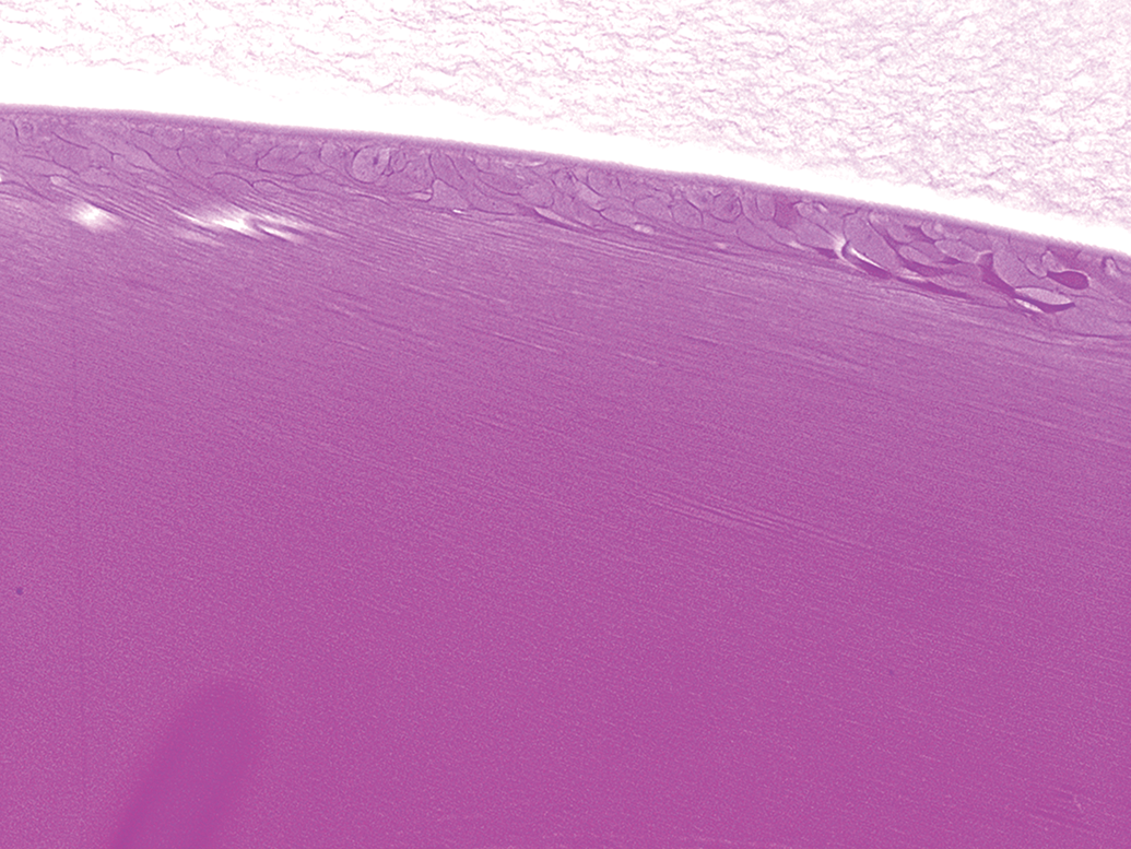
Albino rabbit topical ocular study. Right eye, lens, degeneration, minimal. The posterior lens immediately subjacent to the lens capsule has a layer of swollen lens fibers. This finding was not observed frequently. In this instance, the lens finding correlated with ophthalmic examination finding of lenticular opacity. Hematoxylin and eosin stain. Original objective 10×.
Posterior Segment
Vitreous
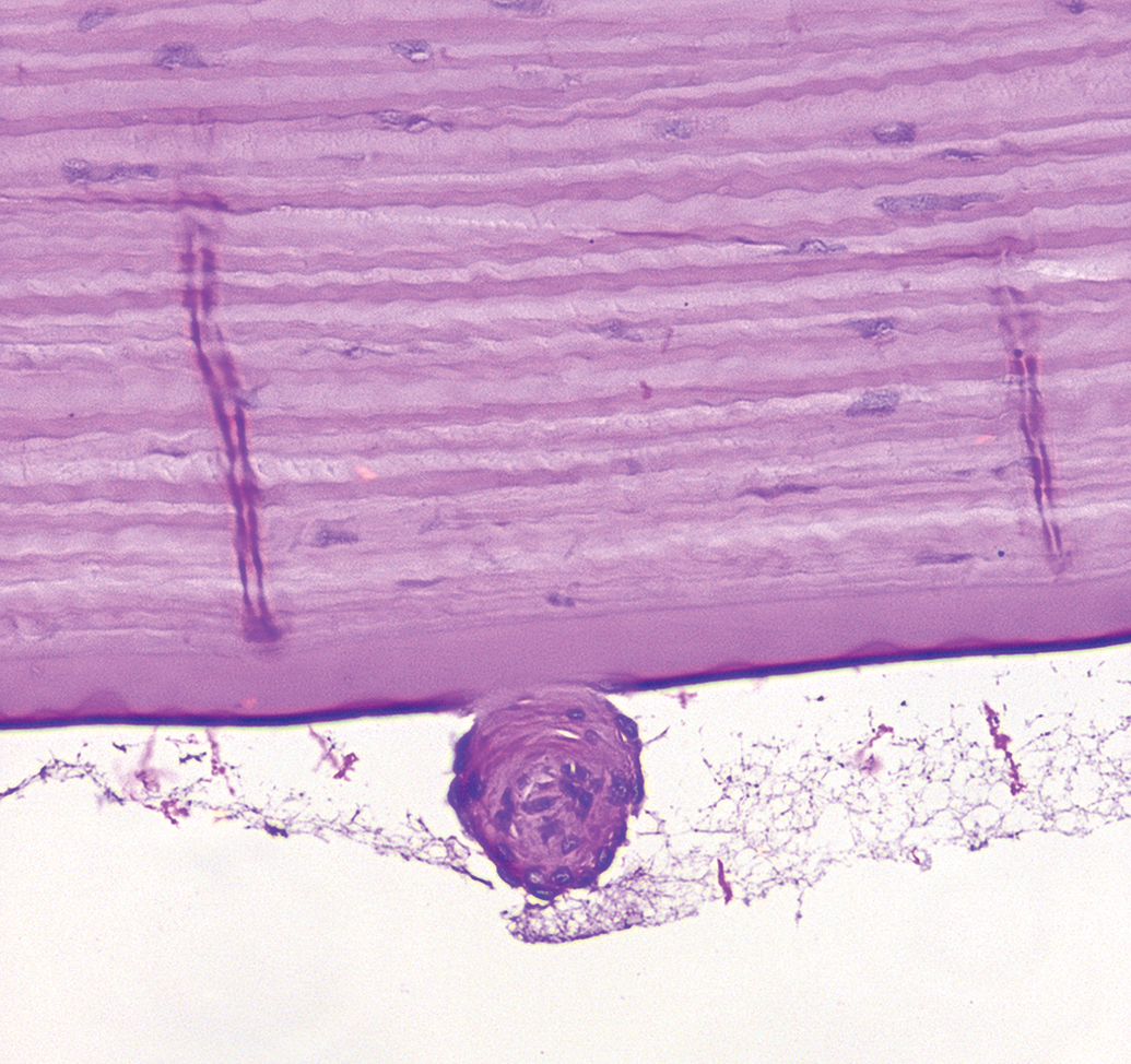
Only one finding, a foreign body in a pigmented topical ocular eye (<1%), was reported in the vitreous. Upon review, it appears that it may instead be an artifact of preparation, with what appears to be a small tag of iris with posterior pigmented epithelium floating free in the vitreal space (Figure 26). These features suggest that it may have been a “floater” on the water bath that was inadvertently incorporated within the ocular section. Therefore, in this analysis, vitreal findings were nonexistent. Spontaneous findings in the vitreous are a rare occurrence.
Pigmented rabbit topical ocular study. Left eye, vitreous, foreign body. This is the only eye in all of the control animals that had this diagnosis. The foreign body appears to be a fragment of iris or ciliary processes that may have been displaced on the water bath during sectioning. Hematoxylin and eosin stain. Original objective 10×.
Retina
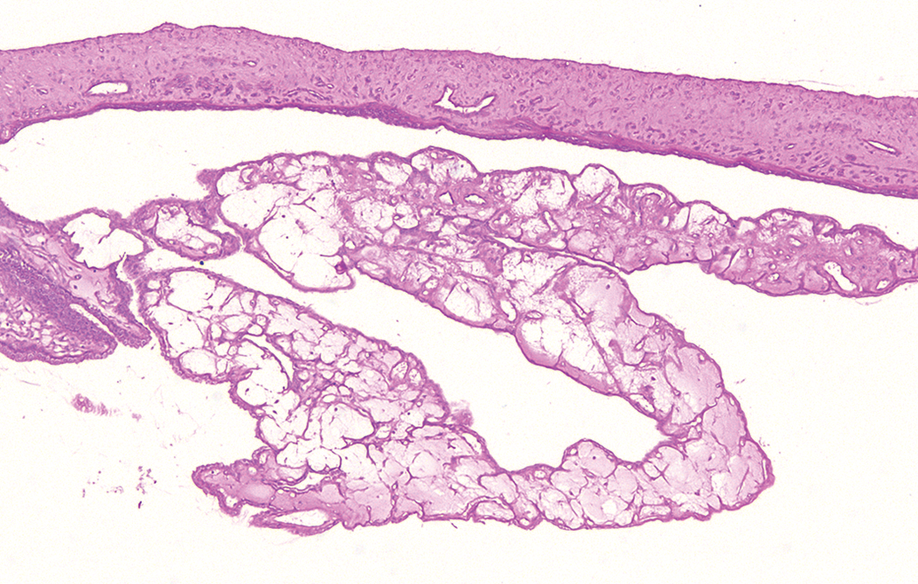
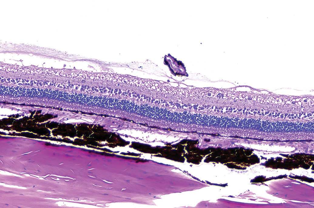
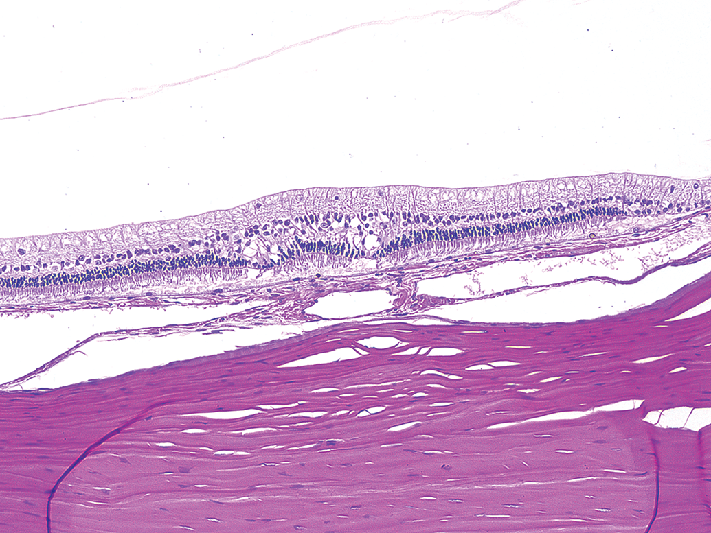
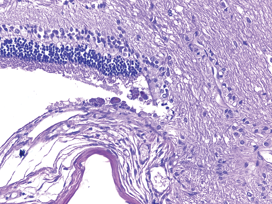
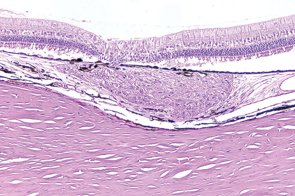
The most common alterations of the retina were folds or rosettes of minimal to mild severity affecting all cohorts at an incidence of up to 18% (Figures 27–31). Some of these findings were termed “dysplasia” in the study pathology reports but further described by the study pathologist as characterized by fold- or rosette-like structures. Given this, and since retinal dysplasias have been included in the diagnoses of retinal rosettes in the rodent INHAND nomenclature (Ramos et al. 2018), they are tabulated into the “fold/rosette” category in the present work. However, some of these warrant additional descriptors as they lack prominent rosette formations or inward projecting folds and may therefore be more accurately described as dysplasia (Figures 30 and 31). In addition, folds, if sectioned on a tangential plane, can look like rosettes (Ramos et al. 2018). Therefore, these findings were categorized together under fold/rosette. Folds/rosettes were commonly observed in the peripheral retina, at or near the ora ciliaris, when retinal location was documented as part of the diagnosis.
Albino rabbit topical ocular study. Left eye, retina, fold/rosette, minimal. The peripheral retina has a fold with loss and disorganization of layers. Hematoxylin and eosin stain. Original objective 10×.
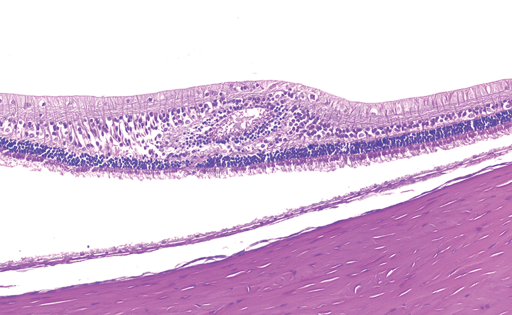
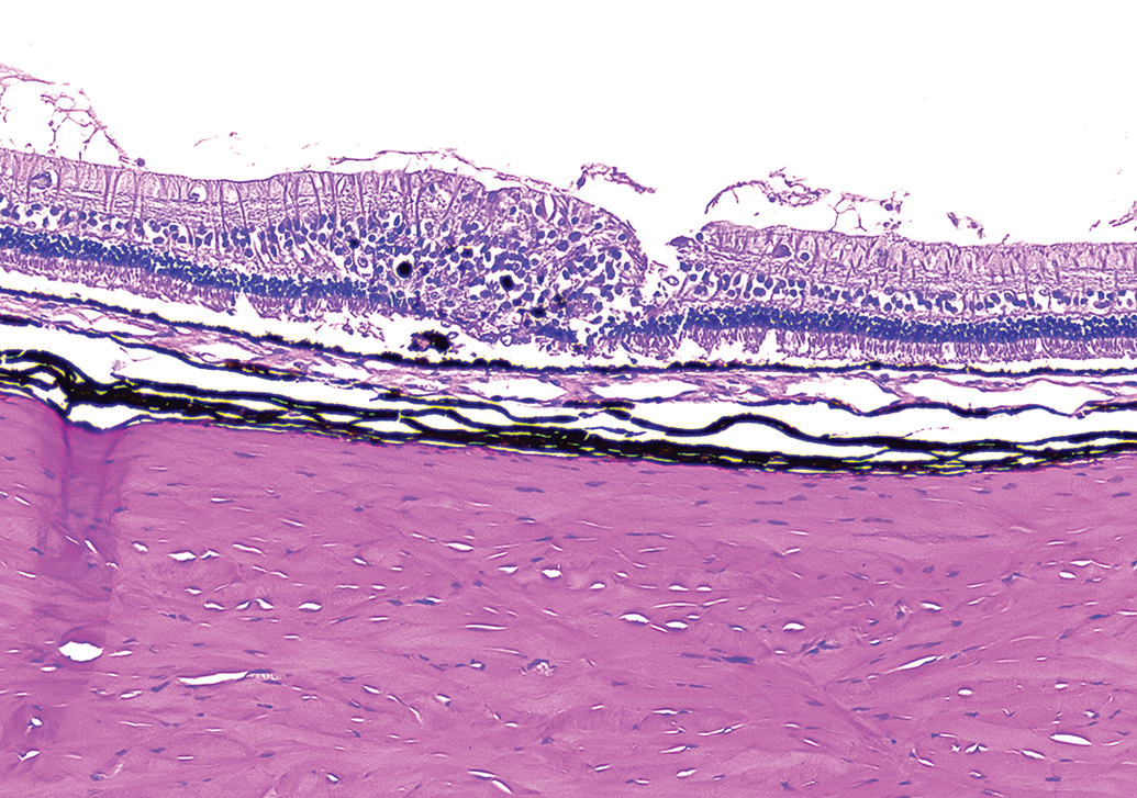
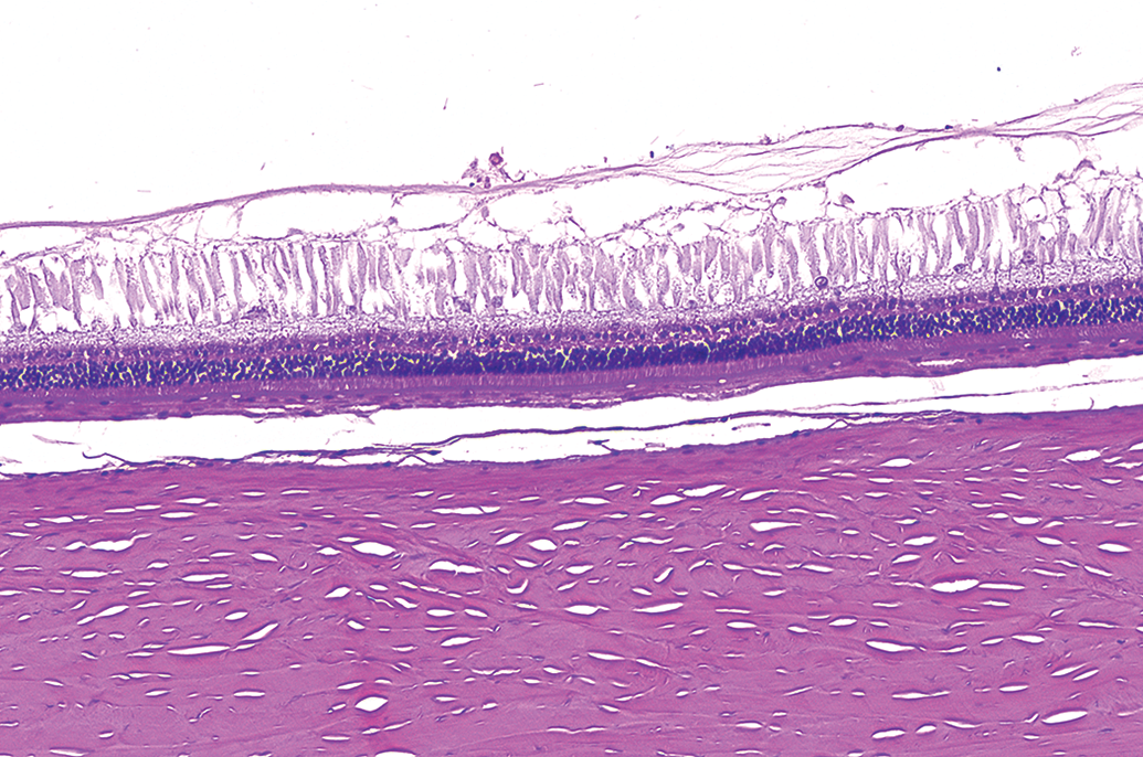
Albino rabbit topical ocular study. Right eye, retina, fold/rosette, minimal. The peripheral retina has a rosette-like structure with increased glial cells in the overlying nerve fiber layer. Hematoxylin and eosin stain. Original objective 10×.
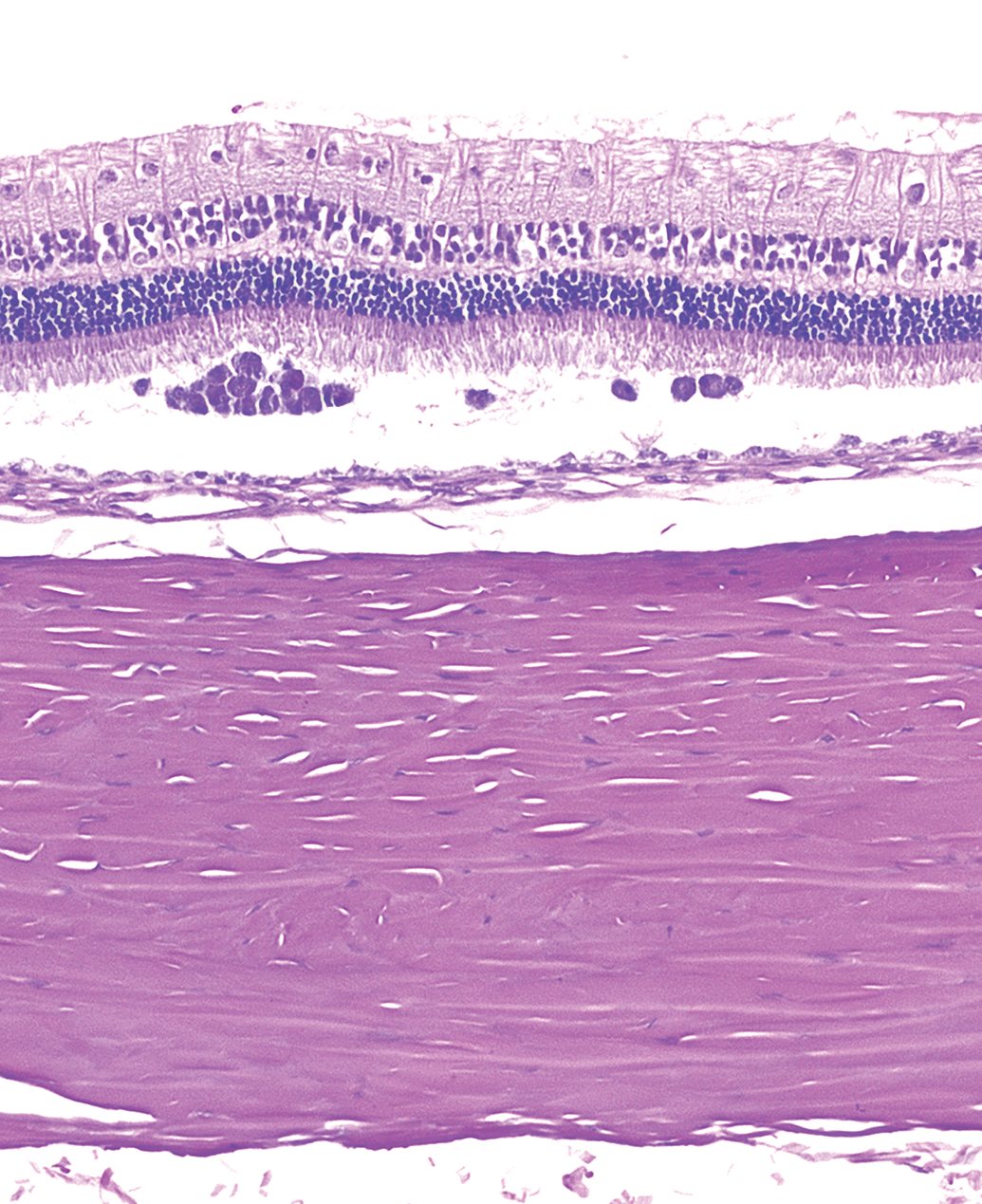
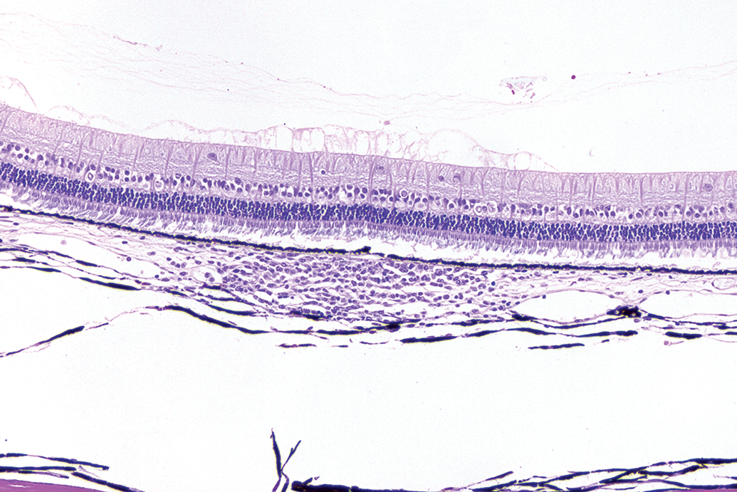
Albino rabbit topical ocular study. Right eye, retina, fold/rosette, minimal. The retina at the medullary ray has a small rosette with general preservation of the retinal layers and a central space lined by the external limiting membrane. Of the lesions diagnosed as retinal rosette, this presentation was the least frequent. Hematoxylin and eosin stain. Original objective 10×.
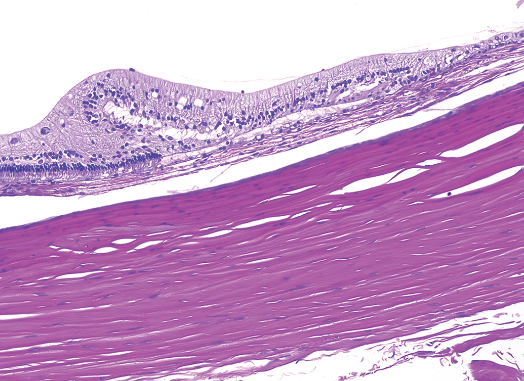
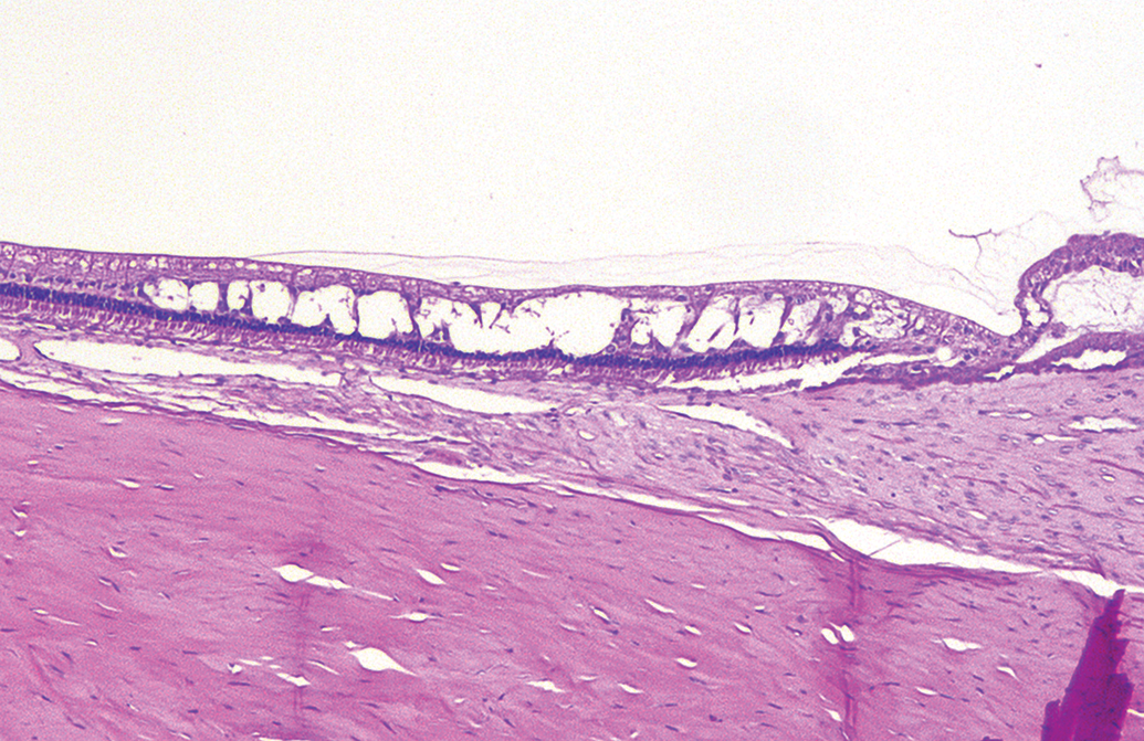
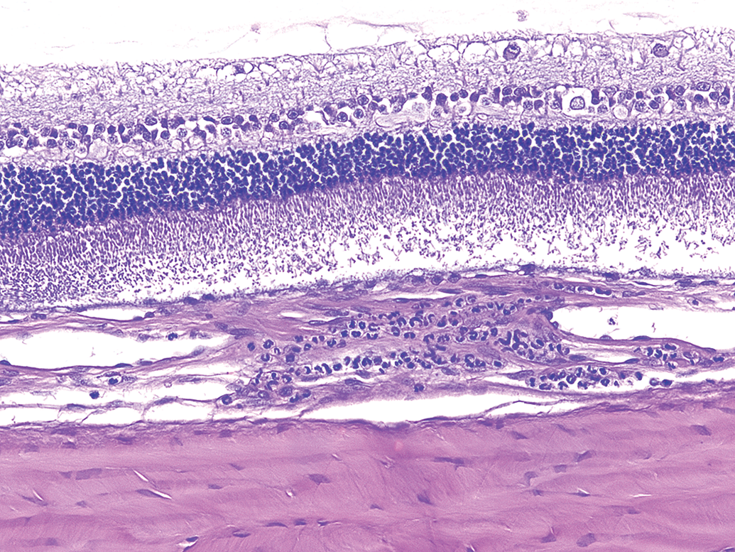
Albino rabbit topical ocular study. Left eye, retina, fold/rosette, minimal. Focal retinal disorganization with vacuolation in the peripheral retina. This retinal finding may be more accurately described as a dysplasia. Hematoxylin and eosin stain. Original objective 10×.
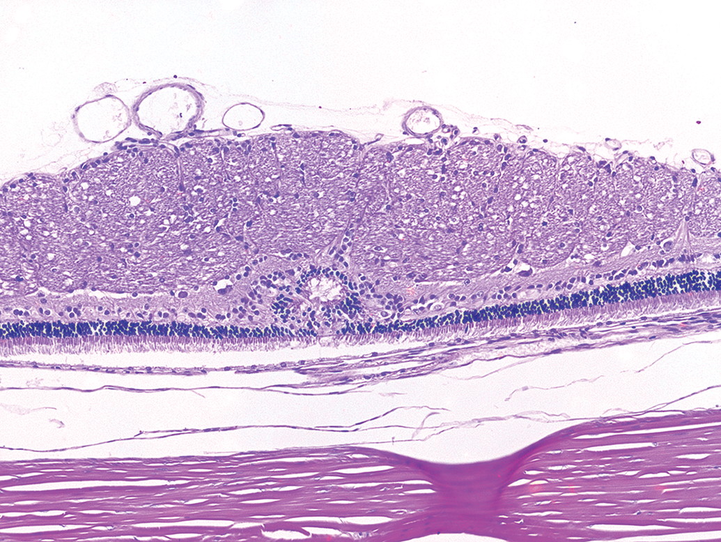
Pigmented rabbit topical ocular study. Left eye, retina, fold/rosette, minimal. A finding initially diagnosed as fold/rosette. The retina has a focal area of disorganization with infiltration of retinal pigment epithelial cells. This retinal finding may be more accurately described as a focal dysplasia. It is uncertain if the break in the retina is artifactual or related to the lesion. Hematoxylin and eosin stain. Original objective 10×.
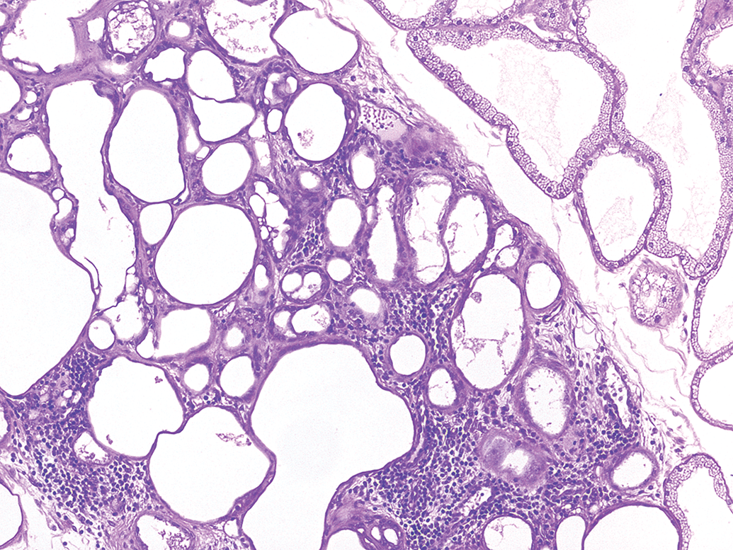
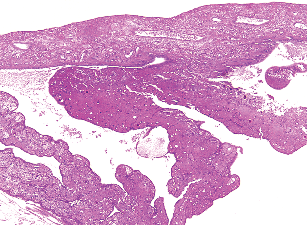
A retinal cyst was observed in a single albino contact lens eye. The finding was characterized by large spaces within the retina consistent with retinoschisis or splitting of the retinal layers (Figure 32).
Albino rabbit contact lens study. Left eye, retina, cyst, minimal. The peripheral retina has multiple clear spaces resembling retinoschisis. Hematoxylin and eosin stain. Original objective 5×.
Other findings in the retina included hypertrophy of the retinal pigment epithelium (RPE), atrophy, axonal swelling, and swelling of the retinal nerve fiber layer (NFL).
Hypertrophy of the RPE (Figures 33 and 34) is relatively commonly observed in rabbits (Schafer and Render 2013b), although it may not be consistently diagnosed due to it being a common spontaneous finding. In the current analysis, we observed this minimal severity finding at less than 1% incidence, affecting one albino topical ocular eye and three albino contact lens eyes.
Albino rabbit topical ocular study. Right eye, retina, retinal pigment epithelium (RPE), hypertrophy, minimal. There are clusters of hypertrophic retinal pigment epithelial cells between the retina and the choroid. The cells have cytoplasmic pigment consistent with lipofuscin, are displaced, and have loss of polarity (i.e., rounded up). Hypertrophic RPE cells are most commonly observed at the ora ciliaris and adjacent to the optic nerve in rabbits and not as a diffuse change. Hematoxylin and eosin stain. Original objective 10×.
Albino rabbit contact lens study. Left eye, RPE, hypertrophy, minimal. Multiple large retinal pigment epithelial cells are located in the subretinal space adjacent to the optic nerve. The cells contain pigment consistent with lipofuscin. Hematoxylin and eosin stain. Original objective 20×.
Retinal atrophy was diagnosed in a total of six eyes (<1%), two pigmented topical ocular and four albino contact lens. When descriptions by the study pathologists included detailed description of the retinal layers affected, it was possible to divide the retinal atrophy findings into “inner retinal atrophy” or “outer retinal atrophy,” according to INHAND recommendations. Inner retinal atrophy includes tissues such as the inner nuclear, inner plexiform, and ganglion cell layers. Outer retinal atrophy includes tissues such as the outer plexiform and photoreceptor layers. When detailed descriptions were not available for a given finding, the general term “retinal atrophy” was used, also consistent with INHAND.
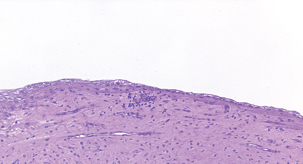
One animal was reported to have axonal swelling of the retina. This finding consisted of swollen axons in the peripheral retina of one eye from a pigmented rabbit in a topical ocular study and was characterized by very few, small, round, homogeneously pale pink structures in the inner nuclear layer. A single case of moderate swelling of the retinal NFL was diagnosed in an albino topical ocular eye (Figure 35). In this case, the inner limiting membrane appears to be pulled away from the NFL, and cell processes in the NFL are notably enlarged. However, in the authors’ experience, this is likely a fixation artifact as it can be produced by fixation in Davidson’s, placement in 70% ethanol for prolonged times (measured in hours), and then placement into 10% neutral buffered formalin.
Albino rabbit topical ocular study. Right eye, retina nerve fiber layer, swelling, moderate. This alteration of the retina was diagnosed as swelling of cell processes in the nerve fiber layer. The inner limiting membrane appears to be pulled away from the nerve fiber layer and cell processes in the nerve fiber layer are notably enlarged. However, in the authors’ experience, this is likely a fixation artifact as it can be produced by fixation in Davidson’s, placement in 70% ethanol for prolonged times (measured in hours), and then placement into 10% neutral buffered formalin. Hematoxylin and eosin stain. Original objective 10×.
Choroid
Findings in the choroid were limited almost entirely to inflammatory infiltrates of minimal severity. Both heterophilic (0.6–2%) and mononuclear cell (0.2–4%) infiltrates were observed. For both types of inflammatory cell infiltrates, they consisted of very small, usually single foci of mononuclear (Figure 36) or heterophilic (Figure 37) inflammatory cells. One pigmented eye in a topical ocular study had a small focus of fibrosis of the choroid with atrophy of the overlying retina (Figure 38). This lesion was of unknown etiology.
Pigmented rabbit topical ocular study. Right eye, choroid, infiltrate, mononuclear cell, minimal. The choroid has a focal aggregate of mononuclear cells. This finding is relatively uncommon in control rabbit eyes. Hematoxylin and eosin stain. Original objective 10×.
Albino rabbit topical ocular study. Right eye, choroid, infiltrate, heterophilic cell, minimal. The choroid has a focal heterophilic infiltrate cuffing choroidal vessels. This finding was exceedingly uncommon. Hematoxylin and eosin stain. Original objective 20×.
Pigmented rabbit topical ocular study. Left eye, choroid, fibrosis, minimal. The choroid has a focal area of fibrosis that is relatively mature with incorporation of low numbers of retinal pigment epithelial cells. The overlying retina has a focal defect, characterized by atrophy of the inner and outer nuclear and plexiform layers. The finding is suggestive of a previous ocular injury. Hematoxylin and eosin stain. Original objective 10×.
Optic nerve
Optic nerve findings were very rare (<1%), consisting entirely of minimal mononuclear inflammatory cell infiltrates in one albino rabbit eye on a contact lens study and one pigmented rabbit eye on a topical ocular study. These cases were characterized by aggregates of cells cuffing vessels within the optic nerve (Figure 39). No intraocular findings were reported for these rabbits.
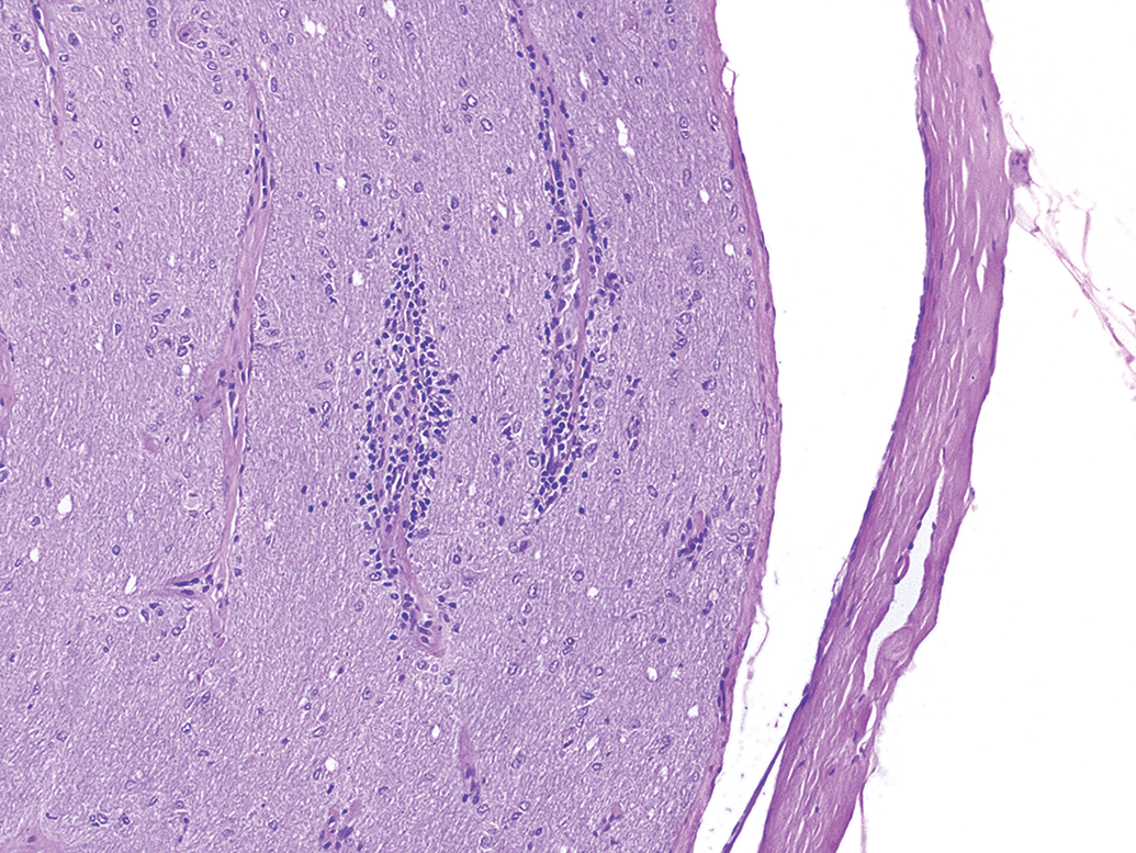
Pigmented rabbit topical ocular study. Right eye, optic nerve, infiltrate, mononuclear cell, minimal. Vessels in the optic nerve have small perivascular cuffs of inflammatory cells. Hematoxylin and eosin stain. Original objective 10×.
Sclera
Findings in the sclera were reported predominantly in the contact lens studies. These findings were all minimal in severity and included heterophilic infiltrates in episcleral tissue (1 eye), mononuclear cell infiltrates in episcleral tissue (1 eye), chronic inflammation of episcleral tissue (2 eyes), and chronic inflammation which was not otherwise specified as to location (1 eye). These were all likely episcleral and not truly affecting the sclera. Also reported was the presence of suture material in 6 eyes, which reflected placement of suture at necropsy to assist with orientation of the eye at tissue trimming; none of these rabbits underwent surgical procedures. Outside of the contact lens cohort, the only findings were two cases of minimal to mild episcleral hemorrhages around the superior rectus muscles, which were also visible during gross examination of the eyes at necropsy.
Discussion
There were no appreciable differences in the incidence or types of findings between similarly treated right versus left eyes or any sex differences.
Overall, the incidence of background findings was highest in the structures anterior to and including the cornea and lower in tissues posterior to the cornea. Most commonly observed were inflammatory cell infiltrates in various tissues such as the eyelids and conjunctiva. This might be expected due to the normal immune response in tissues exposed to the external environment and to minor manipulations and innocuous treatments during a toxicology study. Similarly, higher incidences of background findings were observed in adnexal tissues. A noteworthy exception to the low incidence of findings in the posterior tissues was in the retina where fold/rosettes were somewhat common, as discussed further below.
Upon first glance, it may appear that our data set suggests a higher incidence of background changes in eyes subject to contact lens wear as compared to topical ocular treatment. For example, mononuclear cell infiltration was of a higher incidence in the conjunctiva, Harderian and lacrimal glands, nictitating membrane, and at the corneal limbus in the contact lens cohort. Intuitively, this may make sense, as routine contact lens wear is known to elicit minor changes in the anterior tissues of human eyes (Bruce and Brennan 1990; Ladage et al. 2001). Harderian gland atrophy was also of slightly higher incidence in the contact lens cohort.
However, it is important to consider that different pathologists were assigned to studies. The most common criteria for assignment of studies to individual pathologists was the type of study, topical ocular versus contact lens. This provided for a great deal of longitudinal consistency in the historical data for a given cohort but bears consideration when comparing topical ocular and contact lens data sets. In such a comparison, differences in thresholding of findings or terminology differences between pathologists must be weighed. In fact, if one only takes the contact lens cohort from our data set, it becomes apparent that the incidence of mononuclear cell infiltrates and Harderian gland atrophy are quite similar between eyes fitted with control article–treated contact lenses and contralateral eyes which were untreated. This suggests that the variability among pathologists is a much greater contributor to differences in incidence than the route of administration.
A notable difference was observed in the incidence of retina findings diagnosed as folds, rosettes, and dysplasias in the eyes. When combined and from all groups (untreated, vehicle, and marketed product control groups), the incidence of these retinal findings was 17% for albino rabbits in topical ocular studies, 18% for albino rabbits in contact lens studies, but only 1.5% for pigmented rabbits on topical ocular studies. This difference in incidence in retinal folds, rosettes, and dysplasias between pigmented and albino animals has not been previously described, to the authors’ knowledge, in rabbits or rodents. Retinal folds, rosettes, and dysplasias are thought to be congenital lesions, so there may be a genetic component to their occurrence. Although there are retinal alterations associated with some forms of albinism in humans, such as foveal atrophy, retinal dysplasia is not described (Yanoff and Fine 2002). The pigmented rabbits in these studies were F1 crosses of New Zealand White and Red rabbits, while the albino rabbits were exclusively New Zealand White rabbits. It suggests that, if genetic, the defect is related to the albinism and is likely a recessive trait.
The only other noteworthy difference in incidence of findings between strains was for lymphoid hyperplasia and lymphoid aggregates in the conjunctiva and, to a lesser extent, the subcorneal space. These were more commonly observed in pigmented eyes (45 total cases, 4.5% incidence) as compared to albino eyes (4 total cases, 0.4% incidence). The reason for this difference is not clear but could be variability in diagnostic criteria or thresholding among the pathologists evaluating these studies.
The features of MALT have been described in the rabbit. Specifically, Knop and Knop (2008) and Cain and Phillips (2008) have reported on CALT, while Casteleyn et al. (2010) have described nasal-associated lymphoid tissue. In our review, some of the findings diagnosed as lymphoid hyperplasia are recognized as CALT (e.g., Figure 7). Similarly, some cases of mononuclear cell infiltration in the nasal lacrimal duct were also found to have MALT in section (e.g., Figure 15). It should be acknowledged that, in the absence of other evidence of inflammation or disease/injury, well-formed lymphoid aggregates could be MALT even if germinal centers are not apparent in section. It is possible that some of the diagnoses identified as lymphoid infiltrates may have been from the edge of foci of MALT. In addition, clearly identified MALT was diagnosed as lymphoid hyperplasia. At least one pathologist evaluating these studies used lymphoid hyperplasia of eyelids and conjunctivae to catalogue the occurrence of germinal centers as possible markers of immune activation. It is certainly recognized that there are different practices in approaching tabulation of the incidence of MALT (Woicke et al. 2018; Schuh 2018). This diagnosis of lymphoid hyperplasia is distinct from infiltration which generally was not a well-organized aggregation of lymphoid cells.
Although the overall incidence of degeneration of the lens fibers, consistent with early cataract formation, was quite low in our data set, it is noteworthy that all three cases were observed in albino eyes. No such findings were reported in the pigmented eyes. This higher incidence of spontaneous lenticular changes in albino eyes is consistent with prior investigations (Munger, Langevin, and Podval 2002).
Findings in the nasolacrimal duct were of low incidence and included acute hemorrhages and inflammatory cell infiltrates. This is one of the only tissues in which background lesions were completely absent in untreated eyes and exclusive to eyes treated with innocuous vehicles and marketed product control articles. This may suggest that the routine procedure of dosing can induce subtle changes in this tissue.
Because of the type of studies being conducted, specifically being topical ocular administration or contact lens wear, the preferred strains being used were New Zealand White and New Zealand White × Red F1 rabbits. This strain selection was intentional to avoid the commonly observed corneal dystrophy in Dutch Belted rabbits (Holve, Mundwiler, and Pritt 2011; Moore, Dubielzig, and Glaza 1987), which could become a confounding finding in the interpretation of these types of studies. Consequently, corneal mineralization was not observed and therefore is not reported.
In three eyes, we observed sequelae of findings consistent with congenital buphthalmos. It is noteworthy that the findings were exclusive to albino rabbits in our data set. No such findings were reported in pigmented eyes.
The occurrence of congenitally narrowed filtration angle in rabbits is via a genetic recessive inheritance (Kolker et al. 1963; Greaves and Perkins 1951; Williams 2013) often leading to gross clinical manifestation of buphthalmos (Lindsey and Fox 1994; Barthold, Griffey, and Percy 2016; Tesluk, Peiffer, and Brown 1982; Williams 2013). Animals have a slow, insidious onset of increased intraocular pressure (IOP), often not clearly manifesting until a year of age. The increased IOP is due to a malformation of the drainage angle with insufficient outflow of aqueous (McMaster 1960). Consequently, these animals will often present with buphthalmos and microscopic findings that include hypoplasia of the ciliary body and accumulations of proteinaceous material in the anterior chamber. These associative findings were apparent in our data set. Foci of inflammatory cells in the cornea have also been reported (Bradley 2012), but this change was not evident in the buphthalmic eyes in our data set. Findings in eyes with advanced disease can further include cupping of the optic nerve, and retinal degeneration that starts with the inner layers, among other possible alterations. The 0.2% cumulative incidence (3 cases of 1,222 total rabbits) in the current data set is somewhat lower than the 0.7% incidence observed by Williams (2013). This lower incidence in the current data set is likely due to the careful exclusion of any animal with clinical manifestation of buphthalmos prior to the initiation of an ocular toxicology study.
Rabbits with buphthalmos are often removed from study early due to humane concerns. The occurrence of increased IOP and buphthalmos in animals assigned to test article–treated groups tends to cause a lot of consternation during study execution because it is not until there is microscopic examination of the globe that the cause of the buphthalmos and increased IOP can be determined. Although all three cases of microscopically observed narrowed filtration angle in this data set were predicated by in-life gross or slit lamp biomicroscopic observations of enlarged globes, microscopic examination has been shown to be capable of detecting the congenital anomalies associated with buphthalmos prior to clinical manifestation in this laboratory. Because of the recessive inheritance, once these rabbits are identified as having increased IOP and goniodysgenesis, it is important to notify the animal supplier in attempts to remove those bloodlines from the breeding stock.
A low, 0.3%, incidence of lens fiber degeneration (cataracts) was observed in albino rabbits from our data set. In vivo slit lamp biomicroscopic assessment of rabbits from this same facility, but from an earlier time frame (946 rabbits from studies conducted between 1999 and 2001; Munger, Langevin, and Podval 2002) determined that spontaneous cataracts occurred in 1.1% of pigmented rabbits and in 5.7% of albino rabbits. The discrepancy between the current and earlier data set is attributed to the fact that Munger et al. examined animals in the prescreen phase of study, whereas rabbits in the current data set are only those which were deemed acceptable by prescreen examination, included in the study, and subject to histopathology evaluation. Therefore, after exclusion of any animal found with a cataract at prescreen, only cataracts which developed during the course of a study would be captured in the current data set.
As for terminology, our preference is to use the microscopic term of lens fiber degeneration. Cataract is specifically defined as opacity of the lens, which qualifies it as a gross or ophthalmic examination term. During the fixation and tissue processing that occurs in generating histologic sections of eyes, the lens is made opaque, such that a pathologist sees an entire lens that is opaque. The pathologist sees correlates to the in vivo opacity, such as lens fiber degeneration. In addition, the term cataract carries clinical connotations of an irreversible and often end-stage status to the lens, which is often not what is meant to be communicated in such preclinical toxicology findings.
Retinal folds/rosettes were the most commonly observed finding in the posterior tissues. The majority of cases reviewed in this retrospective evaluation were disorganized, often with small cyst-like central spaces, occasional partial folding of the retina, and frequently gliosis of the NFL. Review of the study pathology reports revealed that most cases of folds and rosettes were grouped together under a single category as “fold” or “fold/rosette.” We believe that segregation of these diagnoses at times may be difficult, although they have differential features as described in the INHAND rodent ocular terminology (Ramos et al. 2018). Specifically, folds may appear as rosettes if sectioned in a tangential plane (Ramos et al. 2018), which could be easily imagined for a structure such as that presented in Figure 27. To demonstrate a spectrum of these findings, we have included representative images of findings that may be classified as folds (Figure 27), rosettes (Figures 28 and 29), and those findings which may be best characterized as general dysplasias (Figures 30 and 31) but were combined into the diagnosis of folds/rosettes.
One finding diagnosed as retinal cyst and some of the folds/rosette findings are similar to some presentations of peripheral retinal cystoid degeneration (PRCD), but these need to be distinguished from each other. PRCD frequently occurs at the ora serrata in primates and at the ora ciliaris of dogs (Martin 2009; Schafer and Render 2013a; Woicke et al. 2018). PRCD may manifest as large, cyst-like structures that replace the retinal architecture as well as may look like retinoschisis. While at times similar to the fold/rosettes observed in these rabbits, PRCD has some distinct differences. The fold/rosettes tended to occur at the periphery of the retina but did not occur exclusively at the most peripheral retina as with PRCD. PRCD forms large cyst-like spaces within the retina, while the central spaces within that of fold/rosettes tend to be smaller and lined by photoreceptor segments and/or outer nuclear layer cells.
Hypertrophy of the RPE is a recognized spontaneous phenomenon in rabbits (Schafer and Render 2013a) and was observed in our data set, though at a low incidence. The hypertrophic cells tend to occur most commonly in the retina adjacent to the ora ciliaris or adjacent to the optic nerve. Therefore, the hypertrophy is a focal or multifocal finding and not a diffuse hypertrophy of the RPE. The hypertrophied RPE cells will often have an eosinophilic to light gray or brown granular cytoplasm. The pigmentation is thought to be due to accumulation of lipofuscin from inefficient breakdown of the rod and cone outer segment platelet membrane (Mecklenburg and Schraermeyer 2007).
The one case of swelling of the NFL was likely an artifact of tissue processing. This alteration can be reproduced by fixation of eyes in Davidson’s solution followed by storage in ethanol. If the calottes of the eyes are then started in tissue processing in 10% neutral buffered formalin instead of ethanol, the retina will have swelling of the NFL with disruption of the inner limiting membrane (K. Schafer, pers. comm.). The distribution of the artifact will usually be multifocal and will not consistently occur in any particular part of the retina. If eyes are randomized across groups for tissue processing, this artifact will occur in treated as well as nontreated groups. Avoiding this artifact requires attention to what the solution is that the eyes are stored in, as well as the tissue processing procedure.
Footnotes
Acknowledgments
The authors wish to acknowledge the following pathologists who, in addition to those contributing as authors to the current work, provided evaluation of tissues at the time of study conduct: Drs. Margarita M. Gruebbel, James K. Maurer, and Rebecca R. Moore of Experimental Pathology Laboratories Inc., and Drs. John E. Sagartz and Kristen J. Nikula of Seventh Wave Laboratories, LLC.
Author Contributions
All authors (NL, KS, OT, BM, RR) contributed to conception or design; data acquisition, analysis, or interpretation; drafting the manuscript; and critically revising the manuscript. All authors gave final approval and agreed to be accountable for all aspects of work in ensuring that questions relating to the accuracy or integrity of any part of the work are appropriately investigated and resolved.
Declaration of Conflicting Interests
The author(s) declared no potential, real, or perceived conflicts of interest with respect to the research, authorship, and/or publication of this article.
Funding
The author(s) disclosed receipt of the following financial support for the research, authorship, and/or publication of this article: Dr. Kenneth A. Schafer reports grants from Alcon Research Ltd., during the conduct of the studies, and grants from Alcon Research Ltd., outside the submitted work.