
Abstract
The important renal tumors that can be induced by exposure of rats to chemical carcinogens are renal tubule tumors (RTTs) derived from tubule epithelium; renal pelvic carcinoma derived from the urothelial lining of the pelvis; renal mesenchymal tumors (RMTs) derived from the interstitial connective tissue; and nephroblastoma derived from the metanephric primordia. However, almost all of our knowledge concerning mechanisms of renal carcinogenesis in the rodent pertains to the adenomas and carcinomas originating from renal tubule epithelium. Currently, nine mechanistic pathways can be identified in either the rat or mouse following chemical exposure. These include direct DNA reactivity, indirect DNA reactivity through free radical formation, multiphase bioactivation involving glutathione conjugation, mitotic disruption, sustained cell proliferation from direct cytotoxicity, sustained cell proliferation by disruption of a physiologic process (alpha 2u-globulin nephropathy), exaggerated pharmacologic response, species-dominant metabolic pathway, and chemical exacerbation of chronic progressive nephropathy. Spontaneous occurrence of RTTs in the rat will be included since one example is a confounder for interpreting kidney tumor results in chemical carcinogenicity studies in rats.
Twenty years have elapsed since publication of my last reviews of mechanisms involved in chemically induced renal carcinogenesis in rodents (Hard 1998, 1999). In that time, the importance of concepts such as key events in tumor pathogenesis and modes of action in risk assessment of chemicals has gained momentum. Also, in the intervening years, much new information has been acquired on this subject, warranting an update on the status of modes of action and mechanisms underlying the occurrence of renal tumors in rats and mice. This review will include consideration of one form of spontaneous renal tumor development because it is a confounder for interpretation of kidney tumor incidence in chemical carcinogenicity studies in rats. Of the broad range of renal tumors in rodents, which include renal tubule tumor (RTT), renal pelvic carcinoma, renal mesenchymal tumor (RMT), renal lipomatous tumors, and nephroblastoma, the review will focus on RTT because this is the most common tumor type occurring in rodents, and the most frequently induced by chemical exposure. Furthermore, the wealth of information available on mechanistic aspects of chemical carcinogenesis pertains to RTT. For this neoplasm, nine mechanistic pathways can be identified involving either the rat or mouse.
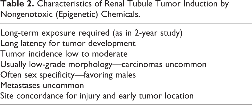
Broadly speaking, chemical carcinogens can be classified as either genotoxic or nongenotoxic depending on the molecular events involved in cancer causation (Pogribny and Beland 2013). Genotoxic carcinogens are agents that interact with DNA, either directly, or after spontaneous metabolic activation, or indirectly as in the case of chemicals inducing free radical formation such as oxidative stress (Pogribny and Beland 2013). These lead to a variety of genotoxic, molecular alterations including DNA damage, DNA adduct formation, and genetic aberrations (Pogribny and Beland 2013). Nongenotoxic carcinogens, also referred to as epigenetic carcinogens, cause neoplastic lesions by mechanisms not involving direct or indirect DNA damage. Epigenetic abnormalities include global genomic hypomethylation, gene-specific DNA hyper- or hypo-methylation, abnormal functioning of DNA and histone-modifying enzymes, altered histone modification patterns, and aberrant expression of gene-controlling microRNAs, which occur in tissues that are susceptible to carcinogen exposure (Pogribny and Beland 2013). The characteristics of renal tumor induction by genotoxic chemicals tend to differ from the characteristics of tumor induction by chemicals acting through epigenetic pathways, as summarized in Tables 1 and 2.
Characteristics of Renal Tubule Tumor Induction by Genotoxic Chemicals.
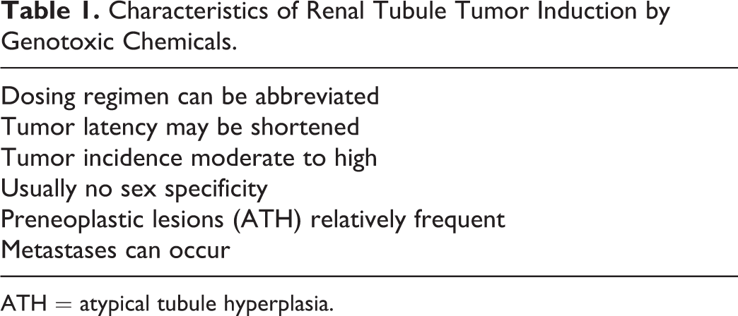
ATH = atypical tubule hyperplasia.
Characteristics of Renal Tubule Tumor Induction by Nongenotoxic (Epigenetic) Chemicals.
In the mechanistic categories considered below, categories 1 and 2 can be classified as genotoxic, while categories 5 to 8 are considered to be epigenetic pathways. Chemicals acting via pathway 4 (multiphase activation) can be genotoxic or nongenotoxic, depending on the weight of evidence supporting on the one hand, metabolic activation to a reactive intermediate, or on the other, evidence in favor of metabolism to a harmless end product that is excreted into the urine. Mitotic disruption might also be considered to have properties similar to genotoxicity because it directly affects tubule cell nuclei.
Data leading to a mechanistic understanding of chemical renal carcinogenesis are generated by private and university-based research laboratories using well-established animal models of renal cancer, as well as by more applied investigations from industry laboratories, and research generated from the carcinogenesis bioassay program of the National Toxicology Program (NTP), which holds one of the largest archives of pathology relevant to toxicology. Up to the end of 2017, almost 600 chemicals or substances had been tested and 61 of those were considered as showing some or clear evidence of carcinogenicity for the rodent kidney. The majority of these substances were associated with RTTs while a very few had induced urothelial tumors of the renal pelvis or ureter. No chemical in the NTP series has induced nephroblastoma, RMT, or lipomatous tumor.
Direct DNA Reactivity
In safety evaluation studies in rats and mice, a test article is deemed to be genotoxic if it is positive in a battery of short-term mutagenicity tests, particularly including the complete Ames test, mammalian cell mutagenesis assay, and in vitro micronucleus test. Quite a number of chemicals can be identified as DNA-reactive renal carcinogens for laboratory rodents, and Table 3 lists some of these. Dimethylnitrosamine (DMN) is a versatile carcinogen inducing both RTT and RMT in high incidence in rats following a single exposure (Hard and Butler 1971). N-nitrosomorpholine (Bannasch, Krech, and Zerban 1980), N-ethyl-N-hydroxyethylnitrosamine (Tsuda et al. 1983), and N-(4′-fluoro-4-biphenylyl)acetamide (Dees et al. 1980) have also been used as classic kidney cancer models for studying the pathogenesis and biology of RTT.
Examples of Genotoxic Renal Chemical Carcinogens in the Rat.
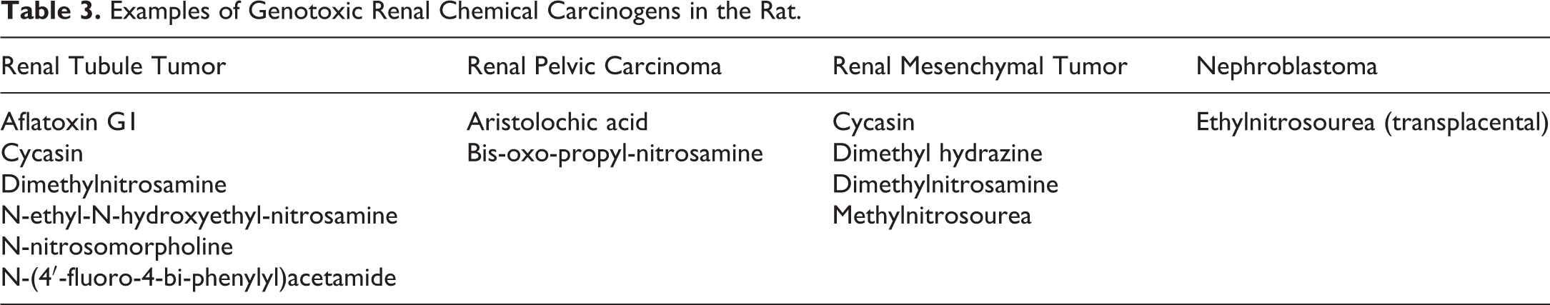
Some chemicals inducing renal tumors via a genotoxic pathway such as methylnitrosourea and ethylnitrosourea can act on the target tissue directly without metabolic conversion, whereas most genotoxic compounds are spontaneously metabolized in the susceptible tissue to a reactive intermediate. For example, DMN is metabolized initially by cytochrome P450 enzyme CYP2E1 in the S2 segment of proximal tubule and then spontaneously metabolized through a series of putative steps, leading to an ultimate carcinogen, methyl carbonium ion, and/or the methyl diazonium cation, one of which is believed to be the active species that methylates DNA (Weekes 1975).
From the work conducted in Dr. Peter Magee’s laboratory in London in 1977, it is known that DMN produces 2 major types of DNA adduct in rat kidney—N7 methylguanine and O6 methylguanine. Nicholl, Swann, and Pegg (1977) showed that N7 methylguanine is rapidly removed by DNA repair processes, while O6 methylguanine is also removed at low noncarcinogenic exposures, but at high doses that produce kidney tumors the adduct persists, and it is the persistence of this potentially mutagenic O6 adduct in rat renal tissue that correlates with carcinogenicity. Downstream from adduct formation neoplasms are believed to arise as a result of an accumulation of mutations involving growth control genes, either growth-promoting nuclear proto-oncogenes or growth-constraining tumor suppressor genes. Point mutations in the von Hippel–Lindau gene (G:C>A:C transition) consistent with persistence of O6 methylguanine have been found in 75% of DMN-induced rat RTT that have clear cell morphology (Shiao et al. 1998) similar to human clear cell carcinoma. There is involvement of the VHL gene in some 60% of clear cell carcinomas in humans.
Indirect DNA Reactivity through Free Radical Formation
Indirect DNA reactivity is mediated by oxidative stress associated with free radical formation. Free radicals are highly reactive compounds by virtue of having unpaired electrons, and this electron imbalance confers an ability to produce cell toxicity and DNA damage involving the self-propagating redox cycle. Examples include the superoxide anion radical, singlet oxygen, and the hydroxyl radical. Hydrogen peroxide has balanced electrons but is itself a reactive molecule. The hydroxyl radical is responsible for lipid peroxidation.
Ferric nitrilotriacetate (FeNTA) and potassium bromate are the main examples of this mechanistic pathway in the rodent kidney. FeNTA (Ebina et al. 1986) and potassium bromate (Kurokawa et al. 1983; DeAngelo et al. 1998) have been recorded as inducing up to 80% to 90% incidence of RTT in rats and/or mice of either sex. In this respect, they are similar to direct DNA-reactive renal carcinogens. For FeNTA, the evidence for oxidative stress occurring in the kidney includes increased levels of 8-hydroxydeoxyguanosine (8-OH-dg), a marker of oxidative damage, in the kidney DNA of FeNTA-treated rats (Umemura et al. 1990), increased 8-hydroxyguanine repair activity in the rat kidney (Yamaguchi et al. 1996), and production of free radical-induced lipid peroxidation (Toyokuni et al. 1994). Lipid peroxidation was monitored biochemically by the formation of thiobarbituric acid reactive substances and of free 4-hydroxy-2-nonenal (HNE). The latter was localized immunohistochemically using a polyclonal antibody against HNE-modified proteins. HNE-modified proteins were observed in degenerating proximal tubule cells following FeNTA exposure (Toyokuni et al. 1994). FeNTA has also been recorded as inducing DNA base modifications in Wistar rat renal chromatin (Toyokuni, Mori, and Dizdaroglu 1994). Furthermore, FeNTA-induced oxidative damage in the rat kidney was prevented by antioxidant administration (Zhao, Khan, and O’Brien 1997). Collectively, these data provide robust evidence supporting the induction of reactive oxygen species as an underlying mechanism for FeNTA renal carcinogenesis in rodents. Potassium bromate has also been shown to produce 8-OH-dg in rat kidney DNA (Kasai et al. 1987).
The complexing chemical, N-nitrilotriacetic acid (NTA) and its salts, are strong chelators, capable of covalently binding to metal ions such as the ferric ion (Fe3+). Because it is highly biodegradable, NTA has been used as a water-softening agent and detergent. Although it is nongenotoxic, NTA alone has been shown to produce renal tubule adenomas and carcinomas in male rats and RTT in both sexes of mice in 2-year carcinogenicity studies (NTP 1977). In a 28-day toxicity study, NTA caused severe vacuolation of proximal tubule cells (Alden et al. 1981) similar to the histopathologic appearance of osmotic nephrosis. To explain NTA’s nephrocarcinogenic property, it has been suggested that, because of its powerful chelating properties, NTA binds to the zinc ion and transports this reactive element into the tubule cell phagolysosomes, where the zinc ion is cleaved from the chelator and accumulates intracellularly (Anderson 1981). Possibly, the Zn ion also causes free radical formation in this setting.
Multiphase Bioactivation Involving Glutathione (GSH) Conjugation
GSH S-conjugation has been mainly associated with the detoxification of xenobiotics and subsequent excretion (Jacoby and Ziegler 1990), but work from several laboratories during the 1990s has demonstrated transformation of some classes of renal toxicants to nephrotoxic GSH S-conjugates, resulting in generation of biologically reactive intermediates (Anders et al. 1988; Monks et al. 1990; Dekant 1993; Lau 1995). This pathway involves multiphase enzymatic bioactivation and interorgan transport processes. Compounds subject to this mechanistic pathway are represented mainly by the halogenated alkenes. The initial site of renal injury with some haloalkenes appears to be the terminal end of the S2 proximal tubule, the medullary ray, and the proximal part of the pars recta tubule (Lau et al. 1990).
A model compound for this pathway is hexachlorobutadiene (HCBD), a common by-product of certain manufacturing processes such as in the rubber manufacturing industry. HCBD is converted in the rodent liver by GSH to a GSH conjugate without causing any liver injury. The conjugate is released into the circulation and carried via bile and blood to the kidney where injury to the tubule epithelium of the S3 segment cells in the proximal outer stripe of outer medulla (OSOM) occurs (Lock and Ishmael 1979). Brush border enzymes on proximal tubules, mainly gamma-glutamyl transpeptidase and cysteinyl glycine dipeptidases, convert the GSH conjugate to a cysteine conjugate. Intracellularly, the cysteine-S-conjugate is either acetylated on a detoxification pathway to mercapturate, followed by excretion, or converted by cytosolic β-lyase in the S3 segment of proximal tubule in the OSOM to reactive sulfur-containing fragments, thiols. These can lead to the formation of thioketenes, which are believed to be nephrotoxic and responsible for the injury to the S3 tubules (Dekant and Henschler 1999). Thioketenes are considered to be carcinogenic. The various enzymes in the pathway represent rate-limiting steps, and threshold effects could apply. There are also competing pathways between detoxification and activation, and therefore, some uncertainty regarding whether a chemical passing through this pathway will exert a carcinogenic effect or not. All of these enzymatic steps exist in humans, but there are quantitative differences between rodent and humans. For example, rats have very high levels of gamma-glutamyl transpeptidase, some 6 to 9 times higher than present in humans (Lau et al. 1990). On a per gram of kidney tissue basis, rats also have 10 times more β-lyase (Lash et al. 1990). Such quantitative differences may make humans less susceptible to the potentially toxic effects of these chemicals.
Other examples acting through this pathway include the fungicide chlorothalonil (Wilkinson and Killeen 1996), the widespread solvent, trichloroethylene (Lock and Reed 2006), and certain quinones (Monks and Lau 1992). For chlorothalonil, thiol metabolites have been detected in the urine of rats exposed to chlorothalonil (Wilson et al. 1990) supporting the notion that a thiol metabolite is the likely proximate carcinogen for haloalkenes acting through GSH conjugation.
Mitotic Disruption
An example of this mechanism is ochratoxin A, a secondary mycotoxic metabolite of several Aspergillus and Penicillium species of fungus. Structurally, it consists of a para-chlorophenolic group containing a dihydroisocoumarin moiety amide linked to
Ochratoxin A has induced a high incidence of renal adenomas and invasive carcinomas in the male F344 rat, with lower incidences of low-grade RTT in female rats (NTP 1989; Boorman et al. 1992). The incidence of adenomas and carcinomas combined in male rats was 72% at the high dose (210 μg/kg body weight) with 60% having carcinomas, making ochratoxin A the most potent renal carcinogen of the NTP carcinogenicity testing series. In contrast, the incidence of adenomas and carcinomas combined in female rats was 16%, with the carcinoma incidence only 6% (NTP 1989). Remarkably, about one-third of the ochratoxin A–induced carcinomas in male rats metastasized, predominantly to the lung (Boorman et al. 1992). Renal changes associated with subchronic ochratoxin A exposure in the rat included degeneration and single-cell death of renal tubule epithelium in pars recta tubules of the OSOM, sustained tubule cell proliferation, and karyomegaly (NTP 1989; Boorman et al. 1992; Rached et al. 2007; Qi et al. 2014). By 9 months, hyperplastic foci consistent with the precursor of RTT had developed (NTP 1989; Dietrich and Swenberg 1991; Boorman et al. 1992).
Apart from the high incidence of RTT, particularly in male rats, the most striking histopathologic change in both the subchronic and long-term studies of ochratoxin A was the presence of karyomegaly in the pars recta proximal tubule and to a lesser degree in the medullary rays extending into the cortex (NTP 1989; Boorman et al. 1992; Rached et al. 2007). This change did not affect all of the tubules in the OSOM but was localized to individual nephrons showing multiple cells with enlarged nuclei (Hard, personal observations). Karyomegaly of the male rat pars recta tubules had dose–response conformity in respect of incidence, severity, and site concordance, with renal tumor induction. Brown, Odell, and Mantle (2007) quantitatively measured the DNA content of renal tubule cell nuclei in male rats dietarily exposed to ochratoxin A and found that some of the enlarged nuclei occurring in renal tubules were aneuploid with up to 16 sets of chromosomes (hexadecaploidy). The Mally/Dekant group at the University of Würzburg, Germany, also measured ploidy status of kidney cells from rats treated for 2 weeks with a range of ochratoxin A doses, and demonstrated a ploidy increase over 4n in DNA content of tubule cells in the renal medulla of rats exposed to higher doses of ochratoxin A (Mally et al. 2005; Rached et al. 2007). Karyomegaly was more severe in male rats than in females, in keeping with the higher tumor incidence in males compared to females (Stemmer and Dietrich 2008).
Karyomegaly represents a morphological manifestation of mitotic disruption resulting from DNA replication without nuclear division and cytokinesis (Jackson 1974). Failures during the mitotic process involving cytokinesis block cause the formation of cells with abnormal ploidy (Hayashi and Karlseder 2013). Ploidy alteration in karyomegaly represents either polyploidy and/or aneuploidy. Polyploidy is an increase in the number of complete sets of chromosomes above the diploid state, whereas aneuploidy is an abnormal number of chromosomes in the cell with a chromosome content that is not a multiple of complete sets (Tanaka and Hirota 2016). Aneuploidy can be a consequence of chromosomal instability (Tanaka and Hirota 2016), but because cells generated through cytokinesis failure are relatively unstable compared to their diploid counterparts, aneuploidy can also promote chromosomal instability (Ganem, Storchova, and Pellman 2007). Chromosomal instability arises from defects during mitosis (Bakhoum et al. 2014), is a hallmark of neoplasia, and can lead to the development of neoplastic lesions (Pfau and Amon 2012). In addition, malfunctions during mitosis and cytokinesis can lead, secondarily, to de novo DNA damage during and after mitosis, damage which has a risk of being propagated (Hayashi and Karlseder 2013).
Ochratoxin A has tested negatively in most microbial and mammalian gene mutation assays, either with or without metabolic activation (NTP 1989; Turesky 2005). Ochratoxin A also proved negative for tumor formation in the engineered p53 heterozygous mouse (Bondy et al. 2015), which is predisposed to tumor development when exposed to genotoxic carcinogens (Tennant et al. 1996). Ochratoxin A is therefore generally regarded as nonmutagenic. Based on the results from a modified comet assay, which demonstrated induction of single strand DNA breaks in cultured mammalian cells and kidney cells of ochratoxin A–treated rats, it was proposed that ochratoxin A–induced DNA damage may be secondary to oxidative stress (Kamp et al. 2005; Domijan et al. 2006). However, Taniai et al. (2013) provided evidence that renal tubule karyomegaly and cell cycle aberrations had no relationship to induction of oxidative stress in rats. Rather, their results confirmed that ochratoxin A-induced cell cycle aberrations and single-cell death as a basis underlying the development of karyomegaly (Taniai et al. 2013). Some studies have shown that the widely accepted marker of oxidative DNA damage, 8-oxo-7,8-dihydro-2′-deoxyguanosine, appears not to be increased in ochratoxin A–exposed rats (Mally et al. 2005; Hibi et al. 2011). Moreover, ochratoxin A did not induce GC:TA transversion mutations in gpt delta rats (Hibi et al. 2011), a genetically modified strain susceptible to oxidatively induced lesions. Mally and Dekant (2009) considered that any oxidative stress response induced by ochratoxin A appeared to be too weak to explain its remarkable carcinogenic potency.
The question of whether ochratoxin A is capable of reacting with DNA to form covalently bound adducts continues to be a controversial topic (Malir et al. 2016). Based on the results using a modified 32P-postlabeling assay which demonstrated adduct spots, mainly in kidney, but also in liver and spleen cells of mice (Pfohl-Leszkovitz et al. 1991, 1993), in kidney of DA and Lewis rats (Castegnaro et al. 1998), and in pig kidney (Faucet et al. 2004), it was proposed that ochratoxin A has genotoxic properties. Although the authors of these findings considered that the results suggested a role for the ochratoxin A phenoxy radical in ochratoxin A–mediated DNA adduct formation in vivo (Faucet et al. 2004), these putative DNA adducts have not been structurally defined and there was no evidence demonstrating that they contained moieties of the ochratoxin A molecule (Mally and Dekant 2005, 2009). The data obtained from the postlabeling studies have also been challenged on the grounds of lack of interlaboratory reproducibility, inconsistencies between studies, nonconformity of temporal and dose–response data, and nonconformity of species- and site-specificity with the carcinogenesis incidence data (Turesky 2005).
Sophisticated methods of detection including liquid scintillation counting and accelerator mass spectrometry (a highly sensitive analytical technique for quantifying extremely low concentrations of radiocarbon) revealed no radiolabeled (3H or 14C) ochratoxin A–related DNA adducts in the kidneys of exposed male F344 rats (Mally et al. 2005; Mally and Dekant 2005). Also, no ochratoxin A–derived DNA adducts were detected in rat kidney using isotope dilution LC-MS/MS (Delatour et al. 2008). It has been pointed out that this absence of adduct production is consistent with biotransformation studies that indicate ochratoxin to be poorly metabolized and incapable of forming reactive intermediates that can interact with DNA (Mally and Dekant 2005). Other research groups also have not been able to demonstrate ochratoxin A–derived DNA adducts using modern analytical chemistry techniques (Gautier et al. 2001; Schlatter, Studer-Rohr, and Rasonyi 1996). On the other hand, gene expression studies consistently demonstrate downregulation of gene expression as the predominant transcriptional response to ochratoxin A (reviewed by Limbeck et al. 2018).
Following the framework approach laid out by the WHO International Program for Chemical Safety for defining modes of action, Mally (2012) has proposed a mode-of-action for ochratoxin A–induced renal carcinogenicity based on mitotic disruption, involving 5 key events.
Site-specific uptake of the compound specifically into the pars recta tubule cells of the outer stripe of outer medulla of male rats after active transport into the kidneys mediated by the kidney-specific rat organic anion transporters OAT1/OAT3 (Tsuda et al. 1999). Inhibition of histone acetyl transferases by ochratoxin A. These enzymes play an important role in regulating a wide range of cellular processes including progression through mitosis, gene transcription, and DNA damage and repair (reviewed by Mally 2012). Recent data suggest a mechanistic link between loss of histone acetylation and the repression of gene expression (Limbeck et al. 2018). Interference with mitosis (observed by live-cell microscopy, Czakai et al. 2011) associated with induction of various mitotic and chromosome aberrations in ochratoxin A exposed tubule cells, such as perturbation of genes involved in mitosis, perturbation of centrosome and spindle checkpoint function (Adler et al. 2009), karyomegaly, asymmetric spindles, aberrant chromosome alignment (Rached et al. 2006), and highly condensed metaphase plates with separated chromatids. Sustained stimulus for attempted compensatory tubule cell proliferation, specifically in the pars recta tubule cells (Rached et al. 2007; Qi et al. 2014). Genetic instability related to the increasing ploidy of karyomegalic tubule cells and consequent RTT development.
Although the molecular initiating event that triggers the interference with mitosis is yet to be established, the data contributions from a number of research laboratories support mitotic disruption as a plausible mode-of-action underlying ochratoxin A renal carcinogenicity. Applying these results to the doses of ochratoxin A used in the NTP carcinogenicity bioassay, mitotic disruption appears to occur at 210 and 70 µg/kg body weight, but not at 21 µg/kg, or lower, and the nonlinearity of renal tumor induction follows the shape of the dose–response curve for mitotic disruption and associated cell proliferation (Mally 2012). The proposed mode-of-action for ochratoxin A therefore is characterized by a threshold for events leading to ochratoxin A renal carcinogenicity (Mally and Dekant 2009).
Another substance which causes a rapid and conspicuous onset of renal tubule karyomegaly in rat proximal tubules, and thus mitotic disruption, is a mycelial extract of Penicillium aurantiogriseum (Barnes et al. 1977; Mantle et al. 1991; Hard and Greig 1991), now classified as P. polonicum (Lund and Frisvad 1994). The fungal specimen was isolated from contaminated grain collected from a dwelling in Yugoslavia where cases of Balkan endemic nephropathy had occurred. The mycotoxic preparation has not yet been tested in a conventional 2-year carcinogenicity bioassay to determine whether it has carcinogenic potential for the rodent kidney, but the histopathological effects including the severity of karyomegaly are very reminiscent of ochratoxin A in the rat.
Sustained Cell Loss and Cell Proliferation Following Direct Cytotoxicity
When a chemical induces injury to proximal renal tubules, there is rapid recruitment of cells from the large reserve pool of cells in the G1 phase of the cell cycle (Vogetseder et al. 2008), aimed at restoring the damaged tubules to normal. With short-term exposures, healthy tubules result from this burst of compensatory regeneration. However, if the chemical insult is persistent, there is sustained compensatory regeneration with continued tubule cell proliferation, and with some chemicals, development of foci of simple tubule hyperplasia in the damaged tubule segment. In this milieu of sustained injury and increased tubule cell proliferation occurring over an extended period in two-year carcinogenicity bioassays, there is a potential for development of atypical tubule hyperplasia (ATH). Simple tubule hyperplasia, involving tubules which retain a single-cell lining, is likely a reversible and not a preneoplastic lesion. ATH, on the other hand, appears to be irreversible and an obligate precursor of renal tubule adenoma (Hard and Seely 2005).
Since recognition that chloroform-induced persistent tubule cell injury and single-cell death accompanied by compensatory cell proliferation that was sustained through a 2-year dosing period in the carcinogenicity bioassay with Osborne-Mendel rats (Hard, Boorman, and Wolf 2000), many chemicals are now known to lead to development of RTT through this mode-of-action (Cohen 1998). Prolonged increased cell proliferation in a tissue could result in “fixing” a spontaneously initiated aberration that would normally be repaired (Cohen and Ellwein 1990). In 2001, fumonisin B1 was used as an example supporting the notion that apoptosis in kidney tubules can represent a key event following chemical injury on the pathway to renal neoplasia (Dragan et al. 2001). Although fumonisin B1 is not DNA-reactive, it induced unusually anaplastic RTTs of an invasive character. They were typified by anaplastic morphology and highly malignant behavior in the form of growth by infiltration as well as by expansion, and metastatic invasion of the lungs in 25% of cases (Hard et al. 2001). The necrosis and cell loss occurring in kidney tubules were entirely in the form of apoptosis, accompanied by an increase in mitotic activity, and simple tubule hyperplasia. In addition, Howard et al. (2001) recorded tubule atrophy and increases in tubule cell proliferation with BrdU, supporting a role for compensatory regeneration. This appeared to be the first example of a chemical-inducing kidney neoplasms by a mode-of-action involving sustained apoptosis, tubule atrophy, and sustained compensatory regeneration (Dragan et al. 2001).
Fumonisin B1 is known to be a potent disruptor of sphingolipid metabolism, and consequently, of sphingolipid cell signaling and function (Müller, Dekant, and Mally 2012). This disrupted state leads to significant elevation of free sphingosine and free sphinganine in the rat kidney, as well as causing depletion of ceramide and complex sphingolipids (Riley et al. 1994; Yoo et al. 1996). Sphingosine is reported to be a strong inducer of apoptosis and growth arrest, as well as interfering with cell contact with the extracellular matrix (Mao and Obeid 2008; Müller, Dekant, and Mally 2012), while complex sphingolipids are involved in regulation of cell adhesion. Complex sphingolipids can be clustered in cell membrane domains, which function as centers for cell signaling and motility (Müller, Dekant, and Mally 2012). While acknowledging that the biological consequences and significance of fumonisin-mediated disruption of sphingolipid metabolism remain incompletely understood, Müller, Dekant, and Mally (2012) propose that renal tubule cell apoptosis is consequent upon disruption of sphingolipid metabolism through accumulation of apoptosis-promoting sphingoid bases. Also, loss of complex sphingolipids involved in maintenance of cell adhesion could play a role in the invasive character of fumonisin-induced renal tumors.
Sustained Cell Loss through Disruption of a Physiological Process
Homeostasis is one of the many important functions of the kidney. The kidney is responsible for absorbing, catalyzing, and recirculating low-molecular-weight (LMW) proteins, which play important roles in many aspects of physiology. Alpha 2u-globulin (α2u-g) nephropathy is a commonly encountered chemically induced mode-of-action in male rats, representing disruption of a normal, physiological process, that of the homeostatic mechanism involving LMW proteins.
Alpha 2u-globulin nephropathy is gender- and species-specific, restricted to the male rat. Classically, the syndrome is characterized initially (within days) by hyaline droplet formation and accumulation in the S2 segments of proximal convoluted tubules. Immunohistochemically the droplets stain positively with antibody to α2u-g. Although some early reports describe the presence of tubule cell necrosis accompanying hyaline droplet formation, it seems that, more usually, cells engorged with α2u-g protein detach or exfoliate into the tubule lumen in a relatively intact state and without the cytoplasmic and nuclear changes associated with single-cell necrosis or apoptosis. Within a few months, granular casts form at the junction of the outer and inner stripes of outer medulla, where the straight segments of proximal tubule narrow into the thin descending limbs of Henle. These granular casts appear to be made up of cellular debris, sometimes containing outlines of nuclei. The casts also stain immunohistochemically for α2u-g, confirming origin from detached cells engorged with droplets. It is likely that the indigestible nature of the detached cells leads to their blockage of the lumen at this point of tubule narrowing. After 6 months or so of continued administration of the inciting compound, granular casts tend to disappear but mineralization becomes evident in the renal papilla. The mineralization, representing calcium hydroxyapatite (Trump et al. 1984), is distinctive in being linear in form, extending along the lumens of the descending limbs of Henle. The mineral represents accumulating cell debris, which becomes blocked at the sharp U-bend where the descending Henle limbs transition into thick ascending limbs. By 2 years, small RTT and/or foci of ATH become evident in the cortex, usually in a relatively low incidence.
One of the problems in recognizing hyaline droplet accumulation is that droplets are not always easy to see with hematoxylin and eosin (H&E) staining. In fact, it is believed that first recognition of the syndrome coincided with the automation of the histological staining process when alcohol-based eosin replaced an aqueous solution of eosin (Luz and Murray 1991). The presence of droplet accumulation is better visualized using a special staining procedure such as Mallory Heidenhain or chromotrope aniline blue stains (de Rijk et al. 2003). Alternatively, examination of H&E-stained tissue by fluorescence microscopy highlights a hyaline droplet response as lysosomes are known to autofluoresce under ultraviolet light (Maunsbach 1966). When grading hyaline droplet accumulation, the use of immunostaining with anti-α2u-g is not recommended; instead, this is better achieved using Mallory Heidenhain or chromotrope aniline blue staining or fluorescence microscopy. In grading hyaline droplet accumulation, it is essential to recognize that the untreated young mature male rat has a normal pattern of hyaline droplets (lysosomes) distributed along the S2 segment of proximal tubule (Hard 2008). This normal pattern is characterized by small rounded droplets with scattered cells in which droplets are concentrated apically in a dense, mulberry-like formation projecting into the tubule lumen. In α2u-g nephropathy, this normal pattern is disrupted and replaced by irregular clusters of more crystalline, brick-shaped or angular inclusions (Hard 2008).
The mechanism underlying α2u-g nephropathy is the noncovalent, reversible binding of the administered chemical or its metabolite to α2u-g, which in the male rat is synthesized in copious amounts in the liver and released into the circulation. Both male and female rats synthesize very small amounts of α2u-g in the secondary sex glands as well as the salivary glands, but the specific isoform of the male rat does not occur in any other laboratory species, or in humans, and the amount of α2u-g eliminated in the urine of female rats is up to 300 times lower than in male rats (Vandoren et al. 1983). Among LMW proteins, where circulating half-lives are usually measured in minutes, α2u-g is poorly digestible with a half-life measured in hours. However, the loose binding of the hydrophobic test article in the lipophilic pocket of the circulating molecule renders the α2u-g molecule even more indigestible. Using an in vitro system with pure cultures of renal cells, Lehman-McKeeman, Riviera-Torres, and Caudill (1990) showed that the time taken for degrading α2u-g bound to a test article ligand was 33% longer than in the unbound state. In the in vivo situation where the test article bound to α2u-g would be competing with many other proteins for lysosomal catalysis, the intracellular digestion would be even more delayed. Consequently, the protein accumulates in the cells of the S2 segment of proximal tubule and can become crystal-like. The resulting sustained cell loss and compensatory regeneration is believed to be the stimulus leading to the development of RTT.
It is important to acknowledge that there is a variation in the intensity of the α2u-g nephropathy response to different chemicals. In the NTP series,
It should also be recognized that not all hyaline droplets observed in rats represent α2u-g protein. For example, eosinophilic hyaline droplets were observed in the cytoplasm of cortical tubules in the 90-day toxicity study of potassium bromate in male F344 rats (Dodd et al. 2013). However, the droplets accumulating in the proximal tubules of potassium bromate-exposed male rats retained a circular shape and a lipid-like pale center morphologically quite dissimilar to the clusters of angular, crystal-like profiles characteristic of α2u-g.
Because α2u-g and α2u-g nephropathy are species- and gender-specific, RTTs associated with this syndrome are not considered to be relevant for extrapolation to humans for risk assessment (Hard et al. 1993; Baetcke et al. 1991; Swenberg and Lehman-McKeeman 1999).
Exaggerated Pharmacologic Response (Involving Carbohydrate Malabsorption, Malnutrition, and Disrupted Calcium Homeostasis)
Several novel classes of nongenotoxic pharmaceuticals have been developed as potential oral medications for type 2 diabetes mellitus. Antidiabetic agents include the alpha-glucosidase inhibitors, and more recently, the sodium-glucose cotransporter-2 (SGLT2) inhibitors. In the family of sodium-glucose transport proteins, SGLT1 is found mainly in the mucosal cells of the small intestine, while SGLT2 occurs in the first 2 segments of the renal proximal convoluted tubule. The kidney has a very important role in glucose homeostasis reabsorbing most of the glucose that is filtered by the glomerulus. Alpha-glucosidase inhibitors, of which acarbose is one example, reversibly bind to alpha-glucosidases in the intestinal brush border, thus delaying digestion of complex carbohydrates and disaccharides to absorbable monosaccharides (Hollander 1992). SGLT2 plays a key role in renal glucose reabsorption and in an insulin-independent mechanism, SGLT2 inhibitors are intended to prevent glucose from being reabsorbed through SGLT2 and reentering the circulation, thus improving glycemic control.
Acarbose administered in the diet was associated with the development of a moderate incidence of RTT, including carcinomas, in male and female Sprague-Dawley rats. There was also a marked reduction, up to 50%, in body weight gain (Schlüter 1988; Bomhard 1996). These authors observed that simultaneous feeding of acarbose and glucose (the absorption of which is not prevented by acarbose), or administering acarbose by gavage after feed intake, negated any renal tumor effect. These data suggested that the “triggering” factor for RTT development was an exaggerated pharmacologic response of glucose deprivation causing conditions of extreme malnutrition and loss of the isocaloric state (Schlüter 1988). Emiglitate, another alpha-glucosidase inhibitor, but molecularly different to acarbose, also induced RTT in rats through a similar mechanism (E. M. Bomhard, 2014, personal communication).
Canagliflozin was the first-in-class SGLT2 inhibitor to be approved by the U.S. Food and Drug Administration (FDA). The drug has been shown to be nongenotoxic in a battery of in vitro and in vivo short-term tests (Ways et al. 2015). Dietary administration of canagliflozin for 2 years was associated with development of RTTs at the high dose level of 100 mg/kg in both sexes of Sprague-Dawley rats fed a standard (glucose) diet (De Jonghe et al. 2014). The renal tumors were highly differentiated with a tubular morphology, perhaps suggesting a mechanism of low potency. At this high dose, the drug also caused inhibition of intestinal glucose uptake, consequent carbohydrate malabsorption, and increased calcium excretion. In a 15-month study, in which glucose was replaced in the standard diet by 40% fructose, carbohydrate malabsorption and renal tumors were prevented. Fructose is transported by GLUT 5 (Barone et al. 2009) and is not inhibited by canagliflozin.
In a 6-month mechanistic study, Mamidi et al. (2014) repeated the dietary exposure of Sprague-Dawley rats to canagliflozin in an attempt to identify the events underlying the renal tumor response. A low incidence of cytoplasmic swelling and vacuolation of renal tubule cells was the only cell injury observed microscopically. However, expression of KIM-1 was increased in the proximal tubules of cortex and OSOM indicative of some epithelial injury. Tubule cell proliferation, demonstrated by BrdU staining, was progressively increased over the 6-month period, indicating occurrence of compensatory regeneration. In addition, there was a subtle increase in sloughed tubule cells in the inner cortex/OSOM. For this author, the sloughed cells were reminiscent of the occasional sloughed cell seen in the normal rat kidney, perhaps suggesting that the carbohydrate malabsorption/malnutrition state enhanced background tubule cell senescence.
In approving canagliflozin, FDA considered that carbohydrate malabsorption associated with high doses of the compound was a necessary proximal event in the development of RTTs in the rat and noted that clinical studies had not shown development of carbohydrate malabsorption in humans at doses higher than the recommended clinical dose (FDA 2013). This difference could be explained by the much greater ratio of SGLT2/SGLT1 inhibition in humans so that glucose malabsorption did not occur. FDA concluded that carbohydrate malabsorption and disrupted calcium homeostasis were associated in rodents with a triad of renal, adrenal, and Leydig cell neoplasms.
Another SGLT2 inhibitor dapagliflozin did not induce RTTs in rats and mice but did induce an increased incidence of foci of ATH (FDA 2013). ATH is considered to be the obligate precursor of renal tubule adenoma, and this result suggests that dapagliflozin initiated the same malabsorption syndrome but lacking the degree of severity seen with canagliflozin.
Species-dominant Metabolic Pathway
Empagliflozin is representative of kidney carcinogenesis linked to a renal metabolic pathway that is unique to a laboratory animal and is not significant in the human kidney. Empagliflozin, also an SGLT2 inhibitor developed for treatment of type 2 diabetes, did not induce RTTs in male or female rats at 700 mg/kg/day, or in female mice, but produced a statistically significant increased incidence of adenomas and carcinomas in male mice at the highest of 3 doses. Oral administration of this compound at 1,000 mg/kg/day to CD-1 male mice for 2 years induced a 10% incidence of RTT (Bogdanffy et al. 2014; Taub et al. 2015).
In the subacute (13-week) study, the neoplastic change was preceded by degenerative tubule alteration and regeneration, including single-cell necrosis and tubule cell proliferation. The degenerative lesions were not observed in female mice or rats of either sex. Minimal single-cell necrosis and minimal increases in mitotic figures were first observed at week 4 of dosing with 1,000 mg/kg. The incidence of these changes increased progressively at weeks 8 and 13, and there was a corresponding increase in Ki-67 positive proximal convoluted tubule cell nuclei indicative of a test article–induced increase in cell proliferation (Knight et al. 2014).
The sex specificity of the renal pathology was not explained by differences in renal uptake of the test article since uptake activity of empagliflozin was not significantly different in isolated kidney slices from mice of either sex (Taub et al. 2015). When [14C] empagliflozin was incubated with male mouse microsomes, there was almost complete metabolism of the test agent, with M466/2 being the predominant metabolite formed. In contrast, the rate of formation of this metabolite in female mouse kidney microsomes was some 31 times lower compared to the activity in male kidney microsomes. M466/2 could not be identified in human kidney microsomes and was minimally present in rat microsomes (Taub et al. 2015). M466/2 spontaneously degraded under physiologic conditions to a reactive species, 4-hydroxycrotonaldehyde (4-OH CTA), and a phenol metabolite (Smith et al. 2017). 4-OH CTA has been shown to be cytotoxic for renal tubule epithelial cells, but is not genotoxic (Smith et al. 2017).
Collectively, these studies define a nongenotoxic mode-of-action for RTT induction involving species- and sex-specific metabolism of a test article by the male mouse that is not relevant to humans.
Chemical Exacerbation of Chronic Progressive Nephropathy (CPN)
CPN is a spontaneous disease that is widespread in laboratory rats, with the potential to act as a serious confounder in the interpretation of both subchronic and long-term safety evaluation studies. It appears to commence at a relatively early stage in the rat’s life span, within 4 months of age (Travlos et al. 2011), and progresses relentlessly into older age when it can cause death from end-stage kidney and renal failure (Gray 1977; Hard and Khan 2004). In terms of incidence and severity progression, male rats are significantly more vulnerable to this disease than female rats. Some strains, such as Sprague-Dawley and F344 are more susceptible than others, for example, the Wistar strain.
The histopathology and biology of rat CPN have been compared with the nature of renal diseases of humans (Hard, Johnson, and Cohen 2009). This work observed that renal disease in humans comprises a range of separate conditions with known causes and different histological features and biology from CPN. In contrast, CPN is a complex but single disease entity of unknown etiology. Because of the various differences, and the consequent lack of a CPN counterpart in humans, it is likely that a mode-of-action involving RTT increase through chemical exacerbation of CPN is not relevant for species extrapolation in human risk assessment (Hard et al. 2013).
Advanced stages of CPN severity have been linked to a marginal increase in RTT development (Hard, Betz, and Seely 2012). Histological reevaluation of the kidneys of untreated control rats from twenty-four 2-year carcinogenicity bioassays in the archives of the NTP has provided qualitative and statistically significant evidence that spontaneous CPN is a risk factor for the development of a marginal increase in RTT in rats in chronic studies (Hard, Betz, and Seely 2012). The kidneys of 2,391 untreated male and female F344 rats were examined and graded for CPN on a sensitive scale of 0 to 8, where grade 1 represented the presence of less than 6 incipient lesions and 8 was end-stage kidney, forewarning imminent death from renal failure. Of the 1,236 males, 170 had end-stage (grade 8) CPN, 43 had a focus of ATH, and 26 had an adenoma. Of the ATH, 23 were in rats with end-stage CPN and 19 in rats with grade 7 CPN. Of the adenomas, 18 were in rats with end-stage and 7 were in rats with grade 7 CPN. In keeping with male gender predisposition, only 6 female rats of 1,155 had end-stage CPN, and there were only 5 rats with foci of ATH and 2 rats with adenomas. Six of these 7 lesions were in rats with grade 7 or 8 CPN. Statistical analysis of these data showed a tight association between grades 7 and 8 CPN and occurrence of an adenoma or precursor hyperplasia. The implication is that chemicals that can exacerbate CPN to end-stage kidney in the absence of any other nephropathy will also likely show a marginal increase over controls in the incidence of adenomas and/or foci of precursor hyperplasia, ATH.
One of the main features of CPN is the high level of cell proliferative activity that occurs throughout its development and progression. The incipient stage of CPN is a small focus of basophilic tubules in the cortex, representing the convolutions of a single proximal tubule, associated with a conspicuously thickened basement membrane. The cells of affected tubules are crowded, indicative of simple tubule hyperplasia, and mitotic figures and sporadic apoptosis are commonly observed. These characteristics indicate an increased state of cell proliferation in the earliest histological stage of CPN. Increased cell proliferation has also been demonstrated with tritiated thymidine at 8 to 10 months of age (Konishi and Ward 1989), and approximately 16 months of age (Short, Burnett, and Swenberg 1989), as well as in end-stage kidney using proliferating cell nuclear antigen (PCNA) in rats from 2-year carcinogenicity studies (Hard and Khan 2004; Hard and Seely 2006). Thus, throughout the rat’s life, CPN is characterized by a high rate of cell proliferation in the affected proximal tubules regardless of the grade of severity. This key event is similar to the increased and sustained tubule cell proliferation induced by chemicals causing repetitive tubule injury with consequent development of RTT. In the CPN case, however, the provoking influence for RTT development is a spontaneous disease process.
Many chemicals appear to have the ability to exacerbate CPN and because CPN is itself a risk factor for RTT, there can be a marginal increase in RTT incidence accompanying this chemically induced exacerbation of CPN. In the absence of any tubule injury present in parenchyma uninvolved with CPN, the possibility that an increase in tumor incidence in such cases is not the direct influence of the administered chemical should be considered (Table 4). In a 2004 review of NTP, carcinogenicity study results involving kidney (Lock and Hard 2004), there were 2 chemicals with sufficient data to permit a conclusion that they were acting through the CPN exacerbation pathway. These chemicals were hydroquinone and ethyl benzene (Lock and Hard 2004). In addition, there was a sizable group of chemicals for which evidence of exacerbated CPN was reported in the NTP reports, but no record of cytotoxicity. No individual animal data were available at that time to prove an association between grade of CPN and presence of adenomas and ATH. Since then, after critical reevaluation of kidney slides in 2-year studies, several more chemicals have been added to the CPN exacerbation category, including quercetin (Hard et al. 2007) and coumarin and primidone (Hard et al. 2013). Chemicals that induce α2u-g nephropathy also cause exacerbation of CPN (Hard et al. 1993). Hence, a marginal increase in RTT observed near the end of a carcinogenicity bioassay with a hyaline droplet-inducing chemical of low potency could be the result of a combination of the two types of nephropathy—α2u-g nephropathy in concert with exacerbated CPN (Lock and Hard 2010).
Characteristics of Renal Tubule Tumor Induction through Exacerbated CPN Mode-of-action.
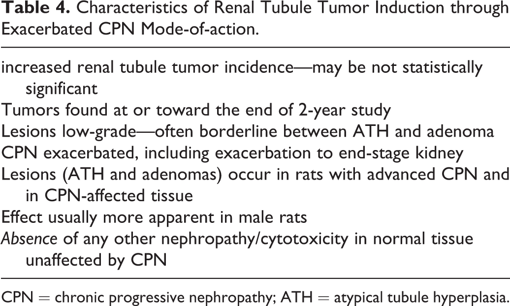
CPN = chronic progressive nephropathy; ATH = atypical tubule hyperplasia.
Spontaneous Tumor Development
Several special rat sub-strains are well-known for having a genetic predisposition for developing RTT. The best known model is the Eker rat, first reported in a Norwegian colony of rats by Eker in 1954. This dominantly inherited renal tumor has been shown to be due to a germline nucleotide insertion in the rat homologue of the tuberous sclerosis 2 (Tsc2) gene (Kobayashi et al. 1995; Yeung et al. 1994). Nearly all rats with this mutation develop RTT by one year of age (Hino et al. 2003). Another example of a Mendelian dominantly inherited predisposition for developing RTTs is the Nihon rat, which was derived from the Sprague-Dawley strain in Japan. This model develops clear cell renal tumors commencing from a very early age due to a single germline nucleotide insertion mutation in the rat Bhd gene (Okimoto et al. 2004). The Long-Evans Cinnamon rat develops an increasing incidence of RTT with increasing age (up to 58% incidence) associated with accumulating copper (Izumi et al. 1994; Kitaura et al. 1999) due to a mutation in the copper-transporting Atpase gene (Wu et al. 1994).
However, from a toxicology perspective, far more important, and not uncommonly encountered in safety evaluation studies, is a spontaneous tumor in rats that has been termed A-V tumor in acknowledgment of its striking amphophilic (or eosinophilic) and vacuolar morphology. This lesion has been observed in all of the conventional breeds of laboratory rat, including the F344, Sprague-Dawley, and Wistar strains, as well as some less common breeds such as the Noble hooded rat. The tumor can be bilateral or multicentric, but there is no record that it is capable of metastasizing. Not only has it been recorded in 2-year studies (Hard et al. 2008) but also in 90-day studies (Hard et al. 1994) and even in younger rats ranging from 12 to 18 weeks of age (Lanzoni et al. 2007).
As indicated above, this tumor is unusual for a rat renal epithelial tumor in having cells with either amphophilic or eosinophilic cytoplasm, coupled with a vacuolar cytology that confers a “moth-eaten” appearance on the tumor. In a survey by Hard et al. in 2008, all of the rat kidneys in the NTP archives that had been diagnosed with a RTT up to that time were reevaluated to determine the frequency of this distinctive RTT. In all, 150 NTP carcinogenicity studies had recorded diagnoses of adenoma, carcinoma, or adenocarcinoma, comprising 1,012 rats (mostly F344) with RTT. Of these studies, almost one-half (74 studies) had one or more rats with proliferative lesions of A-V morphology, and there was a total of 100 rats with these distinctive neoplasms or foci of hyperplasia. Thus, the A-V lesion type appeared in almost 50% of the NTP carcinogenicity studies where an RTT was recorded, and its incidence was approximately 0.1% in the total pool of about 90,000 rats. This neoplastic type showed no strain or gender preference, and statistical analysis demonstrated that it was not linked to chemical treatment, providing unequivocal support for spontaneous origin.
No molecular investigation has been conducted on this tumor type to determine any underlying genetic basis. However, a single report suggests that it is most likely of familial origin. Thurman et al. (1995) from the National Center for Toxicological Research (NCTR) in Arkansas described the occurrence of these tumors in two rats in a long-term study. Unlike most institutions, NCTR had at that time maintained litter records that enabled sibling status to be traced. The two rats bearing A-V renal tumors were littermates, suggesting a hereditary basis for these renal tumors. In addition, the morphology of these tumors strongly resembles some of the aspects of the Eker rat tumor (Everitt et al. 1995). However, unlike the Eker rat tumor, A-V tumors occur in a number of different rat strains, including those from multiple breeding sources.
The unpredictable occurrence of this spontaneous tumor type in carcinogenicity bioassays constitutes a significant confounding influence for assessing tumor incidence associated with chemical exposure. Because of the likely familial nature, and the lack of any association with chemical administration in the many NTP studies evaluated (Hard et al. 2008), lesions with this morphology should, as with oncocytoma, be placed in a separate category and not included in the final (test article–associated) tumor counts.
Concluding Remarks
In summary, nine mechanistic pathways can be identified in relation to the chemical induction of renal tubule cancer in laboratory rodents. Chemicals acting through direct DNA reactivity, indirect DNA reactivity, and mitotic disruption are the most relevant for humans, and generally the linear nonthreshold model is applied as a default assumption for cancer risk assessment. In contrast, chemicals in the category of sustained cell loss/sustained regeneration related to cytotoxicity may be relevant but are treated as having a threshold dose below which toxicity does not occur (Bevan and Harrison 2017). Chemicals in the class involving multiphase bioactivation via GSH could be relevant to humans if they are activated during the metabolic sequence rather than detoxified. The α2u-g nephropathy, exaggerated pharmacologic response, and species-dominant metabolism mechanisms are deemed to have no relevance for humans. Lastly, chemicals causing increased incidence of RTTs through exacerbation of CPN in the absence of other induced tumor types should be regarded as of no concern for humans because CPN is a spontaneous disease of rodents that has no precise human counterpart.
Footnotes
Author Contributions
The author (GCH) contributed to conception or design; data acquisition, analysis, or interpretation; drafting the manuscript; and critically revising the manuscript. The author gave final approval and agreed to be accountable for all aspects of work in ensuring that questions relating to the accuracy or integrity of any part of the work are appropriately investigated and resolved.
Declaration of Conflicting Interests
The author(s) declared no potential, real, or perceived conflicts of interest with respect to the research, authorship, and/or publication of this article.
Funding
The author(s)received no financial support for the research, authorship, and/or publication of this article.