
Abstract
The low background incidence of tumors in rodents from subchronic toxicity studies makes it difficult to assess their relevance, especially when present only in treated animals. This report investigates the occurrence of renal tubule tumors (RTTs), specifically the amphophilic-vacuolar (AV) phenotypic variant, in young Sprague-Dawley (SD) rats from a survey of laboratories conducting subchronic toxicity studies spanning a period of 10 years (2002–2012). This survey establishes a general profile of tumor occurrence; it does not estimate overall incidence or prevalence. AV tumors are spontaneous, nontreatment-related tumors of familial origin, and morphologically distinct from conventional RTTs induced by exposure to renal carcinogens. They are composed of distinct lobules of large, round to polyhedral cells with vacuolated amphophilic to eosinophilic cytoplasm and prominent nucleoli. Data from five collaborating laboratories, representing 37 qualifying studies, are presented. In total, 58 renal tubule neoplasms were recorded in this data set. The AV tumor variant was reported more commonly than the conventional RTT (n = 45 and 13, respectively), and it was recorded in both experimental (n = 32) and control (n = 13) groups. AV tumors occurred more often in females (n = 34) than in males (n = 11); conventional RTTs were recorded more often in males (n = 9) than in females (n = 4). AV tumors often occurred in more than one rat within the same study (up to 7) and were documented to occur in rats as young as 7 to 10 weeks of age. Results from this survey indicate that AV tumors are being reported more commonly in recent years; the majority (n = 33) were reported in studies commencing since 2009. In conclusion, this study reaffirms that AV tumors are spontaneous, nontreatment-related lesions, and suggests that they may be more common than conventional RTTs in young SD rats. The authors propose that AV tumors be recorded separately from conventional RTTs in order to clearly distinguish these two renal tubule neoplasms from one another and allow for appropriate interpretation of a compound’s potential carcinogenic effect in the kidney.
Keywords
Introduction
Due to the low incidence of tumors in rodents from subchronic toxicity studies, it is often difficult to assess their relevance and significance, especially when present only in treated animals. Limited data are available on the occurrence of spontaneous renal tubule tumors (RTTs) in Sprague-Dawley (SD) rats, though they have been reported in both young (Chandra, Riley, and Johnson 1992, 1993; Son and Gopinath 2004; Ikezaki, Takagi, and Tamura 2011) and older animals (McMartin et al. 1992; Nakazawa et al. 2001). This report investigates the occurrence of RTTs, specifically the amphophilic-vacuolar (AV) phenotypic variant, in young SD rats from subchronic toxicity studies (2 weeks to 6 months), and is intended to establish a general profile of their occurrence. Due to the limited nature of this survey, it does not provide an estimate of overall incidence or prevalence of these tumors.
AV tumors are a variant of RTTs that exhibit a morphologic phenotype (Hard et al. 1994, 2008) distinct from that of the conventional RTTs typically seen in carcinogenicity studies, which can occur in rats either spontaneously or as a renal carcinogen-induced lesion. Table 1 summarizes the reported differences between conventional RTTs and the AV RTT variant (adapted from Hard et al. 2008). AV tumors are characterized by a diffuse lobular pattern in which lobules are separated by thin bands of fibrovascular stroma (Figure 1A). Lobules are composed of large, round to polyhedral cells, with amphophilic to eosinophilic cytoplasm, and the cells frequently contain large vacuoles and/or form mini lumens (Figure 1B). Nucleoli are often prominent, and stromal lymphocytic infiltrates are common. AV tumors are often multiple and bilateral, and are often accompanied by regions of atypical tubular hyperplasia. Conventional RTTs (Figure 2A, B) exhibit variable growth patterns and are typically composed of basophilic cells.
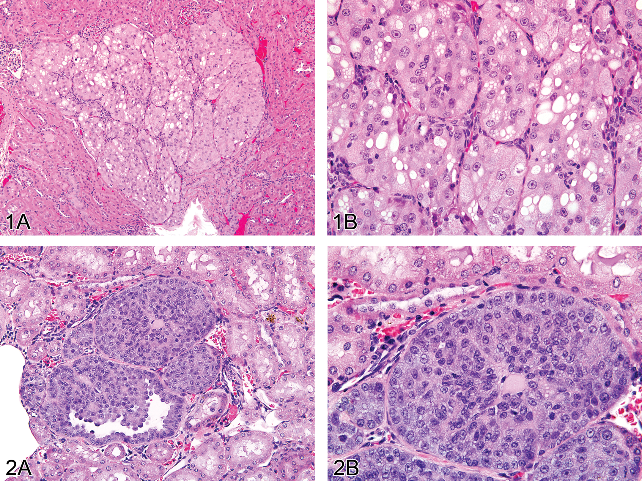
A. AV renal tubule adenoma. Note the distinct lobular pattern, eosinophilic to amphophilic cytoplasmic staining, numerous variably sized intracytoplasmic vacuoles, and lymphocytic infiltrates. H&E. 12.5×. B. Higher magnification of 1A. Note the eosinophilic to amphophilic cytoplasmic staining, numerous variably sized intracytoplasmic vacuoles, and prominent nucleoli. H&E. 40×. F
Comparison of AV-type tumors with conventional RTTs in rats.
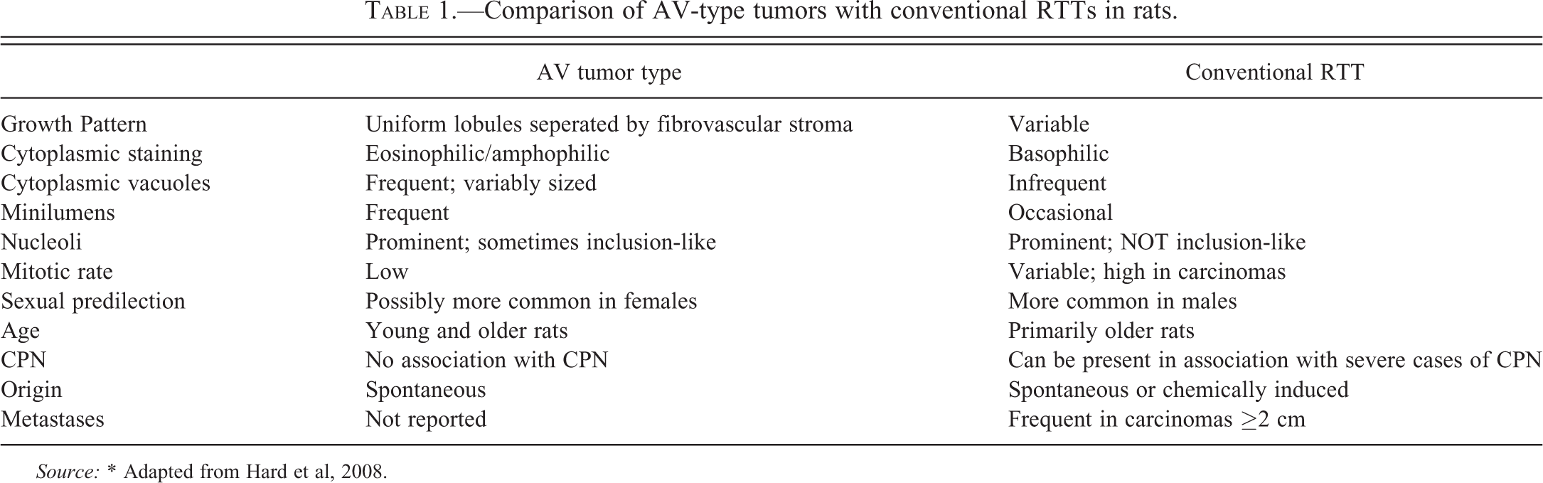
Source: * Adapted from Hard et al, 2008.
AV tumors have previously been demonstrated to be spontaneous, nontreatment-related tumors of familial origin (Hard et al. 2008). They are similar in appearance to the renal neoplasms described in the Eker rat (Everitt et al. 1992). AV tumors have been reported to occur in young rats in 90-day studies (Hard et al. 1994; Hall et al. 2007; Lanzoni et al. 2007), as well as in one group of F344 littermates (Thurman et al. 1995), and occur at a low incidence in 2-year carcinogenicity studies (Hard et al. 2008).
This study presents data from five collaborating laboratories, representing 37 qualifying studies. Laboratories surveyed their databases over a 10-year period, spanning from 2002 to 2012, for rat subchronic toxicity studies in which RTTs and AV tumors were recorded. The distribution of AV tumors between dose groups was examined in order to determine whether they were spontaneous or treatment related.
Methods and Materials
Study Design
Laboratories were contacted individually to seek interest in collaboration. In total, five laboratories (Laboratories A–E) agreed to participate. Each collaborating laboratory was requested to conduct a survey of their databases of subchronic studies. For the sake of this survey, “subchronic” was defined as any study lasting up to 6 months in length, and included two generational studies in which the F1 generation of pups was evaluated. Database searches were confined to a 10-year span, extending from 2002 to 2012, and search criteria were limited to renal tubule adenomas and carcinomas and AV renal tubule adenomas and carcinomas. A study qualified for inclusion if: (1) it was a subchronic study, as previously defined; and (2) at least one animal was recorded with at least one of the aforementioned renal tumor diagnoses. The following studies were not included: studies in which animals were diagnosed with a primary renal tumor other than that previously mentioned (nephroblastoma, transitional cell tumor, etc.), and studies in which animals were diagnosed with a secondary or metastatic tumor (lymphoma, histiocytic sarcoma, etc.). Laboratories were supplied with a standardized format in an Excel spreadsheet in which the following information was requested for each qualifying study: study length, rat strain, sex, number of experimental and control groups, number of animals per group, number of kidneys examined per group, and number of times the specific diagnoses were made per group. Data were received and compiled for evaluation.
In order to provide additional information on the extent of the occurrence of AV tumors among the qualifying studies, collaborating laboratories were requested, whenever possible, to microscopically reexamine slides from animals in which RTTs were recorded. AV tumors were identified by the following characteristic morphologic features: well-circumscribed, non-encapsulated masses composed of well-delineated lobules separated by thin strands of fibrovascular stroma with lymphocytic infiltrates; large round to polyhedral tumor cells with finely granular to vacuolated amphophilic to lightly eosinophilic cytoplasm; large vesiculated nuclei with prominent nucleoli; and frequent central degeneration/necrosis of the lobules. When previously diagnosed RTTs matched these criteria, collaborating laboratories were informed to signify this on their data sheet.
Results
General Summary
Each of the five collaborating laboratories (A–E) surveyed their database(s) over a 10-year period for the occurrence of RTTs and AV RTTs in rat subchronic toxicity studies (2 weeks to 6 months). A total of 37 studies (Table 2) qualified for inclusion; renal tumors were recorded 58 times (Table 3). All studies were conducted with SD rats that ranged in age from 5 to 8 weeks at the study start, depending on the laboratory. Some aspects of the data related to the distribution of the tumor types are summarized in Table 4. Qualifying studies ranged in length from 2 weeks to 6 months and included several two-generational and reproductive toxicity studies (Table 5).
Number and distribution of qualifying studies per collaborating laboratory.
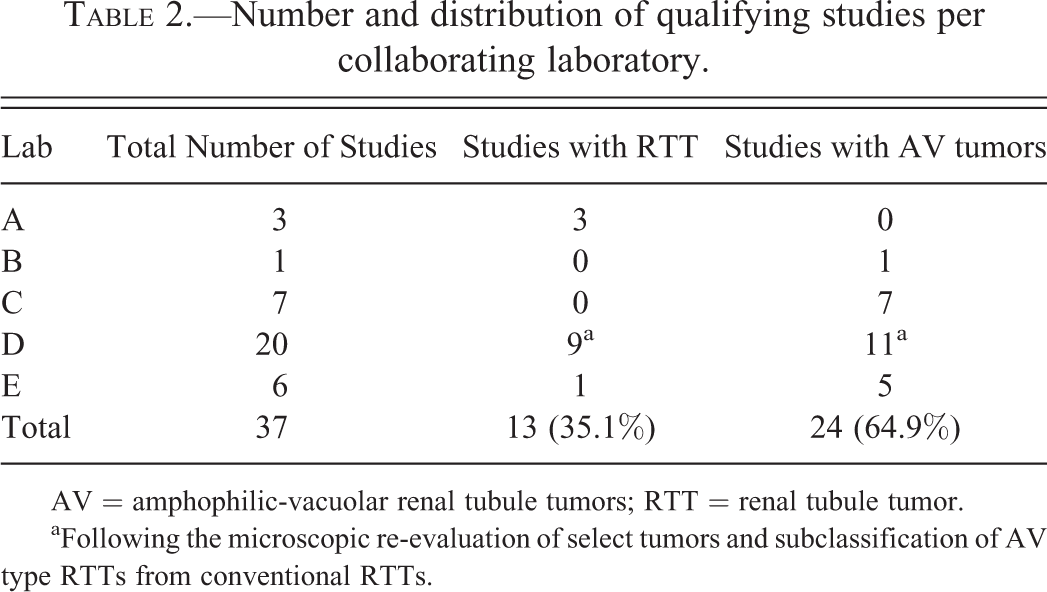
AV = amphophilic-vacuolar renal tubule tumors; RTT = renal tubule tumor.
aFollowing the microscopic re-evaluation of select tumors and subclassification of AV type RTTs from conventional RTTs
Number and distribution of individual tumors per collaborating laboratory.
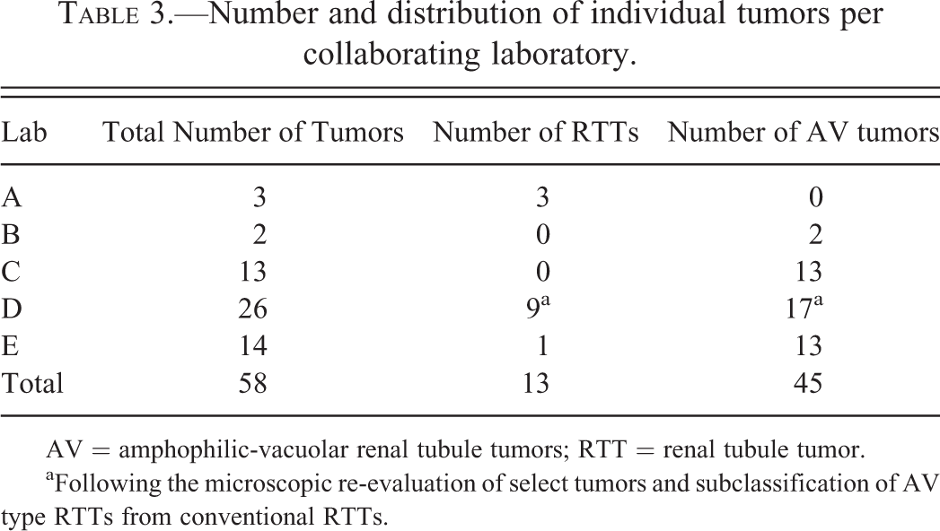
AV = amphophilic-vacuolar renal tubule tumors; RTT = renal tubule tumor
aFollowing the microscopic re-evaluation of select tumors and subclassification of AV type RTTs from conventional RTTs
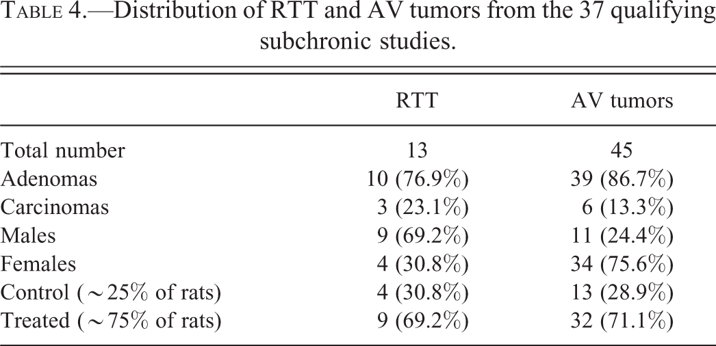
Distribution of RTT and AV tumors from the 37 qualifying subchronic studies.
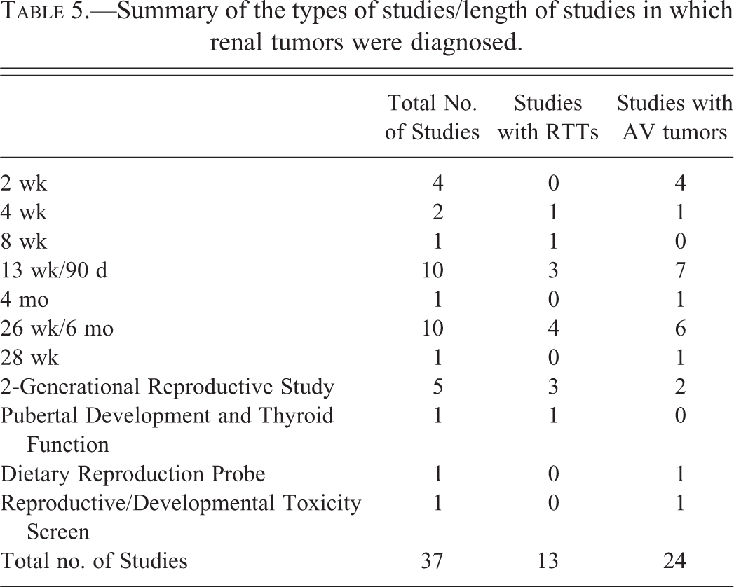
Summary of the types of studies/length of studies in which renal tumors were diagnosed.
Laboratory D microscopically reevaluated a subset of its tumors (18 of the 26). It should be noted that Laboratory D had not previously subclassified AV types of RTTs from conventional RTTs; both conventional and AV types of RTTs had been recorded as “renal tubule adenoma.” However, in studies where RTTs had occurred in treated animals or in multiples, the pathologist often commented in the narrative that the renal tubule adenomas were morphologically consistent with an AV type, emphasizing the spontaneous familial nature of these tumors. Prior to reevaluation, 24 RTT adenomas and 2 RTT carcinomas had been reported by Laboratory D. Seventeen of the 24 RTT adenomas and 1 of the 2 RTT carcinomas were available for microscopic reevaluation. All of the reevaluated tumors, except 1 RTT adenoma, were noted by the reviewing pathologist to be able to be subclassified as an AV type of RTT.
Summary of AV Tumors
Following reevaluation of the subset of tumors from Laboratory D, a total of 39 AV adenomas and 6 AV carcinomas were identified.
AV tumors were recorded in both control (11 adenomas, 2 carcinomas) and experimental (28 adenomas, 4 carcinomas) groups. While AV tumors were recorded 2 to 3 times as often in the experimental groups as compared to the control groups, this reflects that most studies had multiple experimental groups per study as compared to control groups. Within the experimental groups, the occurrence of AV tumors was sporadic and no consistent treatment-related effect was noted by any of the laboratories. Furthermore, Laboratories B, C, and D clearly stated that in all incidences in which AV tumors were discussed in the interpretation, they were specifically determined to be spontaneous, nontreatment-related tumors based upon the young age of the animals, the sporadic occurrence of the tumors, and the lack of coinciding evidence of toxic changes in the tubules.
AV tumors were recorded in females approximately 3 times as often as males (n = 34 and 11, respectively). In comparison, RTTs were recorded more often in males (n = 9) than in females (n = 5).
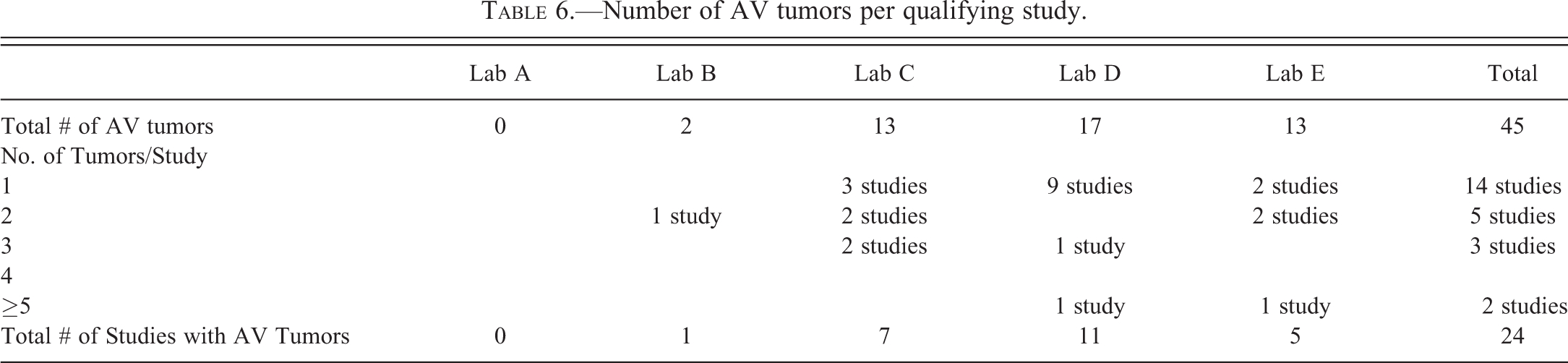
The occurrence of AV tumors within a given study was variable, ranging from 1 to 7 (Table 6). While tumors were most often recorded as isolated incidences (14 studies), it was not uncommon for tumors to occur within multiple rats within the same study and/or group.
Number of AV tumors per qualifying study.
AV tumors were recorded in rats from studies that ranged in length from 2 weeks to 6 months (Table 5). While the exact age of the rats at the time of necropsy for each qualifying study was unknown, animals were 5 to 8 weeks of age at the start of the studies. Therefore, tumors were recorded in rats as young as 7 to 10 weeks of age.
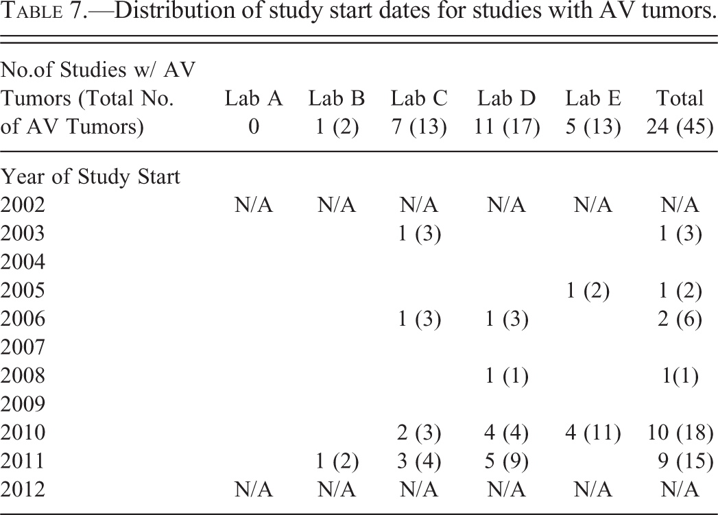
To determine whether AV tumors are more commonly being identified and/or recorded in more recent studies, collaborating laboratories supplied the start dates for each qualifying study. These data are presented in Table 7. Since 2009, there is an apparent increase in the number of studies with AV tumors. From 2002 through 2008, there were a total 14 studies in which renal tumors were reported, 5 of which (37.5%) had AV tumors. In contrast, a total of 23 studies were reported during the time frame of 2009 to 2012, 19 of which (82.6%) had AV tumors. Consequently, the majority of AV tumors recorded occurred in studies that had been initiated since 2009 (n = 33), as opposed to those started prior to 2009 (n = 12).
Distribution of study start dates for studies with AV tumors.
Discussion
RTTs with AV morphologic features were first reported to occur spontaneously in rats nearly 60 years ago (Eker 1954). It is now known that this spontaneous occurrence in the Long-Evans (Eker) rat is due to mutations in the tuberous sclerosis gene (Tsc2; Kobayashi et al. 1995). A morphologically similar tumor has also been observed in SD (Nihon) rat strains and is attributable to mutations in the Birt-Hogg-Dubé tumor suppressor gene (Kouchi et al. 2006). Similar tumors have also been observed in subchronic feeding studies conducted in SD rats (Hard et al. 1994; Hall et al. 2007; Lanzoni et al. 2007). In each case, the occurrence of these tumors was determined to be spontaneous due to the timing of their appearance and the fact that their distinct histological profile clearly differentiated them from chemically induced renal tumors. Chemically induced renal tumors are typically not observed in rats less than 9 to 10 months of age, and only then occur following exposure to extremely potent carcinogens, such as ochratoxin or diethylnitrosamine (Hard et al. 1994). Carcinogen-induced RTTs tend to occur focally and are generally preceded by evidence of nephrotoxicity, even in animals in which tumors do not develop (Hard et al. 1995). In contrast, AV tumors are multifocal and characterized by the presence of large amphophilic and small basophilic cell types, large tumor size, and have been identified in rats as young as 3 weeks of age (Lanzoni et al. 2007; Hard et al 2008; Okimoto et al. 2004).
The occurrence of spontaneous tumors in subchronic toxicity studies has the potential to mislead with regard to causality, particularly when one or more tumors occur in an experimental group. In the case of AV tumors, very little information is available regarding their occurrence in SD rats. Lanzoni et al. (2007) conducted a survey in young (≤18 weeks of age) SD rats from toxicity studies of shorter duration, reporting an overall incidence of 0.66% for preneoplastic and neoplastic lesions in the kidney; however, this study did not recognize AV tumors as a distinct entity and included foci of atypical hyperplasia. Females were affected more commonly than males (0.97% and 0.44%, respectively). Hard et al. (2008) reported the incidence of AV tumors in aged rats from 2 year carcinogenicity at ∼0.1% and demonstrated a relatively equal distribution between the sexes (females: 53.0%; males: 47.0%). However, the F344 rat strain comprised 92% of the rats used in these studies; where as only 7% were accounted for by SD rats.
We surveyed a number of facilities where subchronic rat studies were conducted with SD rats. This survey demonstrated that AV tumors have been observed in numerous studies and in rats from both control and experimental groups. In all incidences, the occurrence of AV tumors was considered by the laboratory to be spontaneous and not related to test substance exposure. This was due to their sporadic occurrence, the young age of the animals, and the lack of additional toxic tubular changes noted within the kidneys. One important observation from this survey is that AV renal tumors are perhaps not as rare as initially presumed. In younger rats, they may in fact be more common than conventional RTTs. This was supported by the reevaluation of several RTTs from Laboratory D. All but 1 of the 18 RTT tumors that were reevaluated was determined to be morphologically consistent with an AV tumor. Another interesting observation from this survey is that it was not uncommon for multiple tumors to appear within the same study. Given that animals within a study are often littermates, this finding supports the familial nature of these neoplasms and stresses the importance of striving for genetic diversity.
This study also suggests that AV tumors may be occurring more commonly in recent years, as the majority of recorded AV tumors occurred in studies commencing since 2009. While it is possible that this represents an increased awareness of this entity due to recent publications, pathologists have been aware of these types of tumors for decades. The results of this survey suggest that the recent increase is real and that there may be an increased prevalence of underlying, predisposing genetic factors within the strains of SD rats currently being utilized.
It is important to note that the results from this report relate to findings from a limited survey. This article is not meant to serve as an incidence article for these tumors, as only subchronic studies in which RTTs had previously been diagnosed were included. Therefore, this subset represents an extremely small portion of the total overall number of subchronic studies conducted at the various laboratories. Due to database limitations, this information was unable to be obtained. Therefore, it would be incorrect to attempt to extrapolate or estimate the overall incidence or prevalence of AV tumors from the data presented within this limited data set.
The results of this survey highlight the need for caution when interpreting results from subchronic studies conducted in SD rats, particularly when it involves the occurrence of tumors in animals from only treated groups. This survey supports previous findings that AV tumors occur spontaneously in both control and treated animals and that, to date, no study has demonstrated a treatment-related effect with regard to occurrence of these types of tumors. As previously suggested by Hard et al. (2008), the authors propose that AV tumors be recorded separately from conventional RTTs, in order to clearly distinguish these two renal tubule neoplasms from one another and allow for appropriate interpretation of a compound’s potential carcinogenic effect in the kidney.
Footnotes
The authors declared no potential conflicts of interest with respect to the research, authorship, and/or publication of this article.
The authors disclosed receipt of the following financial support for the research, authorship, and/or publication of this article: Funding for this work work was provided by DuPont.