
Abstract
Microscopic examination of the brain of adult Beagle dogs from four different general toxicity studies revealed the presence of ectopic choroid plexus tissue in six individual dogs (4 females and 2 males) with ages ranging from 12 to 18 months. In each dog, this finding was characterized by a well-circumscribed mass localized to a region above and along the corpus callosum without any apparent compression of adjacent brain tissue. Each mass was composed of columnar ependymal cells forming tubular structures surrounded by variable amounts of fibrovascular connective tissue and had the appearance of small rests of ependymal cells that had been penetrated by the leptomeninges during neural development. There were no associated clinical signs or macroscopic correlates. Based on morphologic appearance, a diagnosis of spontaneous ectopic choroid plexus with secondary sclerosis was made. To the authors’ knowledge, ectopic choroid plexus has not been reported in Beagle dogs and is rare in humans and horses.
Differentiating between test article–related and spontaneous findings is an important factor in the accurate interpretation of microscopic data from general toxicity studies. To do this, it is critical to recognize and understand the wide range of spontaneous findings that can occur in each species used in these studies. The Beagle dog is one of the nonrodent species widely used in general toxicity studies, and here, we report the observation of a novel spontaneous lesion (ectopic choroid plexus) in the brain of this species. Ectopic choroid plexus tissue was noted incidentally during the routine light microscopic examination of the brain of six different dogs (4 females and 2 males with ages ranging from 12 to 18 months), involving four separate general toxicity studies. In all instances, this finding occurred in the absence of associated clinical signs. Upon completion of each study, the animals were humanely euthanized, and a full necropsy was performed without identification of any correlating macroscopic findings in the brain. Brains were fixed in 10% neutral-buffered formalin and sectioned following recommendations by Bolon et al. (2013). The Beagle dogs from these regulatory studies were purchased from Marshall Farm Marshall BioResources (North Rose, NY) and housed for at least 10 days before the first day of study. All procedures were approved by the animal care and use committee in accordance with federal regulations and the Guide for the Care and Use of Laboratory Animals.
The ectopic choroid plexus in each dog was characterized by a well-circumscribed mass, located above and along the corpus callosum with no compression of adjacent brain tissue (Figure 1). Each mass was composed of columnar ependymal cells forming tubular structures surrounded by variable amounts of fibrovascular connective and adipose tissue and had the microscopic appearance of small rests of ependymal cells that had been penetrated by the leptomeninges during neural development but failed to migrate to the ventricles through choroid fissures (as would have occurred during normal development of the choroid plexus; Figure 2). Based on this microscopic appearance, a morphologic diagnosis of ectopic choroid plexus with secondary sclerosis was made. This finding was considered spontaneous because it was observed sporadically, at low incidence and without dose relationship, including the involvement of a control animal.
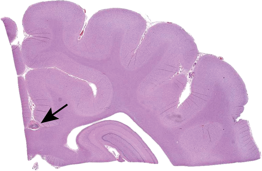
Brain section from a Beagle dog at the level of the dorsal aspect of the thalamus. The black arrow is depicting the ectopic choroid plexus above the corpus callosum.
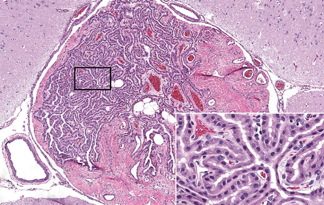
Ectopic choroid plexus located above the corpus callosum and composed of columnar ependymal cells forming tubular structures surrounded (inset) by variable amounts of fibrovascular connective and adipose tissue and had the appearance of small rests of ependymal cells that had been penetrated by the leptomeninges during neural development.
The choroid plexus forms at the end of embryonic development originating from several locations along the dorsal axis of the neural tube, and it has dual embryonic origin including a stromal component of mesenchymal origin (like meninges) and an epithelial component derived from neuroepithelium (roof plate; Liddelow 2015). After neural tube closure, the hindbrain choroid plexus of the fourth ventricle is the first segment to appear, followed by synchronous development of the telencephalic choroid plexus in each lateral ventricle and, finally, the diencephalic choroid plexus in the third ventricle. Because the ectopic choroid plexus in these dogs extended dorsally to the corpus callosum, it is suspected that these were the result of abnormal development of the diencephalic choroid plexus since this segment bifurcates during development, sending a branch through the intraventricular foramina and ultimately fusing into one continuous tissue with the telencephalic choroid plexus (Lun, Monuki, and Lehtinen 2015).
Because this finding was present in two consecutive studies, a genetic association was suspected. Genetic analysis was performed (data not shown and a familial relationship between the affected dogs was not found). To the authors’ knowledge, ectopic choroid plexus has not been reported in Beagle dogs, although it has been rarely reported in humans (Ha et al. 2014) and horses (Baker and Ellis 1981). Sato et al. (2012) reported a similar lesion in the brain of Beagle dogs and called it “hamartoma in the callosal sulcus.” They characterized this finding as a rare lesion that consisted of choroid plexus components (epithelia, fatty tissue, abundant collagen fibers, and small vessels), although they considered the finding to be ectopic tissue.
In conclusion, spontaneous ectopic choroid plexus with sclerosis was an incidental microscopic finding noted in Beagle dogs from four different general toxicity studies. This finding lacked any macroscopic correlates and was not associated with any clinical signs.
Footnotes
Declaration of Conflicting Interests
The author(s) declared no potential conflicts of interest with respect to the research, authorship, and/or publication of this article.
Funding
The author(s) received no financial support for the research, authorship, and/or publication of this article.