
Abstract
Vehicle control Harlan RCCHan™:WIST rats were examined to provide control data for subsequent studies. The rats (180 male and 180 female) were dosed daily via oral gavage with reverse osmosis water for up to 104 weeks. At necropsy, body weights and macroscopic findings were recorded and tissues were collected for histopathology. The mean body weight at terminal sacrifice was 687 g for males and 466 g for females. The overall survival rate was 62% for males and 59% for females. The most common cause of death for males and females found dead or examined following unscheduled euthanasia was pituitary neoplasia with an incidence of 13.9% for males and 18.9% for females. Macroscopic and neoplastic and nonneoplastic microscopic findings are presented by body system.
Keywords
Historical control data are important for the evaluation and interpretation of certain macroscopic, neoplastic, and nonneoplastic findings identified during rodent carcinogenicity studies. For common or uncommon findings with unexpected incidence, historical control data can provide information about the incidence of spontaneously occurring background findings in the species and strain of animal used on test. While the concurrent control group is the most appropriate comparator, historical control data help to support the interpretation of common and uncommon findings (Deschl et al. 2002; Keenan et al. 2009). In addition, historical control data are useful when selecting and assessing the suitability of an animal model (Weber et al. 2011). The purpose of this report is to summarize and present mortality and body weight data and the incidence of macroscopic findings and neoplastic and nonneoplastic microscopic findings for control Harlan RCCHan™:WIST rats from 104-week oral gavage studies.
Methods
Male and female RCCHan™:WIST rats 4 to 8 weeks of age were obtained from Harlan Laboratories, Inc., Frederick, Maryland, in 2 shipments of 180 animals each (90 animals/sex/shipment). Animals were randomized into 1 of 3 groups with group-specific diets (Harlan Laboratories, Inc.). Group 1 received Certified Rodent Diet #2016C (pellet), group 2 received Certified Rodent Diet #2016C (meal), and group 3 received Certified Rodent Diet #2016C (extruded pellet). Animals were housed individually in stainless steel cages in an AAALAC-accredited facility under approved Animal Care and Use Committee guidelines. The study was conducted in compliance with the U.S. Food and Drug Administration Good Laboratory Practice Regulations, and all procedures were in compliance with the Animal Welfare Act, the Guide for the Care and Use of Laboratory Animals, and the Office of Laboratory Animal Welfare. Surviving animals were sacrificed after 104 weeks of daily oral gavage dosing with reverse osmosis water. At the terminal sacrifice, animals were fasted overnight, and terminal body weights were recorded. Animals were anesthetized with sodium pentobarbital, exsanguinated, necropsied, and macroscopic observations were recorded. Tissues (Appendix Table A1) from all animals were collected in 10% neutral-buffered formalin or modified Davidson’s fixative (eye, Harderian gland, optic nerve, and testis), embedded in paraffin, sectioned, stained with hematoxylin and eosin, and examined microscopically. Microscopic findings were recorded and categorized as neoplastic or nonneoplastic providing consistency with a previous paper presenting findings in Harlan RCCHan™:WIST rats at 4, 13, and 26 weeks (Blankenship and Skaggs 2012). Some microscopic diagnoses (e.g., Harderian gland alteration or ventral compression of the brain) were made to confirm and provide microscopic correlates for macroscopic observations recorded by prosectors at necropsy (e.g., white/tan discoloration in the Harderian gland or depressed area in the brain). Given the length of the studies, duration modifiers (e.g., chronic) were applied when appropriate in order to better characterize a finding. Similarly, subanatomic sites (e.g., nasolacrimal duct included under nasal turbinate) were applied to indicate when a specific structure or region was involved. All neoplasms and hyperplasias from all animals and all nonneoplastic findings from a subset of animals (10%) were peer reviewed.
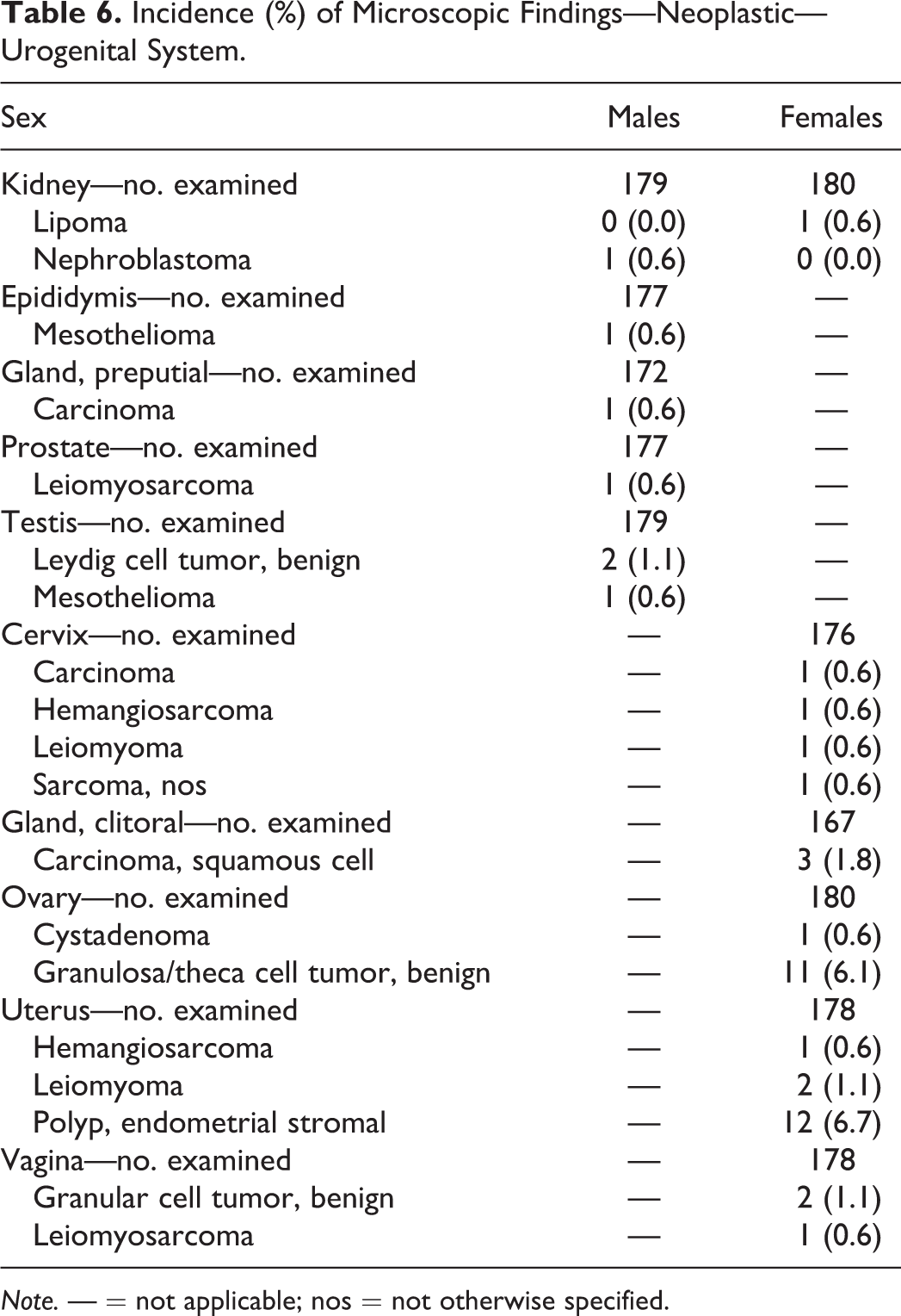
Results and Discussion
Mortality
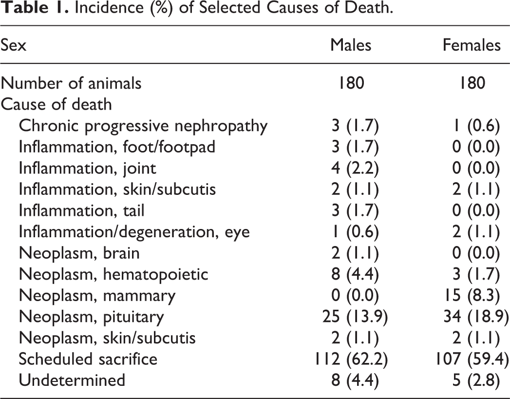
Of the 360 animals evaluated, unscheduled deaths (animals found dead or sacrificed in moribund condition) occurred in 68 males and 73 females, resulting in an overall survival rate of 62% for males and 59% for females. With respect to groups, survival rates were 60%, 72%, and 55% for groups 1, 2, and 3 males, respectively, and 50%, 70%, and 58% for groups 1, 2, and 3 females, respectively. Notably, survival rates were higher in group 2 animals (those fed the meal diet). The most common cause of death for males and females found dead or examined following unscheduled euthanasia was pituitary neoplasia with an incidence of 13.9% in males and 18.9% in females. Selected causes of death (those with an incidence >1%) are presented in Table 1.
Incidence (%) of Selected Causes of Death.
Terminal Body Weights
The overall mean body weight at terminal sacrifice was 687 g for males and 466 g for females. These are similar to the male average (650 g) and slightly higher than the female average (355 g) reported by Weber et al. (2011). Group mean body weight data are presented in Table 2.
Mean Body Weight (g).
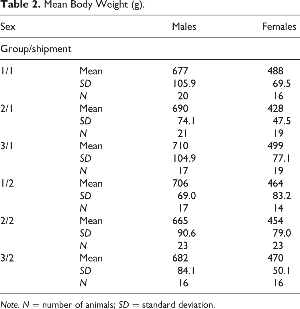
Note. N = number of animals; SD = standard deviation.
Cardiovascular System
Macroscopic findings
The only macroscopic finding observed in the heart was white/tan discoloration which correlated microscopically with cardiomyopathy (Table 3).
Incidence (%) of Macroscopic Findings—Cardiovascular System.
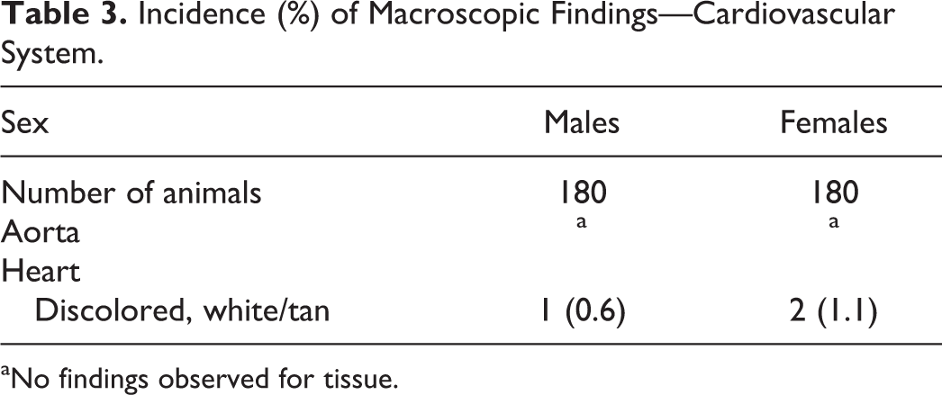
aNo findings observed for tissue.
Microscopic findings—neoplastic
No neoplastic findings were observed in the cardiovascular system. Vascular neoplasms (hemangioma and hemangiosarcoma) occurred in multiple tissues and were reported under the tissue in which they were observed (skin/subcutis, uterus, spleen, mesenteric lymph node, and thyroid).
Microscopic findings—nonneoplastic
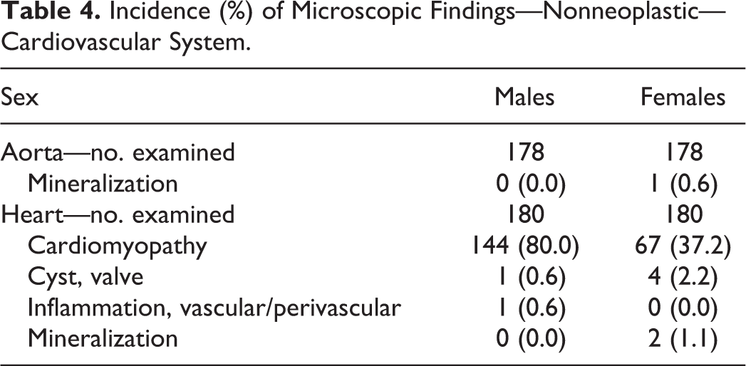
Cardiomyopathy was the most common microscopic finding in the heart (80.0% of males and 37.2% of females; Table 4) and was generally minimal or slight. Cardiomyopathy was characterized by cardiomyocyte degeneration/necrosis, inflammatory cell infiltrate, and/or fibrosis. The diagnosis of the individual components or the use of the aggregate term cardiomyopathy can vary on a study basis (Keenan et al. 2010). Because of the length of this study, the aggregate term cardiomyopathy was used. However, use of the aggregate term may pose translational challenges because of the similarly named human condition.
Incidence (%) of Microscopic Findings—Nonneoplastic—Cardiovascular System.
Urogenital System
Macroscopic findings
Common macroscopic findings included rough kidney surface, large urinary bladder, small seminal vesicle, and small and/or soft testis in males and large renal pelvis and cyst in the ovary and uterus in females (Table 5).
Incidence (%) of Macroscopic Findings—Urogenital System.
Note. — = not applicable.
aNo findings observed for tissue.
Microscopic findings—neoplastic
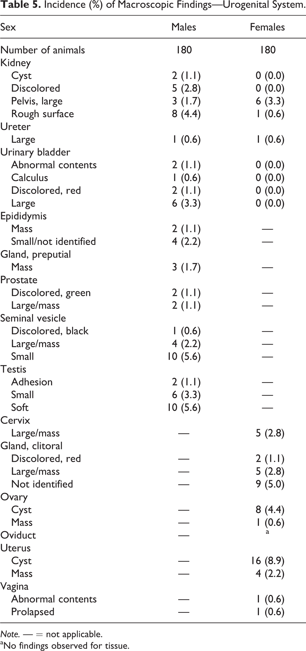
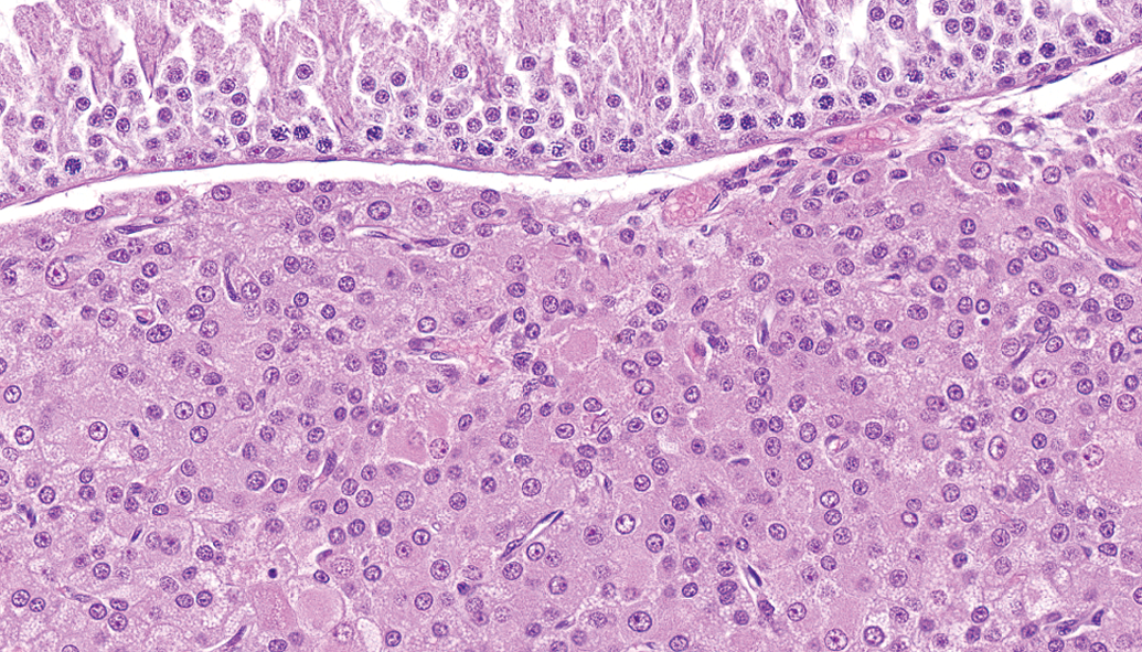
Similar to reports in other rat strains (e.g., Sprague-Dawley, Fischer 344, and Osborne-Mendel; Brix et al. 2005; Solleveld and Boorman 1986), neoplasms in the kidney were rare and included lipoma and nephroblastoma (Figure 1). Leydig cell tumor (Figure 2) was the most common neoplasm in males (1.1%), while endometrial stromal polyp (6.7%) and granulosa/theca cell tumor (6.1%; Figure 3) were the most common in females (Table 6).
Kidney—nephroblastoma, showing basophilic blastema, primitive tubules, and glomeruloid structures. Mitotic figures were common (hematoxylin and eosin).
Testis—Leydig cell tumor. Neoplastic polygonal cells have abundant eosinophilic cytoplasm and little atypia (hematoxylin and eosin).
Ovary—granulosa/theca cell tumor. The predominant neoplastic cells are spindle shaped, arranged in bundles, and have minimal eosinophilic cytoplasm (hematoxylin and eosin).
Incidence (%) of Microscopic Findings—Neoplastic—Urogenital System.
Note. — = not applicable; nos = not otherwise specified.
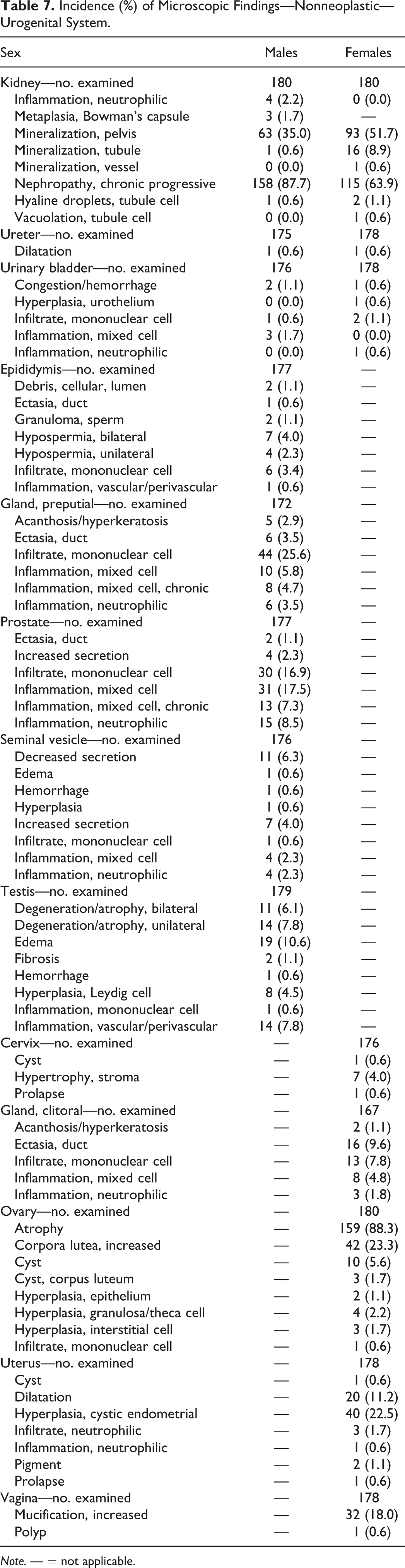
Microscopic findings—nonneoplastic
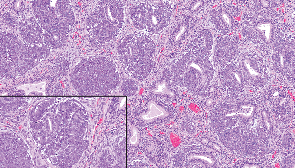
Chronic progressive nephropathy (CPN) was the most common microscopic finding in the kidney affecting 87.7% of males and 63.9% of females (Table 7). At minimal severity, CPN was characterized by basophilic tubules and thickened basement membranes (Seely and Frazier 2015) with the addition of tubule dilatation, hyaline casts, and mononuclear infiltrates at higher severity (Figure 4). CPN severity was generally minimal or slight with relatively few animals reaching higher severity; in general, the severity was lower in females than in males. The higher incidence and severity observed in males are similar to that generally recognized in other strains (Sprague-Dawley and Fischer 344; Hard and Khan 2004). Mineralization in the renal pelvis (typically located in the renal fornices) was also relatively common affecting 35.0% of males and 51.7% of females. Associated with histiocytic sarcoma, hyaline droplets were occasionally observed in the kidney (Figure 5).
Incidence (%) of Microscopic Findings—Nonneoplastic—Urogenital System.
Note. — = not applicable.
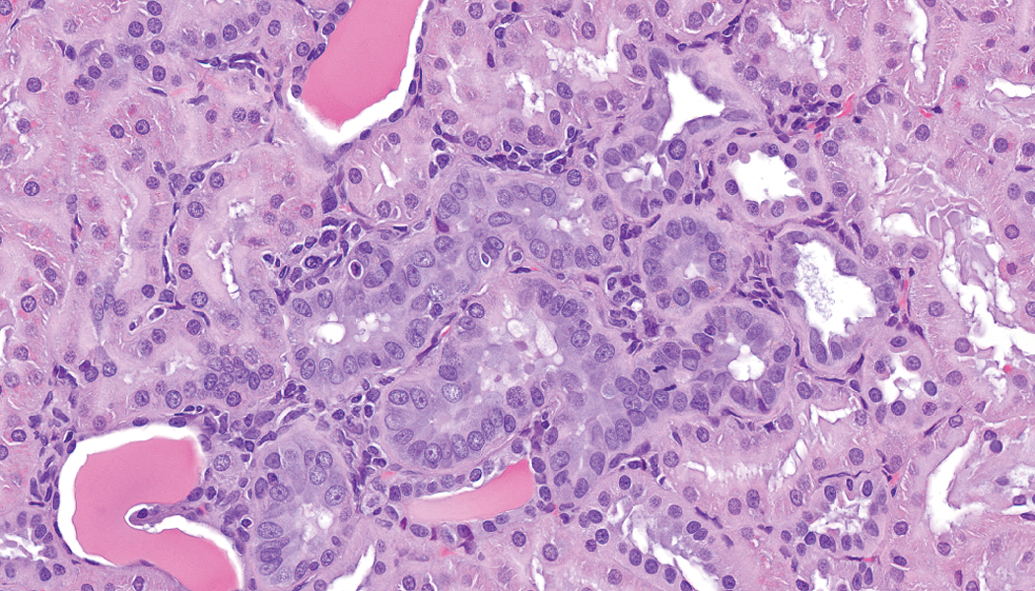
Kidney—chronic progressive nephropathy, basophilic tubules with thickened basement membranes, mononuclear cell infiltrates, tubular dilatation, and hyaline casts (hematoxylin and eosin).
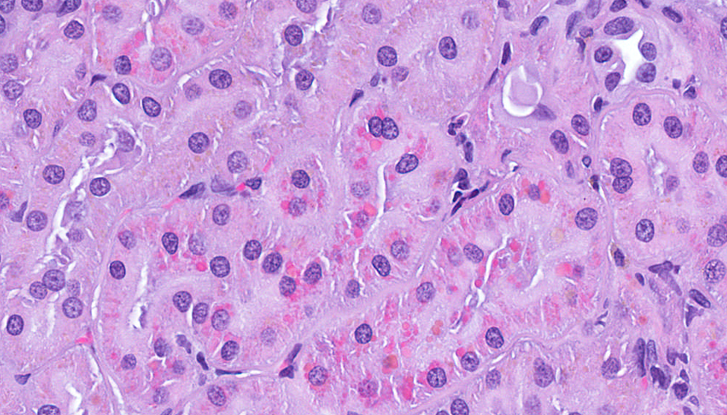
Kidney, tubule cells—histiocytic sarcoma associated hyaline droplets (hematoxylin and eosin).
Ovarian atrophy was present in 88.3% of females by 104 weeks and was characterized by decreased numbers of corpora lutea, atretic follicles, and increased interstitial cells (Table 7).
Respiratory System
Macroscopic findings
White/tan discoloration was the most common macroscopic finding in the lung (Table 8) and generally correlated microscopically with alveolar macrophage infiltrates.
Incidence (%) of Macroscopic Findings—Respiratory System.
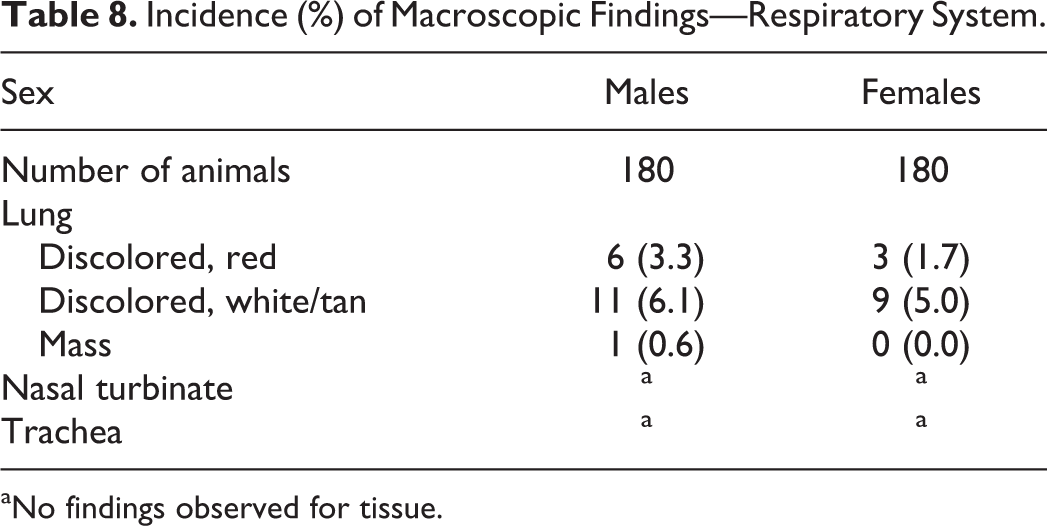
aNo findings observed for tissue.
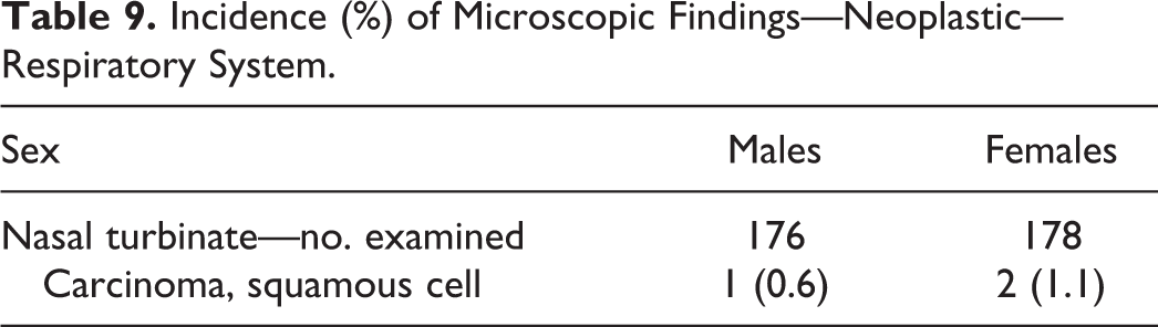
Microscopic findings—neoplastic
The only neoplasms observed in the respiratory system were squamous cell carcinoma in the nasal turbinates; no neoplasms were observed in the lungs or trachea (Table 9). The absence of lung neoplasms in this study was consistent with the low incidence (0.1%) reported in Wistar rats by Poteracki and Walsh (1998).
Incidence (%) of Microscopic Findings—Neoplastic—Respiratory System.
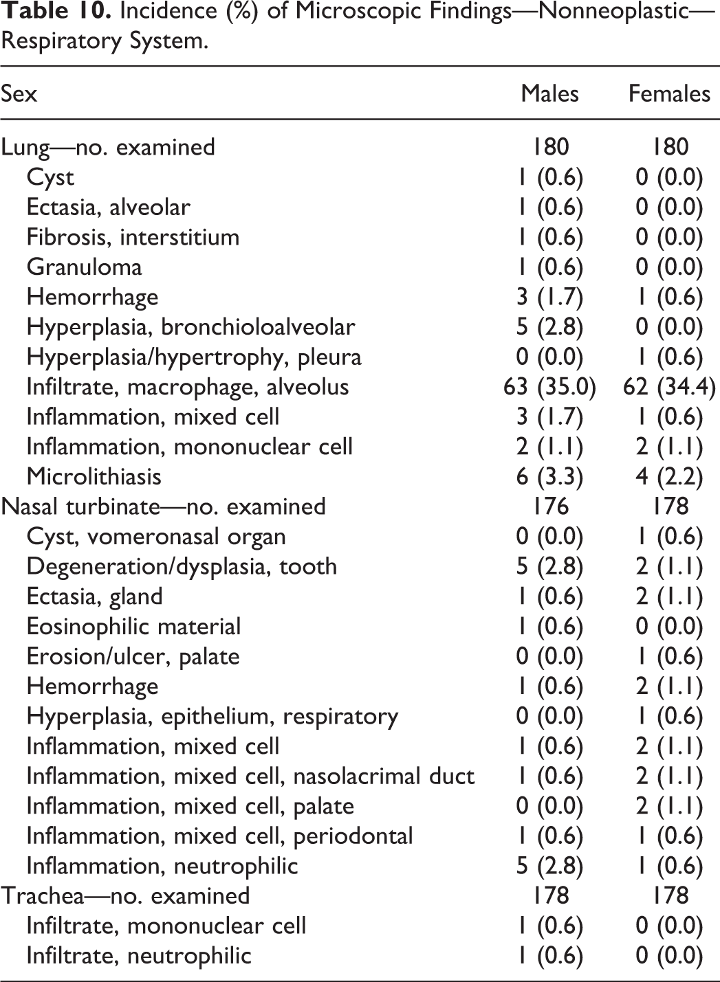
Microscopic findings—nonneoplastic
Alveolar macrophage infiltrates were the most common finding observed in the lungs (Table 10). Affected alveoli contained focal aggregates of macrophages with finely vacuolated cytoplasm, were frequently subpleural or perivascular/bronchiolar, and infrequently associated with additional cellular infiltrates or inflammatory changes.
Incidence (%) of Microscopic Findings—Nonneoplastic—Respiratory System.
Digestive System
Macroscopic findings
Discoloration in the liver and stomach were the most common macroscopic findings in the digestive system (Table 11) and correlated microscopically with vacuolation or foci of cellular alteration in the liver and congestion/hemorrhage in the glandular stomach.
Incidence (%) of Macroscopic Findings—Digestive System.
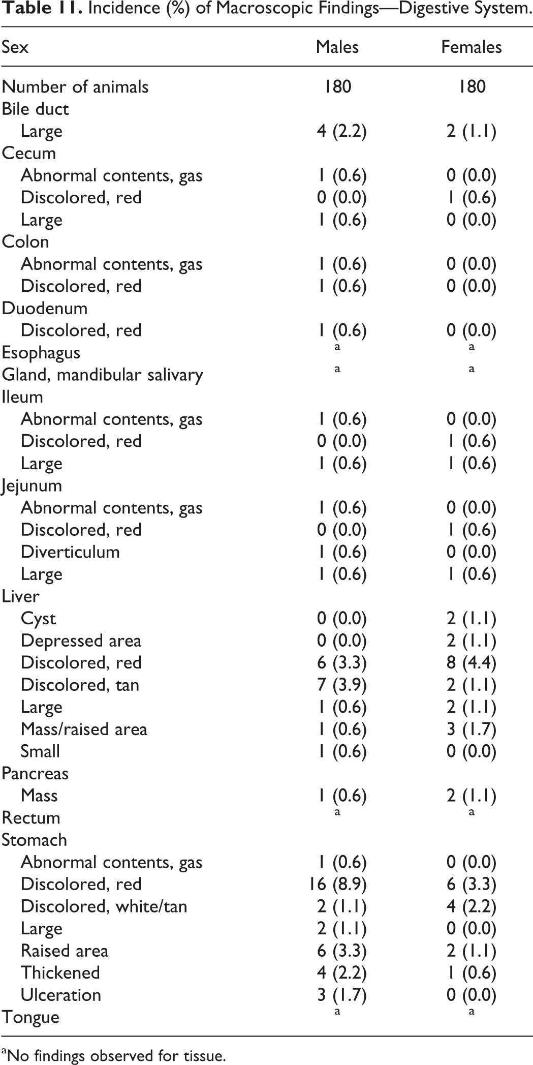
aNo findings observed for tissue.
Microscopic findings—neoplastic
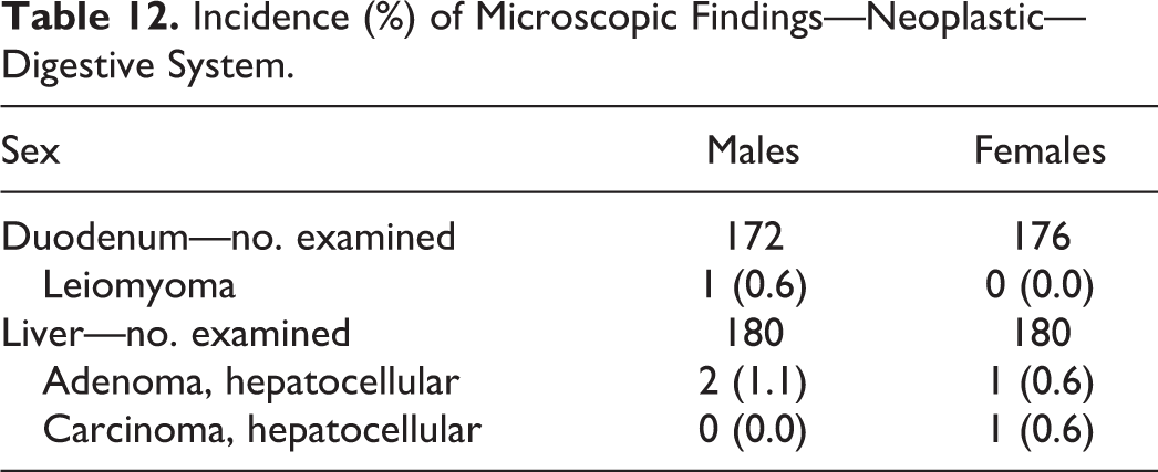
Neoplasms in the digestive system were rare and limited to the liver and duodenum (Table 12).
Incidence (%) of Microscopic Findings—Neoplastic—Digestive System.
Microscopic findings—nonneoplastic
Common microscopic findings observed in the liver included foci of cellular alteration, hepatocellular vacuolation, mononuclear cell infiltrates, periportal fibrosis, and bile duct hyperplasia (Table 13). With respect to foci of cellular alteration, basophilic foci were the most common in females (71.7%) and eosinophilic foci were the most common in males (51.1%). This was similar to that reported in Wistar rats by Walsh and Razmpour (1992) although the incidence is notably higher in the current study; the reason for this is unclear. Bile duct hyperplasia (21.7% in males and 43.3% in females) was similar to that reported in RCCHan:WIST rats by Weber et al. (2011; 28.2% in males and 35.8% in females). In the absence of notable erythrophagocytosis and/or pigment, congestion/hemorrhage in the stomach was considered likely peri/postmortem.
Incidence (%) of Microscopic Findings—Nonneoplastic—Digestive System.
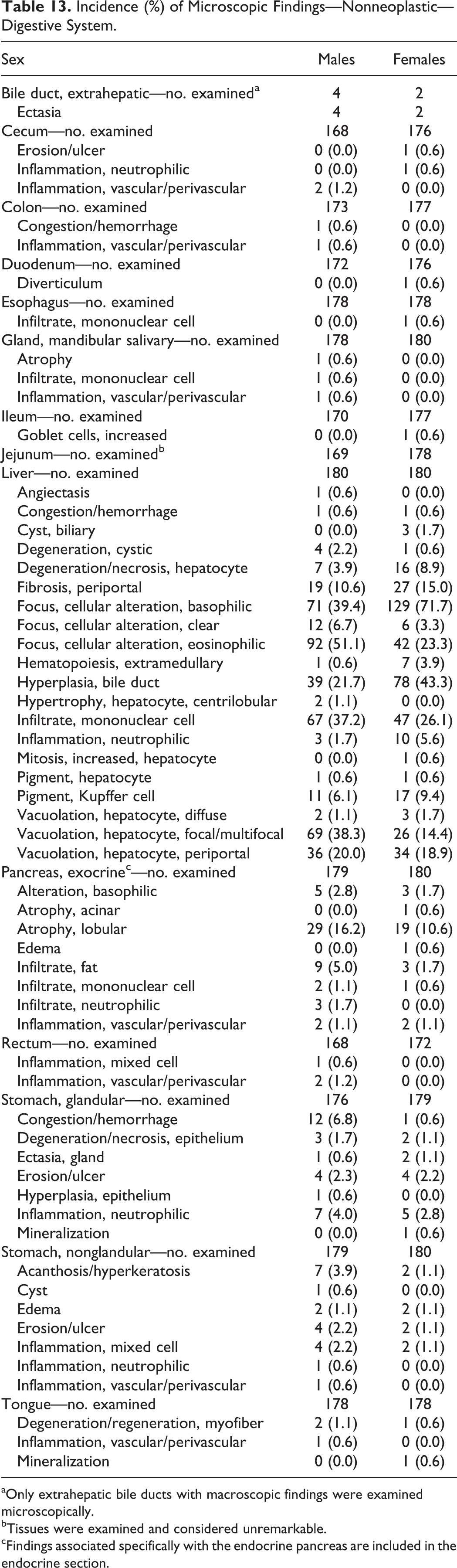
aOnly extrahepatic bile ducts with macroscopic findings were examined microscopically.
bTissues were examined and considered unremarkable.
cFindings associated specifically with the endocrine pancreas are included in the endocrine section.
Hematopoietic/Lymphoid System
Macroscopic findings
The most common macroscopic finding in the hematopoietic/lymphoid system was red discoloration in the mandibular lymph node (Table 14). This correlated with increased erythrocytes in the medullary sinuses and was considered secondary to the terminal blood collection procedure.
Incidence (%) of Macroscopic Findings—Hematopoietic/Lymphoid System.
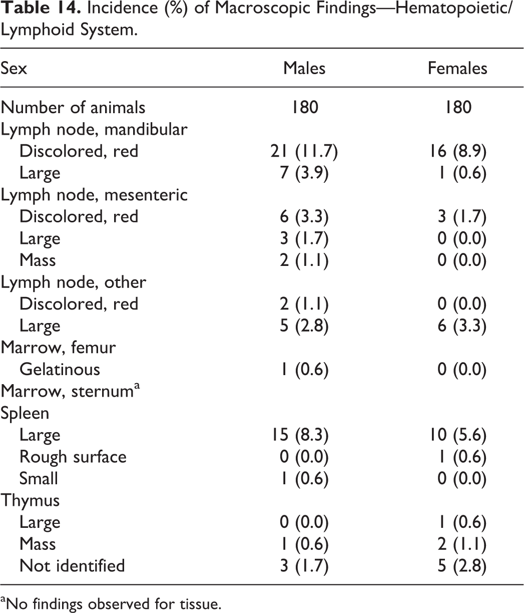
aNo findings observed for tissue.
Microscopic findings—neoplastic
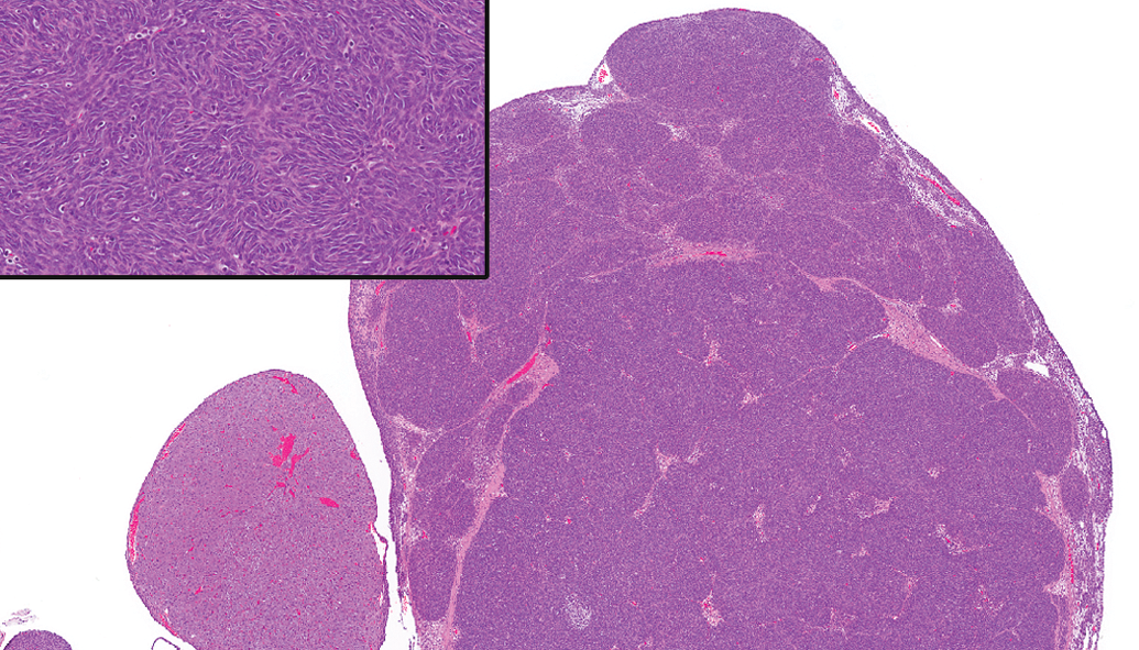
Thymomas were the most common neoplasm in the hematopoietic/lymphoid system (Table 15). Thymomas demonstrated medullary differentiation, were predominantly lymphocytic (Figure 6), and were more common in females than males, similar to the observations made in Wistar rats by Kuper, Beems, and Hollanders (1986).
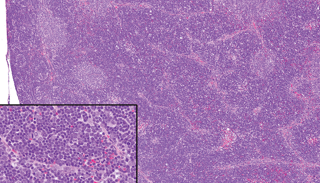
Thymus—thymoma. Mitotic figures were common (hematoxylin and eosin).
Incidence (%) of Microscopic Findings—Neoplastic—Hematopoietic/Lymphoid System.
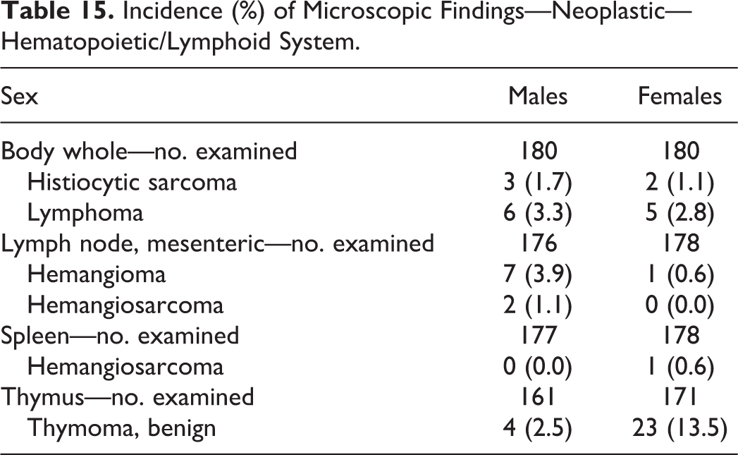
Vascular neoplasms were observed in the mesenteric lymph nodes (Figure 7) and paralleled what was observed by Reindel, Dominick, and Gough (1992); they were more common in males (5.0%) than females (0.6%). The conclusion by Reindel that these vascular lesions represent neoplasms rather than vascular malformation is also supported by the absence of similar vascular neoplasms in previous shorter term studies of 4, 13, or 26 weeks in RCCHan:WIST rats (Blankenship and Skaggs 2012).
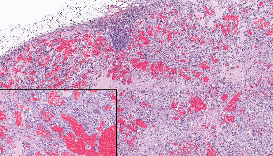
Mesenteric lymph node—hemangioma. Vascular spaces are lined by neoplastic endothelial cells characterized by little cellular atypia and rare mitotic figures (hematoxylin and eosin).
Microscopic findings—nonneoplastic
The most common microscopic findings in the hematopoietic/lymphoid system included increased medullary sinus erythrocytes and increased medullary cord plasma cells in the mandibular lymph node, increased extramedullary hematopoiesis and pigment in the spleen, and epithelial tubule/cyst and decreased lymphocytes in the thymus (Table 16).
Incidence (%) of Microscopic Findings—Nonneoplastic—Hematopoietic/Lymphoid System.
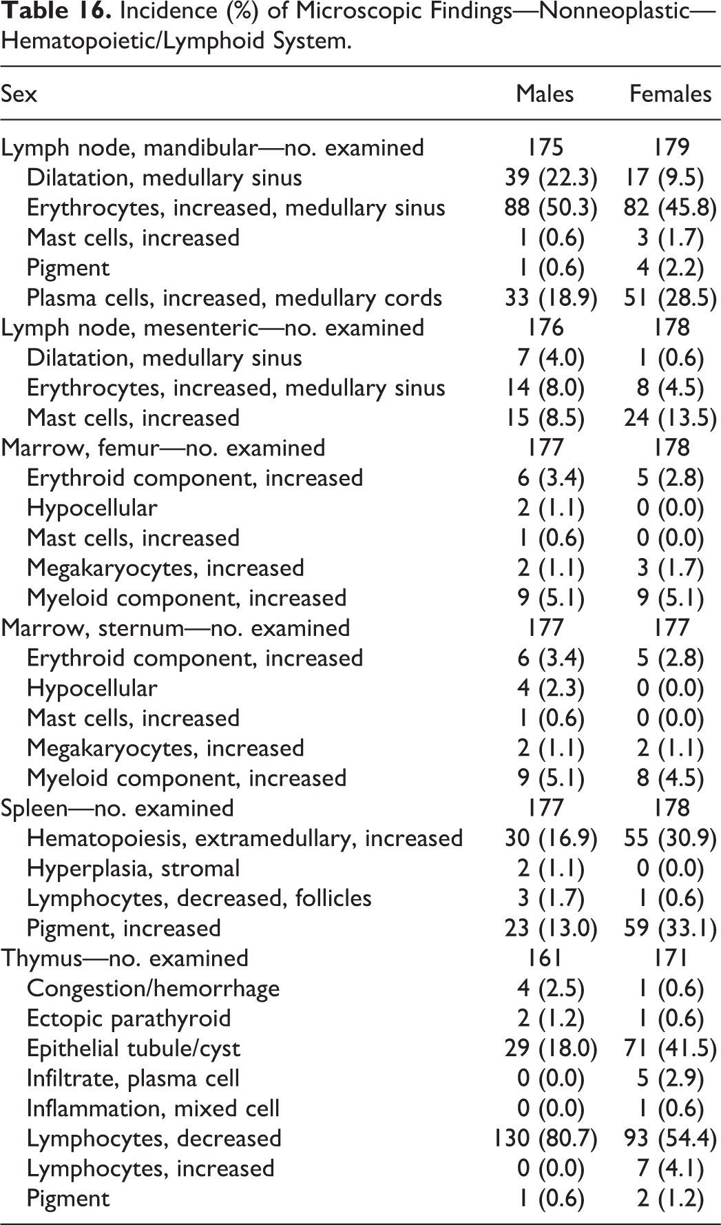
Increased extramedullary hematopoiesis in the spleen was often observed in association (or concurrently) with notable inflammation in other tissues.
In the thymus, the presence of epithelial tubules/cyst and decreased lymphocytes was recorded with a notably increased incidence of epithelial tubules/cyst in females, similar to that observed in Wistar rats by Kuper, Beems, and Hollanders (1986). While both are features of involution (a normal and expected process), for the purposes of this study, the microscopic findings of epithelial tubules/cyst and decreased lymphocytes were diagnosed and graded individually rather than using the aggregate term involution.
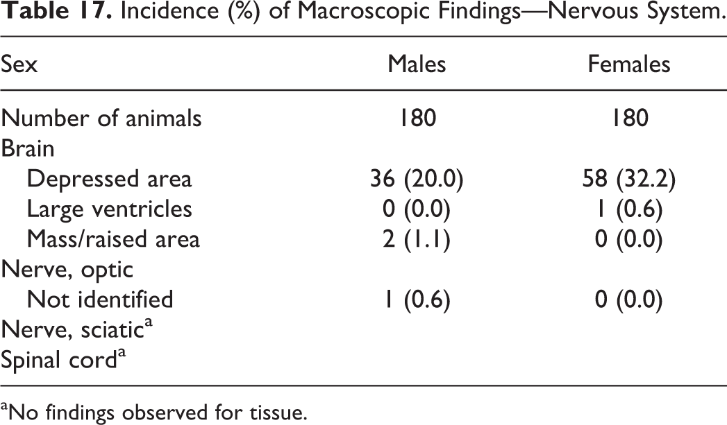
Nervous System
Macroscopic findings
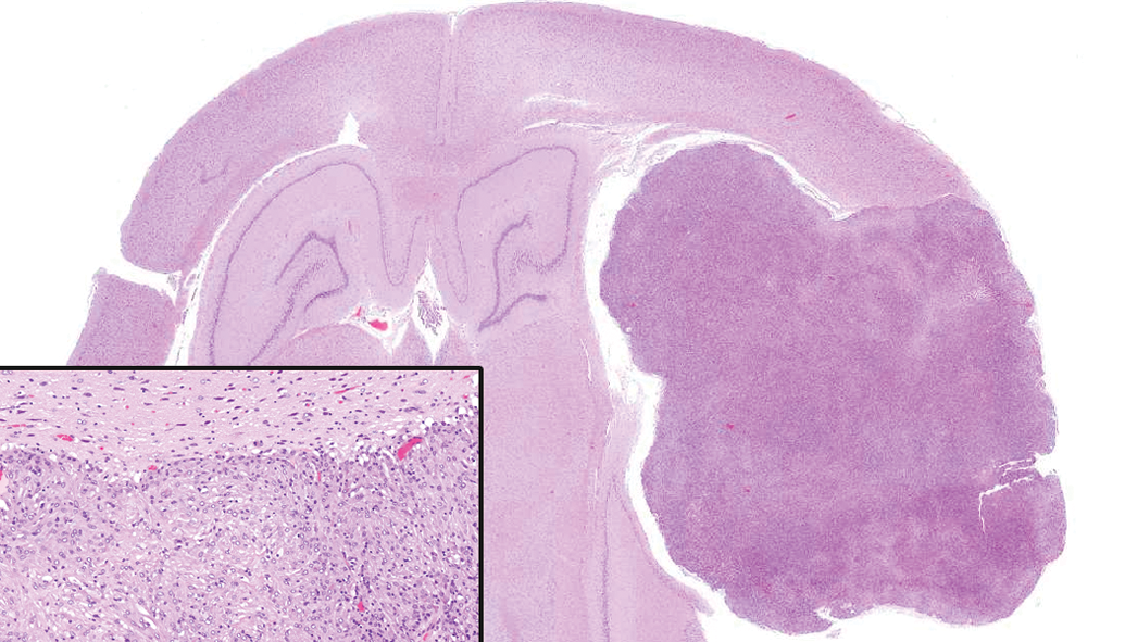
The most common macroscopic finding observed in the nervous system was a depressed area along the ventral surface of the brain (Table 17). This correlated microscopically with ventral compression and the presence of a pituitary adenoma.
Incidence (%) of Macroscopic Findings—Nervous System.
aNo findings observed for tissue.
Microscopic findings—neoplastic
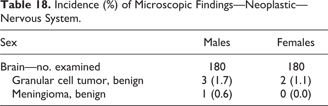
The only neoplasms observed in the nervous system were granular cell tumors and a meningioma (Table 18). The overall incidence of granular cell tumor and meningioma (2.2% in males and 1.1% in females) was similar to that reported in Wistar rats by Weber et al. (2011) and Bertrand, Mukaratirwa, and Bradley (2014). Mitsumori, Maronpot, and Boorman (1987) described the predominant features of both granular cell tumors and meningiomas and their common histologic features. These common histologic features demonstrated a continuum between these two neoplasms and suggest the possibility of a common progenitor meningothelial arachnoid cell. In this study, neoplasms characterized by a predominance of polygonal cells with eosinophilic granular cytoplasm arranged in nests or islands were diagnosed as granular cell tumors (Figure 8), while one neoplasm characterized by a predominance of elongate neoplastic cells with eosinophilic fibrillar cytoplasm arranged in loose fascicles was diagnosed as a meningioma (Figure 9).
Incidence (%) of Microscopic Findings—Neoplastic—Nervous System.
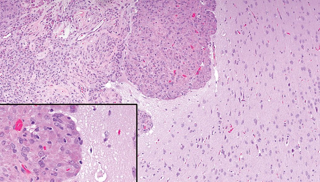
Brain—granular cell tumor. Neoplastic cells are polygonal, contain eosinophilic granular cytoplasm, and are arranged in nests or islands (hematoxylin and eosin).
Brain—meningioma. Neoplastic cells are elongate, contain eosinophilic fibrillar cytoplasm, and are arranged in loose fascicles (hematoxylin and eosin).
Microscopic findings—nonneoplastic
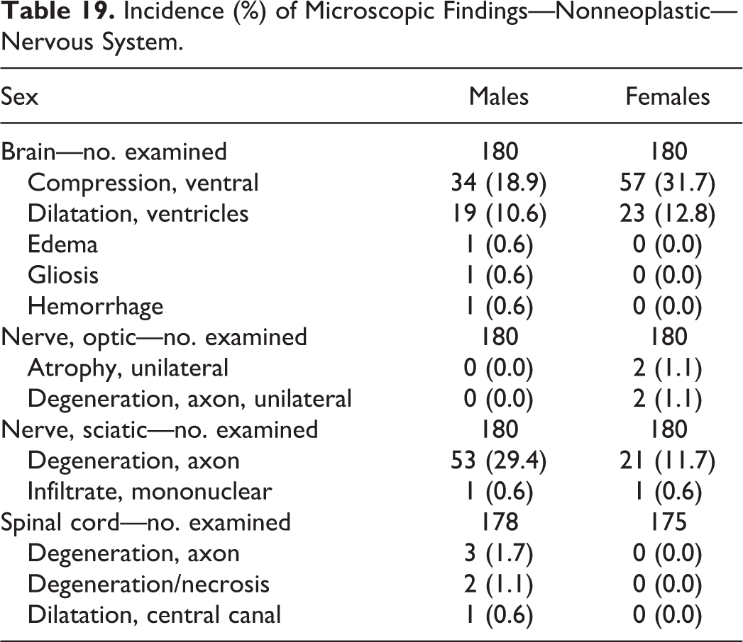
Ventral compression of the brain was observed in 18.9% of males and 31.7% of females and was always associated with a pituitary adenoma; concurrent ventricular dilatation was often present (Table 19). In the peripheral nervous system, sciatic nerve axonal degeneration was observed in 29.4% of males and 11.7% of females.
Incidence (%) of Microscopic Findings—Nonneoplastic—Nervous System.
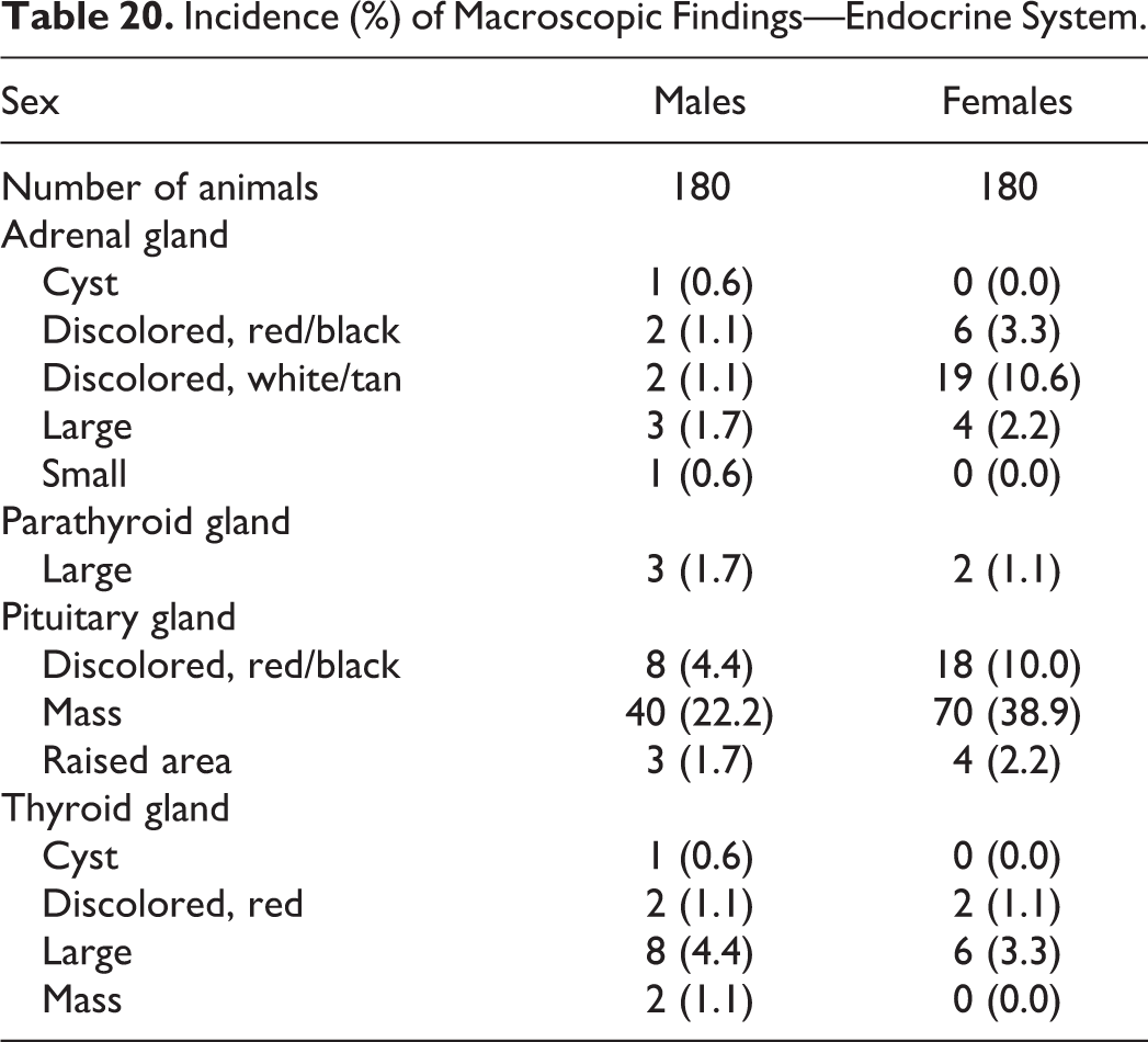
Endocrine System
Macroscopic findings
Red/black discoloration and mass in the pituitary in males and females and white/tan discoloration in the adrenal in females were the most common macroscopic findings in the endocrine system (Table 20) and correlated with pituitary adenoma and focal hypertrophy of the zona glomerulosa in the adrenal gland, respectively.
Incidence (%) of Macroscopic Findings—Endocrine System.
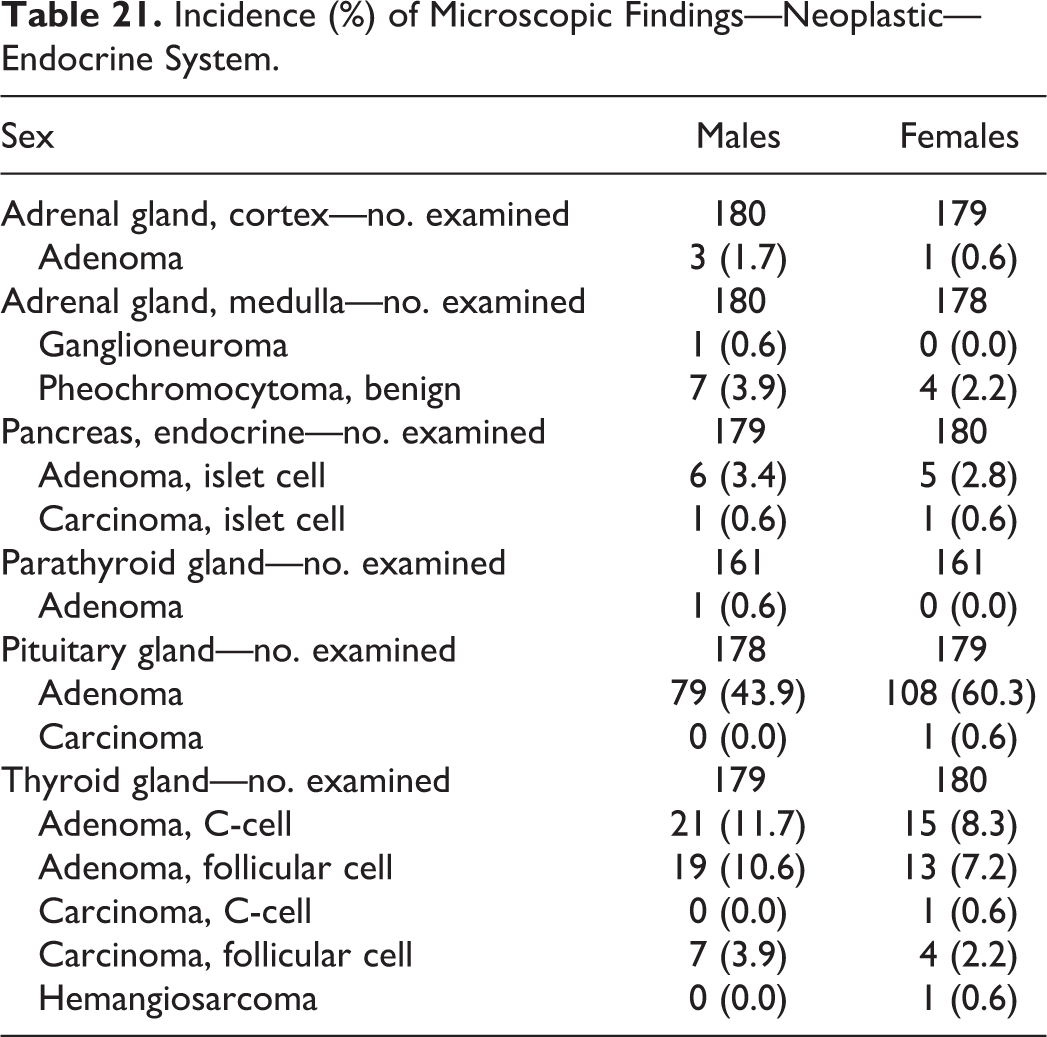
Microscopic findings—neoplastic
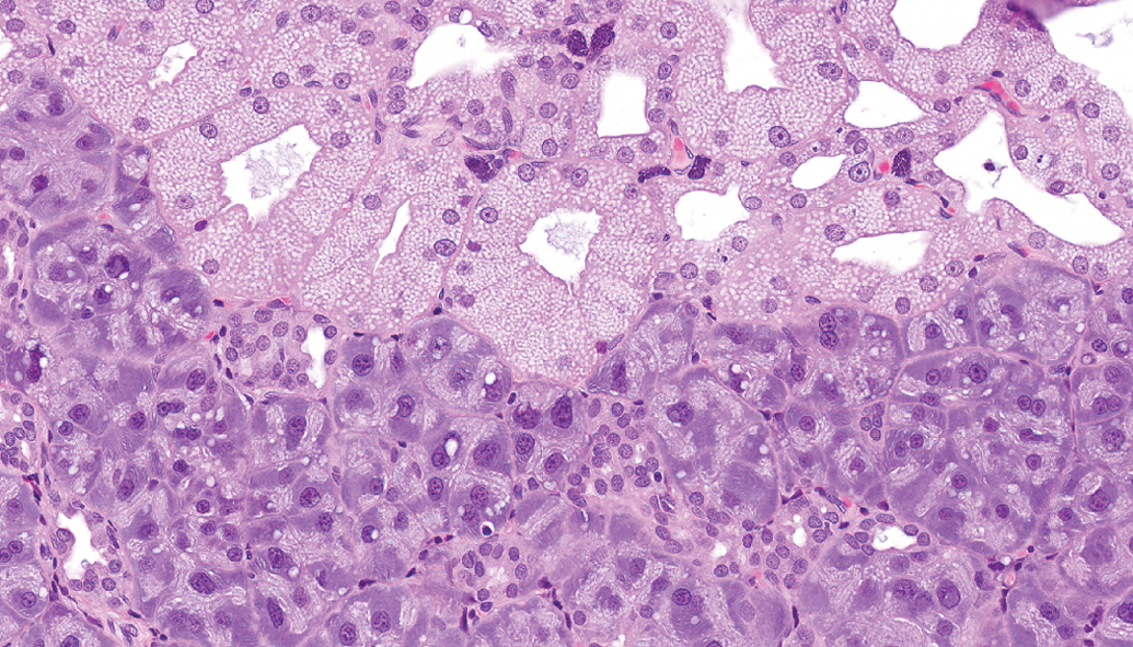
Pituitary adenoma was the most common endocrine neoplasm (Table 21) with an incidence of 43.9% in males and 60.3% in females and accounted for 13.9% and 18.9% of the unscheduled deaths in males and females, respectively. Nearly all pituitary adenomas appeared to arise from the pars distalis except 1 from the pars intermedia (Figure 10). Thyroid C-cell and follicular adenomas were common. Neoplasms observed in the adrenal gland included cortical adenoma, pheochromocytoma (Figure 11), and ganglioneuroma (Figure 12).
Incidence (%) of Microscopic Findings—Neoplastic—Endocrine System.
Pituitary gland, pars intermedia—adenoma. Neoplastic cells contain abundant pale cytoplasm. Moderate atypia and abnormal mitotic figures are present (hematoxylin and eosin).
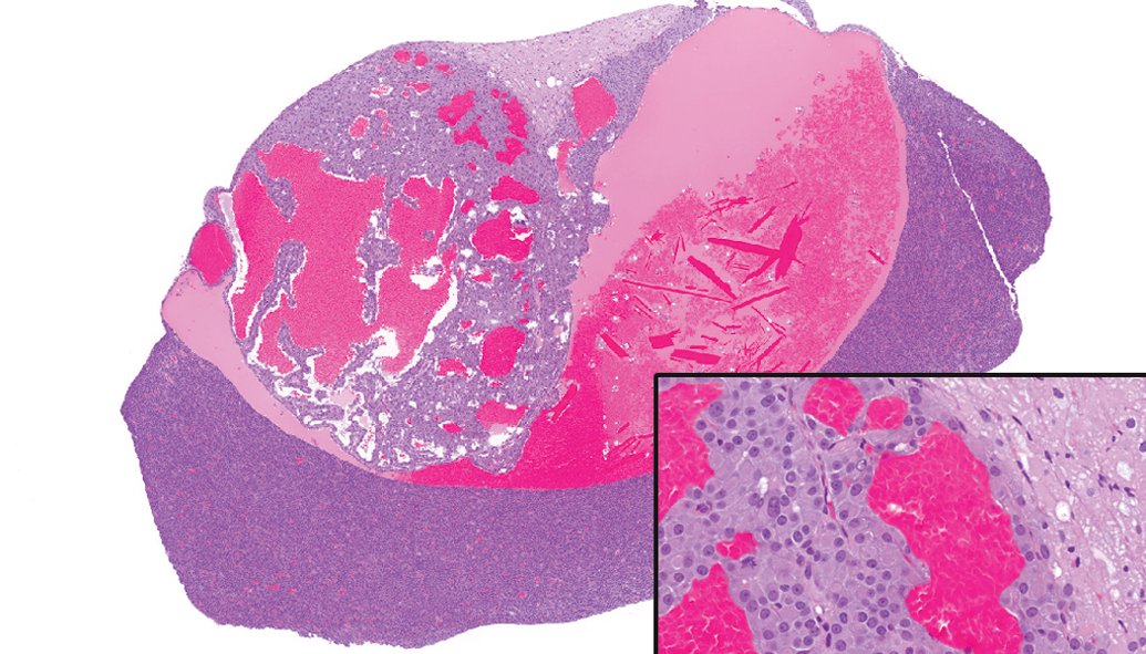
Adrenal gland, medulla—pheochromocytoma. Neoplastic medullary cells are arranged in sheets and cords and compress adjacent cortical cells (hematoxylin and eosin).
Adrenal gland, medulla—ganglioneuroma. Neoplastic ganglion cells in a neurofibrillary matrix compress adjacent cortical cells (hematoxylin and eosin).
Microscopic findings—nonneoplastic
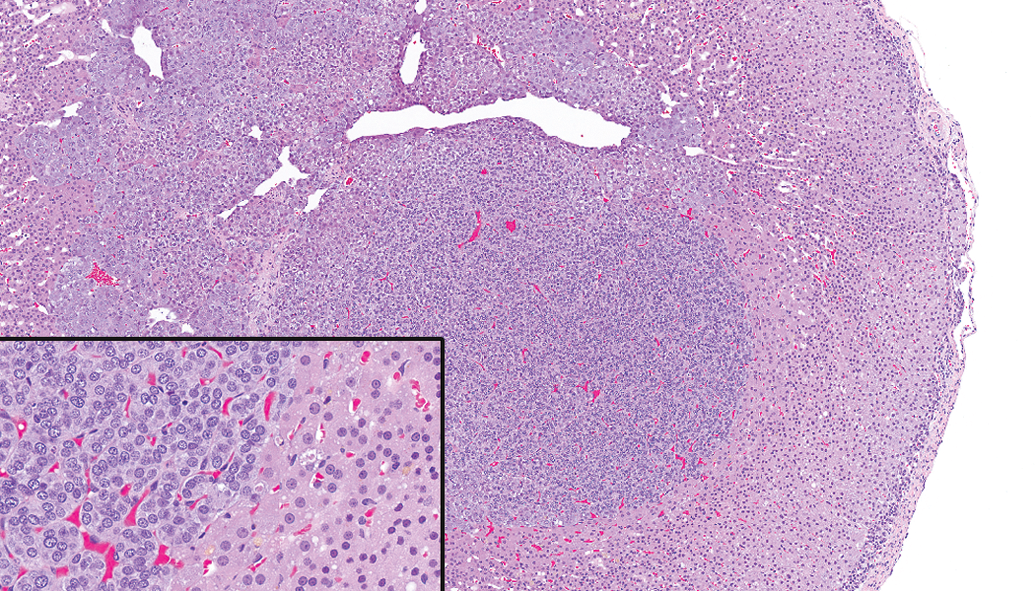
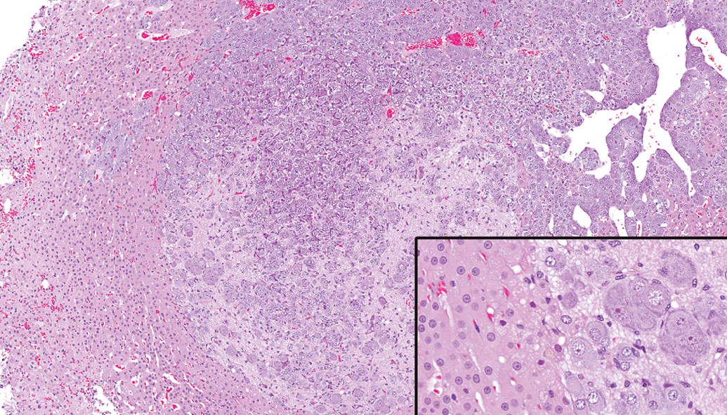
Cystic degeneration in the adrenal cortex represented a continuum of features ranging from foci of few vacuolated cortical cells with associated congestion at minimal severity to larger areas with confluent vacuolated cells, cell loss, cystic spaces, and hemorrhage at higher severity (Figure 13). Cystic degeneration demonstrated a markedly higher incidence (Table 22) in females (64.2%) than in males (6.1%) similar to what was reported in Sprague-Dawley rats by Laast et al. (2014). Similarly, focal hypertrophy of the zona glomerulosa was more common in females (49.7%) than males (24.4%) while, in contrast, focal vacuolation was more common in males (40.6%) than females (10.1%).
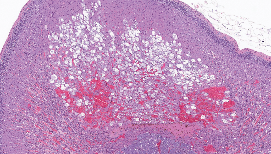
Adrenal gland, cortex—cystic degeneration, showing areas with vacuolated cells, cell loss, cystic spaces, and hemorrhage (hematoxylin and eosin).
Incidence (%) of Microscopic Findings—Nonneoplastic—Endocrine System.
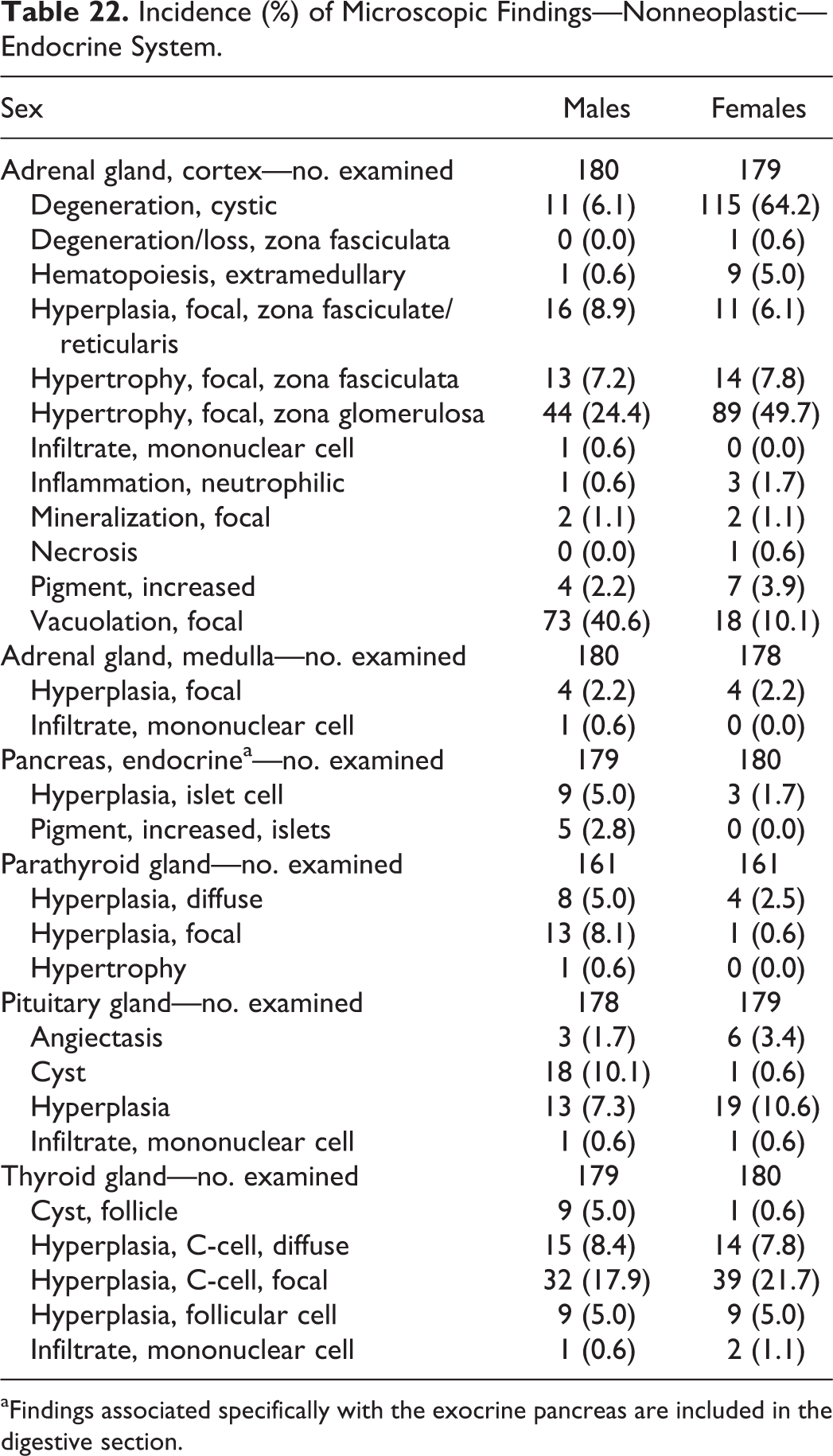
aFindings associated specifically with the exocrine pancreas are included in the digestive section.
Additional common microscopic findings in other endocrine organs included pituitary gland hyperplasia and diffuse and focal C-cell hyperplasia in the thyroid gland (Table 22).
Miscellaneous Tissues
Macroscopic findings
Macroscopic findings were observed commonly in the foot/footpad (e.g., scab, sore) of males and females, mammary gland (e.g., mass, thickened) of females, and lacrimal gland (discoloration) of males (Table 23). In the lacrimal gland, white/tan discoloration correlated with Harderian gland alteration.
Incidence (%) of Macroscopic Findings—Miscellaneous Tissues.
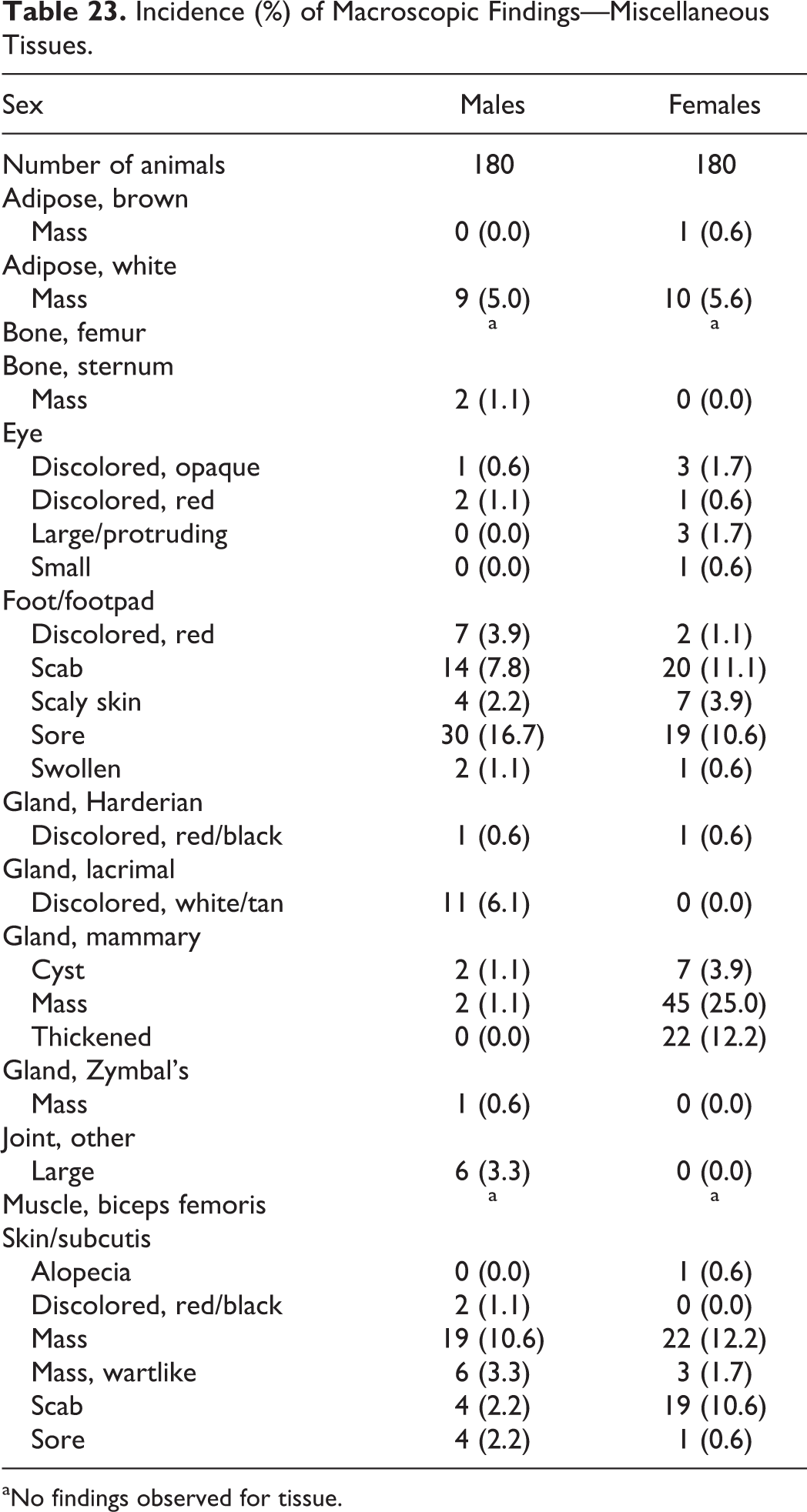
aNo findings observed for tissue.
Microscopic findings—neoplastic
In the mammary gland, fibroadenomas and carcinomas were observed in 16.7% and 15.0% of females, respectively (Table 24). Keratoacanthoma demonstrated a higher incidence in males (6.7%) than in females (1.7%).
Incidence (%) of Microscopic Findings—Neoplastic—Miscellaneous Tissues.
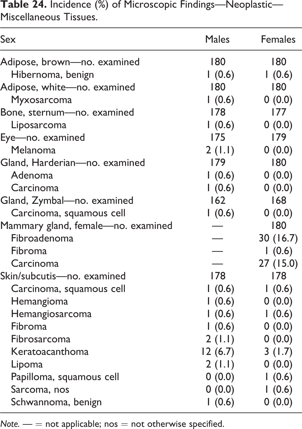
Note. — = not applicable; nos = not otherwise specified.
In the eye, two melanomas were observed.
Microscopic findings—nonneoplastic
In the eye, minimal to slight unilateral and/or bilateral retinal degeneration/atrophy was observed in 15.4% of males and 21.8% of females (Table 25). Minimal retinal degeneration/atrophy was characterized by thinning of the outer nuclear layer to a thickness comparable to the inner nuclear layer, while slight retinal degeneration/atrophy was characterized by an outer nuclear layer that was thinner than the inner nuclear layer (Figures 14 –16).
Incidence (%) of Microscopic Findings—Nonneoplastic—Miscellaneous Tissues.
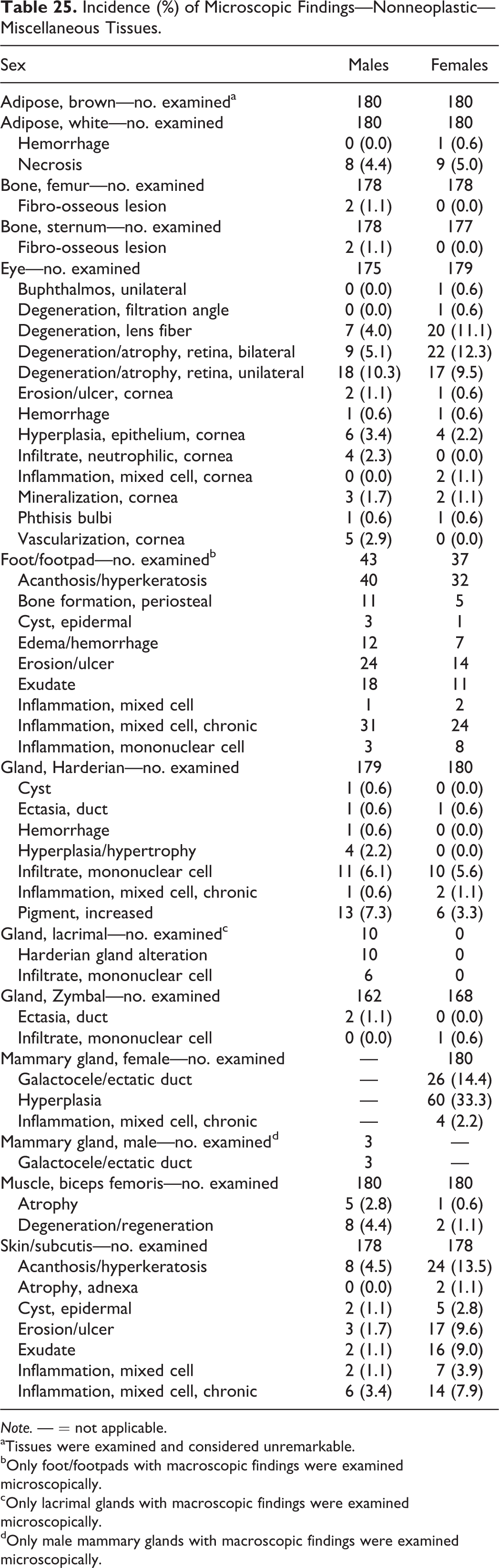
Note. — = not applicable.
aTissues were examined and considered unremarkable.
bOnly foot/footpads with macroscopic findings were examined microscopically.
cOnly lacrimal glands with macroscopic findings were examined microscopically.
dOnly male mammary glands with macroscopic findings were examined microscopically.
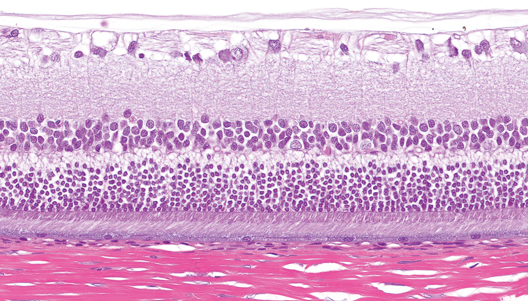
Eye, retina—normal. The inner nuclear layer is thinner than the outer nuclear layer (hematoxylin and eosin).
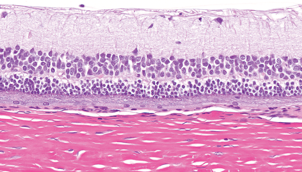
Eye, retina—minimal retinal degeneration/atrophy. The inner and outer nuclear layers are of comparable thickness (hematoxylin and eosin).
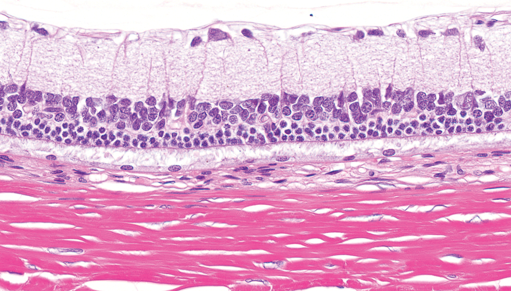
Eye, retina—slight retinal degeneration/atrophy. The outer nuclear layer is thinner than the inner nuclear layer (hematoxylin and eosin).
Harderian gland alteration in the lacrimal gland was only observed in males and was characterized by alveoli lined by plump cuboidal cells containing abundant finely vacuolated cytoplasm that resembled the typical secretory cells in the Harderian gland (Figure 17). Ferrara et al. (2004) previously reported this sex-dependent variation in “harderianization” in aging rats and suggested that it may be due to the disappearance of estrogen receptors from the lacrimal glands of aging male rats. It should also be noted that in this study, lacrimal glands were not examined from all animals. They were only examined if a macroscopic finding was observed at necropsy (e.g., discoloration); Harderian gland alteration was the microscopic correlate.
Lacrimal gland—Harderian gland alteration. Altered cells are characterized by abundant finely vacuolated eosinophilic cytoplasm (hematoxylin and eosin).
In conclusion, survival and body weight data and the incidence of macroscopic findings and neoplastic and nonneoplastic microscopic findings provide support for the interpretation of findings encountered during 104-week carcinogenicity studies and in the evaluation of Harlan RCCHan:WIST rats as a suitable rodent model.
Footnotes
Appendix
Tissues Examined.
| Adipose, browna | Muscle, biceps femoris |
| Adipose, whitea | Nasal turbinates |
| Adrenal gland | Optic nerve |
| Aorta | Ovary |
| Bile ducta | Oviduct |
| Bone marrow | Pancreas |
| Brain | Parathyroid gland |
| Cecum | Pituitary gland |
| Cervix | Prostate gland |
| Clitoral | Gland preputial gland |
| Colon | Rectum |
| Duodenum | Salivary gland, mandibular |
| Epididymis | Sciatic nerve |
| Esophagus | Seminal vesicle |
| Eye | Skin/subcutis |
| Femur | Spinal cord (cervical, thoracic, and lumbar) |
| Foot/footpada | Spleen |
| Harderian gland | Sternum |
| Heart | Stomach, glandular |
| Ileum | Stomach, nonglandular |
| Jejunum | Testis |
| Kidney | Thymus |
| Lacrimal glanda | Thyroid gland |
| Lesions | Tongue |
| Liver | Trachea |
| Lung with large bronchi | Ureter |
| Lymph node, mandibular | Urinary bladder |
| Lymph node, mesenteric | Uterus |
| Mammary gland, female | Vagina |
| Mammary gland, malea | Zymbal’s gland |
aOnly tissues with macroscopic findings were examined microscopically.
Acknowledgments
We would like to thank toxicologist Rachael Avery for study coordination and support and Steve Van Adestine for photographic assistance.
Authors’ Contribution
Authors contributed to conception or design (MS); data acquisition, analysis, or interpretation (BB, JE, GH, MS, AS, SS); drafting the manuscript (BB, JE, GH, AS, SS); and critically revising the manuscript (BB, JE, GH, MS, AS, SS). All authors gave final approval and agreed to be accountable for all aspects of work in ensuring that questions relating to the accuracy or integrity of any part of the work are appropriately investigated and resolved.
Declaration of Conflicting Interests
The author(s) declared no potential conflicts of interest with respect to the research, authorship, and/or publication of this article.
Funding
The author(s) received no financial support for the research, authorship, and/or publication of this article.