
Abstract
Vehicle control Harlan RCCHan™: WIST rats were examined to provide control data for subsequent studies. Sixty male and 60 female rats were sacrificed after 4, 13, and 26 weeks (360 animals total) of daily oral gavage dosing with reverse osmosis water. At necropsy, body weights, organ weights, and macroscopic findings were recorded, and tissues were collected for histopathology. Mean terminal body and organ weight data demonstrated expected age-related trends. Macroscopic findings occurred sporadically, generally at singular or at very low incidence, and with no observable age-related trend. The most frequent observation was discoloration of the stomach mucosa. Neoplastic microscopic findings were uncommon (one endometrial stromal polyp; one hepatocellular adenoma; one C-cell adenoma; and one sarcoma, NOS). The most common and/or notable nonneoplastic microscopic findings included basophilic tubules and mononuclear cell infiltration in the kidney, macrophage infiltration in pulmonary alveoli, and mononuclear infiltration in the liver of males and females, and myocardial degeneration/necrosis and mononuclear cell infiltration in the heart of males. Female reproductive tracts were staged to establish a representative baseline distribution. Diestrus, proestrus, estrus, and metestrus were diagnosed 45.8%, 11.9%, 30.5%, and 11.9%, respectively, at 4 weeks and 27.6%, 13.8%, 50.0%, and 8.6%, respectively, at 13 weeks.
Keywords
Introduction
Historical control data are important for the evaluation of certain organ weight changes, macroscopic findings, and neoplastic and nonneoplastic microscopic findings identified during subchronic rodent toxicity studies. For uncommon findings or findings of uncertain relationship to the test article, historical control data can provide information about the incidence of spontaneously occurring background findings in the species and strain of animal used on test. By providing this information, historical control data help to support the interpretation of common and uncommon findings (Deschl et al. 2002; Keenan et al. 2009). In addition, historical control data are useful when selecting and assessing the suitability of an animal model (Weber et al. 2011). The purpose of this report is to summarize and present body and organ weight data, macroscopic findings, neoplastic and nonneoplastic microscopic findings, and reproductive stage data for control Harlan RCCHan™: WIST rats from 4-, 13-, and 26-week studies.
Method
Male and female RCCHan: WIST rats 4 to 8 weeks of age were obtained from Harlan Laboratories, Inc., Frederick, MD, in three shipments of 120 animals each (60 animals/sex/shipment) approximately 3 months apart. Animals were housed in an AAALAC-accredited facility under approved Animal Care and Use Committee guidelines. The study was conducted in compliance with the US Food and Drug Administration Good Laboratory Practice Regulations, and all procedures were in compliance with the Animal Welfare Act, the Guide for the Care and Use of Laboratory Animals, and the Office of Laboratory Animal Welfare.
Sixty male and 60 female rats (20 males and 20 females from each of the 3 shipments) were sacrificed after 4, 13, and 26 weeks (for a total of 120 animals per time point) of daily oral gavage dosing with reverse osmosis water. All animals (except 2 animals/sex from the 13-week sacrifice) sacrificed at a scheduled interval were fasted overnight, anesthetized with sodium pentobarbital, exsanguinated, and necropsied. Terminal body weights, organ weights, and macroscopic observations were recorded. Macroscopic lesions and over 60 tissues from all groups were collected in 10% neutral buffered formalin or modified Davidson’s fixative (eye, Harderian gland, optic nerve, and testis), embedded in paraffin, sectioned, stained with H&E, and examined microscopically. Formalin-fixed tissue from the tarsal joint of one animal was processed for evaluation by S-100 immunohistochemistry and electron microscopy. Two animals/sex sacrificed after 13 weeks of dosing were fasted overnight, anesthetized with sodium pentobarbital, and perfused (whole body perfusion) with McDowell–Trump fixative. Terminal body weights and macroscopic observations were recorded. Twenty selected tissues were collected in McDowell–Trump fixative for possible electron microscopy and/or embedded in paraffin, sectioned, stained with H&E, and examined microscopically.
Results and Discussion
Mortality
Of the 360 animals evaluated, 357 (99.2%) animals survived until scheduled sacrifice. The moribund condition or death of two unscheduled sacrifices was attributed to the gavage procedure, and the cause of one death was undetermined.
Terminal Body and Organ Weights
Group mean terminal body weight was 316 g for males and 195 g for females at the 4-week sacrifice, 432 g for males and 239 g for females at the 13-week sacrifice, and 503 g for males and 267 g for females at the 26-week sacrifice. Terminal body weight data are presented in Table 1. In general, mean organ weight data demonstrated expected age-related trends. An increasing trend in mean organ weight between Weeks 4 and 26 was observed for the brain, mandibular salivary gland, heart, kidney, liver, lung, and spleen in males and females; testis, thyroid/parathyroid, prostate, and seminal vesicles in males; and uterus and pituitary gland in females (Table 2). A decreasing trend in mean organ weight between Weeks 4 and 26 was observed for the adrenal and thymus in males and females and the ovary in females (Table 2). Increasing trends were generally attributed to overall body growth, while decreasing trends were attributed to organ atrophy. No clear trend was observed in mean organ weight between Weeks 4 and 26 for the pituitary in males and the thyroid/parathyroid in females (Table 2).
Terminal body weight (g).
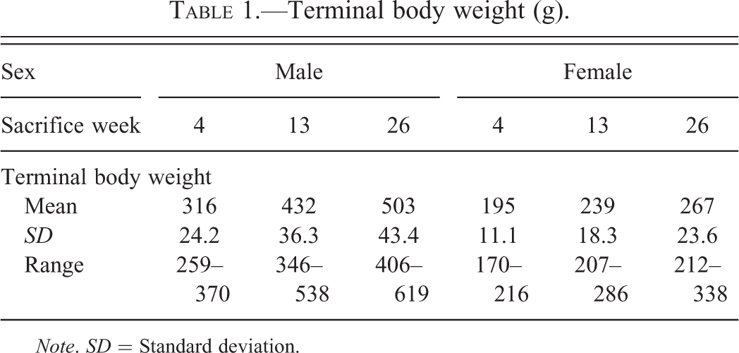
Note. SD = Standard deviation.
Group organ weight (g).
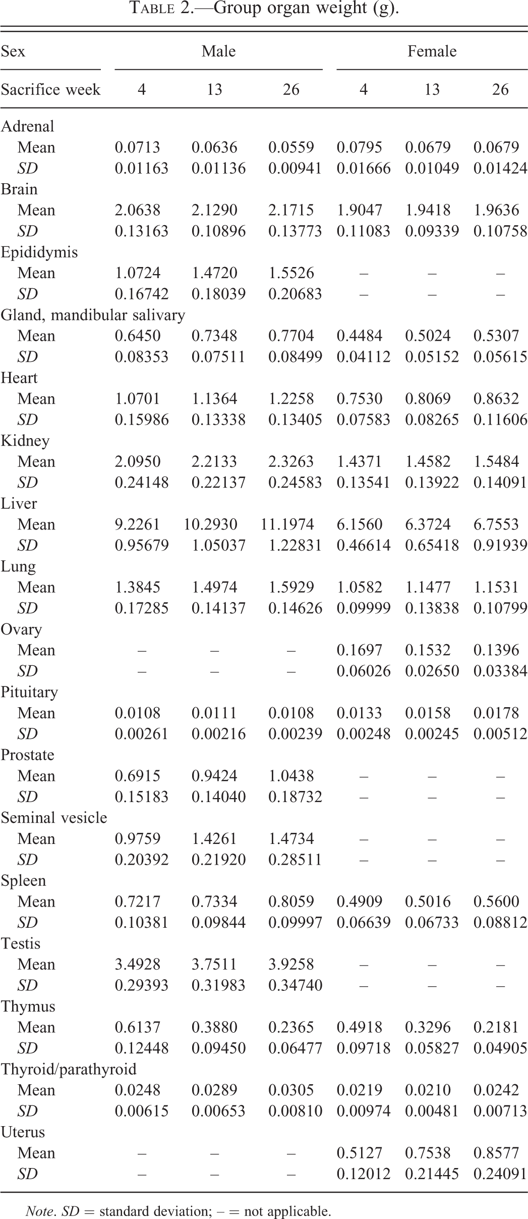
Note. SD = standard deviation; – = not applicable.
Macroscopic Findings
The most frequent macroscopic observation was the discoloration of the stomach mucosa (Table 3). When present, microscopic correlates included hemorrhage and/or congestion. Macroscopic findings occurred sporadically, generally at singular or at very low incidence, and with no observable age-related trend. Tissues with notable incidences of macroscopic observations included the kidney, lung, skin/subcutis, testis, and thymus (Table 3).
Incidence (%) of selected macroscopic findings.
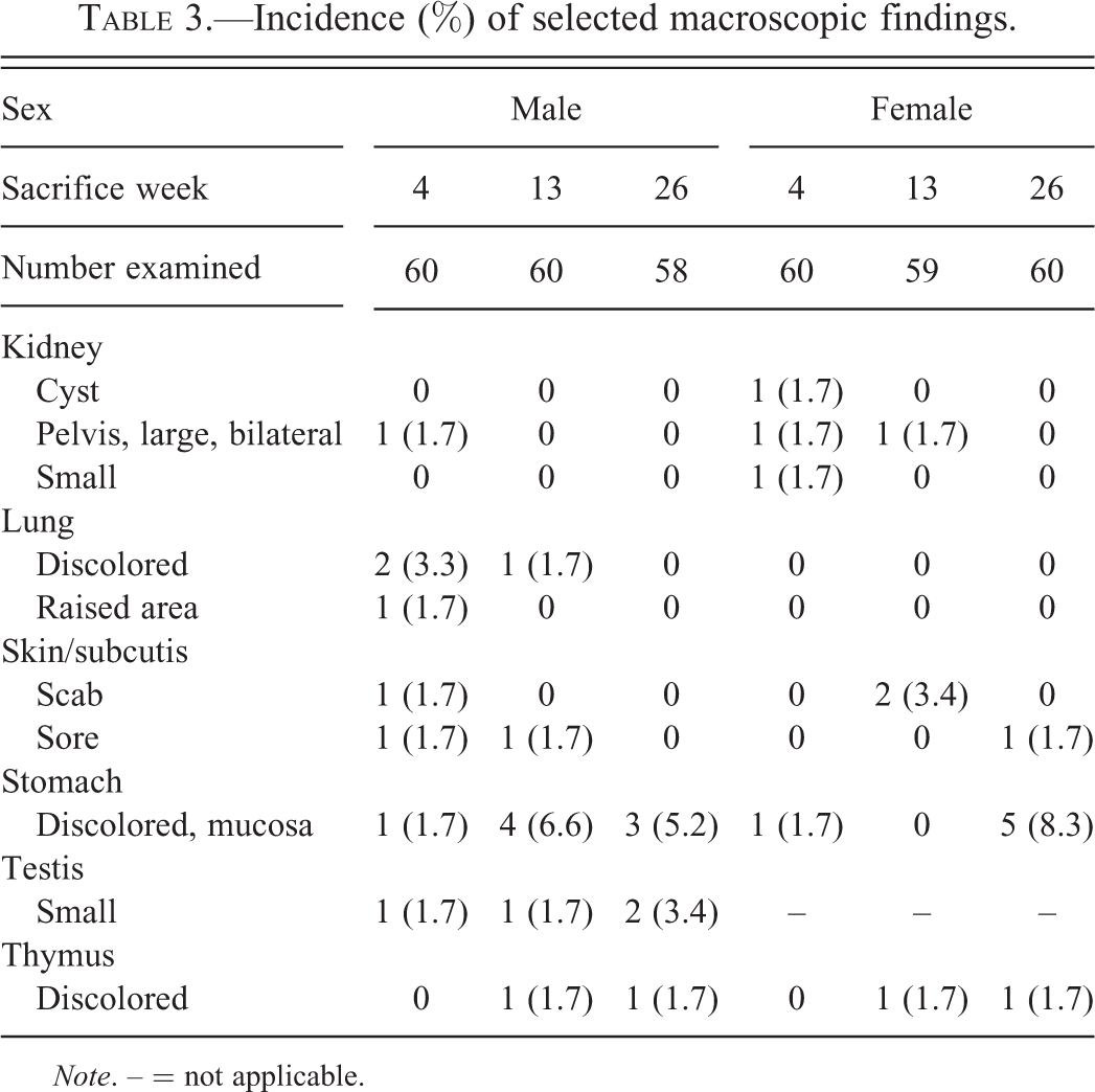
Note. – = not applicable.
Microscopic Findings—Neoplastic
Neoplastic lesions were uncommon and included one endometrial stromal polyp in a 26-week female; one hepatocellular adenoma in a 26-week female; one C-cell adenoma in a 26-week female; and one sarcoma, NOS adjacent to the tarsal joint of a 13-week male. All neoplasms were subject to peer review and diagnosed using standard criteria (Botts et al. 1991; Dixon et al. 1999; Greaves, Faccini, and Courtney 1992, Thoolen et al. 2010). The sarcoma was evaluated further by immunohistochemistry and electron microscopy. By immunohistochemistry, neoplastic cells were S-100 positive; by electron microscopy, defining ultrastructural features suggesting a cell of origin (e.g., external lamina) were not observed. This low incidence of sporadic neoplasms is consistent with reports in the literature. In a previous study (Son et al. 2010), no tumors were reported in control Han Wistar rats from any of the 2-, 4-, 13-, or 26-week studies reviewed, and only two malignant lymphomas and one renal tubular carcinoma in control males from the 16- to 20-week interval from carcinogenicity studies. Dixon, Heider, and Elwell (1995) reported no neoplasms from 90-day studies with Fischer-344 rats.
Microscopic Findings—Nonneoplastic
The incidence of selected nonneoplastic microscopic findings is presented in Tables 4 through 10.
Incidence (%) of microscopic findings—cardiovascular system.
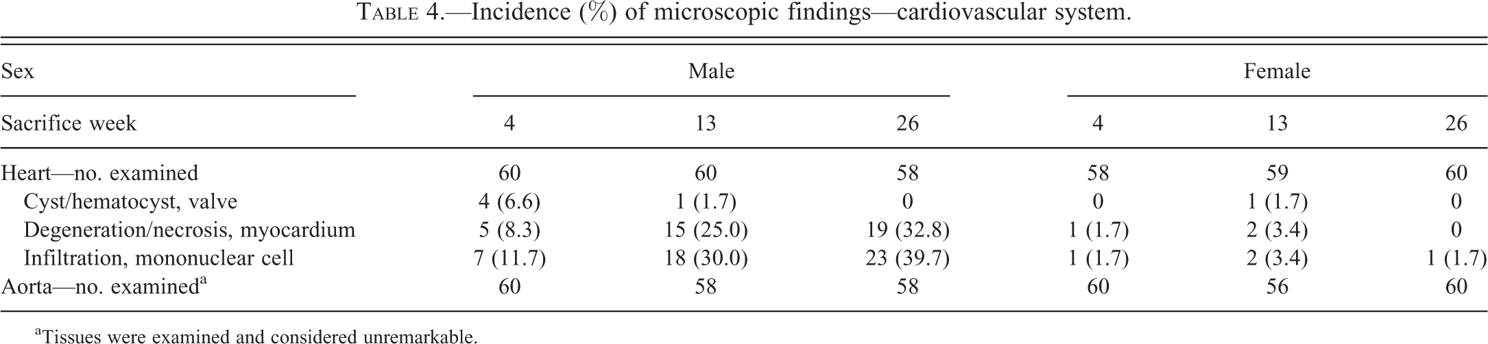
aTissues were examined and considered unremarkable.
Incidence (%) of microscopic findings—urogenital system.
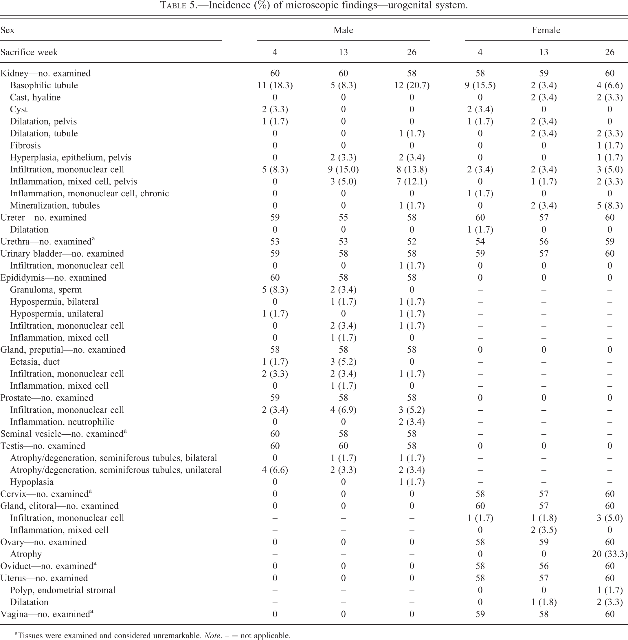
aTissues were examined and considered unremarkable. Note. – = not applicable.
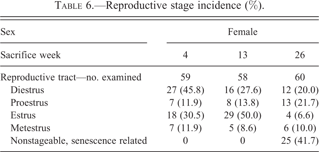
Reproductive stage incidence (%).
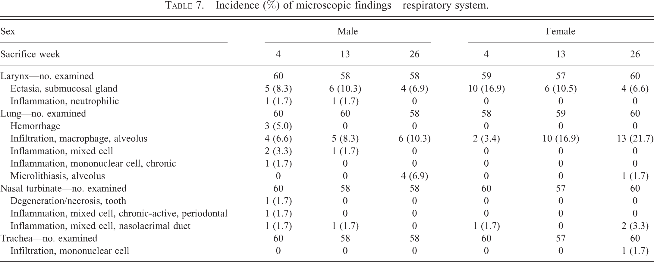
Incidence (%) of microscopic findings—respiratory system.
Incidence (%) of microscopic findings—digestive system.
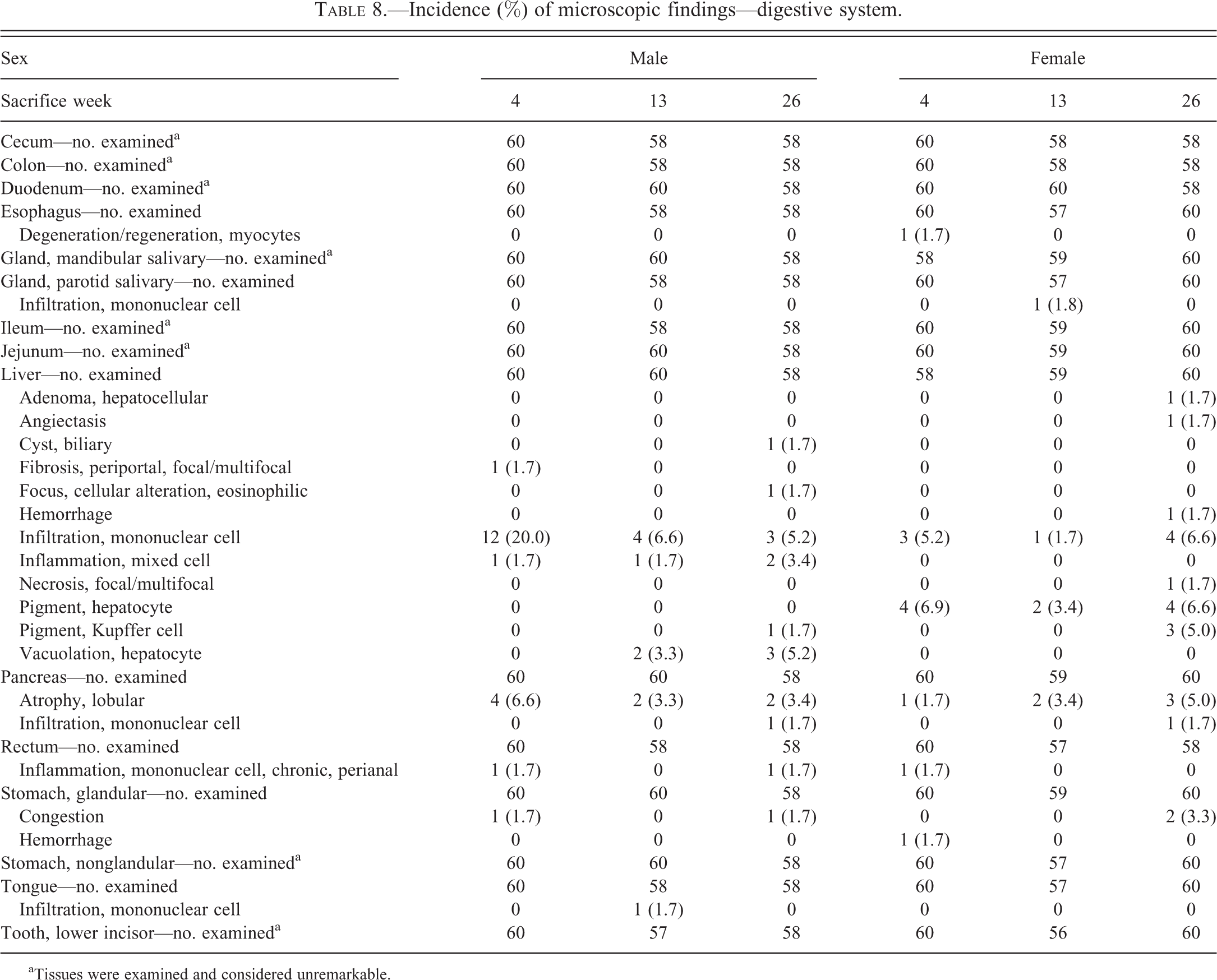
aTissues were examined and considered unremarkable.
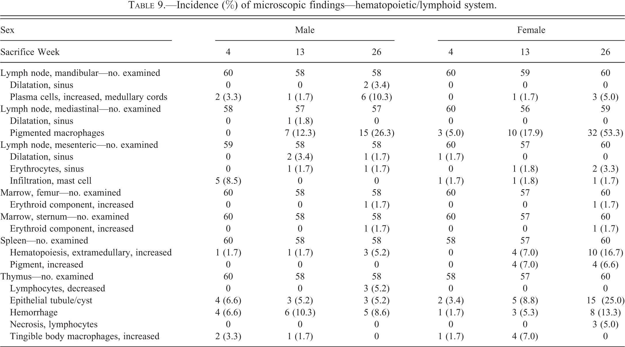
Incidence (%) of microscopic findings—hematopoietic/lymphoid system.
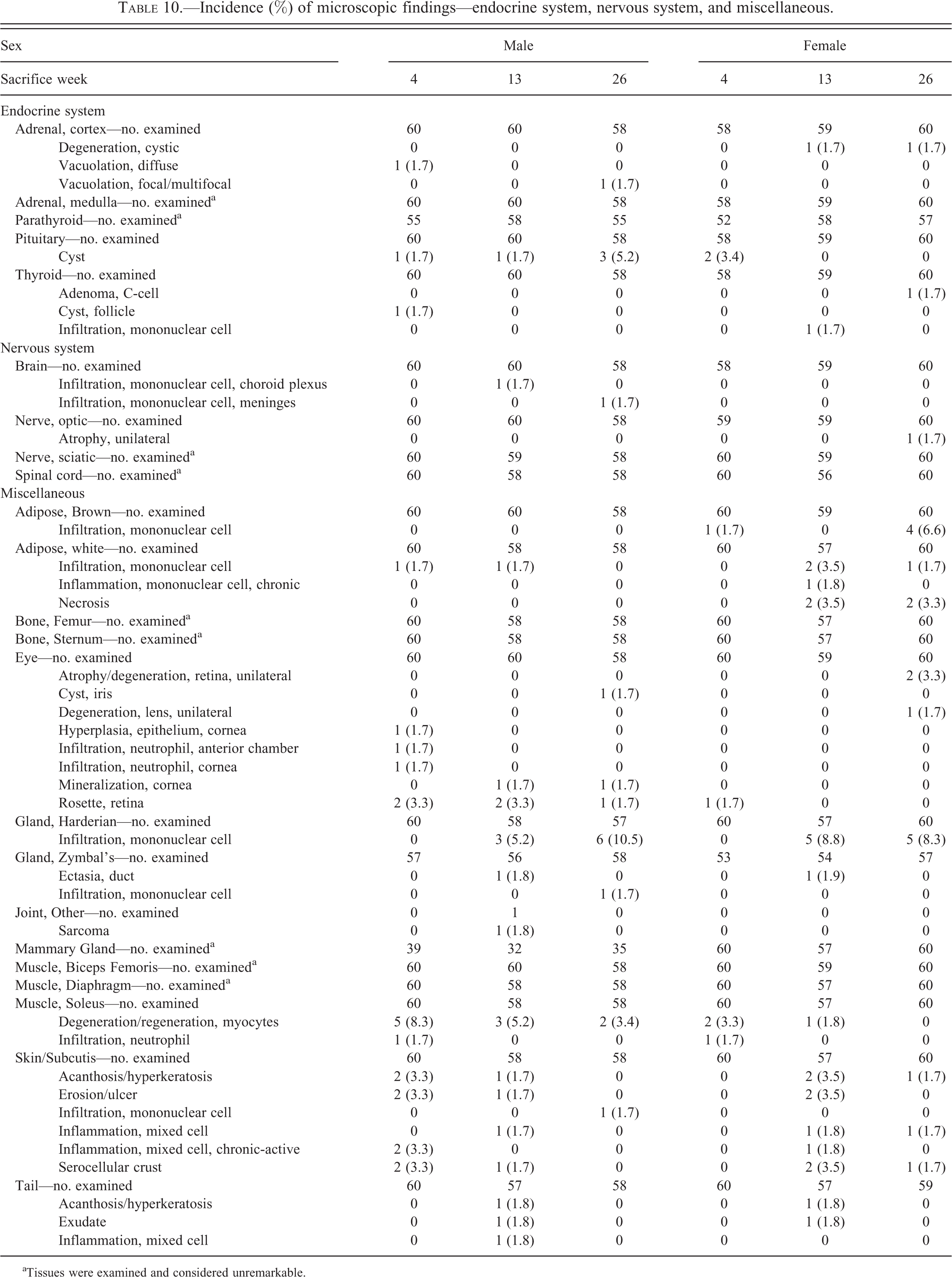
Incidence (%) of microscopic findings—endocrine system, nervous system, and miscellaneous.
aTissues were examined and considered unremarkable.
Cardiovascular System
Myocardial degeneration/necrosis was more common in males than females, increasing in incidence in males between 4 and 26 weeks (Table 4). Myocardial degeneration/necrosis was characterized by one or more hypereosinophilic, occasionally vacuolated, myocardial fibers often lacking cross striations; myocardial degeneration/necrosis was never observed without an accompanying mononuclear cell infiltration (Figure 1). While the use of the term cardiomyopathy is also appropriate, given the variability in individual pathologists’ diagnostic approaches (Keenan et al. 2010), for the purpose of this study, microscopic findings of myocardial degeneration/necrosis and mononuclear infiltration were diagnosed and graded individually rather than using the aggregate term cardiomyopathy. Cysts containing eosinophilic proteinaceous fluid or red blood cells were observed in the valves of some animals (Figure 2). Microscopic findings were not observed in the aorta.
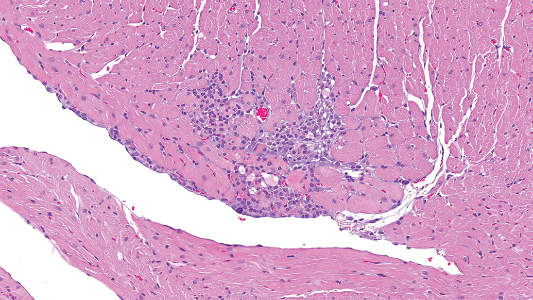
Heart: Myocardial degeneration/necrosis with mononuclear cell infiltrate (H&E).
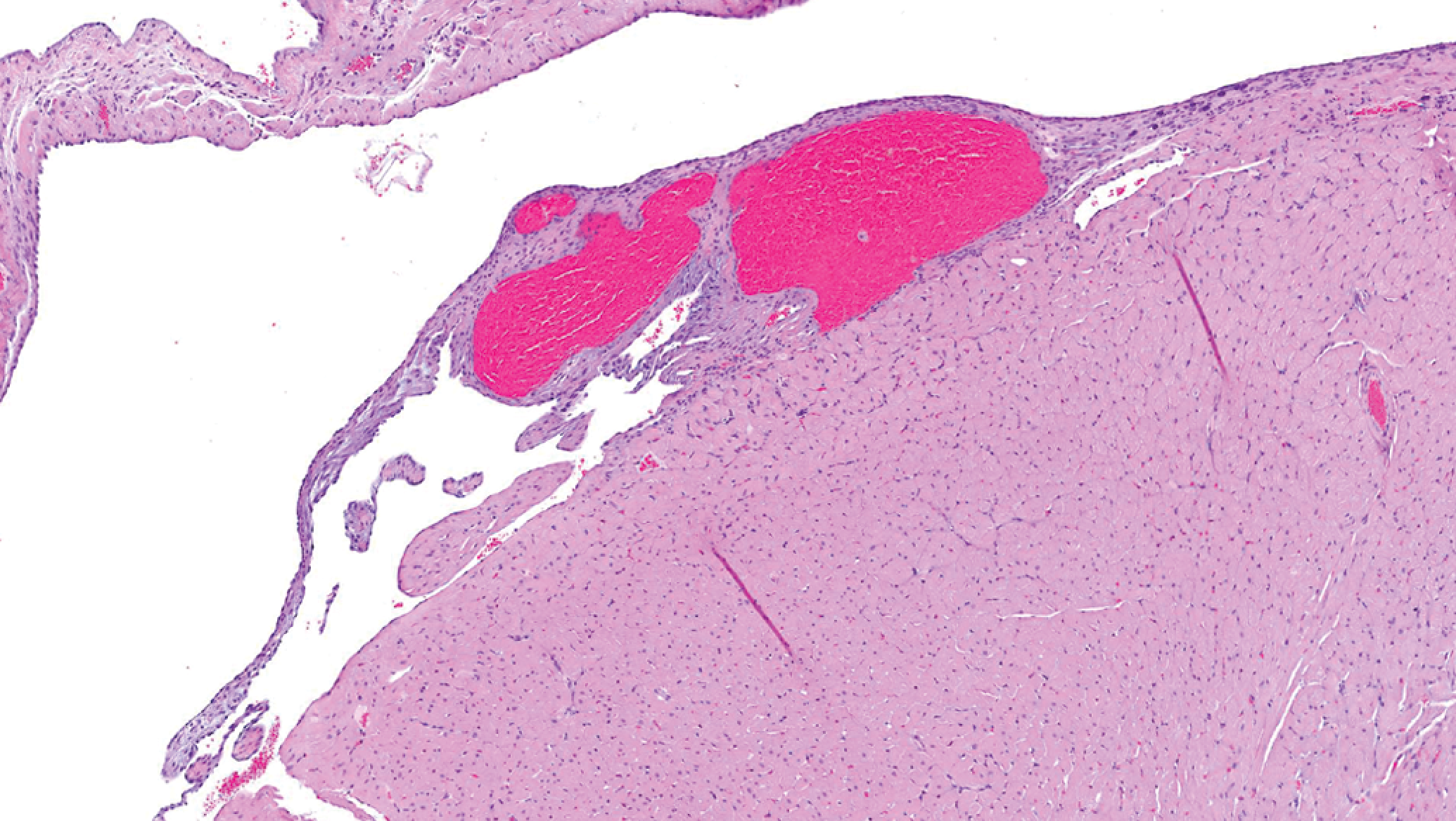
Heart, valve: Hematocyst (H&E).
Urogenital System
The most common microscopic findings observed in the kidney included basophilic tubules (characterized by slightly hypertrophic tubular epithelial cells with cytoplasmic basophilia) and mononuclear cell infiltration. Basophilic tubules likely represented the most reliable early indicator of chronic progressive nephropathy (Hard and Khan 2004), were more common in males than females, and in contrast to carcinogenicity studies (Weber et al. 2011) demonstrated no age-related increase in incidence for studies of this duration. Microscopic findings in the kidney (e.g., basophilic tubules, mononuclear infiltrates, hyaline casts, cyst, and tubule mineralization), some of which are often observed concurrently as components of chronic progressive nephropathy in longer duration studies (Hard and Khan 2004), were rarely observed in combination. Additional findings with an apparent sex-related incidence included mixed cell inflammation in the pelvis of males and hyaline casts and tubule mineralization in females. Sperm granulomas (Figure 3) in the epididymis were relatively common. Testicular hypoplasia, characterized by the loss of spermatogenic cells within a wedge-shaped grouping of seminiferous tubules (Figure 4), was observed in one animal. Other notable microscopic findings associated with the urogenital tract occurred sporadically, at low incidence, and with no clear age-related trend (Table 5). Microscopic findings were not observed in the urethra, seminal vesicle, cervix, oviduct, or vagina.
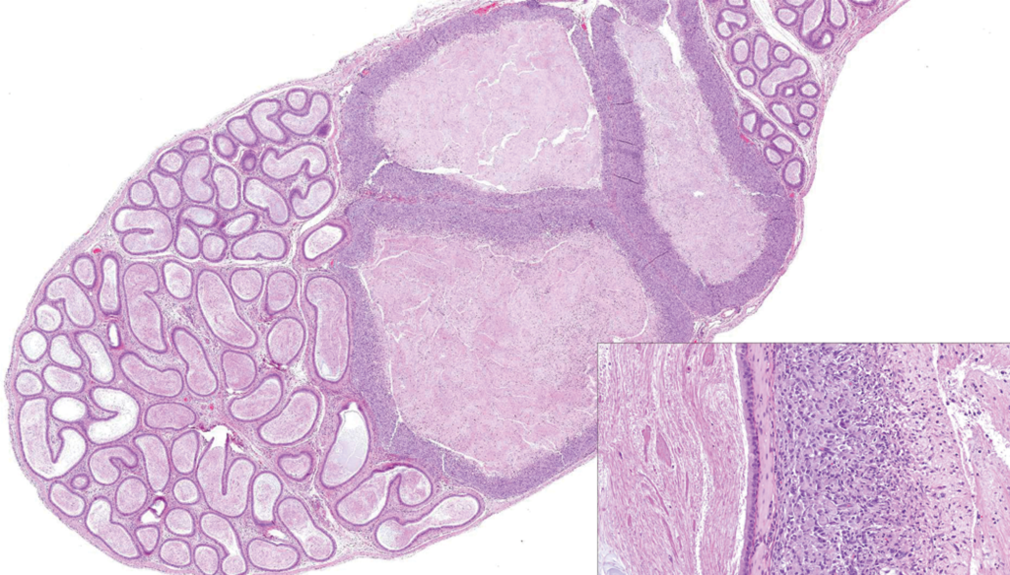
Epididymis: Sperm granuloma (H&E).
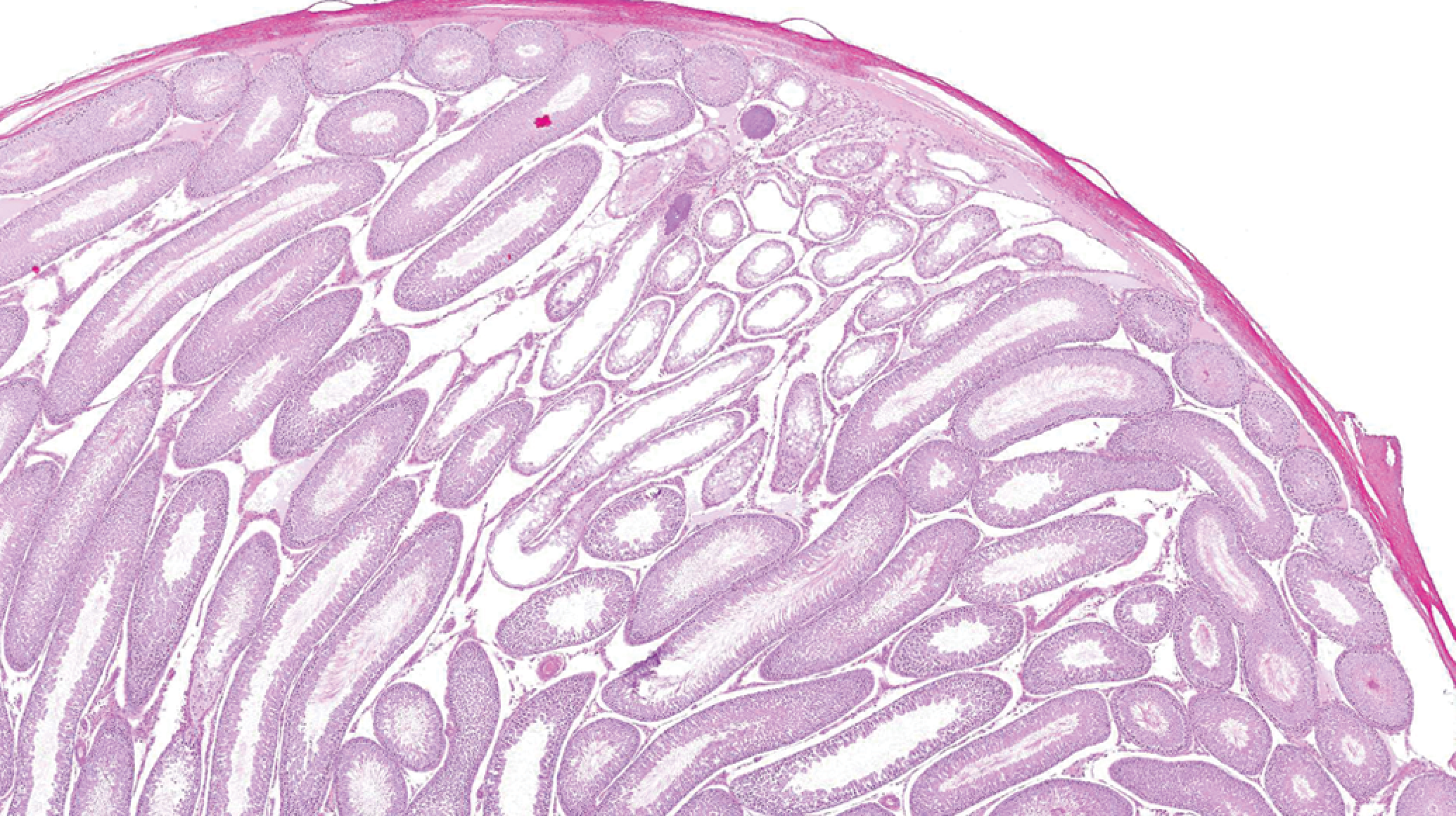
Testis: Hypoplasia: loss of spermatogenic cells within a wedge-shaped grouping of seminiferous tubules (H&E).
Female reproductive tracts were staged to establish a representative baseline distribution. Reproductive tracts (ovary, uterus, and vagina) were evaluated in total, and the appropriate stage (diestrus, proestrus, estrus, or metestrus) was diagnosed using standard criteria (Westwood 2008; Yuan and Carlson 1987; Table 6). At the 26-week sacrifice, 25/60 (41.7%) reproductive tracts were characterized by features attributed to senescence and diagnosed as nonstageable, senescence related. Histologic features (follicular cysts, hypertrophic endometrial epithelium, increased vaginal mucification, and persistent corpora lutea) were consistent with persistent estrus (Figure 5) or repetitive pseudopregnancy (Figure 6; Westwood 2008; Creasey 2012). Because of the high percentage of nonstageable reproductive tracts observed in 26-week females, staging animals in studies greater than 13 weeks of duration is not recommended.
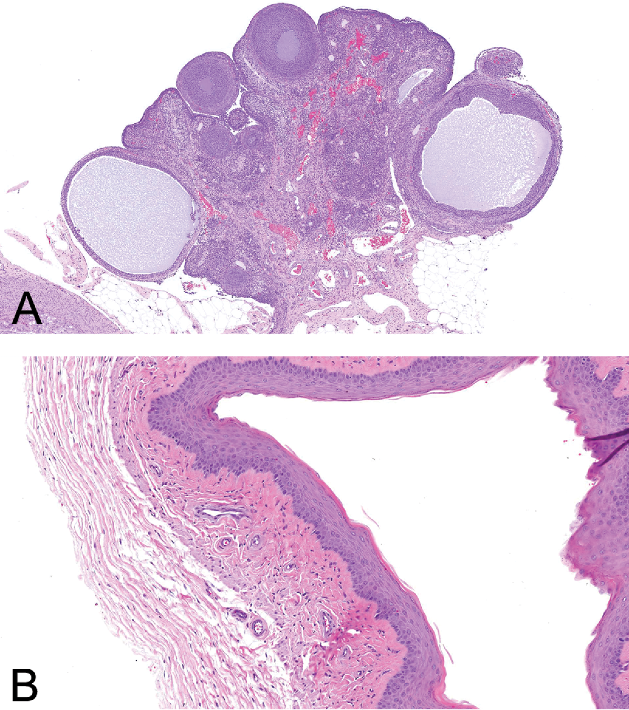
Reproductive tract: Persistent estrus: ovary with cystic follicles and vagina with cornified epithelium (H&E).
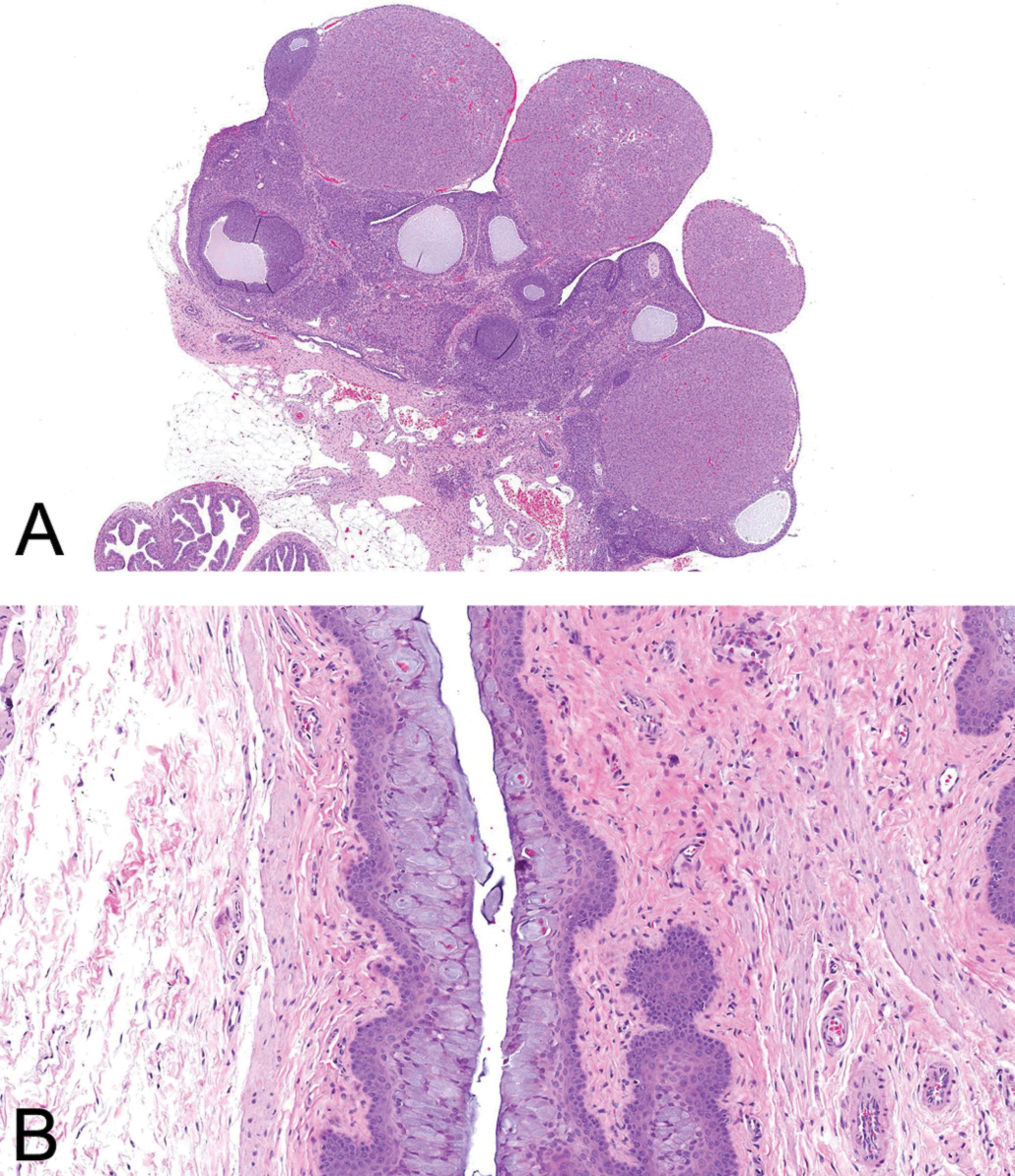
Reproductive tract: Repetitive pseudopregnancy: ovary with persistent corpora lutea and vagina with mucified epithelium (H&E).
Respiratory System
Macrophage infiltration in the alveolus was the most common microscopic finding observed in the lung of males and females, with an apparent age-related increase in females (Table 7). Affected alveoli contained focal aggregates of macrophages with finely vacuolated cytoplasm, were frequently subpleural or perivascular/bronchiolar, and infrequently associated with additional cellular infiltrates or inflammatory changes (Figure 7). Alveolar microlithiasis was characterized by discrete, lamellar, mineralized, concretions expanding alveoli with minimal associated inflammation (Figure 8). No microscopic findings were observed in the nasal turbinates proper, but were occasionally observed in closely associated structures (teeth and nasal lacrimal duct; Table 7).
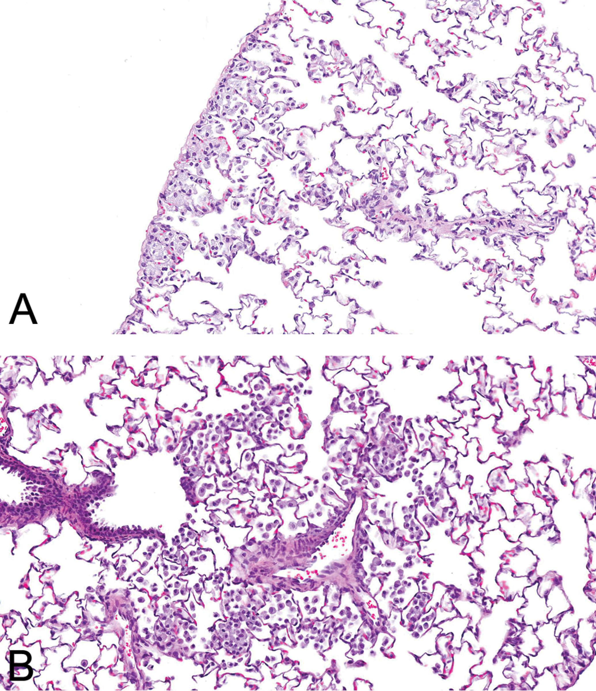
Lung: Alveolar macrophage infiltrates: subpleural and perivascular/bronchiolar distribution (H&E).
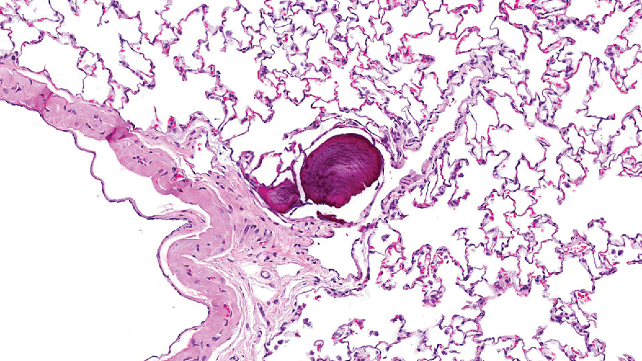
Lung: Alveolar microlithiasis: discrete, lamellar, mineralized, concretions expanding alveoli with minimal associated inflammation (H&E).
Digestive System
Mononuclear cell infiltration was the most common microscopic finding observed in the liver (Table 8). Mononuclear cell infiltrates were multifocal, periportal, or random in distribution and composed primarily of lymphocytes with fewer macrophages and, less frequently, plasma cells. Additional microscopic findings in the liver were sporadic, generally occurred at higher incidence in 13- and 26-week animals, and, with the exception of hepatocellular pigment in females, lacked any strong sex association. Lobular pancreatic atrophy characterized by decreased number and size of acini and increased interstitial connective tissue and mononuclear cell infiltrates (Figure 9) was observed at a relatively uniform incidence independent of animal age (Table 8). Microscopic findings were not observed in the cecum, colon, duodenum, ileum, jejunum, lower incisor tooth, mandibular salivary gland, or nonglandular stomach.
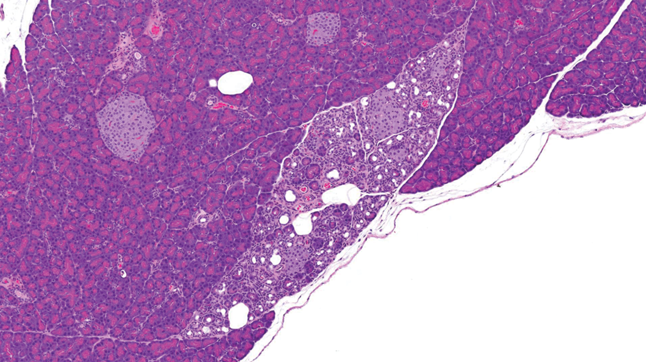
Pancreas: Lobular atrophy (H&E).
Hematopoietic/Lymphoid System
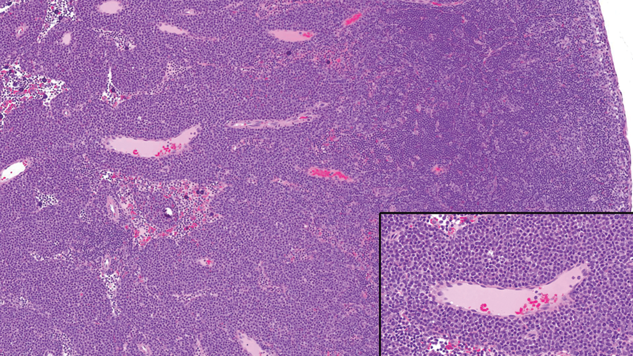
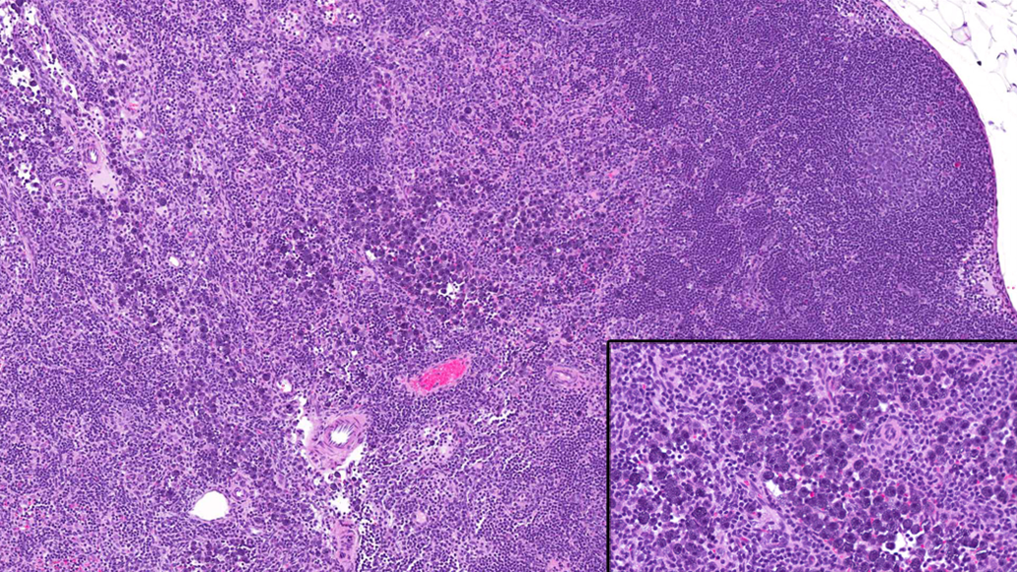
Pigmented macrophages in the mediastinal lymph nodes of males and females, increased pigment and extramedullary hematopoiesis in the spleen of females, and thymic epithelial tubule/cysts in females demonstrated age-related increases in incidence (Table 9). Mandibular lymph node medullary cords were occasionally expanded by increased numbers of plasma cells (Figure 10). Similarly, mesenteric lymph node medullary sinuses occasionally contained increased numbers of mast cells (Figure 11). Thymic epithelial tubule/cyst was characterized by epithelial cells arranged in tubules and nests and lining cystic spaces (Figure 12). Thymic epithelial tubule/cyst demonstrated an age-related increased incidence in females and was more common in females than males. This finding was consistent with the increased incidence of thymic epithelial proliferation and cysts in aging Wistar rats as reported by Kuper, Beems, and Hollanders (1986). Thymic hemorrhage was likely the result of the terminal blood collection procedure.
Mandibular lymph node: Medullary cords expanded by increased numbers of plasma cells (H&E).
Mesenteric lymph node: Medullary sinuses containing increased numbers of mast cells (H&E).
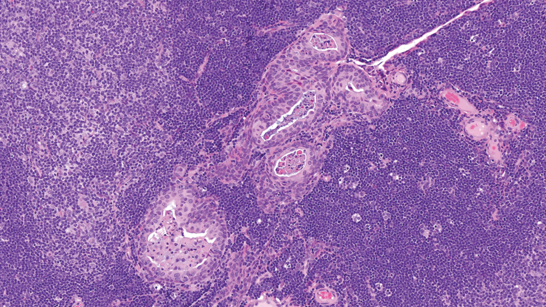
Thymus: Epithelial tubule/cyst (H&E).
Endocrine System, Nervous System, Miscellaneous
Pituitary cyst was the most common finding in the endocrine system. Cysts were simple or multilocular and lined by simple attenuated epithelium and/or ciliated cuboidal to columnar epithelium with intermittent mucous cells (Figure 13). Findings in the eye were uncommon except for the occasional retinal rosette. Corneal mineralization was rare in contrast to the relatively high incidence reported in Fischer-344 rats (Yoshitomi and Boorman 1990). Additional notable microscopic findings are presented in Table 10. These microscopic findings occurred sporadically, at singular or at very low incidence, and with no clear age-related trend. Microscopic findings were not observed in the adrenal medulla, biceps femoris muscle, diaphragm, femoral bone, mammary gland, parathyroid, sciatic nerve, spinal cord, or sternal bone.
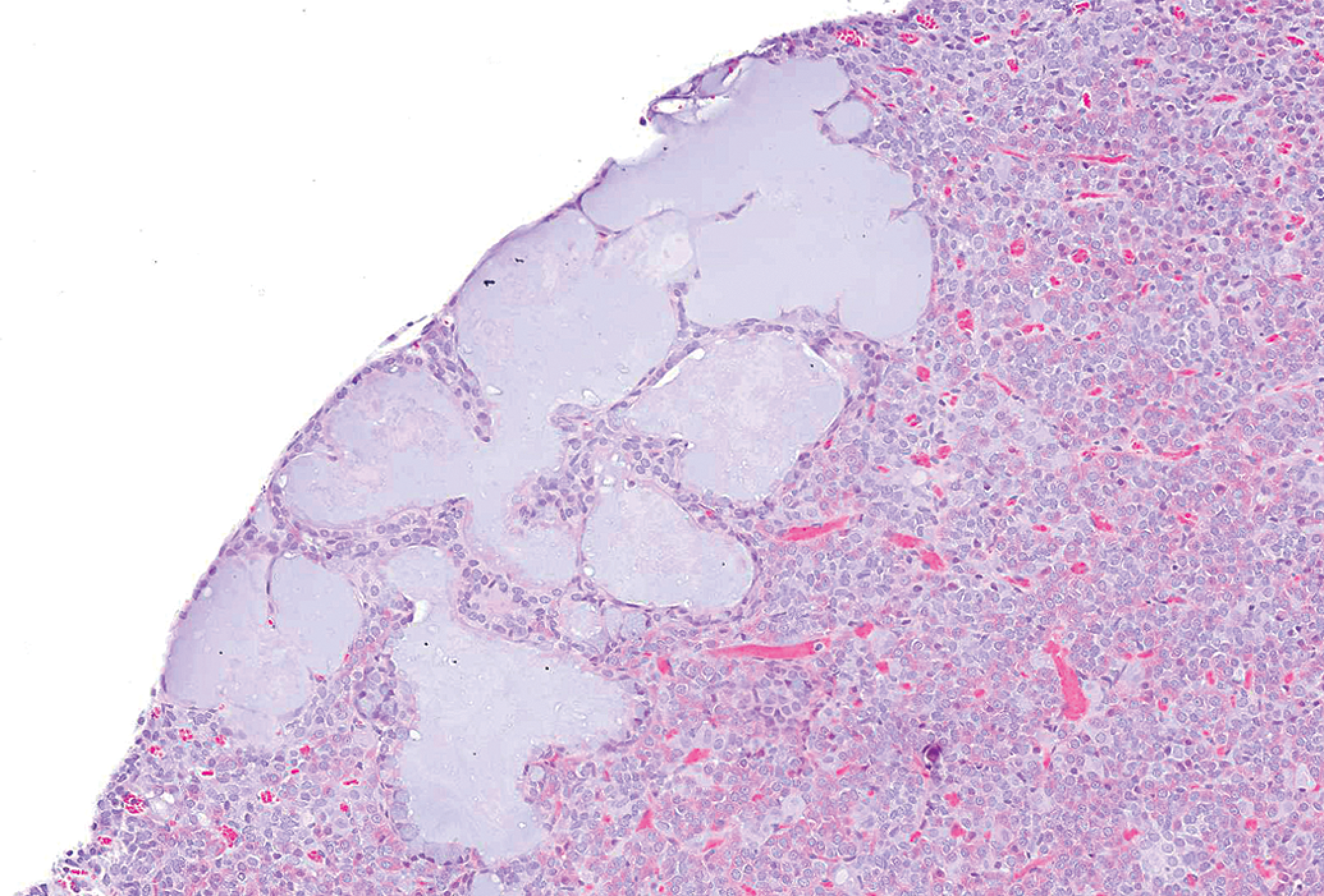
Pituitary: Multilocular cyst lined by ciliated epithelium and mucous cells (H&E).
In conclusion, body and organ weight data, macroscopic findings, neoplastic and nonneoplastic findings, and reproductive stage data provide support for the interpretation of findings encountered during routine 4-, 13-, and 26-week toxicity studies and in the evaluation of Harlan RCCHan: WIST rats as a suitable rodent model.
Footnotes
Abbreviations
Acknowledgments
We thank Drs. Gary Boorman and Mike Elwell for critical review of this article, toxicologist Rachael Avery for study coordination and support, and Steve Van Adestine for photographic assistance.
The authors declared no potential conflicts of interest with respect to the research, authorship, and/or publication of this article.
The authors received no financial support for the research, authorship, and/or publication of this article.