
Abstract
Information about the incidence of spontaneously occurring, nonneoplastic background findings in Syrian hamsters is essential if Syrian hamsters are to be used for toxicity studies. Male and female Syrian hamsters of the strain Han:AURA from the Fraunhofer Institute for Toxicology and Experimental Medicine (ITEM) breeding colony were maintained as control animals for carcinogenicity studies and were examined for the presence of nonneoplastic background findings either when they died or when the study was terminated. The nonneoplastic background lesions observed at an incidence of >50% (high), >25% (moderate), and >10% (low) in either male or female animals or in both sexes in one or more long-term studies are detailed. The results are compared to previous published reports of nonneoplastic, spontaneous background lesions in Syrian hamsters. Background information about the incidence of background lesions in Syrian hamsters on short- and long-term studies is useful to both toxicologists and toxicological pathologists.
Introduction
Historically, rats and mice are used in far greater numbers in biomedical research than Syrian hamsters (Mesocricetus auratus; Strandberg 1987). Information about the incidence of spontaneously occurring, nonneoplastic background lesions in Syrian hamsters is essential if Syrian hamsters are to be used for acute and chronic toxicity studies.
Information about spontaneous background changes in hamsters was published many years ago (Hubbard and Schmidt 1987; Kamino et al. 2001; Pour et al. 1976a,b,c,d; Pour et al. 1979; Schmidt et al. 1983), and recent data on hamster studies have not been published. Breeding methods, mutational drift, and diets may all affect the incidence of neoplastic and nonneoplastic lesions in hamsters and a more recent review of the incidence of nonneoplastic background findings in hamsters is thus overdue.
Materials and Methods
Animals
Male and female Syrian hamsters of the strain Han:AURA from the Fraunhofer Institute for Toxicology and Experimental Medicine (ITEM) breeding colony were maintained as control animals for toxicity/carcinogenicity studies. The hamsters were aged at approximately 6 weeks at the start of the studies. They were housed individually in Makrolon® (polycarbonate) type III cages (EBECO, Germany) with absorbent softwood as bedding material in the cages (Ssniff ¾, Ssniff Spezialdiäten, Soest, Germany). Animal room temperatures were generally maintained at 22 ± 2°C and 40 to 70% relative humidity with a 12-hr light–dark cycle controlled by an automatic timing device. All hamsters had free access to tap water (Hannover city water supplier, Germany) in Makrolon® bottles (approximately 150 ml) with sipper tubes and were fed a hamster diet (V 2144) supplied by Ssniff Spezialdiäten, Soest, Germany. Drinking water and diet were subjected to routine chemical analysis to monitor for contaminants and offered fresh at weekly intervals. Animal health was always monitored by a veterinary officer. All studies were conducted according to Good Laboratory Practice (GLP) and following the regulations of the German animal protection law.
Histopathology
Complete necropsies were performed on all decedent and terminal animals in all of the studies. Moribund animals were always removed from the study and sacrificed and necropsied, and autolysis was minimal. Harvested tissues were fixed in 10% neutral buffered formalin, embedded in paraffin, sectioned at 3–4 µm, and stained with hematoxylin and eosin. Trimming was done according to Ruehl-Fehlert and coworkers (2003), Kittel and coworkers (2004), and Morawietz and coworkers (2004). A complete histopathological examination including the teeth of the maxilla and of macroscopic observations was conducted in all animals.
Study Design
The causes of death in hamsters at different times during the 3 studies are detailed in Table 1. This article is based on 3 hamster studies, that is, three 24-month carcinogenicity studies conducted at the Fraunhofer ITEM, Germany, during the period 1999 through to 2009. The first 24-month study (Table 2) contained 100 control female and 100 control male animals. The second study (Table 2) contained 30 control male and 30 control female animals. The third study (Table 2) contained 60 control male and 60 control female animals. Information was gathered from control groups only. Nonneoplastic background findings were only reported if the incidence exceeded 10% in either male or female animals in 1 or more studies. The incidence of neuroendocrine cell hyperplasia (ectopic C cell hyperplasia) in the larynx and trachea was below 10% but has been included due to the complexity of the lesion and its cell of origin. In addition, the incidence of granular cell aggregates in the rectum and C cell hyperplasia in the thyroid were below 10%; however, these findings were included since they are mentioned in the discussion. The results from the three 104-week studies have been combined in Table 2.
The causes of death in hamsters at different times during the 3 long-term studies.
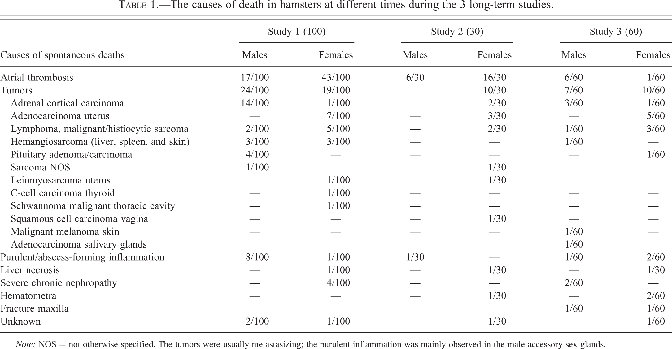
Note: NOS = not otherwise specified. The tumors were usually metastasizing; the purulent inflammation was mainly observed in the male accessory sex glands.
Incidence of common (≥10%) nonneoplastic lesions in control Han:AURA Syrian hamsters in the three 104-week studies including results in separate columns of the combination of all three long-term studies.
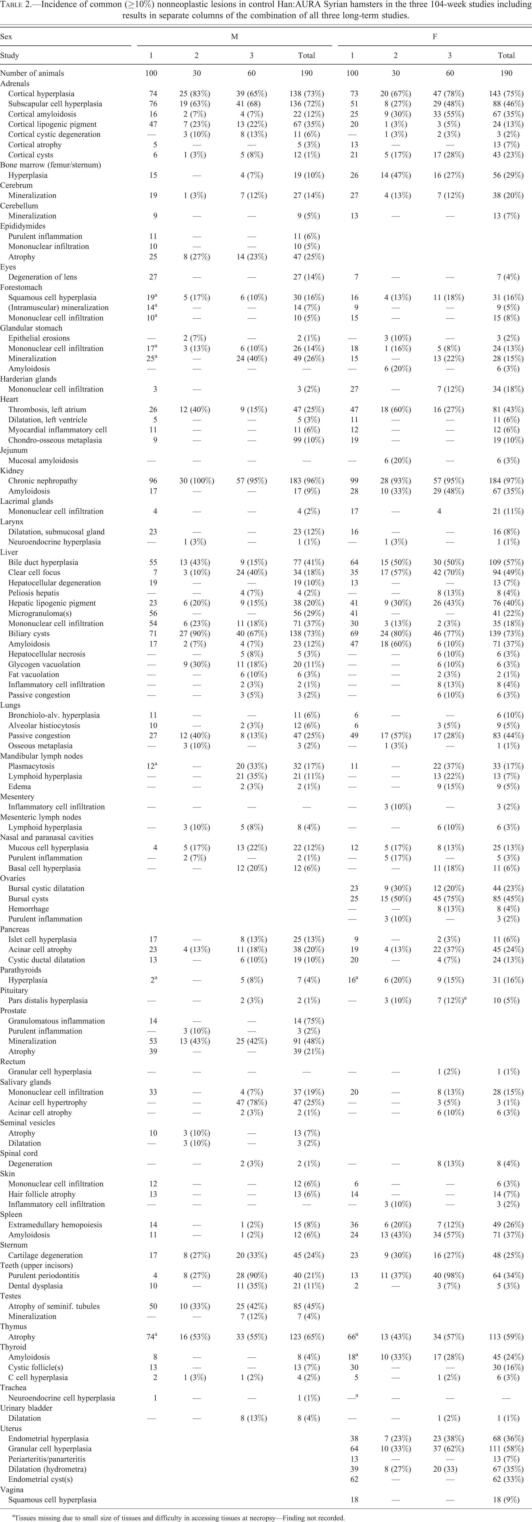
aTissues missing due to small size of tissues and difficulty in accessing tissues at necropsy—Finding not recorded.
Results
Unscheduled Deaths and Decedents
The causes of death in hamsters at different times during the 3 studies are detailed in Table 1. In all 3 studies, left atrial thrombosis was a frequent cause of death in these animals and occasionally tumor development was considered the cause of death.
The incidence of nonneoplastic background findings with a greater incidence of 10% in either male or female animals or in both sexes, in the three 24-month studies, are given in Table 2. The results from the three 104-week studies have also been combined in a separate column in Table 2.
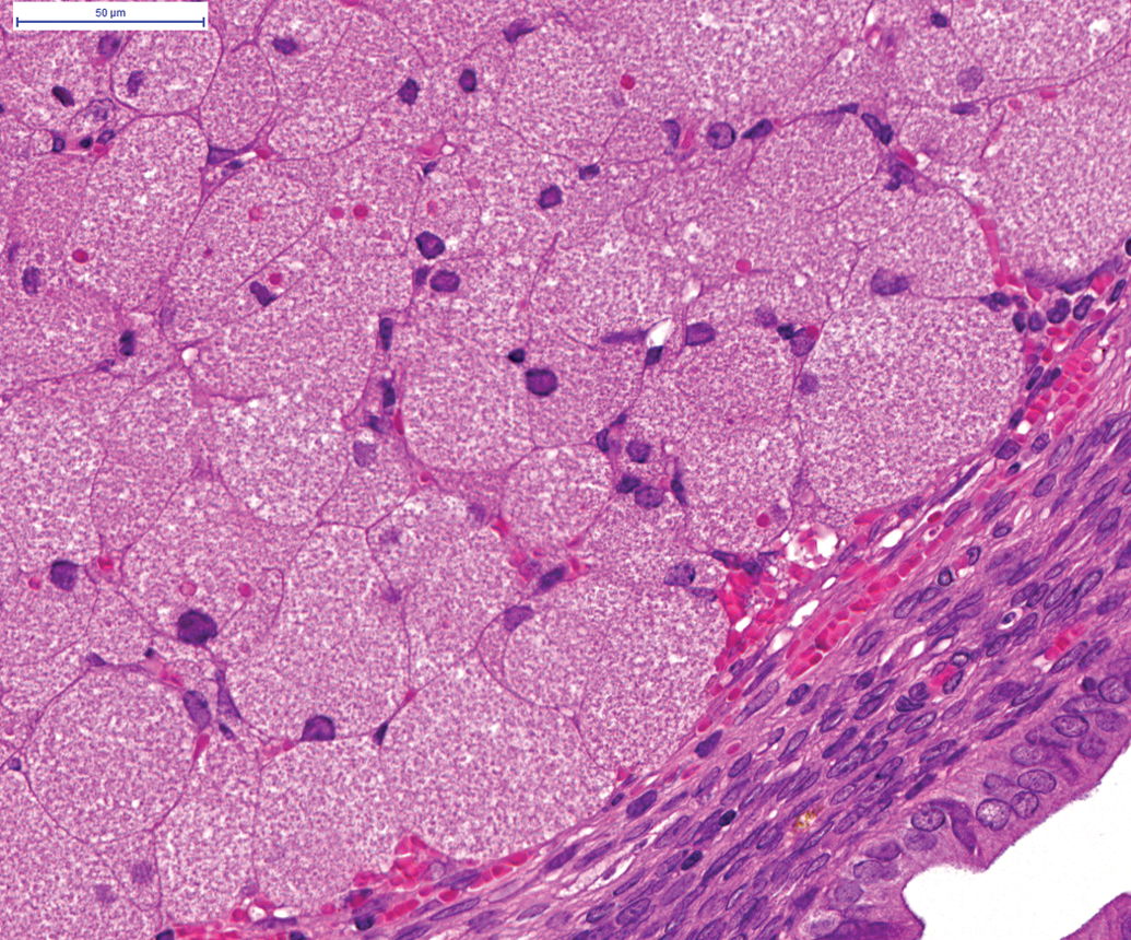
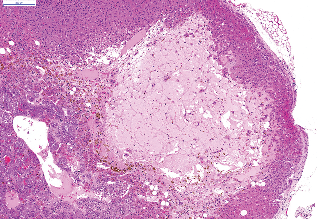
In general, in the three 24-month studies, nonneoplastic background lesions observed at an incidence of >50% (high) in either male or female animals or in both sexes, in one or more studies, included cortical hyperplasia, subcapsular cell hyperplasia, and cortical amyloidosis of the adrenals, chronic progressive nephropathy in the kidneys (Figure 1), bile duct hyperplasia, clear cell focus/foci, microgranulomas, mononuclear/inflammatory cell infiltration, amyloidosis, and cystic dilatation of bile ducts (Figure 2) of the liver. Chronic passive congestion was noted in the lungs (Figure 3) and associated with left atrial thrombosis (Figure 4) which was noted at a high incidence in the hearts of hamsters. Mineralization of the prostate (Figure 5) and atrophy of the seminiferous tubules in the testes (Figure 6) was noted at a high incidence in male hamsters; bursal cysts were noted in the ovaries and granular cell hyperplasia (Figure 7); and endometrial cysts were observed in the uterus in female animals. A high incidence of atrophy of the thymus and amyloidosis of the spleen (Figures 8 and 9) was noted in both male and female hamsters on the three 24-month studies. Purulent periodontitis of the upper jaw and acinar cell hypertrophy of the salivary gland were also noted at an incidence of greater than 50% in female hamsters on one 24-month study.
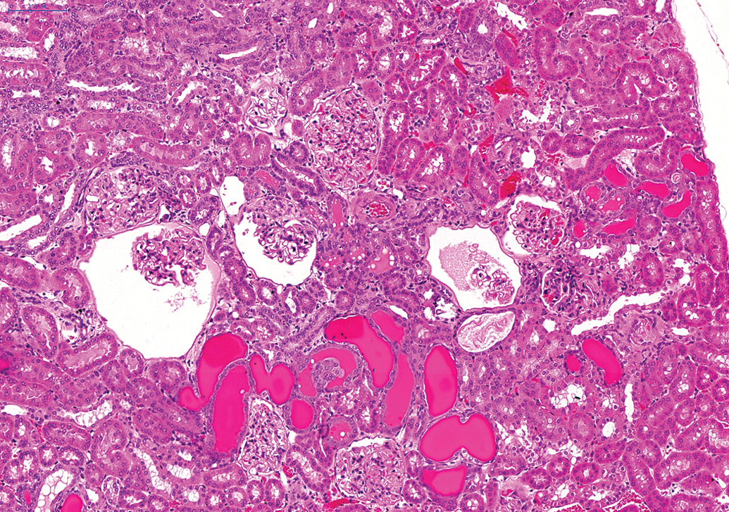
Chronic progressive nephropathy in the kidney. Note the hyaline casts (H&E).
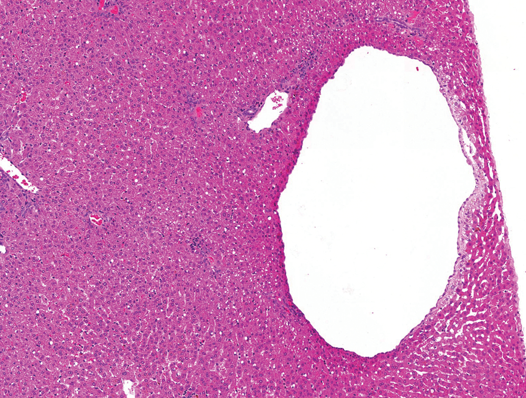
Cystic dilatation of bile ducts in the liver (H&E).
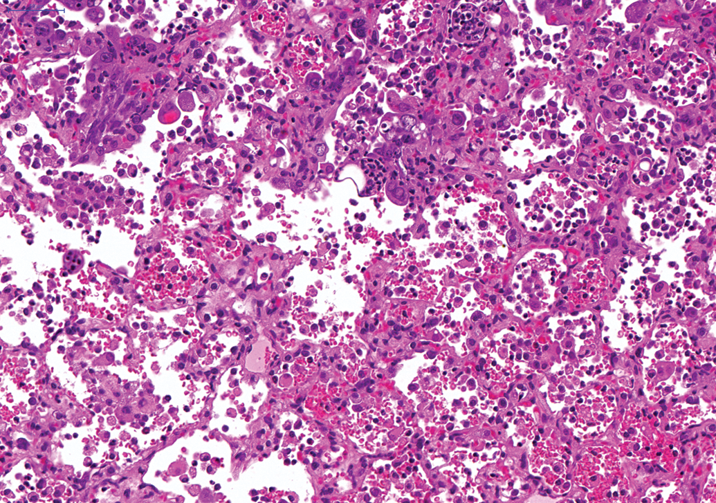
Chronic passive congestion in the lung. Note the effacement of alveolar spaces with histiocytes and erythrocytes (H&E).
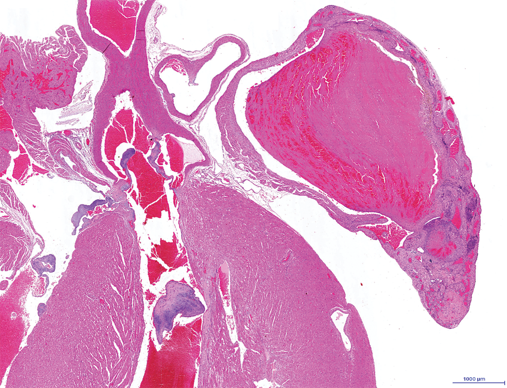
Left atrial thrombosis in the heart. Note the adherent fibrin thrombus in the left atrial chamber and the mineralization in the left atrial wall (H&E).
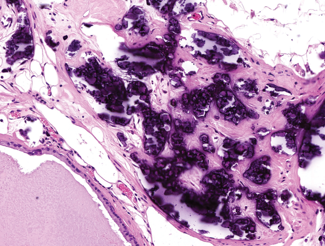
Mineralization of the prostate characterized by basophilic concretions in acini and stroma (H&E).
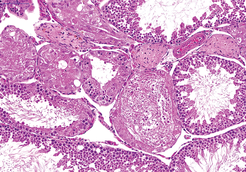
Atrophy, vacuolation, and hypospermatogenesis in the seminiferous tubules of the testis (H&E).
Granular cell aggregate in female uterus. Note the unencapsulated aggregates of large cells with granular cytoplasm and small, eccentric nuclei (H&E).
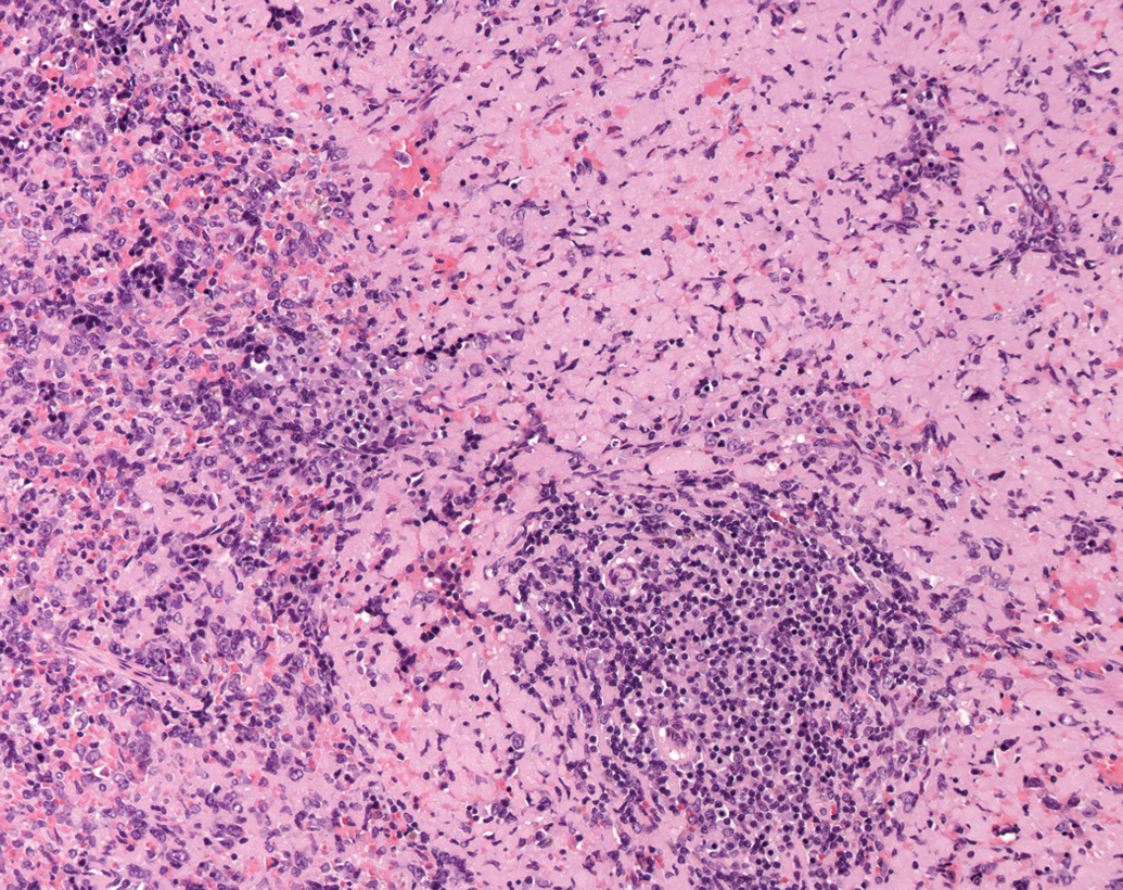
Amyloidosis of the spleen. Note the amorphous pink material in the spleen (H&E).
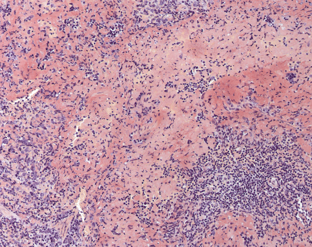
Note the Congo Red-positive amyloid deposits in the spleen (Congo Red stain).
Background lesions noted in the hamsters on the 24-month studies at an incidence level of between 25% and 50% (moderate), in either male or female animals or in both sexes, in one or more studies, included cortical lipogenic pigment deposits and cortical cysts in the adrenal, hyperplasia of the bone marrow of the femur and sternum, “Psamoma body-like” mineralization of the cerebrum (Figure 10), atrophy of the epididymides, degeneration of the lens of the eyes, mineralization of the glandular stomach, mononuclear/inflammatory cell infiltration into the Harderian and salivary glands, amyloidosis of the kidneys, hepatocellular lipogenic pigment deposits, hepatocellular glycogenic vacuolation in both male and female animals, bursal cystic dilatation in the ovaries, endometrial hyperplasia and dilatation of the uterus in female hamsters, atrophy of the prostate in male hamsters, cartilage degeneration of the sternum, amyloidosis of the thyroids of female hamsters, plasmacytosis, and lymphoid hyperplasia of the mandibular lymph nodes, acinar cell atrophy in the pancreas, dental dysplasia of the upper incisors, and increased extramedullary hemopoiesis in the spleen.
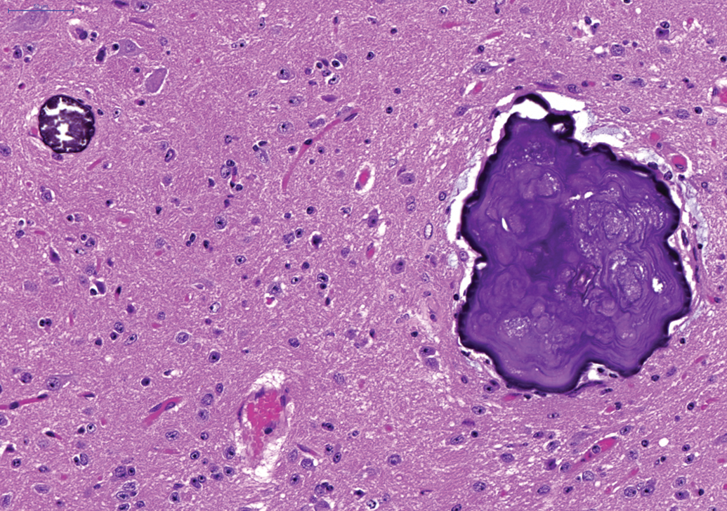
“Psamoma body-like” mineralization of the cerebrum. Note the focal basophilic concretions of calcium (H&E).
Background lesions with a low incidence (between 10% and 25%) in one or more of the three 24-month studies included cortical atrophy and cortical cystic degeneration (peliosis) in the adrenals, “Psamoma body-like” mineralization of the cerebellum, purulent/granulomatous inflammation and mononuclear/inflammatory cell infiltration of the epididymides, squamous cell hyperplasia of the forestomach, mineralization of the forestomach and mononuclear/inflammatory cell infiltration of the forestomach, mononuclear/inflammatory cell infiltration and epithelial erosions of the glandular stomach, dilatation of the left ventricle, myocardial mononuclear/inflammatory cell infiltration, and chondro-osseous metaplasia of the heart, mononuclear cell infiltration into the lacrimal glands, dilatation of submucosal glands in the larynx, hepatocellular degeneration, peliosis hepatis, hepatocellular necrosis, hepatocellular fatty vacuolation, inflammatory cell infiltration and passive congestion of the liver, bronchiolo-alveolar hyperplasia, osseous metaplasia, and alveolar histiocytosis of the lung. Inflammatory cell infiltration of the mesentery is noted in hamsters at a low incidence as well as lymphoid hyperplasia of the mesenteric lymph nodes. Mucous (goblet) cell hyperplasia, basal cell hyperplasia of Bowman’s glands, and purulent inflammation were noted at a low incidence in the nasal and paranasal cavities of hamsters. Purulent inflammation of the ovaries, islet cell hyperplasia and cystic ductal dilatation of the pancreas, hyperplasia of the parathyroid glands, pars distalis hyperplasia of the pituitary, and purulent/granulomatous inflammation of the prostate were also observed at a low incidence in hamsters on one or more of the 24-month studies. Acinar cell atrophy of the salivary glands, dilatation and atrophy of the seminal vesicles, degeneration of the spinal cord, periarteritis/panarteritis of the uterus, and squamous cell hyperplasia of the vagina were noted at a low incidence in hamsters on the 24-month studies. Furthermore, mononuclear/inflammatory cell infiltration and atrophy of hair follicles were noted in the skin at a low incidence in hamsters on the 24-month studies. Amyloidosis of the duodenum, glandular stomach, and jejunum were noted at a low incidence in hamsters on the 24-month studies. Edema of the mandibular lymph node, hemorrhage of the ovaries, mononuclear cell infiltration into the salivary glands, and mineralization of the testes and urinary bladder dilatation were noted at a low incidence in hamsters on the 24-month studies. Adrenal cortical cystic degeneration (peliosis) noted at an incidence of 11/190 (6%) in male hamsters and 3/190 (2%) in female hamsters is illustrated in Figure 11. Neuroendocrine cell hyperplasia or ectopic C cell hyperplasia noted at an incidence below 10% in the respiratory epithelium of the trachea or larynx is illustrated in Figure 12.
Cortical cystic degeneration in an adrenal gland (H&E).
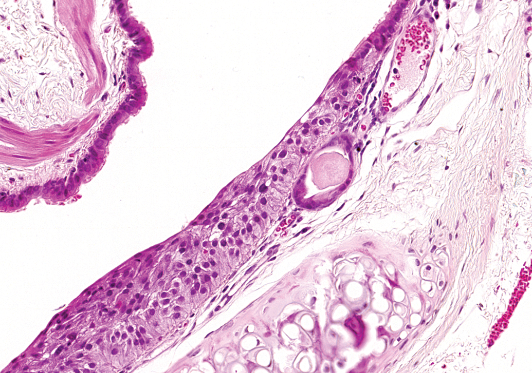
Neuroendocrine hyperplasia or ectopic C cell hyperplasia noted in the respiratory epithelium at the laryngo-tracheal junction (H&E).
Discussion
It is important to have information on the background lesions of hamster, so users may compare new hamster study results to the expected historical control data. The limited and old pathology background findings database of the Syrian hamster is of concern, and new published data on background lesions in the Syrian hamster are of use to all researchers, contract research organizations, and pharmaceutical companies using hamsters as experimental animals.
In general, in the 24-month studies, nonneoplastic background lesions observed at an incidence of >50% (high) in either male or female animals or in both sexes included cortical hyperplasia, subcapsular cell hyperplasia, and cortical amyloidosis of the adrenals. Cortical hyperplasia of the adrenal glands in male Han:AURA hamsters is reported by Kamino et al. (2001) at an incidence of 72% in hamsters over the age of 104 weeks. In addition, hyperplasia of the adrenal cortex in female Han:AURA hamsters is reported at an incidence of 61% in female hamsters over the age of 113 weeks and adrenal amyloidosis occurs at an incidence of 45% at the age of 83 weeks in female hamsters (Kamino et al. 2001). Pour and coworkers (1976b) report that nonneoplastic lesions of the adrenal glands include amyloidosis and cysts in 2 colonies of Syrian hamsters. Hyperplasia of the adrenal cortex, often of the zona glomerulosa, as well as cortical cysts and adrenal amyloidosis are found in 3 hamster strains (white, albino, and cream hamsters; Pour et al. 1979). Acinar cell atrophy in the pancreas was noted at a moderate incidence in the long-term studies analyzed for this article. Pour and coworkers (1976b) report atrophy of the exocrine pancreas in three hamsters from 2 colonies of Syrian hamsters.
Chronic progressive nephropathy is a common nonneoplastic background finding in older hamsters and was noted at a high incidence in our study. Kamino and coworkers (2001) demonstrate an incidence of glomerular cystic degeneration and cortical cysts in the kidneys of male and female hamsters at a frequency of 49% to 70%. Nephrocalcinosis is the most common nonneoplastic renal disease noted in 3 groups of hamster strains with the highest incidence occurring in the white hamster strain (Pour et al. 1979). In 2 colonies of Syrian hamsters, Pour and coworkers (1976b) report amyloidosis, nephrocalcinosis, and pyelonephritis as well as small cortical cysts in the kidneys. Percy and Barthold (2007) state that degenerative renal disease represents an important cause of morbidity and mortality in older hamsters, particularly in female hamsters and that the etiology and pathogenesis of the disease is poorly understood.
Bile duct hyperplasia, clear cell focus/foci (particularly in female hamster livers in all 3 studies), microgranulomas, mononuclear/inflammatory cell infiltration, amyloidosis, and dilatation of the bile ducts of the liver occurred at a high incidence in the 3 long-term studies discussed in this article. Liver cysts and bile duct proliferation occurred in 3 strains of hamster (cream, white, and albino; Pour et al. 1979) and are reported at a relatively high frequency (49–70%) in older male and female Han:AURA hamsters (Kamino et al. 2001). In addition, clear cell foci are reported at an incidence of 35% in female Han:AURA hamsters and are not thought to be a precursor to neoplastic change in the liver (Kamino et al. 2001). Cystic distension of the bile ducts is common in both sexes, particularly in hamsters with liver amyloidosis (Pour et al. 1976d). Pour and coworkers (1976d) noted that cystic bile ducts range from microscopic cysts to macroscopically visible areas between 1 and 15 mm in diameter, often present in the liver periphery. Percy and Barthold (2007) state that the cause of hepatic cysts in older hamsters is thought to be a congenital failure of fusion of the intralobular and interlobular bile ducts or failure of superficial bile ducts to regress.
Passive congestion was noted at a high incidence in the lungs of hamsters on the 3 long-term studies discussed in this article. This lesion is related to the severe cardiac insufficiency caused by left atrial thrombosis. Atrial thrombosis is noted at a high incidence in older hamsters (McMartin 1977). Male and female Syrian hamsters have an equally high incidence of atrial thrombosis and myocardial degeneration, despite the females’ much shorter life span (McMartin 1979). Thrombosis of the cardiac atrium occurred at a high incidence in cream, white, and albino hamsters (Pour et al. 1979) and is reported in both male and female Han:AURA hamsters (Kamino et al. 2001) at an incidence of 30% and 46% in male and female animals, respectively. Female hamsters are more commonly affected than male hamsters, and the syndrome may be associated with amyloidosis (Percy and Barthold 2007). Cardiac thrombosis is noted more often in the Hannover colony of hamsters (Pour et al. 1976a). Percy and Barthold (2007) state that a putative cause of left atrial thrombosis in older hamsters may be cardiac insufficiency since there is often a concurrent myocardial degeneration; however, myocardial degeneration was a rare finding in our Han:AURA hamsters (data not shown), and it is more likely that the atrial thromboses were related to disturbances in blood composition or blood coagulation. Atrial thrombosis was often noted in conjunction with adrenal cortical tumors (data not shown) in our studies.
Mineralization of the prostate and atrophy of the seminiferous tubules were noted at a high incidence in male hamsters, bursal cysts were noted in the ovaries, and granular cell hyperplasia and endometrial cysts were observed in the uterus in female animals in our long-term studies. Prostate calcification is reported in cream, white, and albino hamsters (Pour et al. 1979) and is reported at an incidence of 31% in Han:AURA hamsters. Testicular atrophy is reported in male Han:AURA hamsters at an incidence of 40% (Kamino et al. 2001). Glandular cystic dilatation of the endometrium is reported at an incidence of 11% in Han:AURA female hamster (Kamino et al. 2001).
Granular cell aggregates of the endometrium was noted at an incidence of 64%, 33%, and 62%, respectively, in the 3 long-term studies analyzed for this article. Granular cell foci are visible in the female hamster uterus (incidence of 54%; Kamino et al. 2001). Granular cells resembling those of granular cell tumors were also scattered in the subserosa of the uterus in 3 female hamsters in 2 colonies of Syrian hamsters (Pour et al. 1976b). In addition, tumors composed of granular cells in the intestinal walls of white hamsters have been described by Pour and coworkers (1973). A granular cell aggregate was noted in the rectum of a single female hamster in 1 of the long-term studies analyzed for this article. Granular cell tumors are also described in the hamster peripheral nervous system (Pour et al. 1973).
The cell of origin of granular cell tumors or aggregates has not been definitively described. Up to 23% of control female Sprague-Dawley rats in carcinogenicity studies have granular cell lesions in the distal reproductive tract (Markovits and Sahota 2000a). Markovits and Sahota (2000a) describe granular cell aggregates as nonspace-occupying lesions composed of clusters of typical granular cells with prominent interstitial collagen that are either discrete masses or are difficult to discern from the surrounding tissues. The principal histomorphologic feature of granular tumor cells in B6C3F1 mice is the presence of abundant cytoplasmic eosinophilic granules that stain positive for periodic acid–Schiff with diastase resistance (Veit et al. 2008). Expression of S-100 and neuron specific enolase in rodent granular cell tumors/aggregates may be negative (Veit et al. 2008; Miyajima et al. 2001) or positive (Courtney et al. 1992; Markovits and Sahota 2000a), and uterine granular cell tumors are thought to be of myogenic origin (Veit et al. 2008) or Schwann cell or histiocytic origin (Markovits and Sahota 2000a). Ultrastructurally, uterine granular cell tumors in mice demonstrate prominent electron-dense granules and contain secondary lysosomes (Veit et al. 2008). Markovits and Sahota (2000b) describe the reduction in granular cell tumors in the distal female reproductive tract of Sprague-Dawley rats after aromatase inhibitor treatment. The authors speculate that since these compounds prevent the conversion of androgenic steroids to corresponding estrogens, this may cause the reduction in granular cell tumors, suggesting a role for estrogen in the pathogenesis of these tumors (Markovits and Sahota 2000b).
Hyperplasia of the parathyroid glands was noted at a low incidence in the hamsters on the 3 long-term studies analyzed for this article. Pour and coworkers (1976b) report an incidence of parathyroid cell hyperplasia of 83% in male hamsters and 22% in female hamsters in 2 Syrian hamster colonies, but we did not observe such a high incidence. There was an incidence of about 13% of mainly slight to moderate peri-/panarteritis of the uterus in the long-term hamster studies reported by us. Percy and Barthold (2007) mention an arteriolar nephrosclerosis with fibrinoid change. In our article, in the kidneys, vascular lesions were always summarized (together with other renal pathological components) under chronic progressive nephropathy and thus do not appear as a separate finding in the kidney. Similar vascular changes are also observed in aged rats and mice (Taylor 2011) and are thus not specific to the hamster.
Neuroendocrine cell hyperplasia was noted only in 1 trachea and 1 larynx in the hamsters examined for this study. A continuum exists from minute intraepithelial neuroendocrine cell hyperplasia to malignant neuroendocrine tumors. Since neuroendocrine hyperplasias and tumors are immunoreactive for calcitonin, calcitonin gene–related peptide, and other neuroendocrine markers, these lesions are considered to be ectopic C-cell proliferations (Ernst et al. 1995; McInnes, Ernst, and Germann 2013). The incidence of multifocal C-cell hyperplasia in the thyroid was 2/100 males and 5/100 females in the first long-term study (Table 2), 1/30 males in the second long-term study (Table 2), and 1/60 males and 1/60 females in the third long-term study (Table 2). There was a good correlation between the incidence of C-cell tumors in the thyroid and neuroendocrine tumors in the old hamster studies (Ernst et al. 1995). The areas of thyroid C-cell hyperplasia were more commonly observed than C-cell hyperplasia in the laryngo-tracheal area bordering the thyroids.
“Psammoma body-like” mineralization forming lamellar concentric concretions of the cerebrum were noted at a moderate incidence and “Psamoma body-like” mineralization of the cerebellum was noted at a low incidence in hamsters on the three long-term studies analyzed for this article. Percy and Barthold (2007) report that focal cerebral mineralization may be observed as an incidental finding in the neuropil of the hamster brain with displacement of the adjacent structures and minimal cellular response. In multiple brain and spinal cord transverse sections of 520 hamsters of 1, 3, 6, 12, and 24 months of age investigated using histology and immunohistochemistry, vacuolation of gray matter neuropil and mineralization especially in the brain stem are the most prominent findings (Gerhauser et al. 2013). These findings gradually increase in severity and frequency with age and affect approximately 100% and 50% of 24-month-old male and female hamsters, respectively (Gerhauser et al. 2013).
Purulent periodontitis was also noted at an incidence of greater than 50% in female hamsters on one 24-month study. Pour and coworkers (1979) report dental caries and periodontitis in all 3 strains of hamster (white, albino, and cream) and state that the periodontitis is often associated with abscess and necrosis and extends to the facial region and involves the Harderian glands and brain.
A high incidence of amyloidosis of the spleen was noted in both male and female hamsters on the 24-month study. Hepatic, renal, and splenic amyloidosis is more severe in females than in male Syrian hamsters and becomes so at an earlier age (McMartin 1979). Percy and Barthold (2007) state that amyloid may be detected as early as 5 months but is more common in hamsters at 15 months of age. Pour and coworkers (1979) state that one of the most common generalized lesions in 3 groups of hamsters (inbred cream, linebred white, and linebred albino) is amyloidosis, and female hamsters are affected twice as often as male animals. The disease is noted (in decreasing order of frequency) in the spleen, liver, kidneys, adrenal glands, thyroid glands, glandular stomach, pancreas, and lymph nodes (Pour et al. 1979). The frequency of generalized amyloidosis in the kidneys, adrenal glands, thyroid glands, spleen, liver, and pancreas is more than 5 times greater in female hamsters in 1 colony (Eppley) compared to female animals in another colony (Hannover; Pour et al. 1976a). Systemic amyloidosis is a major cause of death in older hamsters (Gleiser et al. 1971). Amyloidosis was also noted in the jejunum, duodenum, and glandular stomach at a low incidence in hamsters on the 24-month studies described by us.
In conclusion, we report on the incidence of background findings in hamsters on three 24-month studies. Background information about the incidence of spontaneous lesions in Syrian hamsters on long-term studies is useful to both toxicologists and toxicological pathologists and will prove vital for decisions about the suitability of the Syrian hamster in long-term safety studies.
Footnotes
The author(s) declared no potential conflicts of interest with respect to the research, authorship, and/or publication of this article.
The author(s) received no financial support for the research, authorship, and/or publication of this article.