
Abstract
Toxicity studies were conducted by the National Toxicology Program (NTP) to provide information on the potential for toxicity from long-term use of commonly used herbal medicines. Here, we review the findings from these NTP toxicology/carcinogenesis 2-year rodent studies of 7 commonly used herbs. In these studies, the individual herb or herbal product was administered to F344/N rats and B6C3F1 mice by oral administration for up to 2 years. The spectrum of carcinogenic responses ranged from no or equivocal evidence for carcinogenic activity (ginseng, milk thistle, and turmeric oleoresin) to a liver tumor response (ginkgo, goldenseal, kava), thyroid tumor response (ginkgo), or an intestinal tumor response (Aloe vera whole leaf nondecolorized extract). Different mechanisms may be involved in the occurrence of liver (ginkgo, goldenseal, and kava kava) and gastrointestinal toxicity (turmeric oleoresin and Aloe vera whole leaf nondecolorized extract), while the toxic lesion is the same. The results from these hazard identification toxicity/carcinogenesis studies along with those from ongoing National Institute of Health clinical trials of herbal medicines provide more complete information on the risks and benefits from herbal medicine use in the general population.
Introduction
In the United States, natural products are regulated under the Dietary Supplement Health and Education Act (DSHEA) of 1994 (U.S. Food and Drug Administration 2012). A dietary supplement is a product that is ingested and is intended to supplement the diet and contains a “dietary ingredient.” The dietary ingredients in these products may include vitamins, minerals, herbs, or other botanicals (U.S. Food and Drug Administration 2011). In the United States, herbal products are widely used in the general population (Barnes, Bloom, and Nahin 2008). It is estimated that greater than 50% of the U.S. adult population may use complementary or alternative medicines (CAM; Nahin, Dahlhamer, and Stussman 2010). Under DSHEA, additional toxicity studies are not generally required if the herb has been on the market prior to 1994 (National Institute of Health [NIH] Office of Dietary Supplements 2011).
Various government agencies provide information on herbals including use patterns (Center for Disease Control), toxicity information (NIEHS/National Toxicology Program [NTP]), clinical trial data (NIH Office of Dietary Substances, Office of Complementary and Alternative Medicines, and NIH Institutes), and review of reported side effects from herbal medicine use (U.S. Food and Drug Administration 2012).
In this article, we review the results of selected NTP 2-year rodent studies for commonly used herbal medicines including Aloe vera, ginkgo, ginseng, goldenseal, kava kava, milk thistle, and turmeric oleoresin (Table 1). These herbal studies were studied by NTP because of the lack of toxicity and carcinogenicity information reported in the literature. It should be noted that in 2002, the U.S. Food and Drug Administration issued a consumer advisory for kava noting that kava-containing dietary supplements may be associated with liver disease (U.S. Food and Drug Administration 2002). The composition of dietary supplements may vary with the season or region in which the herb is grown, and there is concern regarding the presence of contaminants in over-the-counter dietary supplements (Cohen 2009). A review by experts in the field emphasized the need for more research on the benefits/side effects from herbal medicine use (Ahn et al. 2010).
Herbal products reviewed.
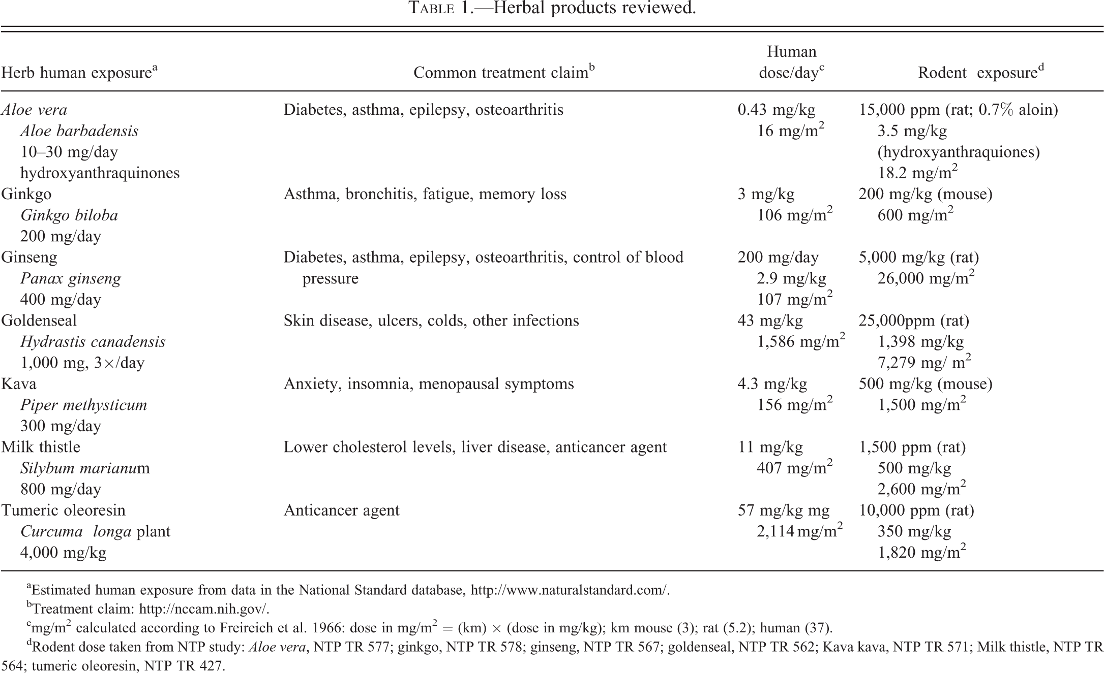
aEstimated human exposure from data in the National Standard database, http://www.naturalstandard.com/.
bTreatment claim: http://nccam.nih.gov/.
cmg/m2 calculated according to Freireich et al. 1966: dose in mg/m2 = (km) × (dose in mg/kg); km mouse (3); rat (5.2); human (37).
dRodent dose taken from NTP study: Aloe vera, NTP TR 577; ginkgo, NTP TR 578; ginseng, NTP TR 567; goldenseal, NTP TR 562; Kava kava, NTP TR 571; Milk thistle, NTP TR 564; tumeric oleoresin, NTP TR 427.
The environment and lifestyles play a role in the risk for cancer (Lichtenstein et al. 2000). While the focus of the NTP herbal toxicity 2-year studies is to identify hazards after long-term exposure to a particular herb, these studies also provided information that suggests that some herbs have antiproliferative activity.
Methods
Characterization of Herbs
The herbal products reviewed in this article were analyzed for various chemical constituents. The NTP used standards to qualitatively or quantitatively identify herbal components using chromatographic techniques. Quantification of individual herbal components (percentage by weight or percentage by peak area) was performed only on selective components. A complete description of the chemical analysis and chromatographic systems used to identify herbal components is provided in the NTP technical reports. The composition of herbal products may vary from batch to batch, and the chemical composition listed below is for the herbal product used in the NTP study.
Aloe vera Whole Leaf Nondecolorized Extract
The Aloe vera leaf whole nondecoloraized extract (also referred to as Aloe vera in this article) used in the NTP study was from Aloe barbadensis Miller plants (Pangea Phytoceuticals, Inc., Harlingen, TX). Leaf weights were a minimum of 400 g at harvest, and the time from harvest to lyophilization was a maximum of 6 hr. The Aloe vera nondecolorized whole leaf extract contained extract from the Aloe vera inner leaf gel and the Aloe vera latex, including the anthraquinones. The content of aloin A was estimated at 5.7 ± 0.2 mg/g to 7.2 ± 0.3 mg/g and the content of aloe emodin was at 70.5 ± 4.5 µg/g (NTP 2011a).
Ginseng
Ginseng extract from the plant Panax ginseng C.A. Meyer was obtained from Plus Pharma, Inc. (Vista, CA), in one lot (3031978). The total ginsenosides in the herb was 7.4 as weight percentage of the total material (NTP 2011b).
Ginkgo biloba Extract
Ginkgo biloba extract was obtained from Shanghai Xing Ling Science and Technology Pharmaceutical Company, Ltd. (Shanghai, China; lot 020703). After chromatography, 37 components were observed to have peak areas greater than or equal to 0.05% of the total peak area, and 3 components were identified: quercetin, kaempferol, and isorhamnetin, which had peak areas equal to 34.08%, 27.77%, and 5.43%, respectively, of the total peak area. The Ginkgo biloba extract used in the present study contained 31.2% flavonol glycosides, 15.4% terpene lactones (6.94% bilobalide, 3.74% ginkgolide A, 1.62% ginkgolide B, 3.06% ginkgolide C), and 10.45 ppm ginkgolic acid (NTP 2012).
Goldenseal
Goldenseal roots were purchased from Strategic Sourcing, Inc. (Reading, PA; HYCA 10/7-10.28.01-C). Weight loss on drying indicated moisture content of 6.35%. The palmatine, berberine, hydrastine, canadine, and total alkaloids contents were estimated at 0.0%, 3.89%, 2.80%, 0.17%, and 6.86% (% by weight; NTP 2010)
Kava Kava Extract
Kava kava extract was obtained from Cosmopolitan Trading Co. (Seattle, WA) in one lot (9077SDK). Kavalactones identified in the extract included methysticin, dihydromethysticin, kavain, dihydrokavain, yangonin, and desmethoxyyangonin (although the percentage of each component was not determined; NTP 2011c).
Milk Thistle Extract
Milk thistle extract was obtained from Indena USA, Inc. (Seattle, WA; 27691/M6). Major alkaloid components qualitatively identified included taxifolin, isosilychristin, silychristin, silydianin, silybin A and B, and isosylibin A and B. Only the total silybin content was quantitated to be approximately 34% (by peak area) or 65% by weight of the test article (NTP 2011d).
Turmeric Oleoresin
Turmeric oleoresin was obtained from Kalsec, Inc. (Kalamazoo, MI) in two lots (2452-A and 2558-A). The material was a purified oleoresin that was produced by extracting turmeric with acetone. Major component was identified as curcumin (1,7-bis (4-hydroxy-3-methoxyphenyl)-l, 6-heptadiene-3, 5-dione [79–84%]) with two other components tentatively identified as 1-(4-hydroxyphenyl)-7-(4- hydroxy-3-methoxyphenyl)-l, 6-heptadiene-3, 5-dione (11.3–16.9%) and 1,7-bis (hydroxyphenyl)-l, 6-hydroxy-3-methoxyphenyl)-l, 6-heptadiene-3, 5-dione (1.3–3.1%; NTP 1993).
Design of 2-year Exposure Studies in F344/N Rats and B6C3F1 Mice
The NTP 2-year studies were conducted in male and female F344/N rats and B6C3F1 mice. The herbs were administered continuously in the feed or by oral gavage (5 days week) for up to a 2-year period. At the start of the study, the animals were 5 to 6 weeks of age. The animals were housed by species and sex, 2 to 3 male rats per cage, 5 female rats per cage, 1 male mouse per cage, and 5 female mice per cage. Tap water and NTP-2000 diet (Zeigler Brothers, Inc., Gardners, PA) were made available ad libitum. The care of animals was according to NIH procedures as described in the “The U.S. Public Health Service Policy on Humane Care and Use of Laboratory Animals, available from the Office of Laboratory Animal Welfare, National Institutes of Health, Department of Health and Human Services, RKLI, Suite 360, MSC 7982, 6705 Rockledge Drive, Bethesda, MD 20892-7982 or online at http://grants.nih.gov/grants/olaw/olaw.htm#pol.” Moribund animals were sacrificed during the course of the study. Complete necropsies were performed on all animals. Tissues were preserved in 10% neutral-buffered formalin, embedded in paraffin, sectioned, and stained with H&E. The following tissues were examined microscopically from male and/or female animals; gross lesions and tissue masses, adrenal gland, bone with marrow, brain, clitoral gland, esophagus, heart, large intestine (cecum, colon, and rectum), small intestine (duodenum, jejunum, and ileum), kidney, liver, lung, lymph nodes (mandibular and mesenteric), mammary gland (except male mice), nose, ovary, pancreas, pancreatic islets, parathyroid gland, pituitary gland, preputial gland, prostate gland, salivary gland, skin, spleen, stomach (forestomach and glandular), testis with epididymis and seminal vesicle, thymus, thyroid gland, trachea, urinary bladder, and uterus. Following the completion of the studies, the accuracy of the histopathologic diagnosis was determined by microscopic reviews of neoplasms and target organs by quality assessment from a pathology working group (Boorman and Eustis 1986; Hardisty and Boorman 1986). The poly-3 test, a test that takes survival differences into account (Bailer and Portier 1988; Piegorsch and Bailer 1997; Portier and Bailer 1989), was used to assess the prevalence of neoplasm and nonneoplastic lesion.
When historical control data were particularly important in the interpretation of the study results, this information is provided (e.g., thyroid gland tumors in ginkgo studies).
Results
Findings from selected NTP toxicity and carcinogenicity 2-year studies of herb products are summarized in Tables 2 and 3. Treatment-related nonneoplastic and neoplastic responses are reported by incidence for Ginkgo biloba extract (NTP 2012), goldenseal (NTP 2010), kava kava extract (NTP 2011c), and Aloe vera (NTP 2011a) in Table 2. Data on the herbs with no evidence or equivocal evidence for carcinogenic activity (ginseng [NTP 2011b], milk thistle [NTP 2011d], or turmeric oleoresins [NTP 1993]) are listed in Table 3.
Selected toxicology and pathology endpoints in 2-year F344/N rat and B6C3F1 mouse studies for herbals causing clear or some evidence of carcinogenic activity (ginkgo, goldenseal, kava).
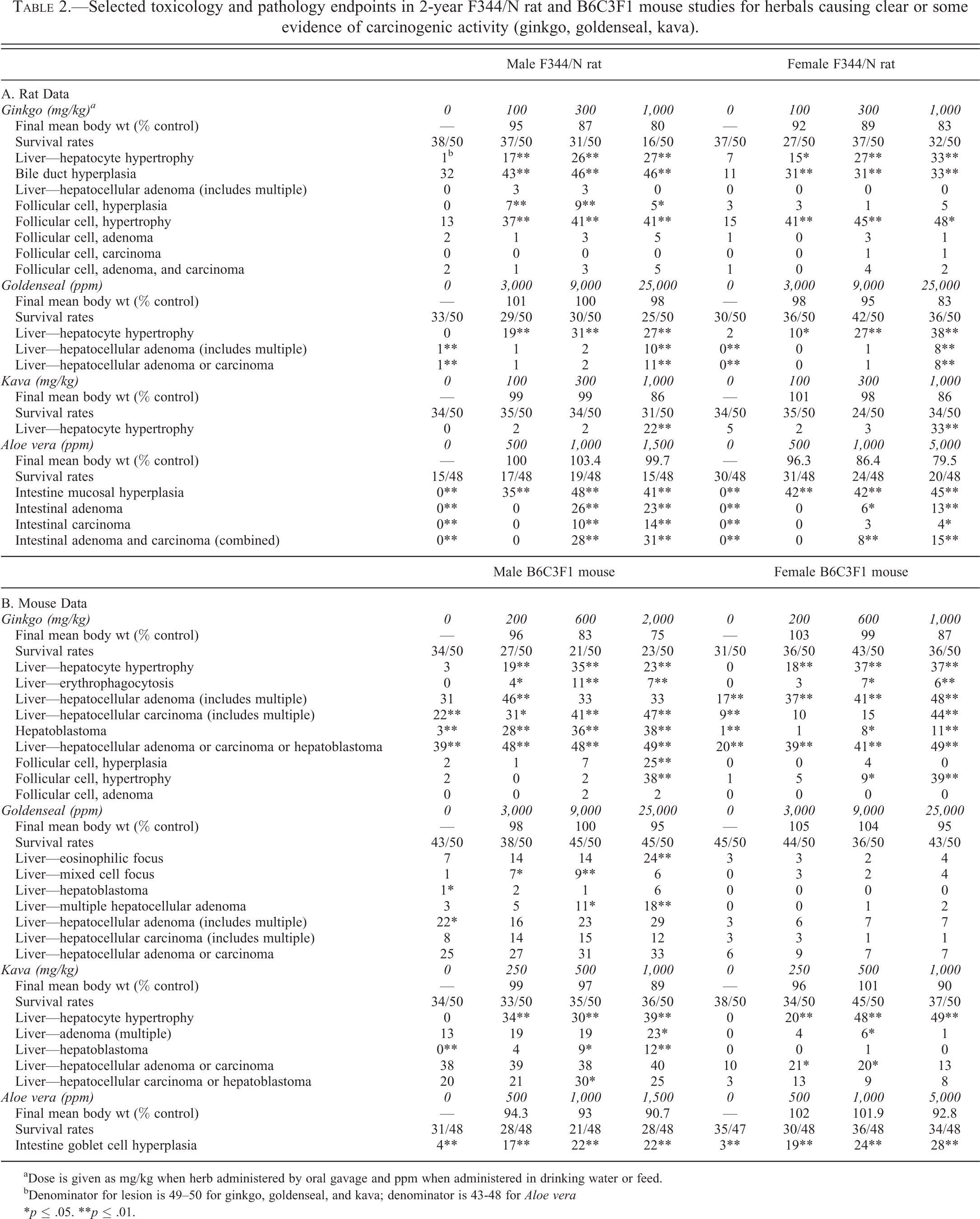
aDose is given as mg/kg when herb administered by oral gavage and ppm when administered in drinking water or feed.
bDenominator for lesion is 49–50 for ginkgo, goldenseal, and kava; denominator is 43-48 for Aloe vera
*p ≤ .05. **p ≤ .01.
Selected toxicology and pathology endpoints in 2-year F344/N rat and B6C3F1 mouse studies for herbals causing equivocal or no evidence of carcinogenic activity (ginseng, milk thistle, and turmeric oleoresin).
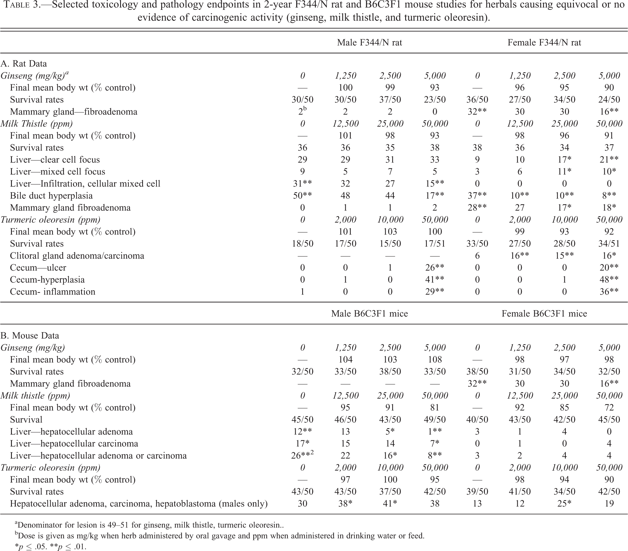
aDenominator for lesion is 49–51 for ginseng, milk thistle, turmeric oleoresin..
bDose is given as mg/kg when herb administered by oral gavage and ppm when administered in drinking water or feed.
*p ≤ .05. **p ≤ .01.
Ginkgo biloba Extract—Male and Female Rats—0, 100, 300, 1,000 mg/kg (Oral Gavage)
There was no clear evidence for a ginkgo carcinogenic response in rats. In male and female rats, there was an increase in centrilobular hepatocyte hypertrophy and treatment-related bile duct hyperplasia (Figure 2). In the liver of males, oval cell hyperplasia and clear cell focus were increased, and in females, fatty change occurred (Figure 1). Focal fatty changes occurred at approximately the same incidence in all dosed groups of females (11/50, 25/50, 30/50, 25/50), although the number of areas displaying focal fatty change tended to increase with dose. This hepatocyte focal fatty change displayed localized microvesicular and macrovesicular fatty change, increased amounts of eosinophilic cytoplasm, large open-faced nuclei, occasional multiple nucleoli, and microgranulomas scattered throughout composed predominantly of macrophages with fewer lymphocytes, plasma cells, and occasional neutrophils. The overall size of the lesion was quite variable. However, target organ toxicity was seen in the liver, thyroid, and nasal cavity (Table 2).
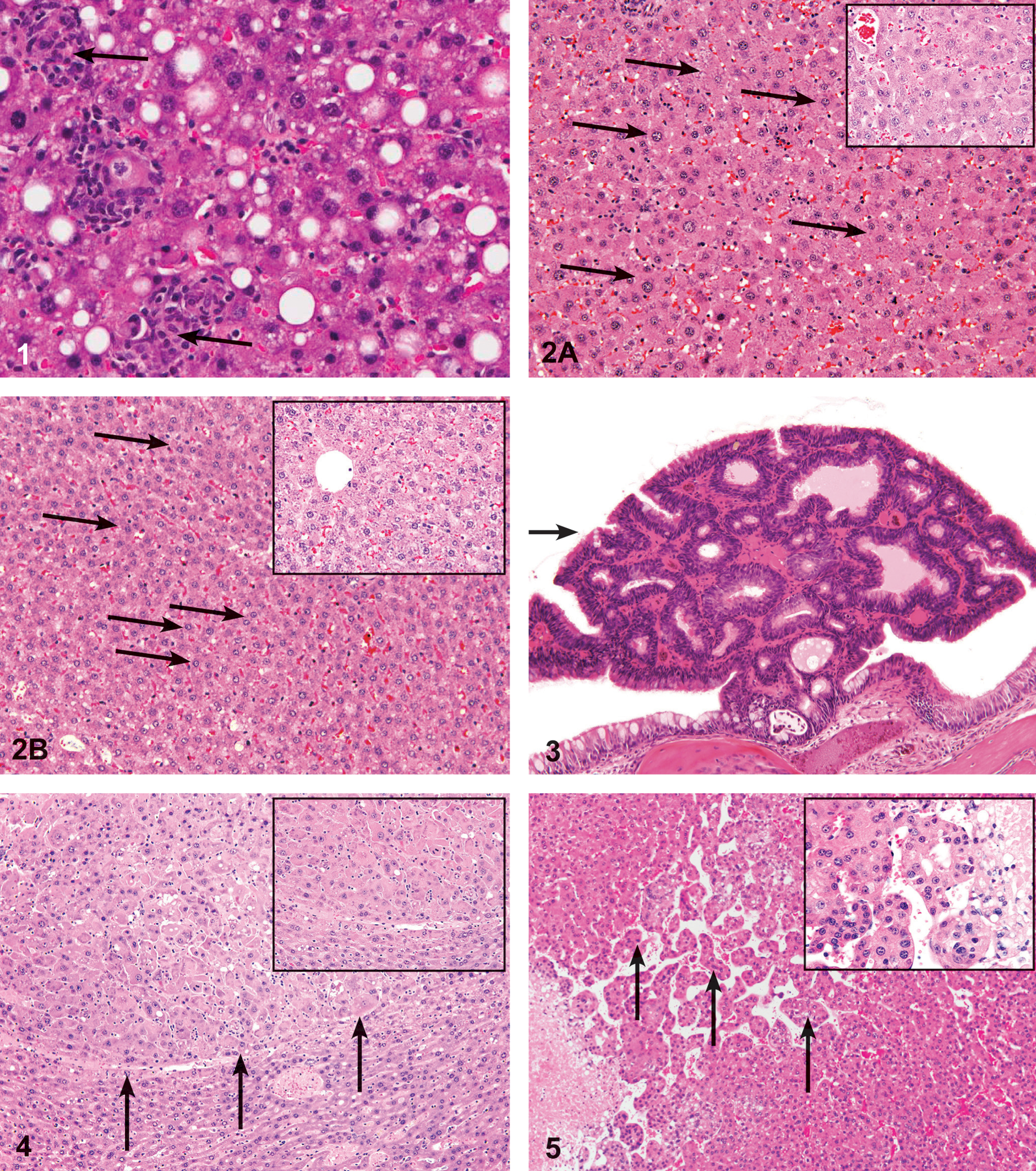
Focal fatty change in the liver of a female F344/N rat administered 2,000 mg/kg of Ginkgo biloba extract by gavage for 2 years. Note hepatocytes displaying microvesicular and macrovesicular fatty change, associated with microgranulomas scattered throughout the lesion, composed predominantly of macrophages with fewer lymphocytes, plasma cells, and occasional neutrophils (arrows). The macrophages often contained fine, acicular clefts (cholesterol clefts). H&E; 10×.
The incidence of thyroid follicular cell hyperplasia and hypertrophy was increased in males and females. While the increase in thyroid gland follicular cell tumors was not significant, the incidence in the high-dose males and mid-dose females was outside of the historical control range (males: 0–6%; females 0–4%) and may have been related to treatment.
In the nose, transitional epithelium, respiratory epithelium hyperplasia and atrophy, respiratory metaplasia, nerve atrophy, and pigmentation in the olfactory epithelium were increased in all dosed groups of males and females (except for 100 mg/kg females). Incidences of goblet cell hyperplasia in the respiratory epithelium were significantly increased in 300 and 1,000 mg/kg males and females, and incidences of chronic active inflammation were significantly increased in 1,000 mg/kg males and females. The incidence of submucosa fibrosis was significantly increased in 1,000 mg/kg males (NTP 2012).
There were no statistically significant increases in nasal cavity neoplasms, although two adenomas of the respiratory epithelium occurred in 300 mg/kg females. These respiratory epithelium adenomas were characterized by an exophytic mass, with a pedunculated or sessile base, growing into the lumen of the nasal passage. The mass consisted of papillary and invaginating, gland-like structures that were composed of pseudostratified epithelium on a scant, fibrovascular stroma (Figure 3). Because this is a rare neoplasm in rats occurring in only 1 (0.1%; range, 0–2%) of 1,196 female rats in the historical controls for all routes of administration, the occurrence of these tumors may have been related to treatment (NTP 2012).
Ginkgo biloba Extract—Male Mice—0, 200, 600, 2,000 mg/kg; Female Mice 0, 200, 600, 1,000 mg/kg (Oral Gavage)
There was clear evidence for a carcinogenic activity in the liver of male and female mice where the incidences of hepatocellular adenoma (Figure 4), hepatocellular carcinoma (Figure 5), or hepatoblastoma (combined; Figure 6) were significantly increased in all dosed groups (Table 2).
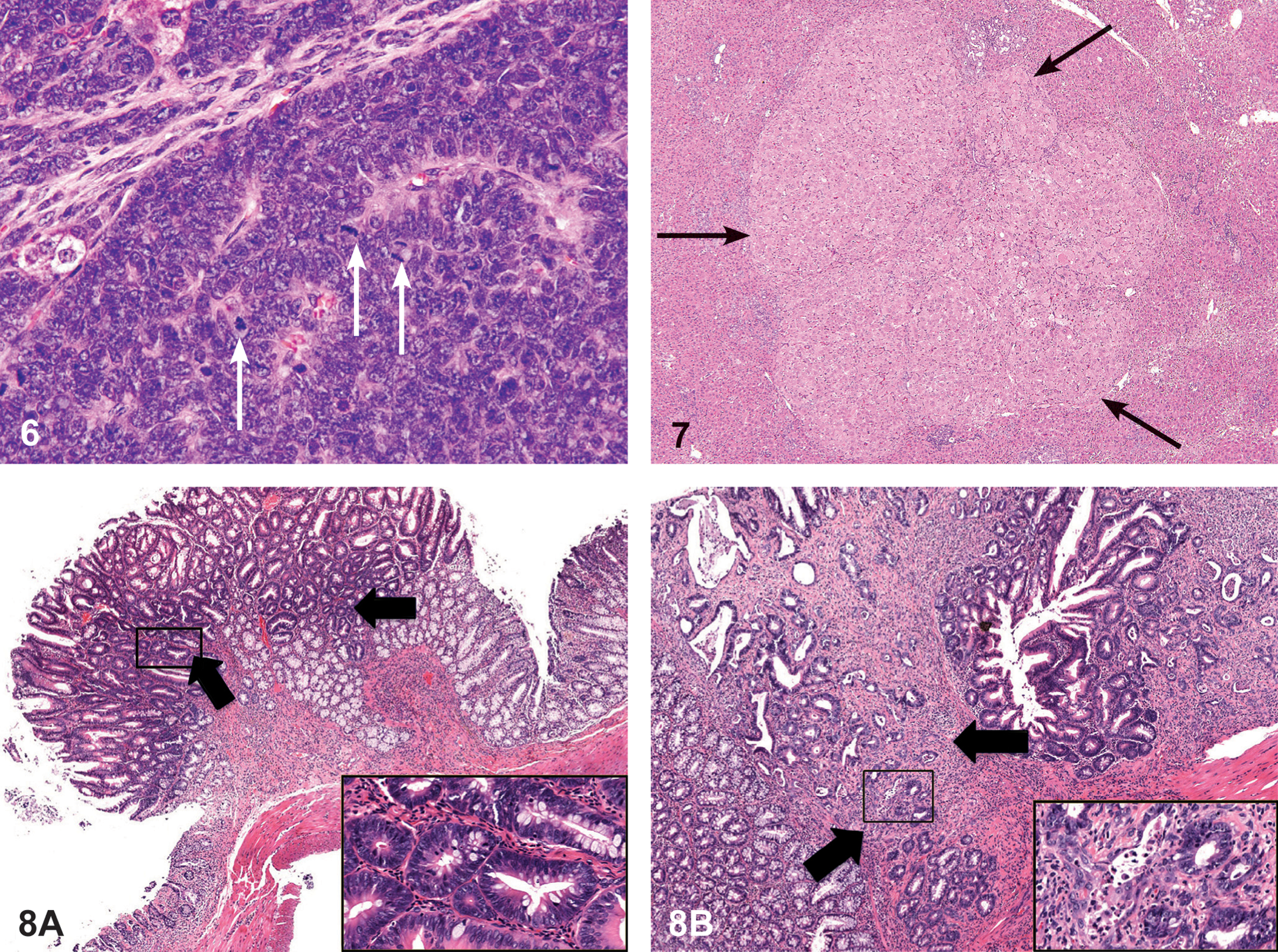
Hepatoblastoma in the liver a female B6C3F1/N mouse administered 2,000 mg/kg of Ginkgo biloba extract by gavage for 2 years. Note the basophilic neoplastic cells arranged in sheets with palisading around vascular spaces. Nuclei are generally irregularly oval to round with a scant amount of basophilic cytoplasm; mitotic figures are numerous (arrows). H&E; 40×.
Liver hypertrophy was seen in all dosed groups of male and female mice. Hepatocytic erythrophagocytosis were significantly increased in all dosed groups of males and in the 600 and 2,000 mg/kg females. Erythrophagocytosis was characterized by enlarged hepatocytes in which the cytoplasm was filled with red blood cells. The nucleus was either marginated to the side of the cell or centrally located surrounded by intracytoplasmic red blood cells. Remaining cytoplasm often formed a distinct band at the peripheral circumference of the hepatocyte.
There was an increase in thyroid gland hyperplasia in males at 200 mg/kg and an increase in follicular cell hypertrophy in males (2,000 mg/kg) and females (600 and 2,000 mg/kg). There were two follicular cell adenomas in the 600 and 2,000 mg/kg males (Table 2).
In the nose, the incidences of hyaline droplet accumulation in the olfactory epithelium were significantly increased in 2,000 mg/kg males and females; the incidences of pigmentation in the olfactory epithelium were significantly increased in 2,000 mg/kg males and 600 and 2,000 mg/kg females (NTP 2012).
Goldenseal—Male and Female Rats—0, 3,000, 9,000, 25,000 ppm (Feed)
In male rats, there was a significant increase in the incidence of hepatocellular adenoma (Figure 7) and hepatocellular adenoma or carcinoma (combined) at the high dose (Table 2). Male rats have a low background rate of liver neoplasms in goldenseal study controls (2%) and in historical feed controls (mean, 2.3%; range, 0–6%). Thus, the incidence of liver tumors (4%) in 25,000 ppm male rats was considered to be related to treatment. Furthermore, the appearance of hepatocellular adenomas in treated groups (study day 612) happened earlier than in the controls (study day 729). In the 25,000 ppm female rats, there was an increased incidence of hepatocellular adenoma. Thus, the occurrence of liver tumors in male and female rats was considered to be clear evidence of carcinogenic activity (Table 2).
Increased incidences of nonneoplastic hepatic lesions in exposed groups of males and females included hepatocyte hypertrophy, hepatocyte degeneration, eosinophilic focus, and mixed cell focus (Table 2). The incidences of eosinophilic focus were significantly increased in 9,000 and 25,000 ppm males and all exposed groups of females. All exposed groups of male and female rats had significantly increased incidences of hepatocyte hypertrophy, and the severity increased with increasing exposure concentration. The incidences of hepatocyte degeneration were significantly increased in all exposed groups of males and in 9,000 and 25,000 ppm females, and the severity increased with increasing exposure concentration (Table 2).
Goldenseal—Male and Female Mice—0, 3,000, 9,000, 25,000 ppm (Feed)
There was an increase in the incidence of hepatocellular adenoma in male mice (Table 2). The incidences of hepatocellular carcinoma were increased, but not significantly, in all exposed groups of males. There were also increases in neoplastic foci in male mice (Table 2).
Kava Kava Extract—Male and Female Rats—0, 100, 300, 1,000 mg/kg (Oral Gavage)
There were no treatment-related neoplastic lesions in rats. The incidences of hepatocellular hypertrophy, centrilobular fatty change were increased in males and females. Cystic degeneration was observed in all dosed groups of males (Table 2).
Kava Kava Extract—Male and Female Mice—0, 250, 500, 1,000 mg/kg (Oral Gavage)
There was a clear evidence for carcinogenic effect of kava in mice, which was based on an increase in hepatocellular tumors. Histologically, the hepatocellular adenomas and carcinomas were similar to those reported in the ginko study.
The incidences of centrilobular hypertrophy in all dosed groups of males and females were significantly increased, and the severities of the lesion tended to increase with increasing dose. Significantly increased incidences of eosinophilic focus occurred in 500 mg/kg males and in 1,000 mg/kg males and females. The incidences of angiectasis increased in a dose-related manner in males, and the increase in the 1,000 mg/kg group was significant. The incidences of hepatocellular necrosis were significantly increased in 250 and 1,000 mg kg males.
Aloe vera Nondecolorized Whole Leaf Extract—Male and Female Rats—0, 500, 1,000, 1,500 ppm (Drinking Water)
Aloe vera nondecolorized whole leaf extract caused a neoplastic response primarily in the large intestine of the rat (Table 2). Additional sampling sites of the rat intestinal tract than those examined routinely in carcinogenicity studies were examined by histopathology in a 2-year study, including the ileo-cecal-colic junction (referred to in the tables as proximal colon), the cecum, and the ascending, transverse, and descending colon site sections.
Histological identification of adenomas (Figure 8A) of the large intestine was based on the pedunculated nodules, polyploid masses that protruded into the intestinal lumen, or sessile lesions that caused thickening of the intestinal wall. Epithelial cells within adenomas were well differentiated and resembled cells in adjacent hyperplastic mucosal epithelium but formed distorted, glandular arrangements often with mild compression of adjacent mucosa. Diagnosis of carcinoma (Figure 8B) was based on invasion of the stroma of the stalk into the submucosa and/or muscularis of the intestinal wall and anaplastic changes in the neoplastic epithelial cells, including hyperchromatic staining and distortion of cellular size and shape.
Treatment-related nonneoplastic lesions occurred primarily in the large intestine and associated mesenteric lymph nodes. Mucosal hyperplasia was a frequent finding in the large intestines of rats that consumed the Aloe vera whole leaf extract in the drinking water. The severities were greater and the incidences of mucosal hyperplasia were higher in the ascending and transverse colon compared to the descending colon sites of the large intestine of rats—the same sites that had increased incidences of neoplasms.
In male and female rats, the administration of the Aloe vera whole leaf extract in the drinking water induced significant dose-related increasing trends in the incidences of mucosal hyperplasia of the proximal, ascending, transverse, and descending colon and cecum. In comparison to the control group, a significant treatment-associated increase in the incidence of mucosal hyperplasia was observed for the proximal colon, the cecum, and the ascending, transverse, and descending colon and rectum of the large intestine at each dose level of the Aloe vera whole leaf. Degeneration and hyperplasia of mesenteric lymph nodes and hyperplasia of the glandular stomach mucosa and the mucosa of the Aloe vera whole leaf extract treatment-related nonneoplastic lesions occurred primarily in the large intestine and associated mesenteric lymph nodes.
Aloe vera Whole Leaf Nondecolorized Extract—Male and Female Mice—0, 500, 1,000, 1,500 ppm (Drinking Water)
There were no treatment-related neoplastic lesions in mice. Treatment-related nonneoplastic lesions appeared primarily in the colon. Goblet cell hyperplasia were observed in the ascending, transverse, and descending colon. In association with goblet cell hyperplasia of the colon, cellular infiltration of the mesenteric lymph nodes showed significant dose-related increasing trends, and significantly higher cellular infiltration was observed in the mesenteric lymph nodes of the 3.0% Aloe vera whole leaf group of male mice when compared with the control group.
Dose-related increasing levels of hyaline droplets (hyaline degeneration) of the nose were also observed in treated male mice. The significance of this lesion is uncertain but is thought to represent a nonspecific adaptive response to the inhalation of irritants.
Ginseng—Male and Female Rats—0, 1,250, 2,500, 5,000 mg/kg (Oral Gavage)
There were no treatment-related increases in tumors in rats. The incidence of mammary gland fibroadenoma (including multiple) in females occurred with a negative trend (Table 3). The incidence of mammary gland fibroadenoma in the 5,000 mg/kg group was less than the historical control range for water gavage studies (94/150, mean ± standard deviation, 63% ± 4%, range, 58–66%); however, the incidence was within the historical range for all routes (701/1,350, 52% ± 15%, range, 24–86%).
Ginseng—Male and Female Mice—0, 1,250, 2,500, 5,000 mg/kg (Oral Gavage)
There were no treatment-related increases in tumors in mice, and there was no clear evidence for other major target organ toxicity.
Milk Thistle Extract—Male and Female Rats—0, 12,500, 25,000, 50,000 ppm (Feed)
There were no treatment-related increases in tumors in rats. Incidences of mammary gland fibroadenoma (single or multiple) occurred with a negative trend in females and were significantly decreased in groups exposed to 25,000 or 50,000 ppm. Mammary gland fibroadenoma is a common finding in aged F344/N rats, with a mean historical incidence of 56% (range, 48–60%) for untreated female rats in feed studies; thus, the incidences of mammary gland fibroadenoma in the 25,000 and 50,000 ppm groups were below the historical control range for feed studies. Because body weights of female rats did not differ across the four groups, the reduction in mammary tumors in the 25,000 and 50,000 ppm female rat groups cannot be attributed to body weight differences.
In the liver of female rats, there were increases in the incidence of clear cell, eosinophilic, and mixed cell foci, but there was no evidence of an increase in liver tumors. A significantly decreased incidence of mixed inflammatory cell infiltration was noted in the liver of 50,000 ppm exposed males; the severity of this lesion was similar in control and treated males. The lesion consisted of randomly distributed foci of inflammation consisting primarily of mixed mononuclear cells that varied from foci of macrophages to a mixture of lymphocytes and the rare plasma cell. Significantly decreased incidences of bile duct hyperplasia (with lowered average severity grade) were noted in 50,000 ppm males and in all exposed groups of females. Bile duct hyperplasia was characterized by the proliferation of biliary epithelial cells within portal areas with occasional minimal extension into the adjacent hepatic lobules.
Decreased incidences of pigmentation were noted in the mesenteric lymph node of all exposed groups of males and females (males: 0 ppm, 45/50 [1.2]; 12,500 ppm, 27/50 [1.0]; 25,000 ppm, 17/50 [1.0]; 50,000 ppm, 9/50 [1.0]; females: 47/49 [1.5], 39/48 [1.2], 29/50 [1.3], 18/50 [1.2]), respectively.
Milk Thistle Extract—Male and Female Mice—0, 12,500, 25,000, 50,000 ppm (Feed)
There were no treatment-related increases in tumors in mice. In male mice, the incidences of hepatocellular adenoma, hepatocellular carcinoma, and hepatocellular adenoma or carcinoma (combined) occurred with negative trends (Table 3). Significantly decreased incidences of hepatocellular carcinoma occurred in 50,000 ppm males; decreased incidences of hepatocellular adenoma and of hepatocellular adenoma or carcinoma (combined) occurred in 25,000 and 50,000 ppm males. All of these decreased incidences were below the respective historical ranges for these tumors. Decreased incidences of clear and mixed cell foci and hepatocytic cytoplasmic vacuolization were noted in the liver of males of all exposed groups (data not shown). Altered cell foci (eosinophilic, mixed, basophilic, and clear) were characterized by a focus of hepatocytes with altered tinctorial properties.
The decreased incidences of hepatocellular adenoma or carcinoma (combined) in males may be attributed to decreases in body weights, but a potential direct effect of the exposure to milk thistle extract cannot be excluded.
Decreased incidences of clear and mixed cell foci and hepatocytic cytoplasmic vacuolization were noted in the exposed groups of males. The incidence of lymphoid hyperplasia was significantly decreased in 50,000 ppm females (9/49, 5/47, 6/46, 0/49).
Turmeric Oleoresin—Male and Female Rats—0, 2,000, 10,000, 50,000 ppm (Feed)
There was no evidence for a carcinogenic response in male rats and equivocal evidence for a carcinogenic response in female rats was based on the presence of clitoral gland adenomas. There was an increase in clitoral gland tumors in female rats. However, because there was no clear dose response and no incidence of clitoral glan hyperplasia, it was uncertain whether this finding was related to chemical administration.
Inflammation was seen in various regions of the gastrointestinal (GI) tract in treated rats. There was an increase in ulceration, hyperplasia, and hyperkeratosis of the forestomach in male rats. These hyperplastic lesions of the forestomach were considered regenerative rather than part of a neoplastic process. There were ulcers, inflammation, and hyperplasia of the cecum and to a minor degree the colon of male and female rats. None of the hyperplasic lesions progressed to neoplasms of the cecum. The mechanisms for these lesions might have been due to direct cytotoxicity of the chemical.
Turmeric Oleoresin—Male and Female Mice—0, 2,000, 10,000, 50,000 ppm (Feed)
There was an increased incidence of hepatocellular neoplasms in male and female mice. However, this increase was not significant by the trend statistic, and there was no corresponding increased incidence of hepatic foci in exposed groups and, thus, this finding was considered to be only equivocal evidence of carcinogenic activity. The histologic features of the hepatocellular tumors were similar to what was reported in the previous studies mentioned above.
Adenomas of the pars distalis of the pituitary gland occurred more frequently in the exposed groups of female mice than in controls (0/46, 2/49, 4/50, 5/50). However, the incidence in the exposed groups was within the range of historical controls female mice (2–36%) and this finding was not considered to be chemically related. Female mice at 50,000 ppm had an increased follicular cell hyperplasia (5/50, 8/50, 7/50, 16/49).
Discussion
Carcinogenic effects in the liver were observed after long-term exposure of rodents to ginkgo, kava kava, or goldenseal. Hepatocellular hypertrophy and/or vacuolization occurred along with the liver neoplastic response. For ginkgo and kava kava, the carcinogenic effect was more pronounced in mice than in rats, including the occurrence of hepatoblastomas, a rare tumor that may occur after exposure to promoters (Diwan, Henneman, and Rice 1995; Turusov et al. 2002). While hypertrophy may predispose rodents to liver cancer, not all chemical exposures that cause this lesion lead to liver cancer (Maronpot et al. 2010). Only 45% of rat studies with chemical-induced liver hypertrophy went on to become rat liver carcinogens in 2-year cancer studies (Allen et al. 2004).
For ginkgo, the positive genotoxic test results (Salmonella studies; NTP 2012) suggest that genotoxic mechanisms play a role in the liver carcinogenic process. Most of the bacterial mutagenic tests were negative for goldenseal, kava kava, and kavalatones (NTP 2010, 2011c; Whittaker et al. 2008). However, berberine (an ingredient in goldenseal) and its metabolite berberrubine inhibit topoisomerase activities (Kobayashi et al. 1995; Makhey et al. 1994). Inhibition of topoisomerase may result in the inhibition of DNA repair processes and, thus, facilitate carcinogenic processes. International Agency for Research on Cancer (IARC) has classified other topoisomerase inhibitors as “carcinogenic to humans—Group 1” (IARC 2011).
Nongenotoxic and genotoxic liver carcinogens give unique liver gene transcript signatures (Auerbach et al. 2010; Fielden et al. 2011; Hoenerhoff et al. 2011), and some rodent liver carcinogens have the same pathway to cancer as found in human liver cancer (Holsapple et al. 2006). Thus, comparative liver gene transcript patterns for ginkgo, goldenseal, and kava may help in identifying the carcinogenic mechanism. These studies are ongoing at the NTP.
Further work is needed to determine whether the mechanisms for the ginkgo-induced thyroid tumors, which were accompanied by increases in thyroid-stimulating hormone (TSH) levels and thyroid follicular cell hypertrophy, are also found in humans (NTP 2012). Thyroid follicular cell tumors arise in rodents from mutations or from perturbations of thyroid and pituitary hormones (e.g., increase TSH; Hill et al. 1998). Chemicals that lack mutagenic activity and cause thyroid proliferation/tumorigenesis related to increases in TSH levels do not always have a similar carcinogenic mechanism in humans (Hill et al. 1998).
In addition to the ginkgo-induced toxic and carcinogenic effects in the liver and thyroid, this herb caused toxicity and tumorigenesis in the nasal cavity. The pathogenesis of these nasal lesions may be related to ginkgo or metabolites reaching the nasal cavity from the blood stream. Olfactory epithelium contains high levels of cyp450 enzymes (Reed 1993; Sells et al. 2007) that could metabolize the herb components into toxic metabolites. Alternatively, the nasal lesions could potentially result from gastric reflux/postgavage reflux through the nasopharyngeal duct (Damsch et al. 2011a, 2011b).
The intestinal tumors occurring in rats after exposure to Aloe vera (nondecolorized whole leaf extract) may be related to anthraquinones in the herb being converted to mutagenic components in the intestine (e.g., aloe emodin; NTP 2011a). Administration of 1-hydroxyanthraquinone caused intestinal tumors in rats (1% in the diet for 480 days; Mori et al. 1990). Chyrsazin (danthron or 1,8-dihyroanthraquinone) also caused intestinal tumors in rats (1% in the diet for up to 16 months; Mori et al. 1985). In contrast, while chyrsazin caused intestinal hyperplasia in mice, there was no evidence for intestinal tumor formation (Mori et al. 1986). 1-Hydroxy-, 1-8-dihydrox-, 1,8-dihydroxy-3-carboxyanthroquinone compounds are all mutagenic compounds (Brusick and Mengs 1997; Morales et al. 2009; Mori et al. 1985, 1986, 1990; NTP 2001), but species variations in the amount of mutagenic components made and/or available in the intestine are factors that may contribute to the intestinal carcinogenic activity.
There was no clear evidence for a carcinogenic response in the ginseng, milk thistle, or turmeric oleoresin 2-year rodent studies. While there have been reports in the literature on the clinical efficacy of these herbs for treating liver disease, cancer, or other disease conditions, these herbs are not licensed as drugs. For this reason, the U.S. government has expanded its support for clinical trials with herbal medicines (National Institutes of Health 2011).
There are over 30 ginsenosides, the putative anticancer agents in ginseng, and studies are underway to determine the ability of ginsenosides to inhibit cell proliferation, tumor cell invasion, and/or metastasis (He et al. 2011; Kim et al. 2012; Luo et al. 2008). Ginsenosides may modulate signaling pathways including cell cycle, inflammatory, or growth factor pathways (Nag et al. 2012).
Milk thistle is being investigated as a treatment for liver disease particularly hepatitis. Safety studies report that there were no adverse affects after administration of silymarin, a major component in milk thistle (Hawke et al. 2011; Miranda et al. 2008; Schrieber et al. 2008, 2010). Other studies report that silymarin (420 mg/kg/day) was well tolerated and reduced the clinical symptoms of hepatitis A, B, or C (El-Kamary et al. 2009). Other clinical trials for the use of milk thistle/silymarin to treat hepatitis are in progress (NIH 2011).
Turmeric oleoresin and its major component curcumin (Gupta et al. 2011, 2012; Shen and Ji 2012; Sung et al. 2012) have antiproliferative properties including inhibition of prostaglandin synthesis (Griesser et al. 2011). Turmeric oleoresin and curcumin are being studied as treatments for irritable bowel syndromes, atopic asthma, colon cancer, and other disease (NIH 2011). The GI toxicity reported in the turmeric oleoresin rodent studies occurred primarily at levels higher than that of the exposures currently being used in the NIH clinical trials.
The NTP studied herbal medicines to identify potential toxicities in a series of rodent studies. A limitation of these hazard identification studies is that a mechanism for the toxic or carcinogenic effect has not always been determined. When there is a proposed mechanism for an observed biologic effect, this may provide a basis for species extrapolation of the finding. For example, there are several studies that report that when herb components are metabolized to anthraquinones, intestinal lesions/tumors may occur. Topoisomerase inhibitors (e.g., components in goldenseal) may contribute to carcinogenic processes. One priority for future herbal toxicity studies would be those with anthraquinone components/metabolites or those with topoisomerase inhibition activity.
These herbal studies along with the clinical trials supported by the NIH allow for a more comprehensive analysis of the risk and benefits from herbal medicine use. A review of all the NIH data after completion of the clinical trials should yield important new insights for herbal medicine use in the United States.
Footnotes
The statements, opinions, or conclusions contained therein do not necessarily represent the statements, opinions, or conclusions of NIEHS, NIH, or the United States government.
The author(s) declared no potential conflicts of interest with respect to the research, authorship, and/or publication of this article.
The author(s) disclosed receipt of the following financial support for the research, authorship, and/or publication of this article: This work was supported by the intramural program of the National Toxicology Program and the National Institute of Environmental Health Sciences (NIEHS), Research Triangle Park, North Carolina.
Abbreviations
Acknowledgments
The authors thank M. Cesta and M. Behl, NIEHS, for their review of the manuscript.