
Abstract
A primary angiosarcoma was found in the tongue of a six-week-old female Wistar rat, sacrificed for humane reasons during the course of a four-week toxicology study. At necropsy, a nodule protruding from the dorsal part of the tongue was found. The nodule displayed microscopically, irregularly shaped vascular spaces separated by collagenous stroma. The spindle-shaped endothelial cells showed pleomorphism, hyperchromatism, and low mitotic activity; large nuclei with one or more nucleoli were present. Multiple metastases were found in the lungs, and the morphology of the cells resembled that of the primary tumor. Immunohistochemically, the primary tumor and the lung metastases were positive for von Willebrand factor and vimentin. The diagnosis of tongue angiosarcoma metastasizing to the lungs was made on the basis of microscopic and immunohistochemical findings.
Angiosarcoma is an uncommon neoplasm of vascular endothelial cell origin that can arise in any anatomic site of the body. In animals, angiosarcomas are not very common, although in some species they are more frequent, particularly in dogs and mice. Three cases of angiosarcoma of the tongue, either alone or in combination with skin angiosarcomas, were reported in a colony of beagle dogs (Culbertson 1982).
Oral angiosarcomas in humans are exceptionally rare, with very few case reports described in the literature (Arribas-Garcia et al. 2008; Tabata et al. 1999). The diagnosis of the tumor may be difficult, especially in poorly vasoformative lesions. In such cases, the differential diagnosis with other types of soft tissue sarcomas or undifferentiated carcinomas is rather challenging and sometimes even impossible on morphological grounds. To the best of our knowledge, this is the first time in the rat, and in rodents in general, that a primary tongue angiosarcoma with metastases to the lungs is reported.
We report on a six-week-old female Wistar rat that was sacrificed for humane reasons following inability to eat and lethargy. The animal was part of a four-week toxicology study conducted in a contract laboratory organization under barrier maintenance conditions. Animal care and handling procedures were carried out in complete accordance with Swiss laws on animal care. The rat had been allocated to the lowest treatment group and received 15 mg/kg/day of a natural antibacterial compound (plant extract). The necropsy performed immediately after the sacrifice revealed a nodule of 10 mm in diameter in the tongue extruding from the dorsal part of the organ. The consistency was firm, and the nodule presented a bluish color on the surface. Many red-brown foci, 1 mm in diameter, were present on the surface of the lungs.
Tongue and lungs were fixed in 10% buffered formalin, processed according to standard operating procedures, and embedded in paraffin wax. Serial sections were cut at 4 to 5 µm thickness and stained with hematoxylin and eosin (HE) or used for immunohistochemistry.
Immunohistochemical reactions were performed using either the streptavidin-biotin-peroxidase complex technique (Vector Laboratories, Inc., Burlingame, CA, USA) or EnVisionTM Dako Visualization Systems (Dako Schweiz AG, Baar, Switzerland) with the following mouse monoclonal or rabbit polyclonal antibodies: anti-α smooth muscle actin (clone 1A4, Dako), anti-pan-cytokeratin (clone PCK-26, Sigma), anti-vimentin (clone V9, Dako), and anti–von Willebrand factor (pAb, Dako).
Microscopic examination of the tissue specimens revealed a neoplasm in the tongue composed of irregularly shaped vascular spaces separated by collagenous stroma (Figure 1A and 1B). The tumor formation presented a pronounced ulceration of the dorsal portion of the tongue mucosa. The spindle-shaped endothelial cells showed pleomorphism, hyperchromatism, and low mitotic activity; large nuclei with one or more nucleoli were present. The neoplastic cells were arranged in bundles and sheets infiltrating the muscle parenchyma. Inflammatory cells, fibrosis, and necrosis were present in the interstitium between the vascular spaces. Many neoplastic apoptotic cells were also present, indicating rapid growth of the tumor. Metastases were found in the lungs (Figure 1C) and exhibited a morphology similar to that of the primary tumor, namely, presence of vascular spaces lined by atypical endothelial cells, pleomorphism, and few mitoses.
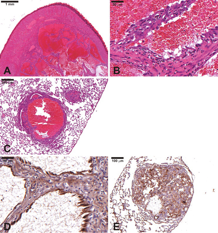
(A) A primary angiosarcoma in the tongue of a six-week-old female Wistar rat. Hematoxylin and eosin (HE). (B) A higher magnification of the tumor composed of irregularly shaped vascular spaces separated by collagenous stroma (a mitotic figure in the endothelial cell visible). HE. (C) Metastases of the angiosarcoma in the lung exhibiting a morphology similar to that of the primary tumor. HE. (D) Von Willebrand factor (an endothelial cell marker) immunohistochemical positivity in the primary tumor. (E) Positively labeled endothelial cells in lung metastasis (von Willebrand factor).
The main clue in the diagnosis of angiosarcoma is the presence of anastomosing channels lined by atypical endothelial cells. In the present case, the positive immunohistochemical results using antibodies against von Willebrand factor (Figure 1D and 1E) and vimentin confirmed the diagnosis of angiosarcoma. Von Willebrand factor is a very well-known marker for endothelial cells. Pan-cytokeratin immunostaining, as expected, was negative, excluding the epithelial origin of the neoplasm. Based on the morphological and immunohistochemical findings, the diagnosis of angiosarcoma arising from the tongue and metastasizing to the lungs was made.
Angiosarcomas are uncommon tumors in animals, and they are rare in humans. In dogs, this tumor occurs more frequently than in other species, particularly in certain breeds, including Italian greyhounds, white boxers, and pit bulls. The skin and soft tissues such as the liver are the most affected. It is published that liver angiosarcoma in the dog is much more frequent than in humans (Priester 1976). Three tongue angiosarcomas in the beagle dog have also been reported (Culbertson 1982).
In aging mice, angiosarcomas are relatively frequent when compared to other animal species. In this species, data reported in the literature indicate that the liver is the most frequent primary site followed by the spleen and bone marrow, and in males the occurrence is higher than in females, 4.5% and 1%, respectively (Chandra et al. 1992). In aged Sprague-Dawley rats, primary benign and malignant vascular neoplasms occur spontaneously in 1.1% of males and in 0.6% of females. The spleen is the organ most affected, and hemangioma is more common than angiosarcoma (Zwicker et al. 1995). In aged Wistar rats, the incidence of angiomas and angiosarcomas is higher than in Sprague-Dawley rats (Poteracki et al. 1988).
Very few cases of human angiosarcomas arising from the tongue have been reported (Tabata et al. 1999). Overall, only four cases (three primary and one metastatic) have been reported in the English literature, and none presented metastases. In general, human angiosarcomas are very rare and usually arise in the skin or in superficial soft tissue of the head, neck, and breast. However, laryngeal angiosarcoma (Loos, Wieneke, and Thompson 2001), oral (lower lip) angiosarcoma (Arribas-Garcia et al. 2008), and primary sinonasal tract angiosarcoma (Nelson et al. 2007) were also reported. The latter tumor usually involved the nasal cavity (in most of the cases) or the maxillary sinus, and all cases were positive for Factor VIII, CD34, CD31, vimentin, and smooth muscle actin, whereas they remained keratin- and S-100 protein-negative. When metastases from the primary angiosarcoma occur, the lung is the most common site, followed by the liver and bone.
Differential diagnosis includes hemangioma, epithelial sarcoma, hemangiopericitoma, fibrous histiocytoma, Kaposi’s sarcoma, malignant melanoma, anaplastic carcinoma, and pyogenic granuloma. Immunohistochemistry is useful in the diagnosis of angiosarcoma, particularly in the poorly vasoformative cases, in which the diagnosis can be problematic (Ohsawa et al. 1995). Von Willebrand factor, CD31 (an adhesion molecule present in endothelial cells, monocytes, and platelets), and vimentin are mostly positive in angiosarcoma (positivity rate between 40% and 100%). By contrast, cytokeratins, S-100 protein, and human melanoma antibody (HMB45) are negative. In the present case, a vasoformative one, the initial diagnosis of angiosarcoma was made on the hematoxylin and eosin specimens and then further confirmed by the immunohistochemical results, which clearly showed positivity for von Willebrand factor and vimentin. The lung metastases were also positive for these markers.
Angiosarcomas can be induced by radiation exposure, such as radiotherapy (Tahir et al. 2006), or by certain chemical substances, particularly vinyl chloride (VC) (Hong, Winston, and Lee 1980). In 1974, hepatic angiosarcomas in workers were associated with the manufacture of polyvinyl chloride (PVC) (Creek et al. 1974). This finding was later substantiated by experimental inhalation studies in laboratory animals with PVC, which reproduced the tumors (Maltoni et al. 1975). In addition, in rodents, VC induces a variety of tumors other than hepatic angiosarcoma, including hepatocellular carcinoma, skin tumors, osteochondroma, bronchoalveolar and mammary gland tumors in mice, and malignant lymphomas in mice and rats (Lee et al. 1978). In man, hepatocellular carcinoma is the only tumor besides hepatic angiosarcoma suspected to be associated with VC exposure (Popper, Maltoni, and Selikoff 1981).
The present case, observed in a young rat, of primary angiosarcoma arising in the tongue and metastasizing in the lungs is a valuable contribution to animal and comparative pathology. The lesion was considered a spontaneous occurrence, as it was observed in the lowest treatment dose group, and the substance was devoid of any tumorigenic potential. In addition, the low dose of 15 mg/kg/day did not cause any treatment-related lesions. The morphological and immunohistochemical features of the tumor in the rat and those described in the oral or nasal cavities in humans were similar.
Footnotes
Acknowledgments
We thank Antje Marcantonio and Susan Gaehler for their excellent technical assistance.