
Abstract
Since the accident at Fukushima Daiichi nuclear power plant, there has been a focus on the impact of low-dose radiation exposure due to nuclear disasters and radiology on human bodies. In order to study very low levels of impact on the human body from low-dose radiation exposure, a system with high detection sensitivity is needed. Until now, the most well-established biological radiation effect detection system in the field of emergency radiation medicine has been chromosomal analysis. However, chromosomal analysis requires advanced skills, and it is necessary to perform chromosomal analysis of a large number of cells in order to detect slight effects on the human body due to low-dose radiation exposure. Therefore, in order to study the effects of low-dose radiation exposure on the human body, it is necessary to develop high-throughput chromosome analysis technology. We have established the PNA-FISH method, which is a fluorescence in-situ hybridisation method using a PNA probe, as a high-throughput chromosome analysis technique. Using this method, the detection of dicentrics and ring chromosomes has become very efficient. Using this technology, chromosomal analysis was performed on peripheral blood before and after computed tomography (CT) examination of patients at Hiroshima University Hospital, and it was possible to detect chromosomal abnormalities due to low-dose radiation exposure in the CT examination. Furthermore, it was shown that there may be individual differences in the increase in chromosomal abnormalities due to low-dose radiation exposure, suggesting the need to build a next-generation medical radiation exposure management system based on individual differences in radiation sensitivity. If techniques such as chromosomal analysis, which have been used for biological dose evaluation in emergency radiation medicine, can be used for general radiology, such as radiodiagnosis and treatment, that will be a contribution to radiology from an unprecedented angle. This article will discuss the clinical application of new biological dose evaluation methods that have been developed in the field of emergency radiation medicine.
1. RADIATION DISASTERS AND MEDICAL/BIOLOGICAL RESEARCH
Among the atomic bomb survivors, leukaemia developed 5 years after the exposure, and various malignant tumours followed. Extensive epidemiological studies of the atomic bomb survivors have shown that carcinogenic risk is correlated with radiation dose. In the Chernobyl accident, children were seen to develop thyroid cancer due to ingestion of radioactive iodine, which has led to subsequent molecular biological studies of thyroid cancer. In the nuclear accident at Tokaimura, a small fuel preparation plant operated by Japan Nuclear Fuel Conversion Co., some victims were exposed to extremely high radiation exposure, and the importance of radiation dose evaluation by chromosome analysis and treatment using regenerative medicine in emergency exposure medicine was recognised. In the accident at Fukushima Daiichi nuclear power plant, operated by Tokyo Electric Power Company Holdings, thyroid cancer was identified by ultrasound screening.
1.1. Atomic bomb
Regarding the health effects on the survivors of the atomic bombing in 1945, a large-scale epidemiological survey of survivors undertaken by Atomic Bomb Casualty Commission (ABCC) and the Radiation Effects Research Foundation has led to elucidation of the relationship between radiation exposure and diseases such as malignant tumours (Kamiya et al., 2015). On the other hand, chromosome analysis, which became possible in the 1960s, has been established as a method for biological dose evaluation of the impact of radiation, such as dicentric analysis (Fig. 1). Furthermore, in a study on the pathogenic mechanism of leukaemia and malignant solid tumours found in the atomic bomb survivors, genetic abnormalities in leukaemia and cancer using molecular biological techniques were identified based on the identification of disease-specific chromosomal abnormalities in leukaemia, and this discovery led to the development of molecular-targeted medicine (John et al., 2004).
Radiation disasters and development of medical and biological research. CT, computed tomography; JCO, Japan Nuclear Fuel Conversion Co.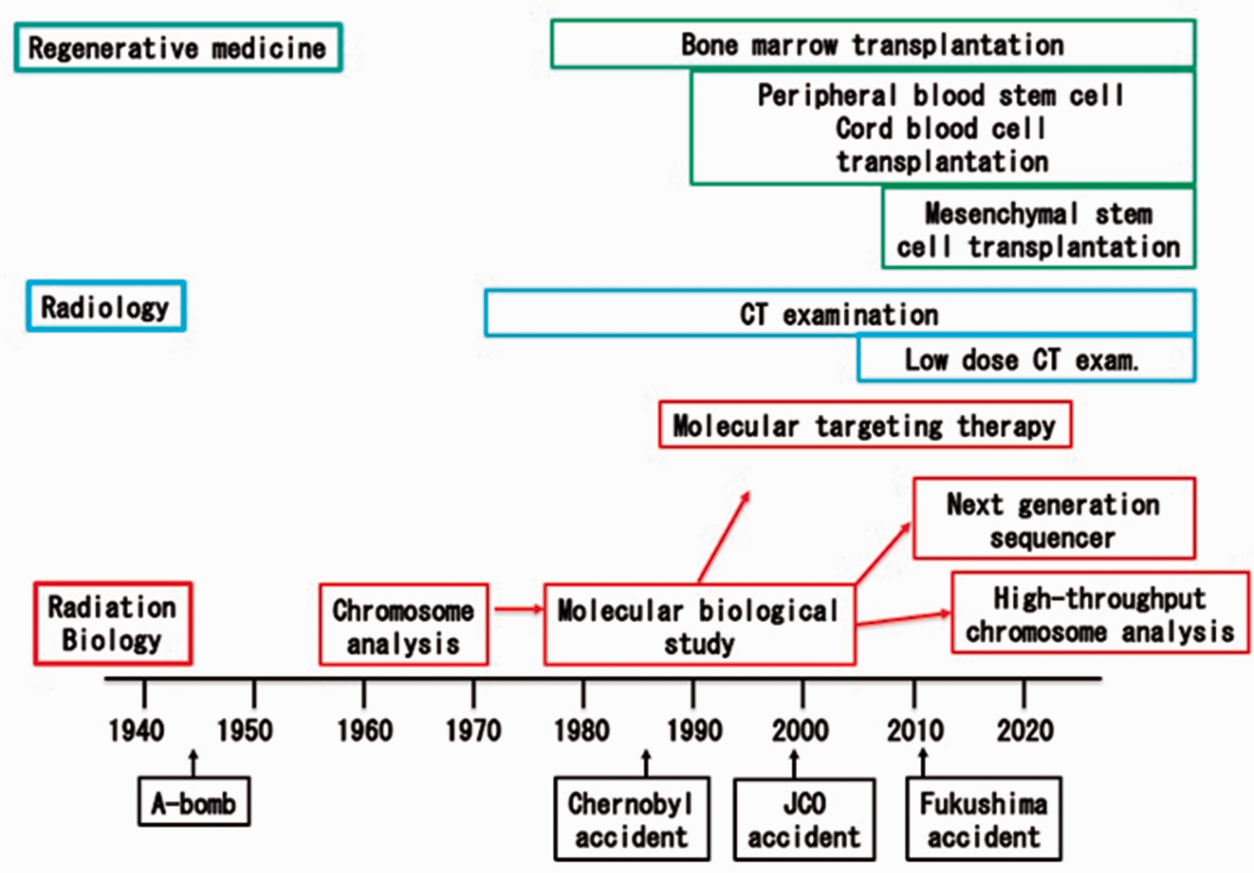
1.2. Chernobyl nuclear accident
In the 1986 Chernobyl accident, workers exposed to very high doses of radiation were treated with bone marrow transplants. Bone marrow transplantation was a medical treatment that had just been developed at that time, and the importance of regenerative medicine was suggested for subsequent treatment of those who were highly exposed. The development of childhood thyroid cancer has become a problem in the general population. Subsequently, advances in molecular biological research on thyroid cancer have led to genetic analyses of patients with thyroid cancer among the victims of the Chernobyl accident, and progress has been made in the elucidation of specific gene mutations (Efanov et al., 2018).
1.3. Tokaimura nuclear accident
In the Tokaimura nuclear accident in 1999, three people were very seriously exposed to radiation and two people were killed. In addition, 667 members of the public were exposed. Regenerative medicine such as peripheral blood stem cell transplantation and cord blood stem cell transplantation was adopted for the treatment of exposed victims, but unfortunately this did not save lives (Fig. 1). However, the experience of this accident re-affirms the importance of biological evaluation of radiation exposure dose by analysis of peripheral blood lymphocyte count and chromosomal aberration frequency, and application of regenerative medicine for the victims of emergency radiation exposure (Hayata et al., 2001).
1.4. Fukushima Daiichi nuclear power plant accident
In the 2011 accident at Fukushima Daiichi nuclear power plant, there were no cases of serious exposure to radiation. Regarding thyroid exposure of general inhabitants, the local countermeasures headquarters measured radioactive iodine in approximately 1000 residents of areas such as Iitate Village, but there were no cases of suspected high-level exposure. The same result was found in the exposure dose estimation of the Fukushima Prefectural Health Survey. However, the thyroid examination of the Fukushima Prefectural Health Survey has identified paediatric cases of thyroid cancer, and debate exists regarding whether this is due to radioactive iodine or overdiagnosis of latent cancer by the examination (Ohtsuru et al., 2019). For this reason, the general public is showing increasing interest in low-dose exposure, including medical radiation exposure. In addition to medical and biological research, such as epidemiological research, the fields of Science, technology and society (STS), the research of the social impact of science and communication between scientists and citizens, are attracting attention to solve these problems.
2. RESEARCH ON THE IMPACT OF RADIATION IN MEDICAL AND BIOLOGICAL RESEARCH
2.1. Research on the impact of radiation and leukaemia/cancer research
In the field of basic biology, functional analysis of genome-repair-related proteins encoded by genes such as RAD51 has been performed since the 1990s by genetic analysis using yeast and Escherichia coli. Since the latter half of the 1990s, research on the mechanism of human DNA repair has made remarkable progress centred on genetic analysis of hereditary diseases (i.e. reverse genetics research), and many human DNA repair-related factors such as ATM in ataxia telangiectasia and MRE11 and NBS1 in related diseases have been discovered. Research on the mechanism of human DNA repair led to the discovery of BRCA1 and BRCA2, which are factors involved in the control of homologous recombination repair of DNA double-strand breaks involved in the development of breast cancer and ovarian cancer. It has also made great contributions to the development of cancer research (McKinnon and Caldecott, 2007).
On the other hand, the Giemsa staining method established in the 1960s has made it possible to identify the breakpoint of chromosome translocations, which led to the discovery of disease-specific chromosomal abnormalities in leukaemia such as Philadelphia chromosome (9:22 translocation) shown specifically in the chromosomal abnormalities of the atomic bomb survivors and patients with chronic myelogenous leukaemia. After that, from the latter half of the 1980s, progress in molecular biological research on chromosomal translocation breakpoints in leukaemia has clarified that the 9:22 translocation of chronic myelogenous leukaemia forms the fusion gene of ABL and BCR, and that the 15:17 translocation, the disease-specific chromosomal aberration of promyelocytic leukaemia, led to fusion of the retinoic acid receptor and PML gene. Disease-specific chromosomal abnormalities have been shown to be very useful in predicting disease prognosis, as well as improving the accuracy of leukaemia diagnosis. Moreover, these findings have led to the development of molecular targeted therapy, such as Imatinib, a specific inhibitor of ABL kinase, for chronic myelocytic leukemia, and all-trans-retinoic acid for acute promyelocytic leukemia. The success of molecular-targeted therapies for leukaemia has led to the development of molecular-targeted therapies for many cancers (Fig. 1) (John et al., 2004).
In cancer research, the advent of next-generation sequencers has enabled genomics, the comprehensive analysis of genomic information. Furthermore, regarding protein analysis, proteomics (i.e. comprehensive protein analysis) has become possible due to recent progress in protein production technology and mass spectrometers. Using the enormous amount of biological information obtained by these new technologies, such as artificial intelligence, we are reaching an age where new and previously unconsidered findings can be obtained. For example, in myelodysplastic syndrome, which is often seen in the atomic bomb survivors, comprehensive analysis of gene mutations has shown abnormalities in gene groups related to splicing (Yoshida et al., 2011). This type of data-driven study will be promoted in the field of radiation research alongside hypothesis-driven research, which has represented the mainstream until now.
2.2. Research on non-cancer diseases and impact of radiation
With regard to the relationship between radiation effects and non-cancer diseases, the effects of radiation exposure on nerve tissues such as atomic bomb microcephaly and the onset of neurological diseases are known; however, the details are not yet fully understood. In ataxia-telangiectasia caused by ATM dysfunction, neuropathy such as progressive ataxia and cerebellar ataxia has been confirmed, and it is known that a gene mutation of DNA double-strand break repair factor MRE11 also causes ataxia-telangiopathy (McKinnon and Caldecott, 2007). Studies have shown that DNA damage and its repair are important for the normal development of nerve tissue, but the details remain unknown. The mechanism of neuropathy due to exposure to radiation remains to be clarified.
It has also been clarified that cerebrovascular impairment, or vascular disorders, occur in the atomic bomb survivors (Shimizu et al., 2010). The relationship between radiation and myocardial damage has attracted attention as ischaemic heart disease has been shown to occur following radiation therapy for breast cancer in the left breast (Darby et al., 2013). On the other hand, in cardiovascular diseases, the involvement of DNA double-strand breaks and their repair mechanism in myocardial regeneration after arteriosclerosis and myocardial infarction has been suggested (Uryga et al., 2016). Elucidation of the relationship between angiopathy and radiation injury, which can cause various diseases, is likely to provide important knowledge not only for understanding the occurrence of diseases, but also for the development of treatment methods. It has also received attention from the perspective of radiation emergency medicine.
2.3. Medical application of biological dose evaluation methods
With technological innovations in engineering such as image analysis technology and the development of high-precision irradiation systems in recent years, radiology – such as radiological diagnosis and radiation therapy – has made remarkable progress. On the other hand, collaboration between radiology and research on radiation effects, especially in the biological field, has not progressed very much to date.
In emergency radiation medicine, analysis of chromosomal abnormalities in lymphocytes has been established as a biological dose evaluation method. This method analyses chromosomes by collecting peripheral blood from an exposed victim, and separating and culturing lymphocytes to obtain cells in the mitotic phase. Chromosome analysis techniques established to date can evaluate exposure dose with high accuracy, but advanced training is required to identify chromosomal abnormalities. Microscopic images are visually analysed, and this places a great physical and mental burden on the technicians. Therefore, we have developed the PNA-FISH method which facilitates the detection of chromosomal abnormalities by colouring the centromeres and telomeres of chromosomes using fluorescence in-situ hybridization with a PNA probe (Fig. 2) (Shi et al., 2012).
Detection of chromosomal abnormalities using the PNA-FISH method. By culturing human peripheral blood lymphocytes for 48 h and then staining centromeres and telomeres by fluorescence in-situ hybridization using a PNA probe, it becomes easy to detect chromosomal abnormalities such as dicentric chromosomes and ring chromosomes. Modified from Shi et al., (2012). © 2021 Radiation Research Society.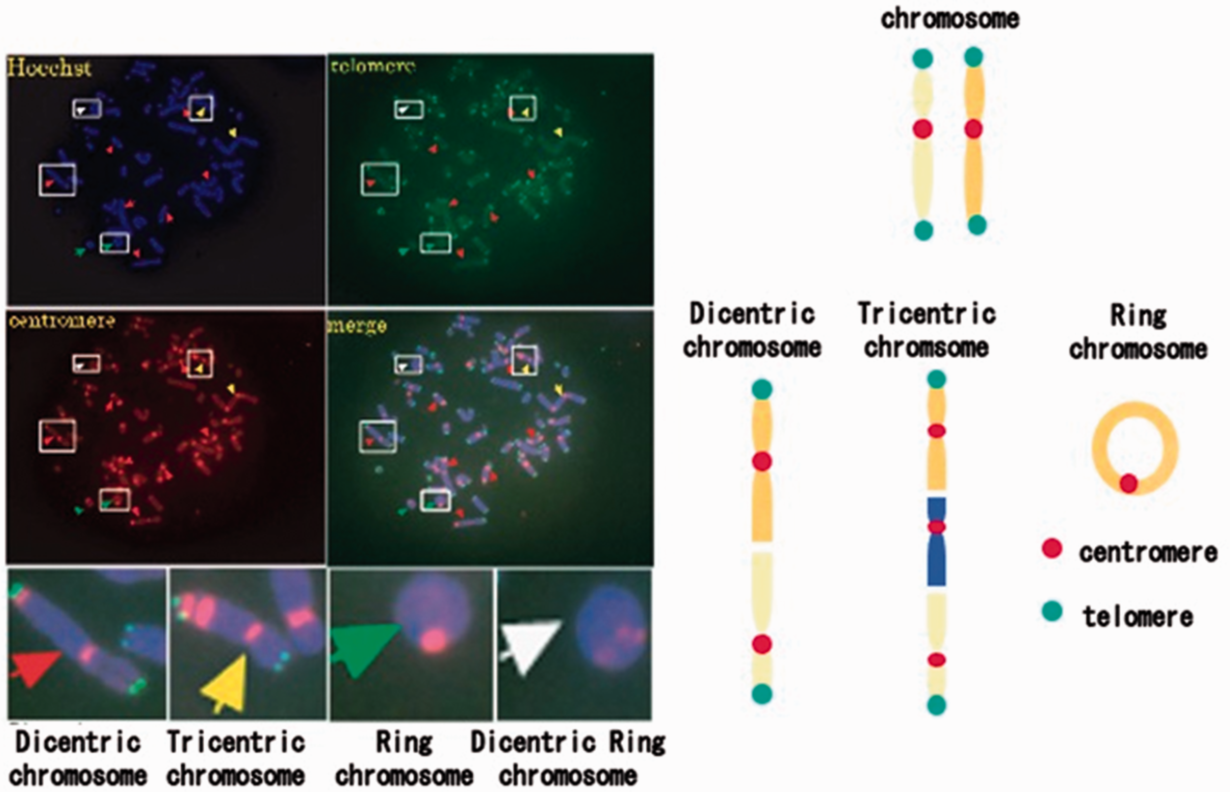
By performing high-throughput chromosomal analysis of 1000 cells or more using this method, it is possible to detect a small number of chromosomal abnormalities such as dicentrics and ring chromosomes formed by low-dose radiation. Using this technology, chromosomal analysis of peripheral blood lymphocytes was performed on patients who underwent normal-dose chest computed tomography (CT) examination, and it was possible to detect an increase in chromosomal abnormalities (Fig. 3) (Shi et al., 2018).
Chromosome analysis by standard-dose computed tomography (CT) examination using the PNA-FISH method. Left: as a result of analysing the chromosomal abnormalities of peripheral blood lymphocytes before and after CT examination by PNA-FISH in 60 cases of non-cancer disease, the number of chromosomal abnormalities increased significantly following CT examination. Right: in cases where there were only a few chromosomal abnormalities (dose length product (DLP) = 0) before CT examination, the increase in chromosomal abnormalities by CT examination was more remarkable than in cases where there were numerous chromosomal abnormalities before CT examination. Modified from Shi et al. (2018). © 2021 Radiation Research Society.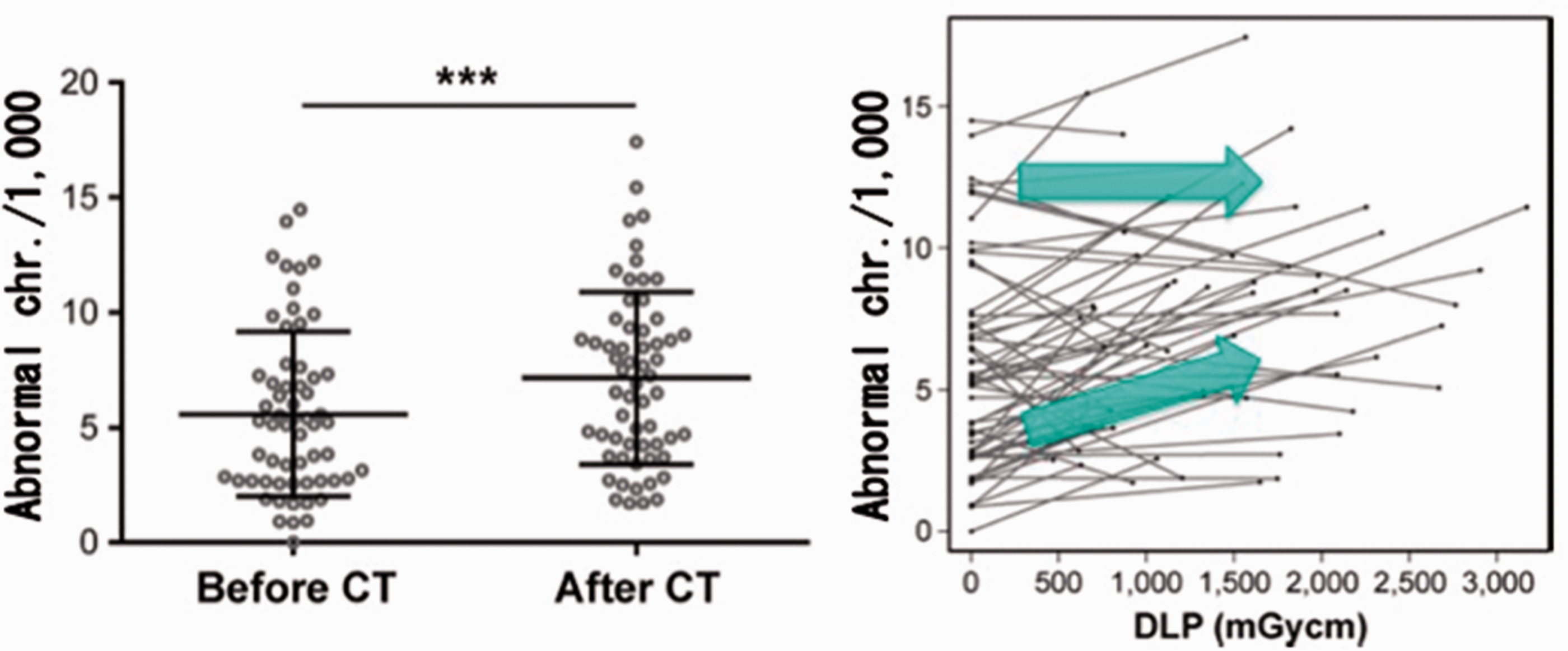
On the other hand, low-dose CT examinations used in lung cancer screening tests, which have recently been shown to reduce lung cancer mortality by approximately 20%, did not show a clear increase in chromosomal abnormalities, even with the PNA-FISH method. The validity of lung cancer screening tests by low-dose CT examination is biologically supported by these findings (Fig. 4) (Sakane et al., 2020).
Comparison of exposure effects of standard-dose and low-dose computed tomography (CT) examinations. The number of chromosomal aberrations in peripheral blood lymphocytes was compared using the PNA-FISH method before and after standard-dose and low-dose CT tests. Standard-dose CT (spectral detector CT) showed a significant increase in the number of chromosomal abnormalities, but low-dose CT did not show a significant increase in the number of chromosomal abnormalities. Modified from Sakane et al. (2020).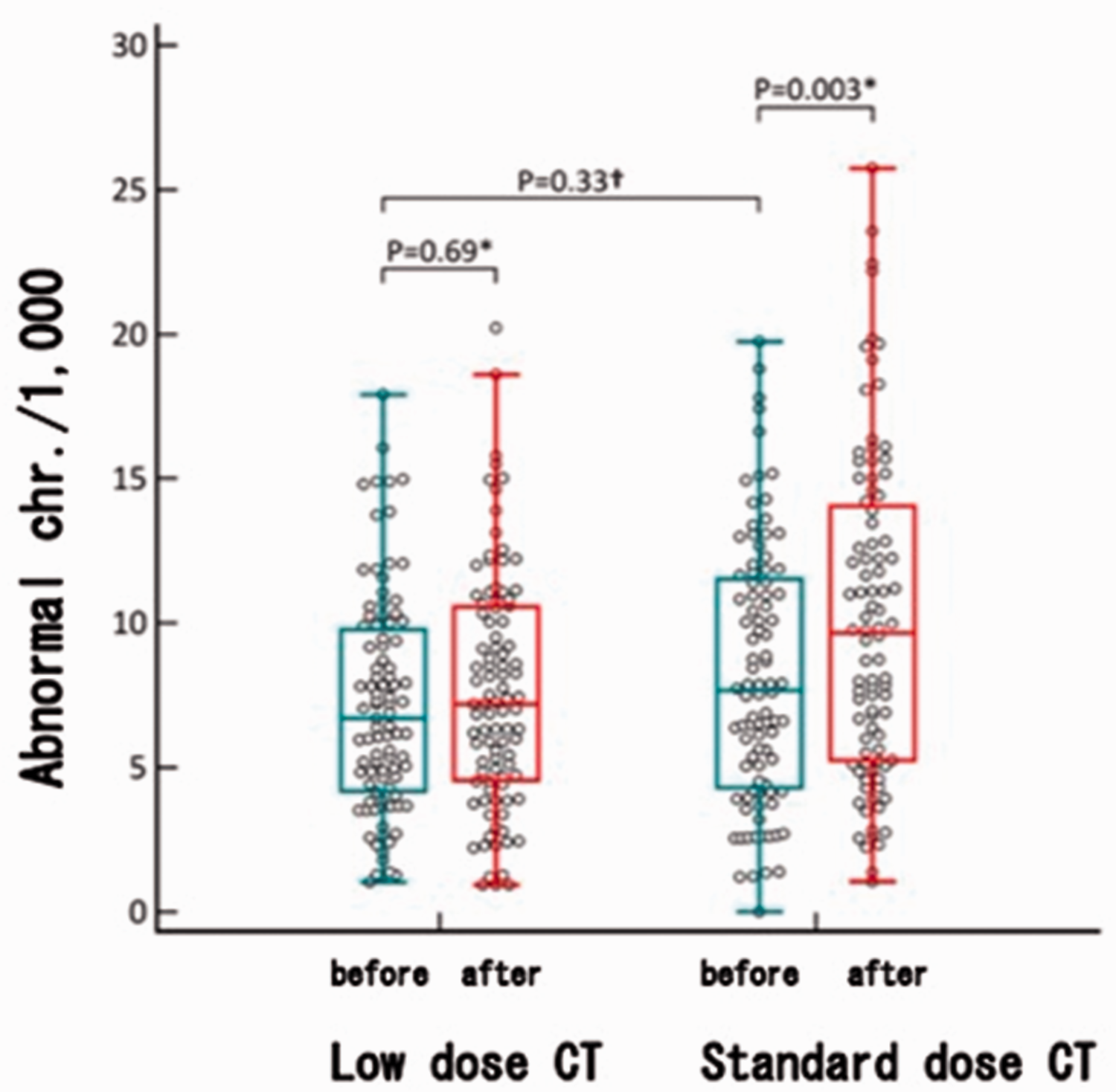
In the future, using high-throughput chromosomal automatic analysis and biological markers developed in radiation effects research, it is anticipated that next-generation radiology will include the development of safe radiological diagnostic technology and methods to predict the side effects and effects of radiotherapy.
3. CONCLUSIONS
In medical and biological science, the importance of research using mathematical life science approaches, or understanding essential life phenomena by constructing models to interpret data is increasing. Originally, radiation research was established by the fusion of biology and physics, and this has evolved using mathematical life science approaches by proposing models based on the observation of radiation-induced cell death, cell proliferation, and chromosomal abnormalities. Therefore, radiation research could be a good model of modern life science.
One of the characteristics of radiation research is that it has a great deal of contact with society. In particular, valuable experience has been gained from the accident at Fukushima Daiichi nuclear power plant regarding risk communication in the event of a radiation disaster. Careful analysis of such valuable experiences is very important to enable scientists and medical professionals to respond appropriately, not only to radiation disasters but also to other disasters including emerging infectious disease pandemics such as coronavirus disease 2019.