
Abstract
Internal doses are calculated using biokinetic and dosimetric models. These models describe the behaviour of the radionuclides after ingestion, inhalation, and absorption to the blood, and the absorption of the energy resulting from their nuclear transformations. The International Commission on Radiological Protection (ICRP) develops such models and applies them to provide dose coefficients and bioassay functions for the calculation of equivalent or effective dose from knowledge of intakes and/or measurements of activity in bioassay samples. Over the past few years, ICRP has devoted a considerable amount of effort to the revision and improvement of models to make them more physiologically realistic representations of uptake and retention in organs and tissues, and of excretion. Provision of new biokinetic models, dose coefficients, monitoring methods, and bioassay data is the responsibility of Committee 2 and its task groups. Three publications in a series of documents replacing the ICRP Publication 30 series and ICRP Publications 54, 68, and 78 have been issued [Occupational Intakes of Radionuclides (OIR) Parts 1–3]. OIR Part 1 describes the assessment of internal occupational exposure to radionuclides, biokinetic and dosimetric models, methods of individual and workplace monitoring, and general aspects of retrospective dose assessment. OIR Parts 2–5 provide data on individual elements and their radioisotopes. Work is also in progress on revision of dose coefficients for radionuclide intakes by members of the public.
1. INTRODUCTION
Occupational and environmental intakes of radionuclides (OIR and EIR, respectively) may occur during routine operations in a range of industrial, medical, educational, and research facilities. They may also occur after an incident involving radioactive material. An adequate assessment of internal exposure resulting from intakes of radionuclides is essential for the design, planning, and authorisation of a facility or activity; for the optimisation of radiation protection of workers; and for the retrospective demonstration of compliance with regulatory requirements.
In internal dosimetry, no operational dose quantities have been defined that provide a direct assessment of equivalent dose or effective dose. Different methods are therefore applied to assess these equivalent or effective doses due to radionuclides in the human body. They are mostly based on various activity measurements and the application of biokinetic and dosimetric models. However, because of the complexity of the overall procedure when calculating internal dose, the International Commission on Radiological Protection (ICRP) provides sets of dose per intake coefficients that allow a direct estimate of the internal dose from knowledge of intake into the body. In addition, for occupational intakes, data are provided to allow intake and dose to be calculated from bioassay measurements. The task of ICRP Task Group 95 on Internal Dose Coefficients is to provide a new set of models and dose coefficients to take into account the latest ICRP recommendations and up-to-date knowledge in biology, physiology, and dosimetry.
2. PREVIOUS PUBLICATIONS ON OCCUPATIONAL AND ENVIRONMENTAL INTAKES OF RADIONUCLIDES
ICRP has been very active for many decades in providing guidance and tools for the calculation of internal doses (ICRP, 1959). Publication 30 (ICRP, 1979, 1980, 1981, 1988) and its supplements gave dose coefficients and values of annual limits of intake (ALI) for workers for intakes of radionuclides by inhalation and ingestion, referencing the recommendations issued in Publication 26 (ICRP, 1977), and the anatomical and physiological data in Publication 23 (ICRP, 1975). Publication 68 (ICRP, 1994b) provided updated dose coefficients for workers following the 1990 Recommendations in Publication 60 (ICRP, 1991). It applied the Publication 66 Human Respiratory Tract Model (HRTM) (ICRP, 1994a) for inhaled radionuclides, the updated basic anatomical and physiological data for the skeleton in Publication 70 (ICRP, 1995b), and revised systemic biokinetic models for selected isotopes of 31 elements given in Publications 56, 67, 69, and 71 (ICRP, 1990, 1993, 1995a,c). Biokinetic models for other elements were taken from Publication 30 (ICRP, 1979, 1980, 1981, 1988) and modified by addition of explicit excretion pathways to improve dose estimates for the urinary bladder and colon walls. Publication 68 (ICRP, 1994b) did not give ALIs because ICRP wished to emphasise the need to take account of all exposures to ionising radiation in the workplace, from external radiation and intakes of all radionuclides.
Publications 54 and 78 gave guidance on the design of monitoring programmes and the interpretation of results to estimate doses to workers following radionuclide inhalation or ingestion (ICRP, 1989, 1997). The guidance was supported by numerical data to enable the assessment of intakes and doses from bioassay data (i.e. measurements of body and organ content, and daily urinary and faecal excretion). These data were provided for a number of radionuclides selected as those most likely to be encountered in the workplace. Predicted values of the measured quantities for various times after a single intake or for routine monitoring were given in terms of the activity of the intake per activity measured. Standard dose coefficients would then be used to calculate effective dose from the assessed intake.
The Publication 56 series (Publications 56, 67, 69, 71, and 72; ICRP, 1990, 1993, 1995a,c, 1996) gave dose coefficients for members of the public for intakes of radionuclides by inhalation and ingestion, referencing the recommendations issued in Publication 60 (ICRP, 1991), and the anatomical and physiological data in Publication 23 (ICRP, 1975). It applied the Publication 66 HRTM (ICRP, 1994a) for inhaled radionuclides, the updated basic anatomical and physiological data for the skeleton in Publication 70 (ICRP, 1995b), and revised systemic biokinetic models for selected isotopes of 31 elements given in Publications 56, 67, 69, and 71 (ICRP, 1990, 1993, 1995a,c). The biokinetic models for the gastrointestinal tract and systemic biokinetic models for other elements were taken from Publication 30 (ICRP, 1979, 1980, 1981, 1988) and modified by addition of explicit excretion pathways to improve dose estimates for the urinary bladder and colon walls.
Finally, Publications 88 and 95 (ICRP, 2001, 2004) gave dose coefficients for the embryo and fetus after intakes of radionuclides by the mother, and for the infant after ingestion of radionuclides in their mother’s milk.
3. CHANGES IN PUBLICATION 103 That Affect The Calculation Of Equivalent And Effective Dose
In the 2007 Recommendations issued in Publication 103 (ICRP, 2007), the concept and use of equivalent and effective dose remained unchanged, but a number of revisions were made to the methods used in their calculation. Changes were introduced in the radiation and tissue weighting factors from the values recommended in Publication 60 (ICRP, 1991). Since radiation weighting factors (wR) for photons, electrons, and alpha particles were unchanged, the only difference of potential importance to internally deposited radionuclides was for neutrons. The changes made do not reflect the availability of additional data, but rather a reconsideration of the appropriate treatment of radiation weighting for protection purposes.
The values of tissue weighting factors (wT) recommended in Publication 103 (ICRP, 2007) were changed from the values given in Publication 60 (ICRP, 1991), reflecting improved knowledge of radiation risks. The main sources of data on cancer risks are the follow-up studies of the Japanese atomic bomb survivors, used to derive risk coefficients averaged over seven Western and Asian populations with different background cancer rates (ICRP, 2007). The new tissue weighting factors were based on cancer incidence rather than mortality data, adjusted for lethality, loss of quality of life, and years of life lost. Weighting for hereditary effects was based on estimates of disease in the first two generations rather than at theoretical equilibrium. The main changes in tissue weighting factors in the 2007 Recommendations (ICRP, 2007) are an increase for breast (from 0.05 to 0.12), a decrease for gonads (from 0.2 to 0.08), and inclusion of more organs and tissues in a larger ‘remainder’ (from 0.05 to 0.12). The remainder dose is now calculated as the arithmetic mean of the doses to the 13 organs and tissues for each sex. Tissue weighting factors continue to represent averages across the sexes and across all ages.
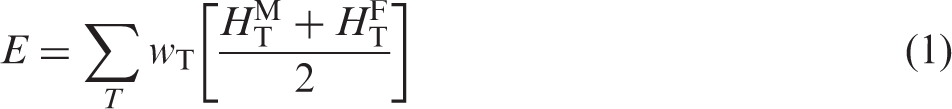
A further important change introduced in the 2007 Recommendations (ICRP, 2007) is that doses from external and internal sources are calculated using reference computational phantoms of the human body (ICRP, 2009). In the past, the Commission did not specify a particular phantom, and various mathematical phantoms such as hermaphrodite Medical-Internal-Radiation-Dose-type phantoms (Snyder et al., 1969), the sex-specific models of Kramer et al. (1982), and the age-specific phantoms of Cristy and Eckerman (1987) have been used. Voxel models, constructed from medical imaging data of real people, give a more realistic description of the human body than afforded in mathematical (or stylised) phantoms. Thus, ICRP decided to use voxel models to define the reference phantoms to be used in the calculations of dose distribution in the body for both internal and external exposures. These models (or computational phantoms), described in Publication 110 (ICRP, 2009), represent Reference Adult Male and Female, and have organ masses in compliance with the reference anatomical values compiled in Publication 89 (ICRP, 2002a). These phantoms are designed specifically for calculation of the radiological protection quantities corresponding to the effective dose concept of the 2007 Recommendations (ICRP, 2007). Equivalent doses to organs and tissues, HT, are calculated separately for Reference Adult Male and Female, and then averaged in the calculation of effective dose, E:
It is made clear in Publication 103 (ICRP, 2007) that effective dose is intended for use as a protection quantity on the basis of reference values, and relates to reference persons rather than specific individuals. The main uses of effective dose are in prospective dose assessment for planning and optimisation in radiological protection, and retrospective demonstration of compliance for regulatory purposes. Sex averaging in the calculation of equivalent and effective doses, implicit in the past use of hermaphrodite mathematical phantoms, is now explicit in the averaging of equivalent doses to adult male and female phantoms. Sex and age averaging in the derivation of tissue weighting factors can be seen to obscure differences in estimates of absolute radiation detriment between men and women, and between adults and children. However, practical protection would not be improved by calculating effective dose separately for males and females, and to do so might give a misleading impression of the precision of these quantities.
4. NEW BIOKINETIC MODELS DEVELOPED BY ICRP
In parallel to these changes, ICRP has continuously developed biokinetic models that describe the behaviour of the radionuclides in the body. Biokinetic models of the alimentary and respiratory tracts are used to define the movement of radionuclides within these systems, resulting in absorption to blood and/or loss from the body. The behaviour of radionuclides absorbed to blood is described by element-specific systemic models that range in complexity. These models are intended both for the derivation of dose coefficients and the interpretation of bioassay data. The models used in the new series of ICRP publications (OIR series and EIR series) are as described, in brief, below.
4.1. Human Respiratory Tract Model
The HRTM, described in Publication 66 (ICRP, 1994a), was updated in Publication 130 (ICRP, 2015) to take account of data accumulated since its publication, although the basic features of the model remain unchanged. Inhaled particles containing radionuclides deposit in the extrathoracic (ET) airways (nose, larynx, etc.), the bronchial (BB) and bronchiolar (bb) airways of the lung, and the alveolar–interstitial (AI) region, with deposition in the different regions being mainly dependent on particle size (ICRP, 1994a, 2002b). Removal from the respiratory tract occurs mainly by dissolution and absorption to blood, and the competing process of transport of particles to the throat followed by their entry into the alimentary tract. The proportions absorbed to blood or cleared by particle transport depend on the speciation and the solubility of the material, and on the radioactive half-life of the radionuclide. The ICRP model for the respiratory tract is also applied to gases and vapours, and to inhalation of radon and its radioactive progeny.
For absorption to blood, the main changes introduced in Publication 130 (ICRP, 2015) are:
redefinition of the Type F, M, and S absorption defaults: larger rapid dissolution fraction (fr) values for Types M and S of 0.2 and 0.01, rather than 0.1 and 0.001, respectively, with lower rapid dissolution rate (sr) values of 3 day−1 for Types M and S, and 30 day−1 for Type F, rather than 100 day−1; material-specific parameter values for fr, sr, and the slow dissolution rate (ss) in selected cases where sufficient information is available (e.g. forms of uranium); element-specific values of sr and the bound state parameters, fb and sb, where sufficient information is available; and revised treatment of gases and vapours in which solubility and reactivity are defined in terms of the proportion deposited in the respiratory tract. The default assumption is 100% deposition (20% ET2, 10% BB, 20% bb, and 50% AI) and Type F absorption. The SR-0, -1, and -2 classification described in Publication 66 (ICRP, 1994a) has not been found to be helpful and is not used.
For clearance by particle transport, the main changes are:
more realistic clearance from the nasal passage, including transfer from the anterior to the posterior region, based on recent human experimental studies; revised characteristics of slow particle clearance from the bronchial tree based on recent human experimental studies. It is now assumed that it occurs only in the bronchioles rather than as a particle-size-dependent phenomenon throughout the bronchial tree; and longer retention in the AI region of the lung, with a revised model structure based on recent data including long-term follow-up of workers exposed to insoluble 60Co particles and plutonium dioxide.
4.2. Human Alimentary Tract Model
The Publication 30 (ICRP, 1979) model of the gastrointestinal tract has been replaced by the Human Alimentary Tract Model (HATM) described in Publication 100 (ICRP, 2006). The main features of the HATM can be summarised as follows:
inclusion of all alimentary tract regions: oral cavity, oesophagus, stomach, small intestine, right colon, left colon, and rectosigmoid (sigmoid colon and rectum); a default assumption that absorption of an element and its radioisotopes to blood occurs exclusively in the small intestine (i.e. the total fractional absorption equals the fractional absorption from the small intestine); a model structure that allows for absorption in other regions, where information is available; a model structure that allows for retention in the mucosal tissues of the walls of alimentary tract regions, and on teeth, where information is available; and explicit specification of the location of target regions for cancer induction within each alimentary tract region.
Publication 100 (ICRP, 2006) gave preliminary values of electron and alpha particle absorbed fractions for stem cell layers in alimentary tract regions. Publication 133 (ICRP, 2016a) provided new calculations for both particle types, and for both content and wall sources. For regions within the small intestine, new models of segment folding were also implemented.
4.3. Systemic models
A systemic model describes the time-dependent distribution and retention of a radionuclide in the body after absorption to blood and systemic circulation, and its excretion from the body. In contrast to ICRP’s current and past biokinetic models describing the behaviour of radionuclides in the respiratory and alimentary tracts, ICRP’s systemic models have generally been element specific with regard to model structure as well as parameter values. A single generic model structure that depicts all potentially important systemic repositories and paths of transfer of all elements of interest in radiation protection would be too complex to be of practical use. However, generic model structures have been used in previous ICRP documents to represent the systemic biokinetics of groups of elements, typically chemical families, known (or expected) to have qualitatively similar behaviour in the body. For example, Publication 20 (ICRP, 1973) introduced a generic model formulation for the alkaline earth elements calcium, strontium, barium, and radium, but provided element-specific values for most model parameters. In Parts 1–3 of Publication 30 (ICRP, 1979, 1980, 1981), a model developed for plutonium, including parameter values as well as model structure, was applied to most actinide elements. The use of generic systemic model structures was increased in ICRP publications on doses to members of the public from intake of radionuclides (ICRP, 1993, 1995a,c), and is further expanded in recent ICRP publications because it facilitates the development, description, and application of systemic biokinetic models. An important development is that, as the availability of data allows, models have been made to be more physiologically realistic with regard to the dynamics of organ retention and excretion, so that they can be applied more reliably to the interpretation of bioassay data as well as the calculation of dose coefficients.
5. METHODOLOGY FOR DOSE CALCULATIONS: THE ICRP DOSIMETRY SYSTEM
ICRP publishes dose coefficients for the inhalation or ingestion of individual radionuclides by workers and members of the public, giving both equivalent doses to organs and tissues, and effective dose (ICRP, 1991, 2007). Biokinetic models are used in conjunction with reference physiological data, computational phantoms, and radiation transport calculation codes for the calculation of dose coefficients (ICRP, 2007). The steps in the calculation can be summarised as follows:
biokinetic models that are developed for individual elements and their radioisotopes are used to calculate the total number of transformations occurring within specific tissues, organs, or body regions (source regions) during a given period of time (usually 50 years for adults, or to age 70 years for children) by determining the time-integrated activity in each source region; dosimetric models, based on male and female reference computational phantoms and Monte Carlo radiation transport codes, are used to calculate the deposition of energy in all important organs/tissues (targets) for transformations occurring in each source region, taking account of the energies and yields of all emissions (ICRP, 2008). At this stage, sex-specific absorbed doses in each target organ or tissue resulting from a nuclear disintegration in each source region are calculated; the radiation weighting factors are applied to determine sex-specific committed equivalent doses to an organ or tissue; the sex-specific committed equivalent doses are sex averaged; and the tissue weighting factors are applied to determine the sex-averaged committed effective dose.
Dose calculations involve the use of nuclear decay data, anthropomorphic phantoms that describe the human anatomy, and codes that simulate radiation transport and energy deposition in the body. The data provided in the new OIR series (see Section 6) are calculated using revised decay data (ICRP, 2008), the ICRP reference computational phantoms of the adult male and female based on medical imaging data (ICRP, 2009), and specific absorbed fractions (ICRP, 2016a) calculated using well-established Monte Carlo codes such as MCNPX, PHITS, and EGSnrc (Pelowitz, 2008; Kawrakow et al., 2009; Niita et al., 2010).
For all dose calculations, radionuclides are assumed to be uniformly distributed throughout source regions, although these can be whole organs (e.g. liver) or a thin layer within a tissue (e.g. bone surfaces). Similarly, target cells are assumed to be uniformly distributed throughout target regions that vary in size from whole organs to layers of cells. Doses from ‘cross-fire’ radiation between source and target regions are important for penetrating photon radiation. For ‘non-penetrating’ alpha and beta particle radiations, energy will, in most cases, be largely deposited in the tissue in which the radionuclide is deposited. Photon and electron transport are followed for most source and target combinations. Additionally, special considerations are taken into account for alpha and beta emissions in a number of important cases. These include:
doses to target cells in the walls of the respiratory tract airways from radionuclides in the airways (ICRP, 1994a); doses to target cells in the alimentary tract from radionuclides in the lumen (ICRP, 2006); and doses to cells adjacent to inner bone surfaces (50 -µm layer; see below), and all red marrow from radionuclides on bone surfaces and within bone mineral.
6. NEW SERIES OF PUBLICATIONS: THE OIR AND EIR SERIES
The changes in methodology introduced in the 2007 Recommendations (ICRP, 2007), including revised tissue weighting factors and the introduction of reference phantoms, required the recalculation of all previously published dose coefficients. As explained in preceding sections, considerable improvements have also been made to other aspects of dose calculation methodology and biokinetic models.
The OIR series replaces the Publication 30 series (ICRP, 1979, 1980, 1981, 1988) and Publication 68 (ICRP, 1994b) to provide revised dose coefficients for OIR by inhalation and ingestion. The revised dose coefficients have been calculated using the Publication 100 (ICRP, 2006) HATM and a revision of the Publication 66 (ICRP, 1994a) HRTM which takes account of more recent data. In addition, information is provided on absorption to blood following inhalation and ingestion of different chemical forms of elements and their radioisotopes. In selected cases, it is judged that the data are sufficient to make material-specific recommendations. Revisions have been made to many of the models that describe the systemic biokinetics of radionuclides absorbed to blood, making them more physiologically realistic representations of uptake and retention in organs and tissues, and of excretion.
The publications in the OIR series provide data for the interpretation of bioassay measurements as well as dose coefficients, replacing Publications 54 and 78 (ICRP, 1989, 1997). In assessing bioassay data such as measurements of whole-body or organ content or urinary excretion, assumptions have to be made about the exposure scenario, including the pattern and mode of radionuclide intake, physical and chemical characteristics of the material involved, and the elapsed time between the exposure(s) and measurement. The OIR publications provide some guidance on monitoring programmes and data interpretation.
OIR Part 1 has been issued as Publication 130 (ICRP, 2015), and includes chapters on control of occupational exposures, biokinetic and dosimetric models, monitoring methods, monitoring programmes, and retrospective dose assessment. Subsequent publications provide data on individual elements and their radioisotopes, including biokinetic data and models, dose coefficients, and data for bioassay interpretation. OIR Parts 2 and 3 have been issued as Publication 134 (ICRP, 2016b) and Publication 137 (ICRP, 2017), and OIR 4 is nearing completion. In these publications, each element section provides: dose coefficients (committed effective dose and committed equivalent doses to organs or tissues per Bq intake (Sv Bq−1) for inhalation and ingestion of all relevant radioisotopes; dose per content functions [committed effective dose per predicted activity content in the body or in a given organ or per daily excretion (Sv Bq−1)]; and reference bioassay functions [values of activity (Bq) retained in the body or specific organs, or excreted in urine or faeces, at various times after unit intake (i.e. 1 Bq) by inhalation or ingestion].
Data are provided in the printed publications and in electronic annexes. The data provided in the printed publications are restricted to tables of committed effective dose per intake (Sv Bq−1), tables of committed effective dose per content (Sv Bq−1), and graphs for reference bioassay functions. Data are provided for all absorption types of the most common isotope(s), and for an activity median aerodynamic diameter (AMAD) of 5 µm. In cases for which sufficient information is available (principally for actinide elements), lung absorption is specified for different chemical forms, and dose coefficients and bioassay functions are calculated accordingly. The electronic annex that accompanies the OIR series contains a comprehensive set of committed effective and equivalent dose coefficients, dose per content functions, and reference bioassay functions for most of the isotopes presented in Publication 107 (ICRP, 2008), and for a range of physicochemical forms and aerosol AMADs. Data for intake by ingestion and for direct input to the blood are also given. The electronic annex provides a set of data files which may be accessed by the user directly or by using the accompanying Data Viewer. The Data Viewer permits rapid navigation of the dataset and visualisation of the data in tabulated and graphical formats, such as graphs of the time series of dose per content function values, or predicted activity content per dose (Bq Sv−1) as a function of time after intake.
The EIR series will replace the Publication 56 series (ICRP, 1990, 1993, 1995a,c, 1996) to provide revised age-dependent dose coefficients for members of the public for environmental intakes of radionuclides by inhalation and ingestion. The revised dose coefficients are being calculated using the Publication 100 (ICRP, 2006) HATM and the revision of the Publication 66 (ICRP, 1994a) HRTM described in Publication 130 (ICRP, 2015). As for the OIR series, revisions have been made to many of the models that describe the systemic biokinetics of radionuclides absorbed to blood, making them more physiologically realistic representations of uptake and retention in organs and tissues, and of excretion, and considering the age dependence of biokinetics.
The first publication in the EIR series will provide an introduction to the series and include sections on biokinetic and dosimetric models, plus data on individual elements and their radioisotopes, including biokinetic data and models, and dose coefficients. As for the OIR series, each element section will provide dose coefficients: committed effective dose and committed equivalent doses to organs or tissues per Bq intake (Sv Bq−1) for inhalation and ingestion of all relevant radioisotopes. Data will be provided in the printed publications and in electronic annexes. The data provided in the printed publications will be restricted to tables of committed effective dose per intake (Sv Bq−1). For intakes by inhalation, data will be provided for all absorption types of the most common isotope(s) and for an AMAD of 1 µm. The electronic annex that accompanies the EIR series will contain a comprehensive set of committed effective and equivalent dose coefficients for most of the isotopes presented in Publication 107 (ICRP, 2008), and for a range of aerosol AMADs and activity median thermodynamic diameters. The electronic annex will provide dose coefficients and other radionuclide-specific data as a set of data files which may be accessed by the user directly or by using the accompanying Data Viewer.
An important question is whether the improvements made to biokinetic and dosimetric models have substantial impacts on the numerical values of dose coefficients. An analysis of the data published in OIR Part 1 (ICRP, 2016) shows that, for inhalation of reference forms of radionuclides (aerosols of 5 µm; Type F, M, or S), the majority of new dose coefficients are slightly lower (within a factor of 2) than those published in the Publication 30 series (ICRP, 1979, 1980, 1981, 1988). In some very rare cases (14CO, 14CO2, 59Fe Type F, 90Sr Type S, 60Co Type S), dose coefficients have increased by a factor of approximately 2 because of the revision of the biokinetic models, and a better description of radionuclide retention and distribution in tissues. For ingestion, the new dose coefficients are similar to or a few percent lower than the previous dose coefficients. The most significant decrease is for moderately soluble forms of 90Y, where the dose coefficient is now lower by a factor of 5. In this specific case, the older coefficient could be seen as very conservative.
It is reassuring that differences between the old and the new data are mainly small, confirming that the protection of workers was already reliably based on existing data. The increased sophistication and realism of the new biokinetic and dosimetric models gives us additional confidence in the data provided, and contributes to reductions in uncertainties. It also means that they are readily applied to the interpretation of bioassay data. It should also be noted that the new data in the OIR series extend the existing data sets, providing specific coefficients for isotopes and chemical forms that were not described previously, contributing to improvements in exposure and dose assessments, and the protection of workers. Furthermore, this series provides physiologically based biokinetic models that can be used for applications other than radiation protection, including in toxicology, pharmacology, and medicine.