
Abstract
The recommendation from the International Commission on Radiological Protection that the occupational equivalent dose limit for the lens of the eye should be reduced to 20 mSv year−1, averaged over 5 years with no year exceeding 50 mSv, has stimulated a discussion on the practicalities of implementation of this revised dose limit, and the most appropriate risk and protection framework to adopt. This brief paper provides an overview of some of the drivers behind the move to a lower recommended dose limit. The issue of implementation in the medical sector in the UK has been addressed through a small-scale survey of doses to the lens of the eye amongst interventional cardiologists and radiologists. In addition, a mechanistic study of early and late post-irradiation changes in the lens of the eye in in-vivo-exposed mice is outlined. Surveys and studies such as those described can contribute to a deeper understanding of fundamental and practical issues, and therefore contribute to a robust evidence base for ensuring adequate protection of the eye while avoiding undesirable restrictions to working practices.
1. Introduction
The International Commission on Radiological Protection (ICRP) published its report on tissue reactions in 2012 (ICRP, 2012). This report is an extensive and comprehensive review of the effects of ionising radiation on tissues; it also considers the dose thresholds, below which the incidence of any given tissue reaction occurs in less than 1% of an exposed group. In many cases, there was little indication that threshold doses were much different from those previously recognised. However, evidence had accumulated from epidemiological studies indicating that the threshold dose for effects on the lens of the eye had been overestimated. Previously, absorbed dose thresholds for cataract were considered to be approximately 2 Gy for acute exposures and on the order of 4–5 Gy for fractionated and protracted exposures; the newer evidence suggested that the threshold dose was around 0.5 Gy for both acute and protracted/fractionated exposures. On the basis of this new evidence, ICRP considered the dose limit for the lens of the eye and, prior to Publication 118 (ICRP, 2012), issued a statement on tissue reactions (ICRP, 2011) which recommended that the dose limit for occupational exposure should be reduced from 150 mSv year−1 to 20 mSv year−1 averaged over 5 years, with no individual year exceeding 50 mSv.
The conclusion that the threshold dose for the lens of the eye had previously been overestimated was based largely on analysis of epidemiological studies of cataract and lens opacities, particularly those of Japanese atomic bomb survivors who had extended post-exposure follow-up periods, and Chernobyl liquidators in whom exposures were protracted. These studies have been considered in recent reviews (Ainsbury et al., 2009; Bouffler et al., 2012) which also concluded that the absorbed dose threshold for cataract is around 0.5 Gy in the case of acute exposures, with protraction or fractionation of exposure having little effect on the value. A particularly important study is that of Neriishi et al. (2007), which reports on the prevalence of cataracts requiring surgical intervention among atomic bomb survivors. This study reports a 0.5-Gy threshold dose for significant excess relative risk; the confidence intervals on the 0.5-Gy threshold value included 0 Gy, indicating that a non-threshold relationship might apply. This study therefore indicates that radiation exposure is associated with lens opacities that impair vision, not just smaller pre-cataractous lesions that have a minimal effect on vision.
ICRP’s recommendation of a reduced dose limit for the lens of the eye has caused considerable concern in some occupational areas, particularly interventional radiology and cardiology, where compliance with a new dose limit may be difficult and may require modifications to working practices (Englefield, 2011; Martin, 2011; Broughton et al., 2013).
In considering potential mechanisms of radiation-associated lens cataract, Bouffler et al. (2012) observed that the dose–response slope for cataract and lens opacities reported in the more recent epidemiological investigations is not as steep as conventionally considered to apply to tissue reactions, and several studies have indicated that incidence can be described by a non-threshold dose–response relationship. This suggests that effects on the lens of the eye may be governed by processes other than cell killing or functional inactivation, as generally accepted to be important for the induction of tissue reactions. Ionising radiation produces a characteristic posterior subcapsular cataract that is distinct from, for instance, a nuclear cataract that would be typical of an age-related cataract phenotype (Little, 2013). Therefore, the mechanistic detail associated with the development of posterior subcapsular cataract and the involvement of the underlying cortex (Merriam and Worgul, 1983) will likely involve specific events that are associated with cataractogenesis in general. Oxidative stress clearly plays an important role in the development of cataract, and reduction in anti-oxidant capacity is associated with age-related cataract. The incidence of cataract is strongly age-related, and it has been suggested that radiation acts in cataractogenesis by accelerating the ageing process, which in itself is likely to include an effect of oxidative stress (Pendergrass et al., 2010). There is good evidence for a role of genetic factors in determining cataract sensitivity (Churchill and Graw, 2011; He et al., 2013). In experimental models, genes that play important roles in suppression of tumourigenesis are also demonstrated to affect cataract formation (e.g. Wiley et al., 2011), and DNA-repair-related genes are reported to affect radiation-induced cataract (Kleiman et al., 2007). These observations lend weight to the argument that the process of cataract formation following radiation exposure has similarity with mechanisms of stochastic effects. However, further mechanistic investigations are required to define the mechanisms that lead to cataract following ionising radiation exposure.
On the basis of the brief discussion above, two main issues relevant to the revised ICRP recommendation for a reduced occupational dose limit for the eye stand out as requiring further attention: (1) investigation of exposures in the medical sector to identify any difficulties there may be in compliance with a reduced dose limit; and (2) further investigation of the mechanisms contributing to radiation cataractogenesis to help clarify how radiation causes cataract, as well as whether radiation cataractogenesis is best treated as a stochastic effect or a tissue reaction for low dose risk estimation. Two studies that consider these issues are summarised below.
2. Small-Scale Survey of Eye Doses in the Uk Medical Sector
As noted above, concerns have been expressed that doses to the lens of the eye received by those working in the medical sector may be high enough to present some difficulties in implementation of the recommended 20-mSv occupational equivalent dose limit for the eye. In particular, concern has been noted in the interventional cardiology and radiology fields, as highlighted by Vañó et al. (2009). To address this issue, a small-scale study of doses to the lens of the eye was undertaken recently in three UK radiology and cardiology departments (Ainsbury et al., 2014).
In brief, this survey targeted three busy departments offering a full range of radiology services including computed tomography, fluoroscopy, mammography, magnetic resonance imaging, nuclear medicine, ultrasound, and x ray. Within these centres, consultant and registrar interventional cardiologists and radiologists were the focus as these clinicians were predicted to be among the most exposed. Radiation doses to the lens of the eye were assessed over a 4-week period using the Public Health England (PHE) eye lens dosimeter (Gilvin et al., 2013). This dosimeter is supplied as a headband worn just above the eye, and measures the Hp(3) operational quantity using a tissue equivalent thermoluminescence dosimeter behind a filter equivalent to a 3-mm thickness of tissue (Fig. 1).
Illustration of Public Health England’s eye lens dosimeter (Gilvin et al., 2013).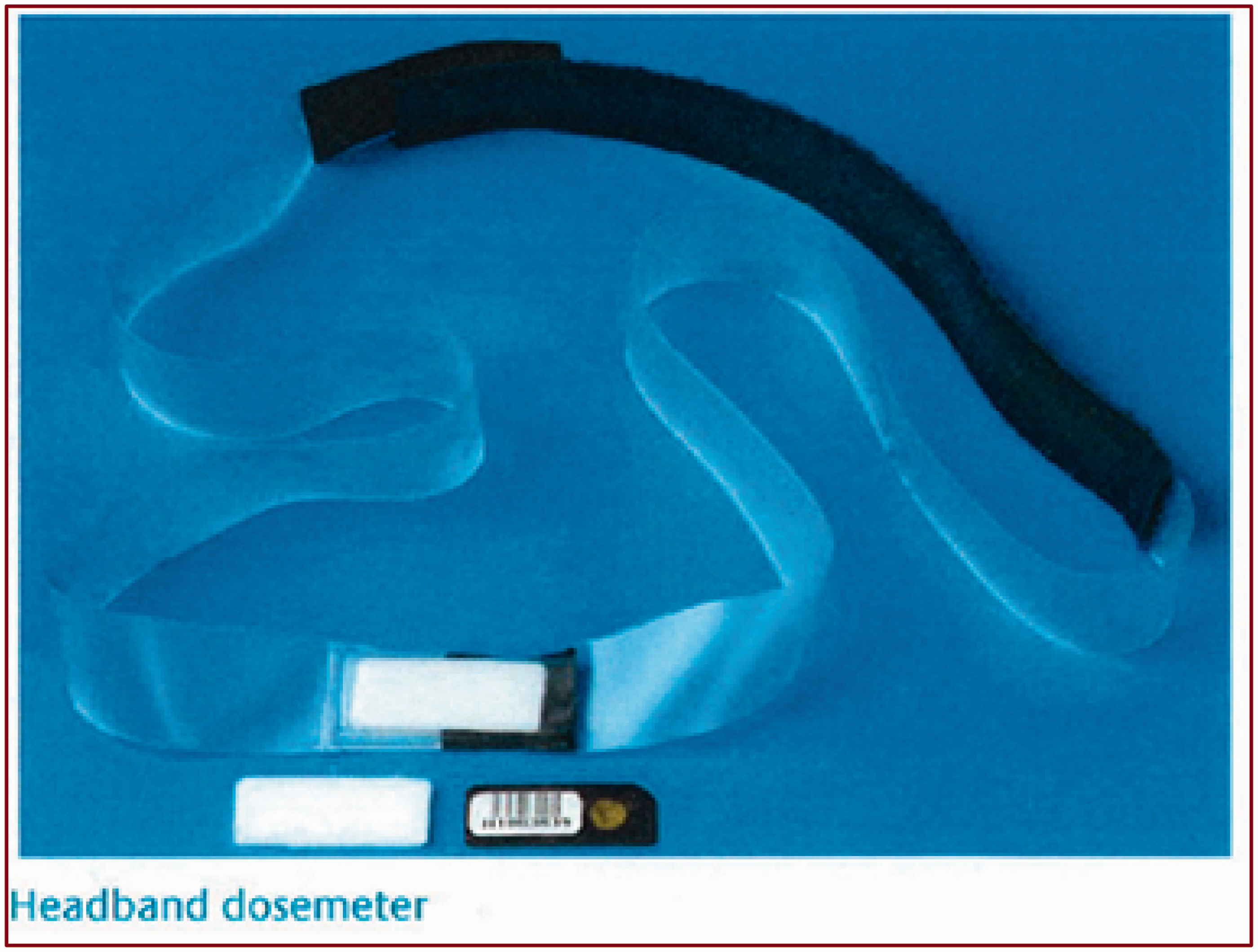
Sixty-eight dosimeters were provided to the hospitals along with instructions for use and a questionnaire. The questionnaire aimed to establish:
if dosimeter use followed the supplied instructions; job title of user; types and numbers of procedures undertaken during the survey period; and personal protective equipment (PPE) used.
Hospital-based radiation protection specialists distributed the dosimeters to hospital staff, collected the dosimeters and questionnaires, and returned them to PHE for analysis. Of the 68 distributed dosimeters, 61 were returned along with 58 completed questionnaires.
Full results are presented elsewhere (Ainsbury et al., 2013); however, to summarise:
Doses received by staff carrying out a full range of interventional procedures, including coronary angiogram, cardiac catheterisation, transcatheter aortic valve implantation, angioplasty, and pulmonary vein isolation, were monitored. Over 1000 procedures were carried out by the monitored staff. A median of 15 procedures were carried out per monitored individual, with a wide range of 1–70 on the median value. Reported use of PPE was good, although only nine individuals reported the use of lead glasses. Thirteen of the returned dosimeters recorded doses above the detection limit of 0.15 mSv. The median recorded dose over the 61 dosimeters was below the detection limit of 0.15 mSv. Two returned dosimeters recorded doses of potential concern (1.60 and 1.58 mSv, respectively).
If the two staff with recorded doses of 1.60 and 1.58 mSv over the 4-week period continued to be exposed at the same level over 1 whole year, doses just above the revised dose limit (equivalent yearly doses of 20.80 and 20.54, respectively) could accumulate. In these cases, one participant reported that lead glasses were not worn, while the other participant reported that lead glasses were used but the dosimeter was worn outside the glasses. This indicated that doses to the eye in these two cases would be below 20 mSv year−1 if doses were recorded behind properly worn lead glasses that can provide a 5–10-fold reduction in dose (Koukorava et al., 2011).
Overall, this small-scale, three-centre survey of doses to the lens of the eye in the UK suggests that compliance with the ICRP recommended dose limit (ICRP, 2012) should not be difficult where there is good uptake and correct use of PPE. However, the survey has limitations in the number and types of staff surveyed, and the number of centres covered. It was also notable that no obvious relationship was observed between the number of procedures carried out and recorded doses to the lens of the eye. Therefore, there appear to be individual factors that affect eye exposures and, although not observed in this survey, there may be variation in practice and therefore eye exposures in different hospitals. Nonetheless, the doses recorded in this survey are comparable with those reported elsewhere (Donadille et al., 2011; Efstathopoulos et al., 2011).
3. Mechanistic Study of Radiation Cataractogenesis
Given the uncertainty on the risk of radiation cataract at low doses and the need for a sound basis for risk extrapolation, mechanistic studies can contribute to providing a robust scientific basis for the protection of humans against radiation-associated cataract. With this aim in mind, a study of the dose–response for radiation cataract and early lens changes has been initiated. The main part of the study uses C57BL/6 J mice exposed to 20 mGy–2 Gy whole-body x rays. Eye lenses are removed at times between 1 hour and 10 months following irradiation. Multiple endpoints are under investigation in anterior marginal lens epithelial cells (i.e. the region of the lens where lens fibres are formed), including DNA damage assessed by the presence of gamma-H2AX staining, cell proliferation, cellular senescence, cell cycle arrest, and lens fibre morphogenesis. Additional tissues are being stored from the mice exposed in the study to allow investigation of comparative radiosensitivity in a range of tissues (including blood, skin, and whiskers).
This collaborative study between PHE and Durham University is at an early stage; experimental assays are being refined but it is clear that the mouse lens provides an accessible system for the study of early- and late-phase effects related to radiation exposure and cataract formation. An important aspect of the study is sampling over an extended timeframe, so that early changes can be related to later changes more evidently involved in cataract formation. In the longer term, the availability of well-defined experimental mouse models with a wide range of genetic conditions will help to characterise relevant pathways and susceptibility factors that contribute to cataract formation at low doses.