
Abstract
Level of evidence: 5 – expert opinion
Introduction
Haller and Onodi cells are anatomical variants of ethmoid air cells, well-known for their clinical significance in otolaryngology. Discovered over a century ago, the origins of their discovery and the stories of the pioneers who found them have become obscured by time.
The Life of Albrecht von Haller
Albrecht von Haller (1708-1777) was born in Bern, Switzerland. He studied medicine in Tubingen, and later Leiden, learning under anatomy luminaries Hermann Boerhaave and Bernhard Albinus. After graduation, he traveled to study in London, Oxford, Paris, Strasbourg, and Basel, before returning to Bern to open a private practice in 1729. Due to his reputation as an anatomist, he was appointed professor of anatomy, botany, and surgery at the newly-founded University of Göttingen in 1736. During his tenure, he published many influential works, including Praelectiones academicae, a 7-volume commentary on Boerhaave’s lectures; Icones anatomicae, an 8-part anatomical study; and Primae lineae physiologiae, a textbook on human physiology. Homesick, Haller eventually moved back to Bern in 1753 to serve as an administrator. Ever the tireless writer, Haller continued to publish numerous works on botany, medicine, physiology, and philosophy before his death in 1777. 1
Discovery of the Haller Cell
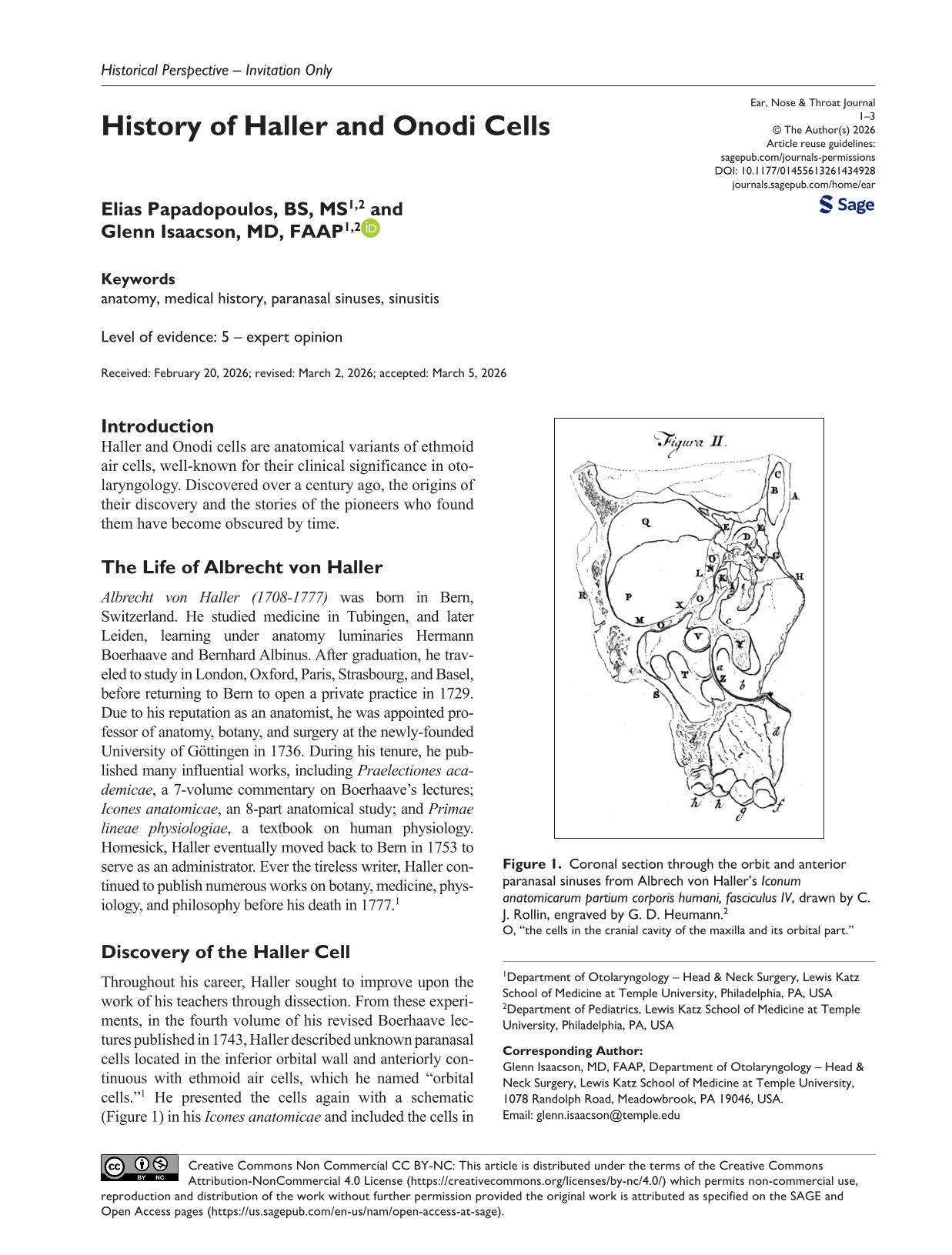
Throughout his career, Haller sought to improve upon the work of his teachers through dissection. From these experiments, in the fourth volume of his revised Boerhaave lectures published in 1743, Haller described unknown paranasal cells located in the inferior orbital wall and anteriorly continuous with ethmoid air cells, which he named “orbital cells.” 1 He presented the cells again with a schematic (Figure 1) in his Icones anatomicae and included the cells in his textbook, Elementa physiologiae corporis humani.1,2 In his texts, Haller recognized that the sinuses and cells fill with mucus, which may become difficult to expel, an early nod to their clinical significance. 3
Coronal section through the orbit and anterior paranasal sinuses from Albrech von Haller’s Iconum anatomicarum partium corporis humani, fasciculus IV, drawn by C. J. Rollin, engraved by G. D. Heumann. 2
These “orbital cells” remained in relative obscurity until 1925, when German otorhinolaryngologist Ludwig Grünwald proposed that the “Cellula Halleri” may narrow the ethmoid infundibulum and consequently the ostium of the maxillary sinus. In the 1970s, Walter Messerklinger, a pioneer of functional endoscopic sinus surgery (FESS), confirmed that large Haller cells can obstruct the infundibulum and intensify maxillary sinus inflammation. With the advent of computed tomography, the significance of Haller cells became appreciated for their roles in acute rhinosinusitis.1,4
The Life of Adolf Onodi
Adolf Ónodi (1857-1919) was born in Nikolos, Hungary. After graduating from the University of Budapest in 1880, he worked as a teaching assistant in the Institute of Anatomy, forging a distinguished career as a surgeon and laryngologist. He was appointed honorary lecturer at the university in 1887 and later associate professor of the Department of Rhinolaryngology and hospital director in 1919. He was an elected member of the Hungarian Academy of Sciences and was one of the founding members of the Hungarian Society of Laryngologists and Otologists in 1893. Ónodi’s career abruptly ended following the fall of the Hungarian Soviet Republic in 1919. Due to his political involvement, he was expelled from the university and emigrated to Vienna, where he died that same year. 5
Discovery of the Onodi Cell
Adolf Ónodi’s scientific contributions focused on paranasal sinus anatomy. He was particularly interested in the relationship between sinus disease and visual impairment. In his 1904 work, Die Sehstörungen und Erblindung nasalen Ursprunges, Ónodi mapped out the topographic relationship of the optic nerves to the paranasal sinuses. 6 While the proximity of the optic nerve to the sphenoid sinus was already known, Ónodi was the first to demonstrate a frequent relationship between the optic nerve and the most posterior ethmoid cell, now known as the Onodi cell (Figure 2). He documented 38 variations of the relationship between the optic nerve and the ethmoid air cells and sphenoid sinus, proposing that sinus disease may cause visual disturbances due to this proximity. Ónodi’s findings gained fame, and in 1910 he published an English translation featuring 50 illustrations reproduced from photographs of his anatomic specimens. 7 Following the rise of FESS and CT imaging in the late 20th century, identification of Onodi cells became crucial to avoid damaging the optic nerve and the internal carotid artery during sinus surgery. 4
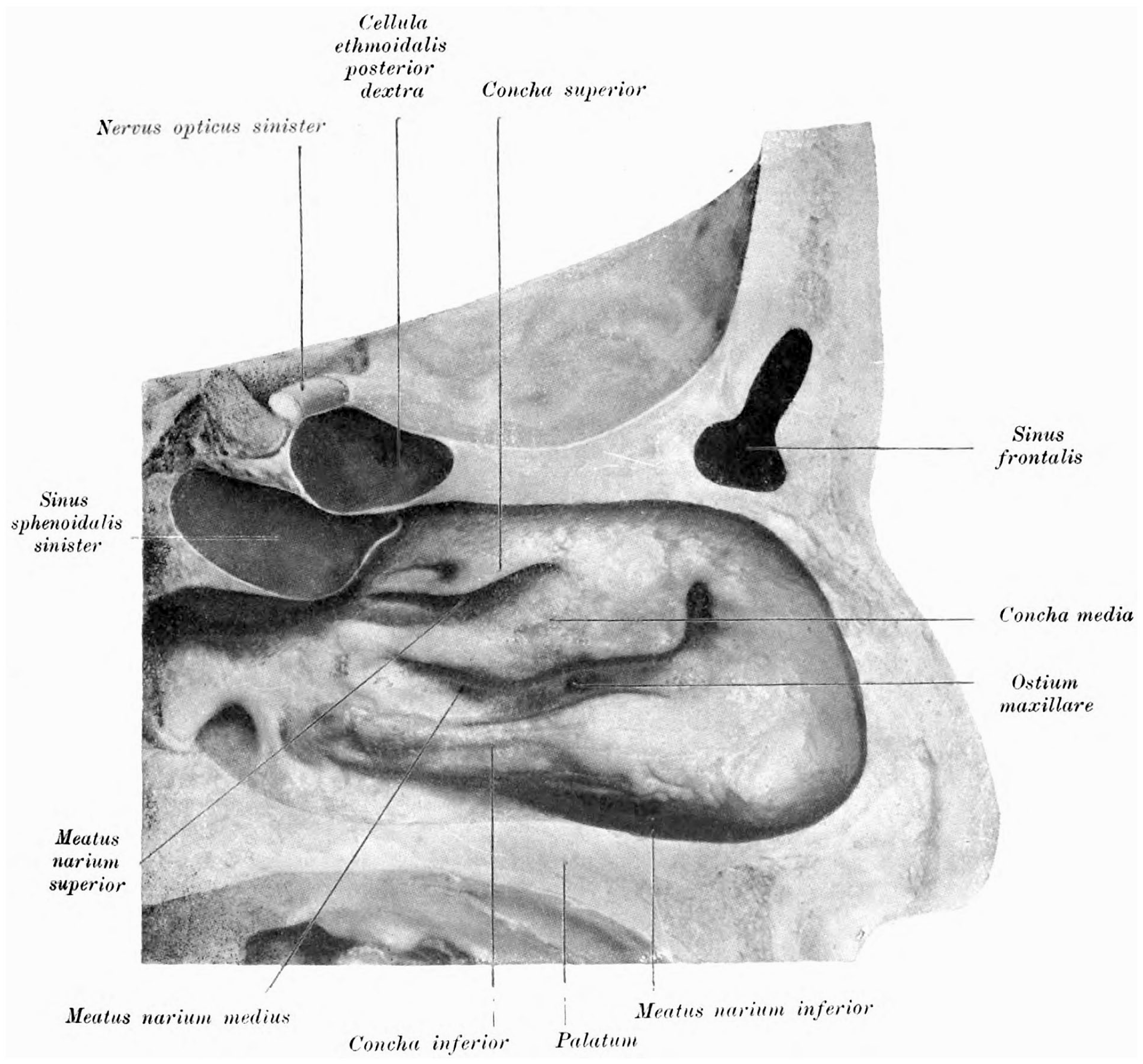
Illustration from Adolf Ónodi’s The optic nerve and the accessory sinuses of the nose. The “posterior ethmoidal cell,” later known as the Onodi cell, in close contact with the optic nerve. 7
It should be noted that current trends in medicine favor the use of non-eponymous terminology. Still, attribution for insightful observations such as those of Haller and Ónodi must not be forgotten.
Footnotes
Ethical Considerations
This article does not contain any studies with human or animal participants.
Funding
The authors received no financial support for the research, authorship, and/or publication of this article.
Declaration of Conflicting Interests
The authors declared no potential conflicts of interest with respect to the research, authorship, and/or publication of this article.
Data Availability Statement
This paper contains no new data. All data included are available through conventional library sources. The authors would be pleased to assist interested researchers via the corresponding author’s email.