
Abstract
Neck pain is a common reason for primary care visits, and its differential diagnosis should consider various conditions. The reported incidence of hyoid bone fractures is extremely low, accounting for only 0.002% of all fractures. The most common causes of hyoid bone fractures include strangulation attempts and motor vehicle accidents. We report a case of an uncommon complication of manual therapy of the cervical spine. A 76-year-old woman complained of neck pain that worsened during speaking and swallowing, originating from a neck physiotherapy session. The otolaryngological examination revealed tenderness on the right side of the neck. Flexible nasal endoscopy demonstrated a shallow right piriform recess and asymmetry of the arytenoid cartilages. Computer tomography scan of the neck showed an isolated fracture of the right greater horn (cornu major) of the hyoid bone. The treatment was nonsurgical, with the use of a Schantz collar and pain relief drugs. Reported symptoms of hyoid bone fractures include dysphagia, odynophagia, and neck pain. In most cases of hyoid fractures, conservative management suffices, involving rest, analgesic and anti-inflammatory treatment, and neck immobilization. Surgical treatment is often necessary in the cases of fractures accompanying other injuries.
Introduction
The hyoid bone is an unpaired bone situated in the anterior part of the neck at the level of the C3 vertebra, in the space between the mandible and the thyroid cartilage. It consists of a body, 2 greater horns (cornua majora) and 2 lesser horns (cornua minora). All parts of the hyoid originate from the second and third pharyngeal arches. While it is not directly connected to other bones, it is closely associated with an extensive tendon-muscular complex. The hyoid bone plays a relevant role in the orofacial complex. It is involved in phonation due to its connection to the larynx and also contributes to swallowing, prevention of regurgitation, and tongue movement. Its functions include maintaining the airway between the oropharynx and the tracheal rings, as well as assisting in head posture due to its complex connections with the mandible and cervical spine. 1 These 2 (the mandible and cervical spine) provide the hyoid with effective protection, which, in conjunction with its high mobility, makes the hyoid resistant to injuries. 2 The most frequently documented injury is a fracture, although it is typically identified in postmortem, occurring in 17% to 76% of cases in individuals who have been strangled or hanged. 3 Fractures in these cases are usually the result of direct trauma, while fractures identified during the lifetime of the individual usually occur after indirect trauma (so-called muscular fractures). 4 It is worth noting that such injuries can also occur, particularly during activities such as combat sports. 5 In addition, they may manifest following excessive hyperextension of the cervical spine and as an iatrogenic consequence, for instance, in cases of surgical removal of a median branchial cyst. 4 The reported incidence of hyoid bone fractures is extremely low, at 0.002% of all fractures. However, this statistic may not accurately represent today’s society, considering the increasing popularity of combat sports and associated head and neck injuries. 5
The symptoms of hyoid bone fractures are nonspecific and can vary. They may range from mild neck pain that worsens during swallowing or coughing to severe neck pain accompanied by odynophagia or dyspnea. The most common presenting signs typically include tenderness and swelling in the anterior neck. 6
It is important to mention that nonspecific neck pain is a common musculoskeletal condition, with an age-standardized prevalence rate of 27.0 per 1000 population in 2019. 7 It is a frequent reason for primary care visits, and its differential diagnosis should consider various conditions, including inflammatory and degenerative joint diseases, neck muscle strains, and life-threatening conditions such as cervical spine injuries and neoplastic changes. 8
The aim of this study is to present a case of neck injury resulting in the fracture of the greater horn (cornu major) of the hyoid bone in a 76-year-old woman, which occurred during a physiotherapy procedure (manual therapy). In addition, a literature review on this topic will be provided.
Case Report
A 76-year-old woman was referred to the otolaryngological clinic with a complaint of pain in the neck region. She reported neck pain, localized on the right side, worsening during speaking and swallowing. The patient, a former actress, emphasized the significance of her speech organ’s optimal function for her profession. Notably, the symptoms appeared immediately after a neck physiotherapy session, that involved manual therapy, which she underwent yesterday, prompting her to seek medical attention today. In the otolaryngological physical examination, no posttraumatic changes were found; only palpation tenderness was observed on the right side at the level of the hyoid bone. Flexible nasal endoscopy demonstrated a shallow right piriform recess and asymmetry of the arytenoid cartilages. In addition, slight limitations in the mobility of the right vocal fold were observed during full glottal closure. The neck ultrasound examination did not show any signs of trauma or hematoma. In order to provide a more accurate assessment, a neck computed tomography (CT) scan was performed. It revealed an isolated fracture of the right greater horn (cornu major) of the hyoid bone with mild edema of the surrounding soft tissues (Figure 1). The results of the CT scan and the patient’s symptoms were decisive for the treatment strategy. She was treated nonsurgically with the use of the Schantz collar. As part of the conservative management, a liquid diet, rest, and pain relievers were also implemented. After 8 weeks, a follow-up neck CT scan was performed, which revealed features of bone union at the hyoid bone fracture site (Figure 2).
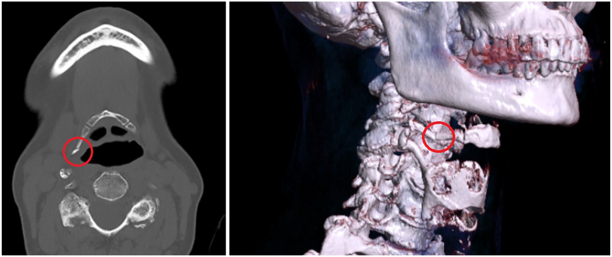
Computed tomography scan with reconstruction shows a fracture of the greater horn of the hyoid bone (red circle).
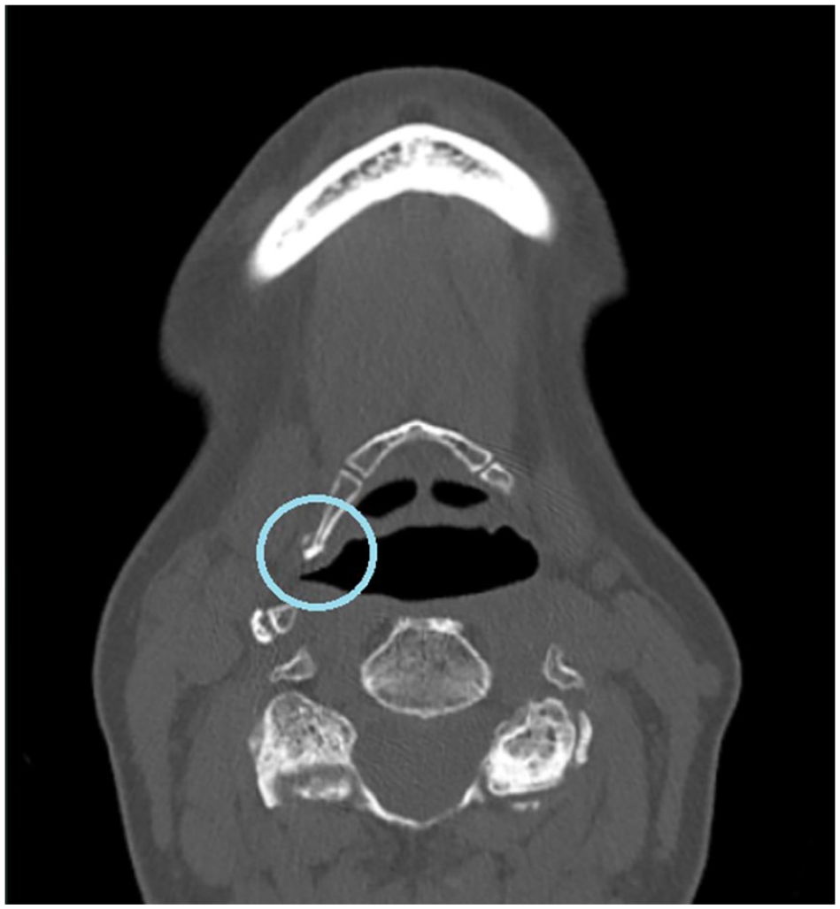
Computed tomography scan performed 8 weeks after injury shows features of osteogenesis at the hyoid bone fracture site (blue circle).
Discussion
The hyoid bone is a well-protected bone in the neck and is not susceptible to fractures due to its secure position surrounded by muscles and its mobility. Fractures resulting from injuries other than strangulation occur sporadically and, according to the literature, constitute only 0.002% of all fractures. 9 Nevertheless, it is crucial to bear in mind the potential occurrence of rare fracture locations and dislocations within the hyoid-larynx complex. 10
Patients with a hyoid bone fracture may present with a range of symptoms, spanning from asymptomatic cases and mild symptoms causing temporary discomfort to life-threatening manifestations. 6 In the case presented, the primary symptom was exacerbated neck pain during speaking and swallowing. It is noteworthy that the patient was an actress and the optimal function of the speech organ held significant importance in her profession.
If there is a clinical suspicion of a hyoid bone injury, it is crucial to conduct both a clinical examination and imaging for an accurate diagnosis. Diagnosis of this condition can be challenging due to its rarity. Performing a flexible nasoendoscopy is recommended as it can help identify pharyngeal lacerations, vocal cord hematoma, edema, and airway obstruction. 2 Cervical CT scan should be performed to confirm the diagnosis, 11 and to detect the presence of other possible bone traumas in neck region. In our case, a neck CT scan demonstrated a displaced hyoid bone fracture with mild edema of the underlying soft tissues with no evidence of an airway compromise.
There are no guidelines for intervention or standard observation time due to the rarity of the hyoid bone fractures. Various clinical manifestations of the fracture require different management strategies. Conservative therapy is appropriate for patients with mild symptoms and no compromise to their airways. This approach involves rest, observation, dietary changes, and appropriate analgesia. Using a neck collar for immobilization to relieve pain may be helpful. For patients without symptoms, only observation and rest are required. In cases of respiratory distress and airway perforation, patients may necessitate endotracheal intubation or an emergency surgical airway procedure, such as tracheostomy. 2 In order to that, all patients with a hyoid bone fracture should be closely monitored for 48 to 72 hours because individuals who were previously asymptomatic may experience a sudden onset of symptoms such as hemoptysis, edema, ecchymosis, and spasm, which can lead to life-threatening asphyxia. 3
The presented case is discussed due to the rare occurrence of isolated hyoid bone fractures, which, however, should be considered in the differential diagnosis of causes of neck pain. They should be taken into account in cases of traffic accidents, blunt neck trauma, sports injuries, and even as a consequence of physical therapy procedures, especially when the patient also experiences neck swelling, hoarseness, or dyspnea. It is worth emphasizing that these injuries can result in life-threatening airway obstruction.
Footnotes
Data Availability
The data that support the findings of this study are available on request from the corresponding author: Karolina Dzaman.
Declaration of Conflicting Interests
The author(s) declared no potential conflicts of interest with respect to the research, authorship, and/or publication of this article.
Funding
The author(s) disclosed receipt of the following financial support for the research, authorship, and/or publication of this article: The authors received no financial support for the research and authorship. APC was funded by the Centre of Postgraduate Medical Education in Warsaw.
Ethical Approval
Ethical approval is not applicable for this article.
Statement of Human and Animal Rights
This article does not contain any studies with human or animal subjects.
Informed Consent
Verbal informed consent was obtained from the patient for their anonymized information to be published in this article.