
Abstract
Primary hyperparathyroidism (PHPT) is a common endocrine disorder characterized by hypercalcemia and elevated or abnormally normal parathyroid hormone (PTH) levels, due to excessive secretion of PTH by 1 or more parathyroid glands. In this report, we discuss the diagnostic and therapeutic challenges posed by ectopic parathyroid adenomas, a rare but atypical presentation of PHPT. We present the case of a 36-year-old female with PHPT due to an ectopic parathyroid adenoma located in the submandibular region. The patient presented with bone pain and was initially evaluated with routine imaging studies, which were negative. [18F] F-choline positron emission tomography (PET)/Computed tomography revealed the ectopic adenoma, leading to successful surgical management. Ectopic parathyroid adenomas are rare but can occur in various locations, and functional imaging modalities such as choline PET can aid in their detection. Surgical resection remains the definitive treatment for parathyroid adenomas, with intraoperative PTH monitoring guiding the extent of resection. Proper evaluation and management of PHPT is essential to avoid significant morbidity. Our case adds to the growing body of literature on the importance of considering ectopic locations of parathyroid adenomas in patients with PHPT.
Introduction
Primary hyperparathyroidism (PHPT) is a common endocrine disorder characterized by hypercalcemia and elevated or abnormally normal parathyroid hormone (PTH) levels due to excessive secretion of PTH by 1 or more parathyroid glands. 1 In most cases, the cause of PHPT is a benign adenoma (80-85%), while hyperplasia and carcinoma are rare causes (10-15% and <1%, respectively).1,2 Ectopic parathyroid adenomas are an unusual cause of PHPT, accounting for less than 5% of cases. 3 They are defined as parathyroid glands located outside their normal anatomic position. 4 PHPT is most frequently caused by a solitary adenoma, which is usually located within the cervical region, but up to 5% of cases can present with ectopic parathyroid adenomas.5,6 Ectopic adenomas can be found in various locations such as the mediastinum, retroesophageal space, thymus, submaxilar space, thyroid gland, carotid sheath, and intrathymic parathyroid gland. 6 Although ectopic adenomas are less common than cervical adenomas, they can pose a diagnostic challenge due to their atypical location and presentation.
Parathyroid adenomas located in ectopic sites can present with different clinical manifestations than cervical adenomas. They may be associated with a higher rate of multiglandular disease, more severe hypercalcemia, and a higher likelihood of being missed on imaging studies.5,7 Delayed diagnosis and treatment of these adenomas can lead to persistent hypercalcemia, recurrent disease, and a higher risk of osteoporotic fractures. 7
In this report, we present the case of a patient with PHPT due to an ectopic parathyroid adenoma. We also discuss the diagnostic and therapeutic challenges posed by this atypical presentation and review the current literature on the management of ectopic parathyroid adenomas.
Case Report
A 36-year-old female presented with bone pain lasting for 1 year, without any other associated symptoms. She had no medical history of lithiasis, gastritis, or family history of hyperparathyroidism. Physical examination was unremarkable. A bone densitometry showed low mineral bone density with a bone mineral density Z-score L1-l4 of −3.1. Initial laboratory investigations revealed low vitamin D (22 ng/mL), high ionic calcium (1.65 mmol/L), high 24-hour urine calcium (423 mg/24 hours), creatinine of 0.57 mg/dL, low serum phosphate (1.4 mg/dL), and elevated PTH (497 pg/mL). A Technetium 99m sestamibi scintigraphy with Single-photon emission computed tomography (SPECT)/CT scan was negative for adenoma, and an ultrasound of the cervical region did not show any suggestive lesions and neither did the neck and chest CT. However, a [18F] F-choline PET/CT scan revealed right submandibular uptake, leading to the diagnosis of an ectopic parathyroid adenoma in the submandibular region (Figure 1).
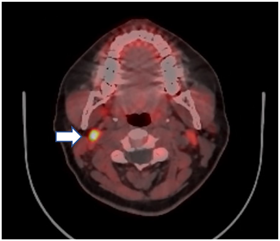
Choline PET image showing right submandibular uptake suggestive of ectopic parathyroid adenoma (white arrow).
The patient underwent surgery, and a 2.5-cm right submaxillary parathyroid adenoma was found (Figure 2). Intraoperative PTH was 650 pg/mL, which decreased to 61 pg/mL at 10 minutes and 40 pg/mL at 15 minutes after adenoma removal. The pathological examination confirmed the presence of a parathyroid adenoma.
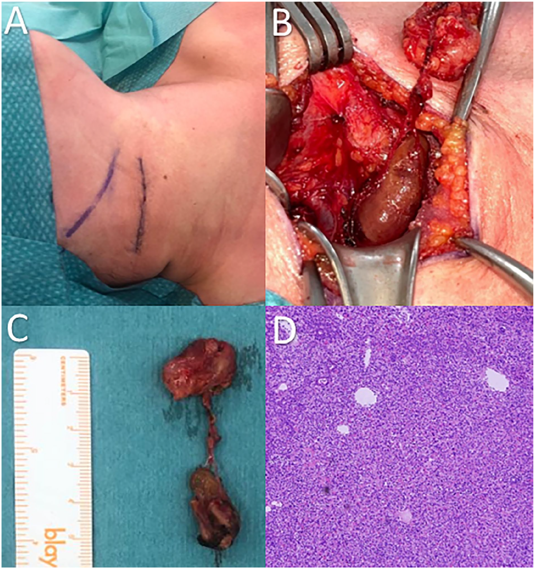
(A) Right submandibular incision for area exploration. (B) Right submandibular parathyroid adenoma. (C) 1.5-cm parathyroid adenoma with associated adenopathy. (D) Image with hematoxylin and eosin staining, magnification 10× and 100×. Parathyroid tissue composed of principal cells with adenomatous change due to the absence of associated adipose tissue.
At 6-month follow-up, there was normalization of PTH and calcium levels, as well as symptomatic improvement in bone pain.
Ethical Component
This case report was carried out within the ethical principles for medical research in humans according to the Declaration of Helsinki—59th General Assembly, Seoul, Korea, October 2008. The national regulations of the Ministry of Health and Social Protection of Colombia Resolution 8430 of 1993 regarding Chapter I “Of the ethical aspects of research in human beings” were taken into account. This research is classified within the research category without risk, and with the informed consent of the patient.
Discussion
Ectopic parathyroid adenomas are uncommon but can develop in various locations. During embryonic development, parathyroid glands normally migrate to the posterior aspect of the thyroid gland. However, in some cases, parathyroid tissue cells become trapped in other regions due to abnormal migration. In the case of submandibular parathyroid adenomas, these cells become trapped in the submandibular region and multiply, eventually forming an ectopic adenoma in that location. 8
In cases where routine imaging is negative, functional imaging modalities such as [18F] F-choline PET/CT can aid in the detection of ectopic adenomas. PET choline is a molecular imaging technique that uses a radiolabeled form of glucose to visualize the metabolic activity of tissues. The utility of PET choline in the identification of parathyroid adenomas is due to its high sensitivity and specificity for detecting small tumors and accurately locating their position in the body. 9
Surgical resection remains the definitive treatment for parathyroid adenomas, with intraoperative PTH monitoring guiding the extent of resection. Our case highlights the importance of considering ectopic locations of parathyroid adenomas in patients with PHPT who have negative imaging studies. The use of [18F] F-choline PET/CT scan in our case proved to be useful in localizing the ectopic parathyroid adenoma, leading to successful surgical management.
In addition, our case emphasizes the need for proper evaluation and management of PHPT, as it can lead to significant morbidity if left untreated. Studies have shown that surgical management of PHPT can lead to a decrease in morbidity and mortality. 10 In conclusion, our case adds to the growing body of literature on the importance of considering ectopic locations of parathyroid adenomas in patients with PHPT. The use of [18F] F-choline PET/CT in our case was a valuable tool in localizing the adenoma, leading to successful surgical management. It is essential to evaluate and manage PHPT appropriately to avoid significant morbidity.
Footnotes
Data Availability
The case information was obtained in an authorized manner by the patient and the treating surgical group.
Declaration of Conflicting Interests
The author(s) declared no potential conflicts of interest with respect to the research, authorship, and/or publication of this article.
Funding
The author(s) received no financial support for the research, authorship, and/or publication of this article.