
Abstract
Carotid body tumors (CBTs) originate from the paraganglionic tissue in the bifurcation of the common carotid artery. Magnetic resonance (MR) imaging is a commonly used diagnostic method in the preoperative diagnosis of these tumors. In this study, we demonstrated an isthmus between the right and left carotid body tumors in a patient with bilateral CBT. The left CBT also was associated with a saccular aneurysm of left external jugular vein.
Introduction
Carotid body tumors (CBTs) arise from the paraganglionic tissue in the bifurcation of the common carotid artery. Sometimes dysphagia and carotid pulse may be associated with the mass. Tumor may cause invasion of carotid artery. However, internal jugular vein invasion is unusual. Bilateral CBT is a rare entity and usually associated with familial forms. 1 Magnetic resonance (MR) imaging is a commonly used imaging technique for diagnosis and preoperative evaluation of these tumors and provides the necessary information for surgical planning. 2 To the best of our knowledge, magnetic resonance (MR) imaging findings of the conjugation via a retropharyngeal isthmus between the right and left CBTs have not been mentioned in the prior literature. We present MR imaging and MR angiography findings of a 61-year-old woman with conjoined CBTs. This rare condition was also associated with a saccular aneurysm of left external jugular vein.
Case Presentation
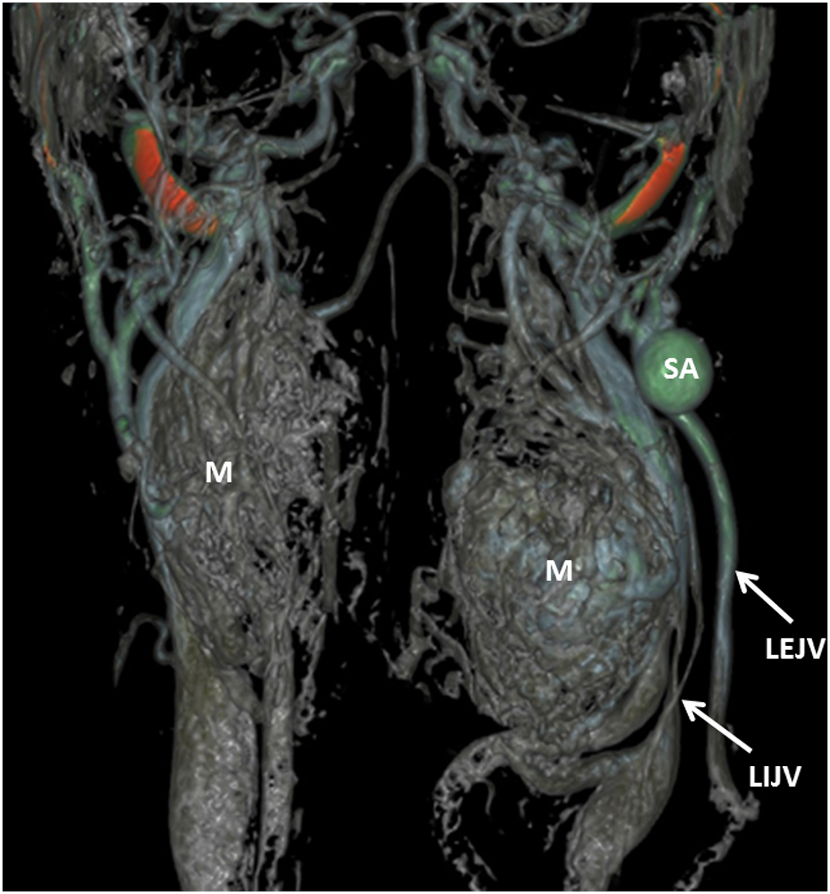
A 61-year-old woman was referred to our radiology department for further examination of dysphagia and hypertension with a 3-month history. The patient had no other medical history. On physical examination, the arterial blood pressure was 180/100 mm Hg in both arms. There was pulsatile swelling in both sides of the neck with palpation. Because of suspicion for a vascular neck mass, MR imaging and MR angiography were performed. MR scans demonstrated bilateral CBTs. MR sequences also showed an isthmic structure between right and left CBTs (Figure 1). On angiographic images, the left internal jugular vein was invaded by left CBT. There also was an aneurysm of the left external jugular vein likely associated with vascular redistribution from left internal jugular vein invasion (Figure 2). Axial post-contrast fat suppressed T1 MR image shows the retropharyngeal Isthmus connection between right and left carotid body tumors (arrows). Images also reveal an invasion and compression in the left internal jugular vein (dashed arrow) and dilated left external jugular vein (arrowhead). Coronal contrast enhanced 3D reformat MR angiography shows bilateral carotid body tumors and left external jugular vein aneurysm. SA = saccular aneurysm, M = carotid body tumor, LIJV = left internal jugular vein, LEJV = left external jugular vein.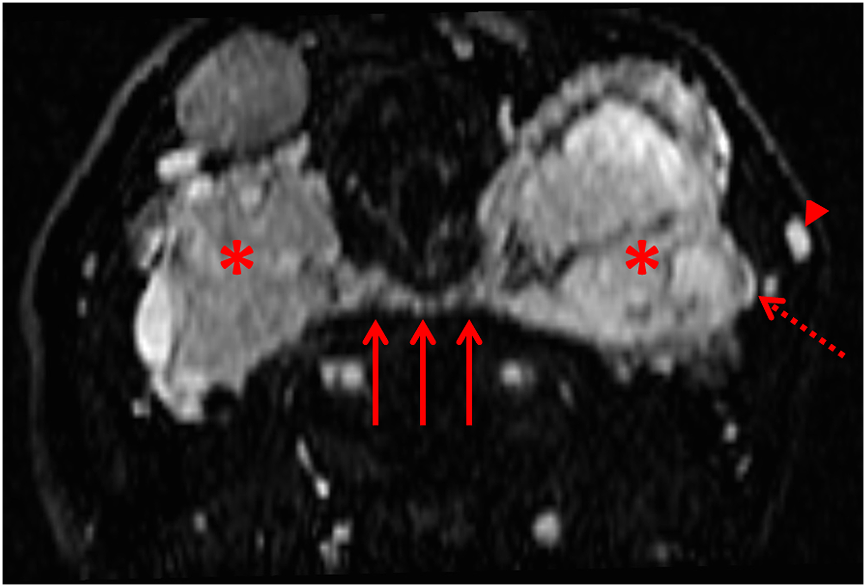
Conclusion
Carotid body tumors constitute more than 50% of head and neck paragangliomas. They often occur as slow-growing masses in the neck. The most common origination (60%) is carotid bifurcation. Additional origins often include jugular bulb, nodose ganglion, vagal nerve, and middle ear. 3 The incidence of CBT is <1: 30,000. The malignant potential of these tumors is about 5% to 10%. 4 They may be familial and bilateral, associated with autosomal dominant inheritance pattern. 5
Clinical findings and various imaging modalities are usually used for diagnosis. Although catheter angiography is the gold standard for the diagnosis and management of CBT, it is an invasive method. A combination of MR imaging and MR angiography provides important information for diagnosis and surgical planning. 3 Nowadays, these imaging techniques are successfully used for this condition.
Although CBTs can invade internal and external carotid arteries, jugular vein invasion is unusual. Moreover, the retropharyngeal fusion of the CBTs across midline is an unexpected condition. In present case (Figure 3), the left internal jugular vein had compression and invasion by the CBT. There also was a saccular aneurysm of the left external jugular vein. In our case, we suspect that the left external jugular vein aneurysm occurred due to vascular redistribution from invasion of the left internal jugular vein. Schematic drawing of the carotid body tumors. Illustration demonstrates an isthmus (arrow) between both carotid body tumors, invasion (dashed frame) of the left internal jugular vein, and saccular aneurysm (asterisk) of the left external jugular vein.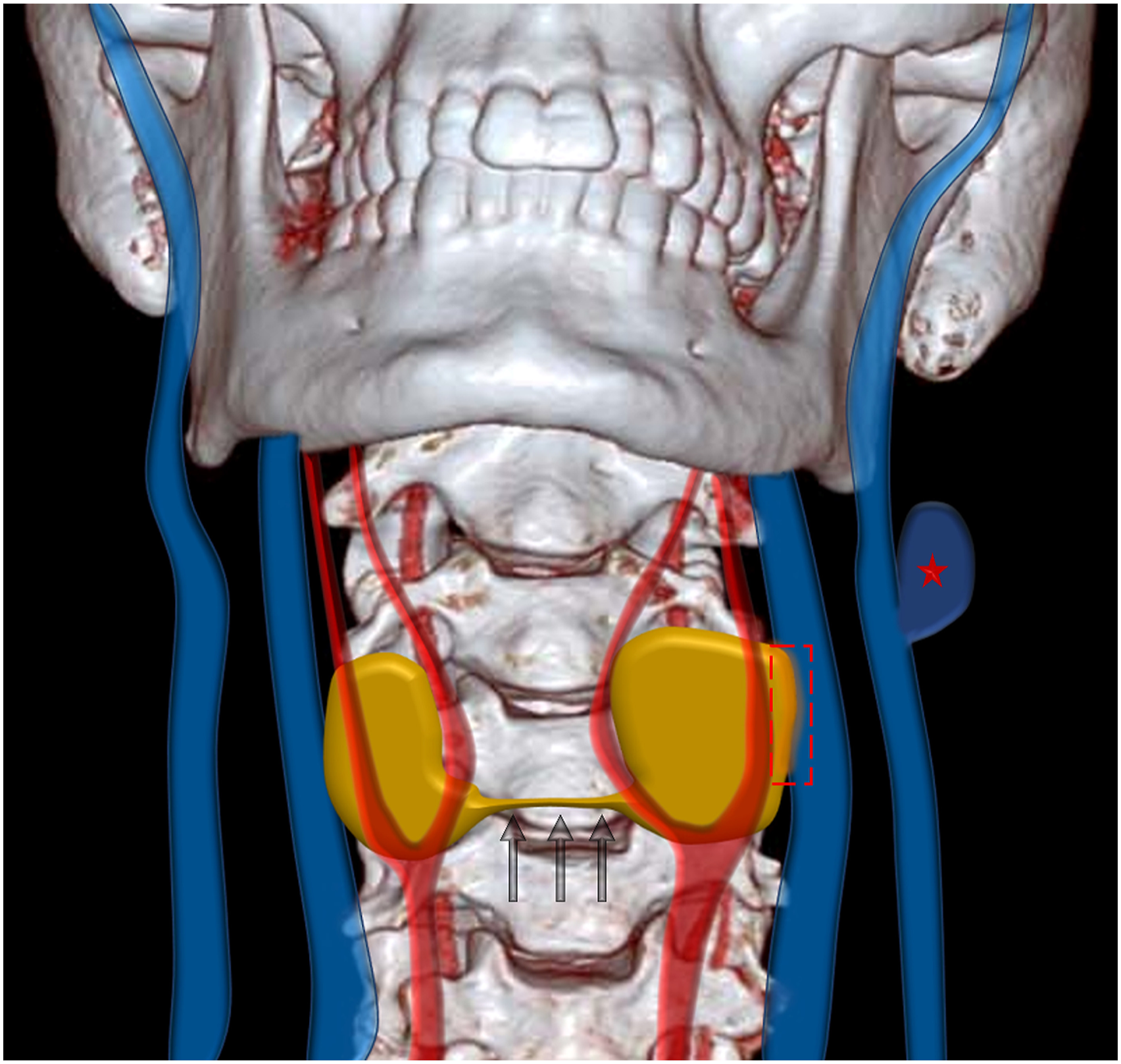
Although it is uncommon, bilateral CBTs diagnosis should always be kept in mind in patients admitted with mass in the neck, and it should be known that other pathologies may be present in these patients.
Footnotes
Declaration of Conflicting Interests
The author(s) declared no potential conflicts of interest with respect to the research, authorship, and/or publication of this article.
Funding
The author(s) received no financial support for the research, authorship, and/or publication of this article.