
Abstract
Cartilaginous choristoma is a rare benign lesion of the external auditory canal, generally found incidentally on physical exam in an asymptomatic patient. Our patient had the largest cartilaginous choristomas described in the literature to date. Additionally, this reviews the nomenclature and differential diagnoses of masses within the external auditory canal.
Case description
A 25-year-old male tenor singer and immigrant from Puerto Rico presented to the office for dysphonia. He was a non-smoker, consumed edible marijuana, and reported no alcohol use.
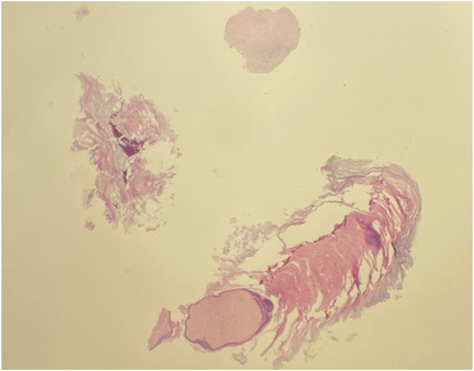
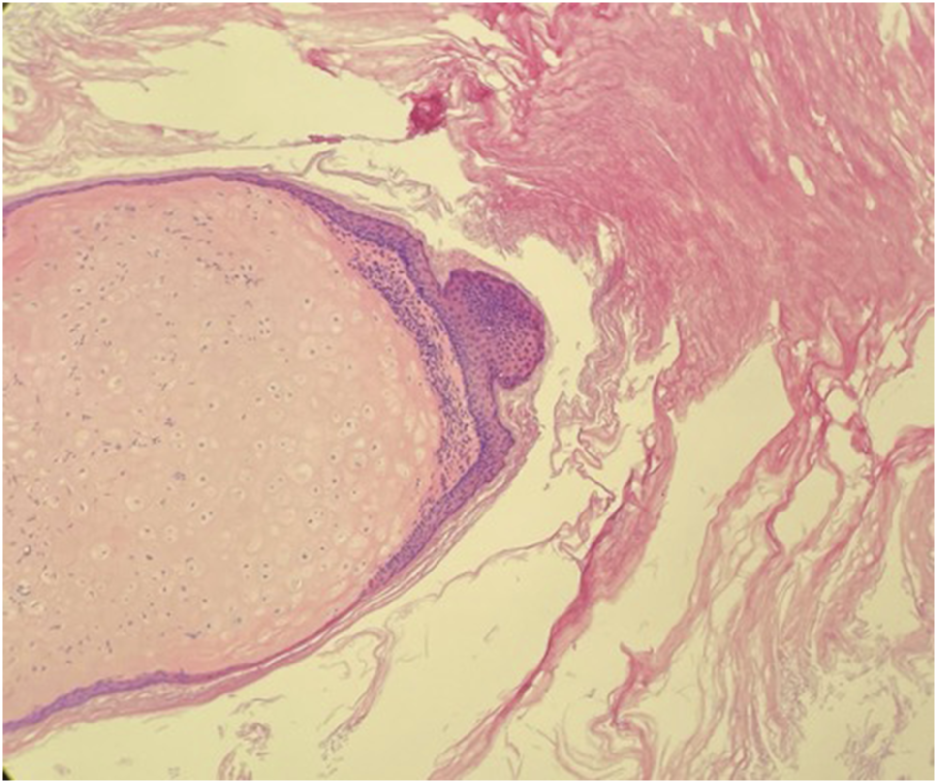
Examination revealed a white, spike-like mass blanketed in cerumen within the right external auditory canal (EAC) protruding from the anterior-superior portion of the bony canal measuring approximately 15 mm × 5 mm (Figure 1). The remainder of the otologic examination was normal in both ears. A CT demonstrated no evidence of cholesteatoma or other abnormalities. The patient underwent a microdirect laryngoscopy with excision of a vocal fold mass, and concurrently, the ear lesion was excised. Pathology analysis on a low-power view revealed focal cartilage tissue surrounded by squamous epithelium with keratin (Figure 2). A high-power analysis also demonstrated cartilage with surrounding squamous epithelium and keratin (Figure 3). No atypia or malignancy was noted. There was no recurrence of the lesion at his 4-month follow-up office visit. Cartilaginous choristoma of the EAC with squamous epithelium and with keratin. Histologic section showing cartilaginous choristoma. Mass shows focal cartilage tissue is surrounded by squamous epithelium and with keratin (original magnification ×2). Histologic section showing cartilaginous choristoma. Mass shows cartilage surrounding squamous epithelium and keratin (original magnification ×20).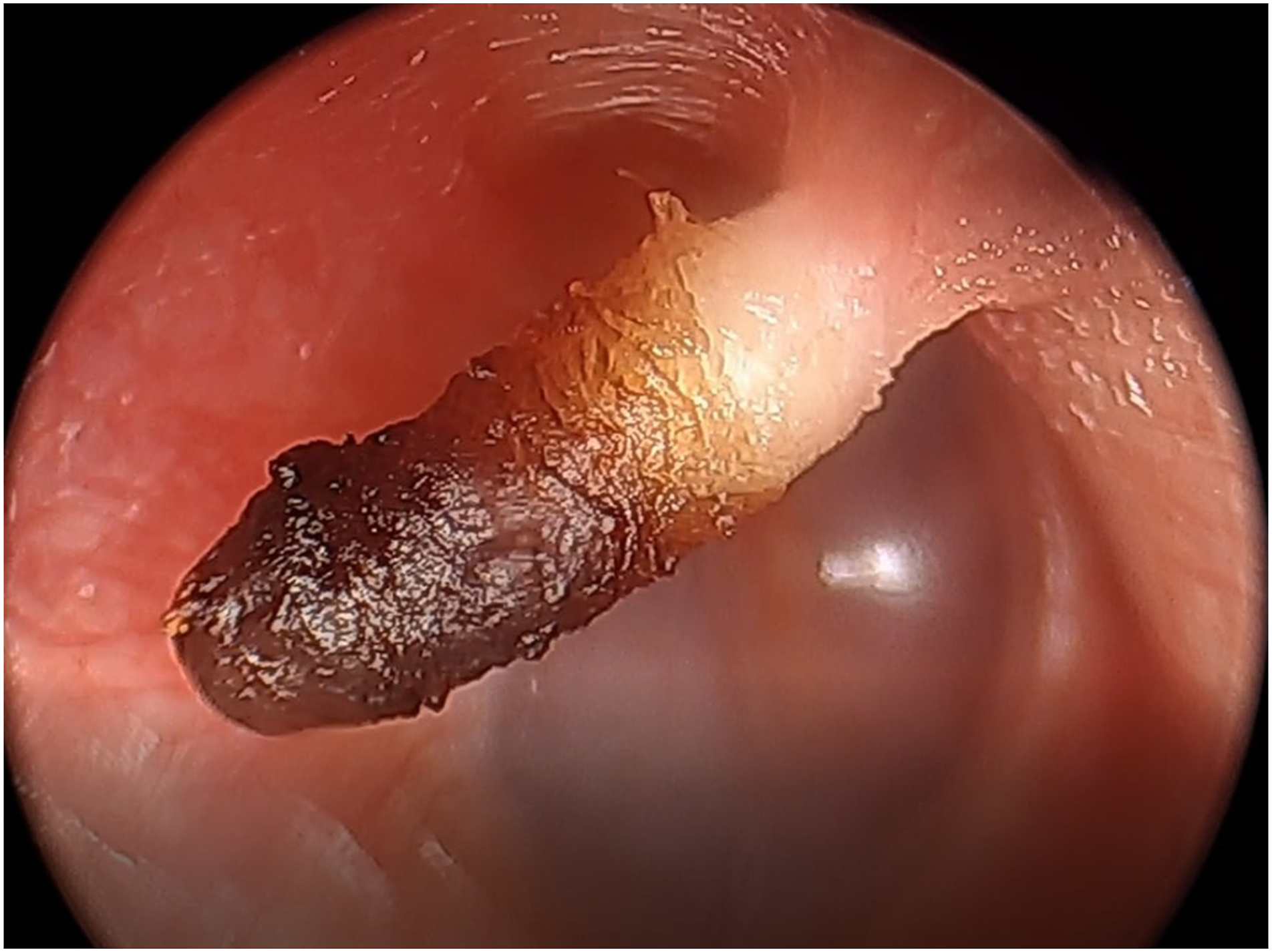
Choristoma is a developmental tumor-like growth of microscopically normal tissue in an abnormal location. The most common type of oral choristoma is composed of bone, cartilage, or both. Cartilaginous choristoma is typically found incidentally in asymptomatic individuals. 1 The lesion described appears to be the largest cartilaginous choristoma reported in the literature.
Historically, cartilaginous lesions of the EAC were previously referred to as chondromas. However, cartilaginous choristoma is the revised nomenclature. The new terminology characterizes the lesion better as a tumor-like growth with histologically normal tissue. 1 -3 Furthermore, the incidental finding of such a mass in an anatomic site different from where one would normally expect to find the tissue in question solidifies the label of choristoma. 1 -3 For choristomas of the EAC, a key differentiating factor is whether the mass originates from the underlying periosteum. 2 A chondroma would be histologically similar (both lesions have normal, non-neoplastic, hyaline cartilage), but it would be in direct contact with the bony portion of the EAC rather than the cartilaginous portion. 2
Grossly, these lesions usually are small (1-2 mm), solitary, white, smooth, and firm. 1,3 The shape can vary from round to club- or horn-shaped. 1,3 Cartilaginous choristoma of the EAC is a rare finding, with only about 50 cases reported in the literature. 1 Choristomas have been reported at other anatomic sites in the head and neck, notably epibulbar and intraoral. 2,4 In the ear, typically they are located at the medial portion of the anterior wall of the EAC. 3 The majority of cases have been diagnosed in the second, third, or fourth decade of life and the vast majority of reported cases occurred in Asians. 1,2 The mainstay of treatment is local excision, and there have been no reported cases of recurrence. 1 That being said, cartilaginous choristomas have little growth potential and there have been no cases of malignant transformation, suggesting that observation is a viable option. 1 -3
There are multiple other lesions that can present similarly and should be included in the differential diagnosis of a solitary mass of the external auditory canal. Exostoses can be an incidental finding in the EAC. However, usually they are found bilaterally in patients with frequent aquatic exposure and are broad-based with complete tympanic bone involvement. 1,3,5,6 Osteomas can also present in the EAC and are typically found attached to the tympanosquamous suture, lateral to the isthmus. 5,6 Another possibility is cholesteatoma, which rarely presents in the EAC. 7 Although a keratin horn usually arises in sun-exposed areas, it has been previously reported as an incidental finding of the EAC. 8 Malignancies also occur in the EAC.
Careful otoscopic examination, review of patient risk factors, and imaging can help diagnose choristomas and differentiate this relatively inconsequential lesion from serious masses.
Footnotes
Declaration of Conflicting Interests
The author(s) declared no potential conflicts of interest with respect to the research, authorship, and/or publication of this article.
Funding
The author(s) received no financial support for the research, authorship, and/or publication of this article.