
Abstract

Otoscopic Clinic
A 63-year-old woman who denied a history of ear trauma, infection, or surgery presented to our ENT clinic with intermittent left-sided aural fullness that had persisted for several years. Otoscopy indicated a bulging soft tissue over the anterior wall of the left EAC (Figure 1A), and the mass vanished when the patient’s mouth was opened (Figure 1B and video). High-resolution computed tomography (HRCT) of the temporal bone revealed a small bony defect in the left inferior and anterior EAC, with a soft tissue herniated from the TMJ into the EAC (Figure 1C). A dynamic physical examination of masticatory movement and radiological imaging indicated spontaneous TMJ herniation into the EAC. (A) Otoendoscopy indicated a protruding mass-like lesion on the anterior wall of the left external auditory canal (EAC). (B) This lesion vanished when the patient’s mouth was opened. (C) Axial view in high-resolution computed tomography of the temporal bone revealing a soft-tissue nodule herniated from the temporomandibular joint via the bony defect of the EAC.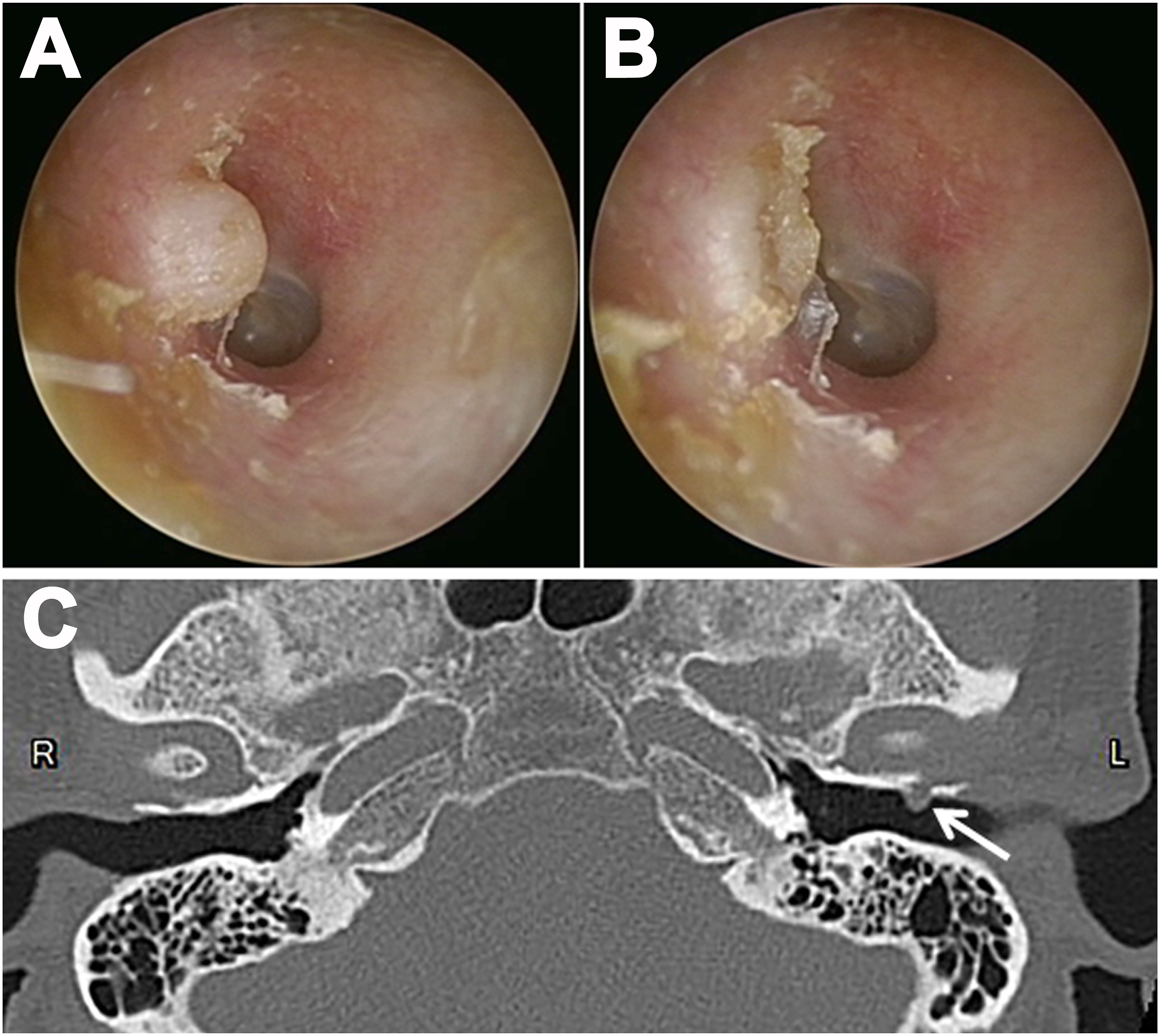
Few studies have reported spontaneous TMJ herniation into the EAC, which was first discovered by German anatomist Hawke and colleagues in the 1980s. 1 The foramen of Huschke, which separates the anteroinferior aspect of the EAC and the TMJ, is an anatomical change in the tympanic segment of the temporal bone. 2 -4 It usually decreases in size after birth and closes at around 5 years of age. 2 -5 If the bony dehiscence persists, the soft tissues of the TMJ may herniate through the patent foramen of Huschke (PFH) into the EAC. Approximately 80% of patients with TMJ herniation through the PFH are over 50 years of age, and accumulated stress from chewing may lead to erosion and may enlarge the foramen with age. 2,5,6 Most patients are asymptomatic, whereas others may present with otalgia, otorrhea, clicking tinnitus, hearing impairment, or aural fullness. 3 -6 The modalities for diagnosis are HRCT, magnetic resonance imaging, or otoscopy. 2 -4 In this case, we used zero-degree ear endoscopy to obtain an excellent view and record the dynamic image during masticatory movement. Surgical intervention with reconstruction of the bony defect is recommended if the patient is symptomatic. 5,6 Studies have reported excellent outcomes of foramen repair through preauricular or endaural surgical approaches. 5,6 However, our patient requested conservative care at follow-up.
Footnotes
Declaration of Conflicting Interests
The author(s) declared no potential conflicts of interest with respect to the research, authorship, and/or publication of this article.
Funding
The authors disclosed receipt of the following financial support for the research, authorship, and/or publication of this article: This work was supported by Tri-Service General Hospital, Grant Number TSGH-D-110081.
Data Availability
The data presented in this study are available on request from the corresponding author.
Significance Statement
Temporomandibular joint (TMJ) herniation into the external auditory canal (EAC) is rare. We describe a case of spontaneous TMJ herniation into the EAC using otoscopic, dynamic, and radiological imaging. The findings of this case report provide reference for clinical physicians to quickly make diagnoses.