
Abstract
Mucosal melanoma arising in the middle ear or eustachian tube is uncommon. We present a patient with hearing loss and otalgia found to have mucosal melanoma which occurred in the eustachian tube with extension into the middle ear cavity and external ear canal. Otologic clinics was consulted and biopsy of the mass located at the external canal was performed to ascertain the pathological diagnosis. The patient refused immunotherapy and surgery instead of undergoing radiotherapy and died from hepatic metastasis 8 months later. The mucosal melanoma originated from the eustachian tube with extension into the external ear canal is exceedingly rare, and the differential diagnosis should be considered for tumors in external ear canal.
Introduction
Mucosal melanoma accounts for only 0.8% to 3.7% of all melanomas, and more than half occur in head and neck region. 1 Of these, the mucosa of the sinonasal and oral cavities are the most commonly involved sites. According to previous reports, the 5-year overall survival for patients with mucosal melanoma ranges from 20% to 35%. 1 Melanoma arising in the middle ear or eustachian tube is exceedingly rare. 2 There are only 21 patients of eustachian tube melanoma reported up to date, 2 we thereby describe a patient with hearing loss and otalgia as the major presenting symptoms in this report. Pathological diagnosis of mucosal melanoma was ascertained by the biopsy from the mass in the external ear canal.
Case Report
A female patient (57 years old) had a 1-year history of intermittent left earache with hearing loss which worsened over time. When the patient presented to otolaryngology clinic, a mass in the left external canal was identified. This lesion was characterized by red-purple tissue with some black content admixed (Figure 1A). Biopsy was taken at this location, and the pathology returned as malignant mucosal melanoma.
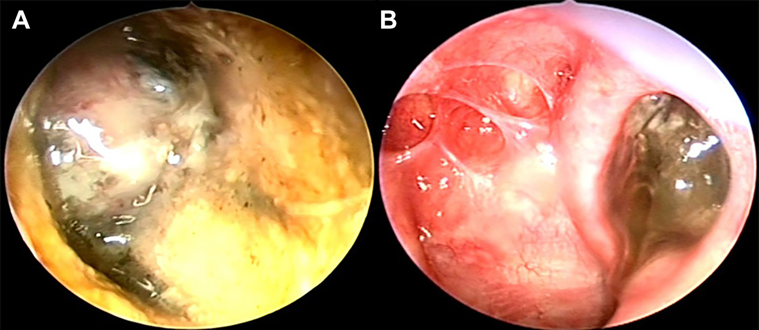
(A) Mass with heterogeneous color in left external ear canal; (B) mass in the left eustachian tube orifice.
Further examination with flexible nasopharyngoscopy showed there was also a mass in the orifice of the left eustachian tube (Figure 1B). Another piecemeal biopsy in the left eustachian tube orifice was performed, and the pathology was also consistent with malignant mucosal melanoma (Figure 2).
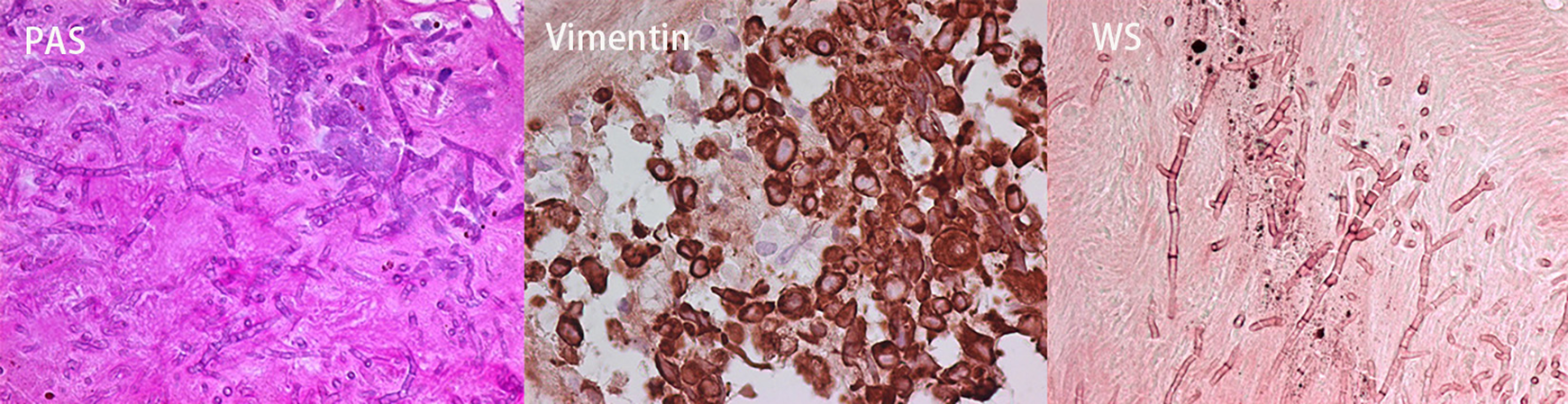
The pathologic manifestation with staining of PAS, Vimentin, and WS, respectively. PAS, Periodic Acid-Schiff; WS, Warthin Starry.
Temporal bone computed tomography (CT) showed that the mass was simultaneously located in the left eustachian tube and left middle and external ear canal (Figure 3A and B). Moreover, the wall of the bony segment of eustachian tube and petrous internal carotid artery (ICA) were infiltrated by the tumor (Figure 3C and D), and the petrous ICA was encroached by the tumor. On magnetic resonance imaging (MRI), the tumor signal was heterogenous in all images; the signal intensity on T1WI was higher than that on T2WI (Figure 4). On positron emission tomography (PET)/CT, the Standardized Uptake Value maximum (SUVmax) for the lesion in nasopharyngeal was 10 and the SUVmax for that in middle ear was 6.1. There were no regional or distant metastases detected.
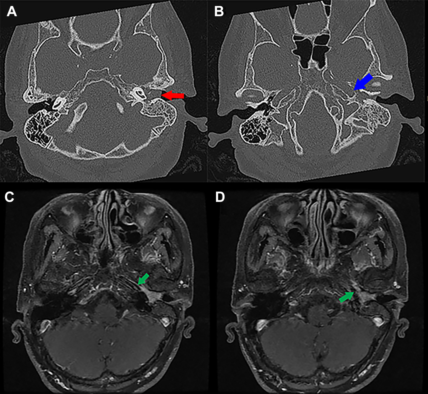
(A) Computed tomography scan of the melanoma in middle ear cavity (arrow); (B) CT scan of the melanoma infiltrating the ICA; (C and D) MRI scan of the melanoma infiltrating the ICA (arrows). CT indicates computed tomography; ICA, internal carotid artery; MRI, magnetic resonance imaging.
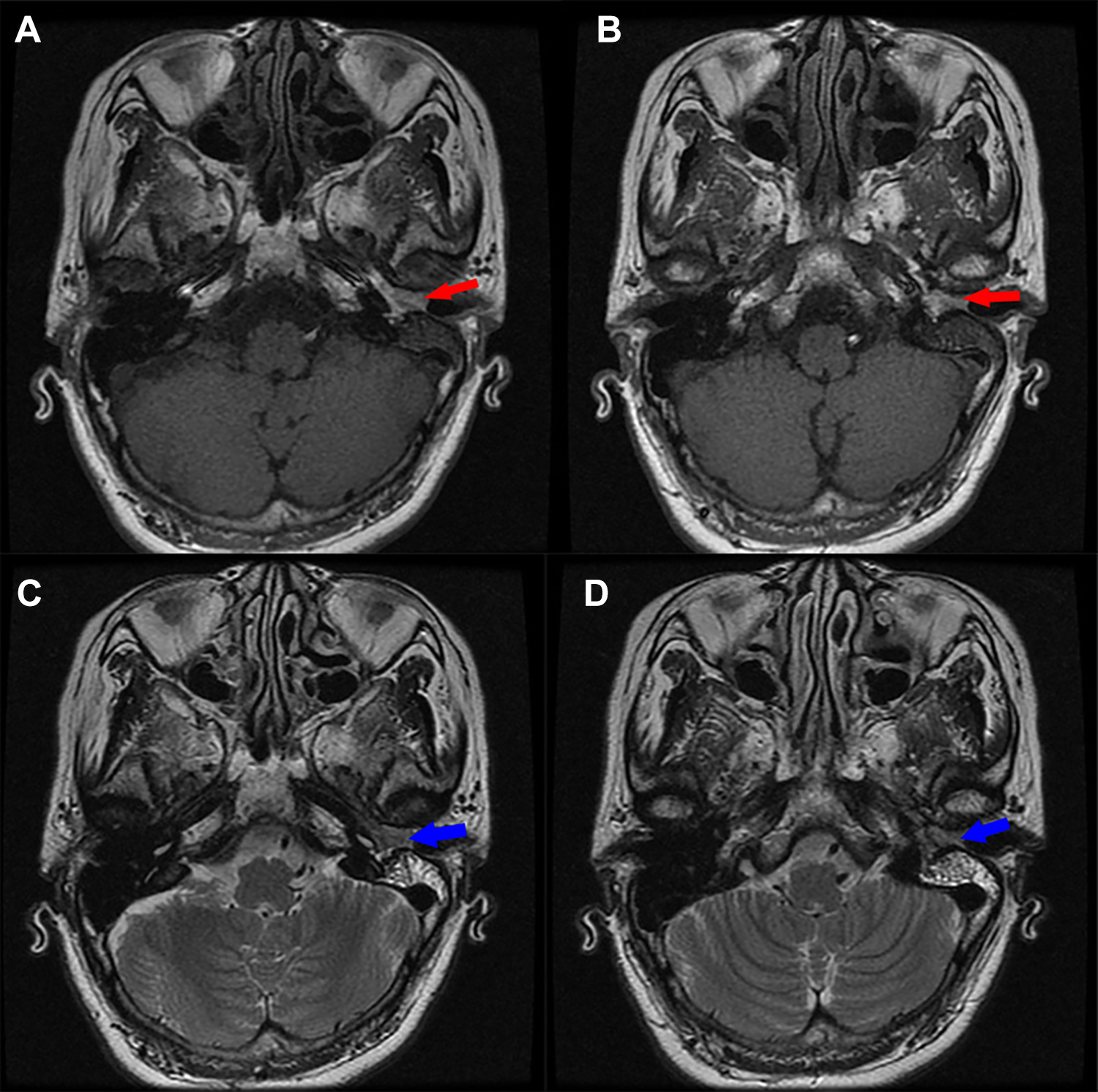
The signal intensity of melanoma on T1WI (A and B, arrows) is higher than that on T2WI scans (C and D, arrows).
The complementary cerebral vascular flow was assessed via digital subtraction angiography examination, and the inverse flow pressure was only 15 mm Hg. The patient could not tolerate ICA balloon occlusion for more than 10 seconds.
The patient refused melanoma-related genetic testing and molecular-targeted immunotherapy and chose to be treated with palliative radiotherapy ultimately and died from hepatic metastasis 8 months after ascertainment of the diagnosis.
Discussion
Mucosal malignant melanoma in head and neck is rare, and there are about 21 cases of mucosal malignant melanoma in eustachian tube and 3 cases with extension into the middle ear were reported. 2 -4 Patients with mucosal melanoma in the eustachian tube frequently present due to the obstructive symptoms such as otitis media or hearing loss. 5 To the authors’ knowledge, this is a first case with otalgia and hearing loss were the major symptoms, and the tumor was first discovered as a mass in the external canal by an otologist. The differential diagnosis of tumors originating in the external or middle ear may include ceruminous carcinoma, squamous cell carcinoma, and glomus tumor, especially coexisted with a dark appearance. 1
The radiological appearance of the melanoma was in accordance with the characteristics reported by Yang et al 6 and Kim et al. 7 On CT scan, the bony part of eustachian tube was partially destroyed, and the petrous ICA was also encased by tumor. The encroachment of the petrous ICA might constitute the contraindication for radical dissection of the tumor. Yang et al also demonstrated that the signal intensity of the lesion largely depends on the concentration and distribution of melanin in the tumor. Therefore, the signal intensity on T1WI was higher than that on T2WI, and a heterogeneous enhancement after contrast.
Wei et al demonstrated, 8 the characteristic markers such as Periodic Acid-Schiff (PAS), Warthin Starry (WS), Vimentin, and Human Melanoma Black (HMB)-45 was positive for mucosal malignant melanoma. The same pathological and immunohistochemical characteristics could also be detected from biopsies both in eustachian tube orifice and ipsilateral external canal, which proved the same origin of tumor in adjacent areas.
With respect to the origin of the tumor, Peters et al reported a case of malignant melanoma in the eustachian tube with extension into the middle ear cavity, 5 and they considered that the eustachian orifice as the original site. The signal intensity on MRI was determined by the content of the melanin. 6 Moreover, the SUVmax in PET/CT may also indicate the vitality of tumor. The higher the SUVmax, the higher degree of malignancy of the tumor. The SUVmax was 10 in eustachian tube orifice, which was much higher than that 6.1 in middle ear cavity. Therefore, we speculated that the eustachian tube was the original site of the melanoma.
Due to the intimate relationship between petrous ICA and eustachian tube, the encasement of petrous ICA by tumor made radical dissection of tumor challenging, especially when the cerebral vascular flow could not be compensatory by the contralateral side. Molecular-targeted immunotherapy, followed by surgical resection, chemotherapy, and carbon-ion radiotherapy were reported as the current treatment strategies for mucosal melanoma. 9,10 As immunotherapy becomes an increasingly used option that one may consider first using immunotherapy and potentially reserving surgery for resectable lesions that didn’t respond well to other approaches. 9,10 However, a surgical resection is still the main and first-line treatment strategy for mucosal melanoma in our institution. The cross-sectional cohort studies to investigate the compositive efficacies for eustachian tube melanomas will be anticipated in future. In conclusion, the mucosal melanoma originated from the eustachian tube with extension into the external ear canal is exceedingly rare, and the differential diagnosis should be considered for tumors in external ear canal.
Footnotes
Authors’ Note
N. R. London holds stock in Navigen Pharmaceuticals which is currently of no value.
Acknowledgments
The authors would like to thank Dr Honggang Liu from the department of Pathology for the assistance of pathological analysis.
Declaration of Conflicting Interests
The author(s) declared the following potential conflicts of interest with respect to the research, authorship, and/or publication of this article: N. R. London was a consultant for Cooltech, Inc.
Funding
The author(s) received no financial support for the research, authorship, and/or publication of this article.