
Abstract
Introduction:
Smoking is a public health problem that has been proven to have adverse effects on human health. Aerobic exercise has positive effects on the human body, especially on the respiratory system.
Objective:
The aim of this experimental animal model study was to determine whether regular aerobic exercise has a protective effect against the harmful effects of cigarette smoke on the nasal mucosa of rats.
Methods:
A total of 24 male Wistar albino rats were randomly separated into 3 groups of 8: group 1 (cigarette smoking), group 2 (cigarette smoking and exercise), and group 3 (control group). At the end of the experiment period, histopathological (light and electron microscopy) and immunohistochemical (GSTA 1, CYP1A1, and CYP2E1) evaluations were made of the nasal mucosa of the animals.
Results:
Goblet cell loss and basal membrane thickening were significantly lower in group 2 and group 3 compared to group 1. In the electron microscope evaluation, the inflammatory expressions of the goblet cells were observed in a very small area in group 2. In group 1, these were distributed over large areas between the mucosal cells. There was seen to be significant swelling of the mitochondria in group 1 compared to the other groups. No statistically significant difference was determined between the groups with respect to GSTA1, CYP2E1, and CYP1A1 scores (P > .05).
Conclusion:
The results of this study showed that regular aerobic exercise has a protective effect against the harmful effects of smoking on the nasal mucosa of rats.
Introduction
Despite development of many methods for stopping smoking and widespread public awareness of this subject, it remains a significant public health problem, and smoking rates are increasing. 1 Cigarette smoke has been proven to cause serious damage to the respiratory system, not only from blood circulation but also directly from the inhaler pathways, which may cause diseases such as heart attacks, strokes, cardiovascular disease, chronic obstructive pulmonary disease, and emphysema. 2
Cigarette smoke is formed from approximately 7000 chemical components known to be potentially carcinogenic. 2,3 Respiratory tract diseases are the result of the first reaction of these toxic substances found in cigarette smoke with the cells and tissues of the respiratory tract. 4 As cigarette smoke can cause many diseases from infection to cancer, it is known to be a significant cause of morbidity and mortality in terms of public health. 5 The most effective means of preventing these harmful effects created by cigarettes is to remain distanced from cigarettes and smoke. However, when this is not possible, some protective precautions should be taken. In this context, regular aerobic exercise, N-acetyl-L-cysteine, vitamin C, β-carotene, and lycopene have been reported to have a protective effect against cigarette smoke. 6 -8
Although many studies have researched the effect of cigarette smoke on the bronchoalveolar system, there are insufficient studies that have evaluated the effects on nasal mucosa. 9,10 The nasal mucosa is the first part of the respiratory tract exposed to irritant substances such as infectious agents, dust, and smoke. 4 Therefore, it is of great importance to examine in detail the harmful effects of smoking on the nasal mucosa. Moreover, there are no studies investigating the effect on the nasal mucosa of methods that may reduce the harmful effects of smoking, such as regular aerobic exercise. The aim of this experimental animal study was to determine through histopathological evaluation and immunohistochemical analysis whether regular aerobic exercise has a protective effect against the harmful effects of cigarette smoke on the nasal mucosa.
Materials and Methods
Experimental Material and Animal Care
Approval for this experimental study was granted by the local ethics committee (Prot. no:16.08.2016-436). All procedures complied with experimental ethical principles and animal protection laws. The study included a total of 24 male Wistar albino rats, aged 10 to 12 weeks, each weighing approximately 250 g. The animals were anesthetized with ketamine hydrochloride (50 mg/kg, intramuscular). They were kept in a room at a constant temperature of 20°C ± 2°C, with controlled humidity of 60% ± 5% under a 12-hour light–dark cycle with ad libitum access to food and water.
Experimental Design and Animal Groups
The rats were randomly divided into 3 groups of 8 rats. The study protocol was developed with reference to previous experimental animal studies that had investigated the effects of smoking and exercise. 11,12 Group 1 rats were exposed to the equivalent of smoke from 15 cigarettes in an isolated cigarette smoke cabin for 30 minutes twice a day, 5 days a week, for 3 months. Group 2 rats were exposed to cigarette smoke at the same rates as the smoking group and were also forced to run at an average speed of 18 m/min on a treadmill (4-path treadmill for rats) for approximately 40 minutes a day, 5 days a week, for 3 months. Group 3 served as the control group and was not administered any treatment. At the end of the 3-month experimental period, the rats were killed. The nasal mucosa samples of all groups were taken from the same nasal septum region. Histopathological and immunohistochemical evaluations were conducted by specialists who were blinded to the experimental groups.
Light Microscopy
Nasal mucosa tissues were removed and fixed in 10% buffered formalin, then dehydrated through graded alcohols, and embedded in paraffin blocks. Sections of 5-µm thickness were cut and then stained with hematoxylin and eosin (H&E), PAS-Alcian blue, and Masson trichrome according to the standard protocols. The specimens were examined using a light microscope (Leica DM6000B; Leica, Wetzlar, Germany) with a digital camera (DC490; Leica) and evaluated according to changes in the type of the epithelium, loss of cilia and goblet cells, and the presence of edema, congestion, and inflammation. Each specimen was scored from 0 to 3 according to the findings, as absent = 0, mild = 1, moderate = 2, and severe = 3. The mean value of the scores was used for statistical analysis.
Electron Microscopy
For transmission electron microscopic examination, the nasal mucosa of the rats was dissected and sectioned. The sections were fixed in 2.5% glutaraldehyde for 48 to 72 hours, postfixed with 1% osmium tetroxide in phosphate buffer (pH 7.4) for 2 hours, dehydrated in increasing concentrations of alcohol, washed with propylene oxide, and then embedded in epoxy resin embedding media. Semi-thin sections of approximately 2 mm in thickness and ultrathin sections of approximately 60 nm in thickness were cut with a glass knife and an ultramicrotome (LKB-Produkter AB, Bromma, Sweden). The semi-thin sections were stained with methylene blue and examined under a light microscope (Nikon Optiphot; Nikon Corporation, Tokyo, Japan). Following this examination, the tissue blocks were trimmed, and ultrathin sections were obtained using the same ultramicrotome and then stained with uranyl acetate and lead citrate. Following staining, the ultrathin sections were examined using a transmission electron microscope (JEM-1200EX; Jeol Ltd, Tokyo, Japan).
Immunohistochemical Staining
Tissues were fixed in 10% buffered formalin and embedded in paraffin blocks. Sections were cut in 4-µm thickness, and 1 section was stained with H&E to observe the tissue morphology. For immunohistochemistry evaluations, endogenous peroxidase activity was blocked by incubating the sections in 1% hydrogen peroxide (vol/vol) in methanol for 10 minutes at room temperature (RT). The sections were subsequently washed in distilled water for 5 minutes, and antigen retrieval was performed for 3 minutes using 0.01 M citrate buffer (pH 6.0) in a domestic pressure cooker. After washing in distilled water, the sections were transferred in 0.05 M Tris-HCl (pH 7.6) containing 0.15 M sodium chloride (Tris-buffered saline [TBS]). The sections were incubated at RT for 10 minutes with super block [SHP125 (ScyTek Laboratories, Inc., Logan, UT, USA)] to block nonspecific background staining. The sections were then covered with the primary antibodies at dilutions of 1:500 for GSTA1, 1:200 for CYP1A1, and 1:500 for CYP2E1 in TBS at 4° overnight. After washing in TBS for 15 minutes, the sections were incubated at RT for biotinylated link antibody (SHP125; ScyTek Laboratories). Then, treatment with Streptavidin/HRP complex (SHP125) was applied. Diaminobenzidine was used to visualize peroxidase activity in the tissues. Nuclei were lightly counterstained with hematoxylin, and then the sections were dehydrated and mounted. Both positive and negative controls were included in each run.
Light microscopy examination of the immunohistochemically stained sections was performed by 2 pathologists blinded to the study groups. The distribution, localization, and characteristics of immunostaining were recorded. A brown color in the cytoplasm and/or nucleus of the epithelial cells was evaluated as positive staining. Scoring was applied by observers blinded to the study groups. Scoring differences between observers were resolved by consensus. For each antibody, the intensity of the reaction was scored as negative (−), weak (1+), moderate (2+), or strong (3+) to describe the immunoreactions.
Statistical Analysis
Data obtained in the study were analyzed statistically using IBM SPSS Statistics v20 software. Descriptive statistics of continuous variables were given as mean and standard deviation values, and categorical variables were given as number and percentage. The χ2 test and Fisher exact test were used to compare categorical variables in the 3 groups. Kruskal-Wallis variance analysis was used for continuous variables. A value of P < .05 was accepted as statistically significant.
Results
Light Microscopy
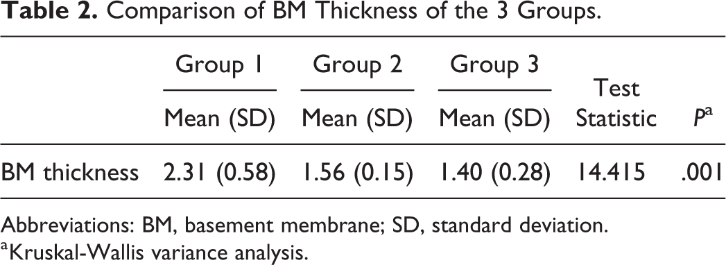
In group 1, reactive changes secondary to inflammation were observed. In some areas, the respiratory epithelium was replaced by stratified squamous epithelium. Mitosis was observed in the epithelium. There was severe loss of cilia and goblet cells. The existing goblet cells were filled with secretion indicating hyperactivity. Moderate congestion and severe inflammatory cell infiltration were observed in the lamina propria. Intraepithelial lymphocyte and neutrophil infiltration was also observed. The basal membrane was 2.359-µm thick in group 1.
The loss of cilia and goblet cells was mild in group 2. Moderate congestion was observed, and in some areas, the blood vessels were filled with inflammatory cells. Although infiltration was observed both in the lamina propria and in the epithelium, the infiltration was less in the epithelium. The mean thickness of basal membrane was 1.564 µm.
In group 3, the nasal mucosa showed normal characteristic features with goblet cells within pseudostratified ciliated columnar epithelium overlying the lamina propria containing glands, blood vessels, and nerve fibers. The mean thickness of basal membrane was 1.383 µm. The arrangement and organization of collagen fibers in the lamina propria was detected to be similar in all groups under Masson trichrome staining (Figure 1).

Different light microscopic appearance of nasal mucosa in groups. (A) Group 3; (B) group 1; (C) group 2 (I: hematoxylin–eosin X400; II: PAS-Alcian blue X1000; III: Masson trichrome X400). Stratified squamous epithelium in group 1. Severe neutrophil and lymphocyte infiltration in lamina propria and epithelium both in group 1 and group 2 (white arrows). Goblet cells filled with secretion in group 1 and group 2. Thicker basement membrane in group 1 (black arrow).
Goblet cell loss was determined to be at a statistically significantly lower level in group 2 and group 3 compared to group 1 (P < .05). The levels of inflammation and vascular congestion were statistically significantly lower in group 3 than in group 1 and group 2 (P < .05). The levels of cilia loss were statistically significantly lower in group 3 than in group 1 (P < .05) and were lower in group 2 than in group 1 but not at a statistically significant level (P > .05; Table 1). The thickness of the basal membrane was significantly greater in group 1 than in both group 2 and group 3 rats (P < .01; Table 2).
Comparison of Ciliary Loss, Goblet Loss, Vascular Congestion, and Inflammation Scores of Rats in the 3 Groups.
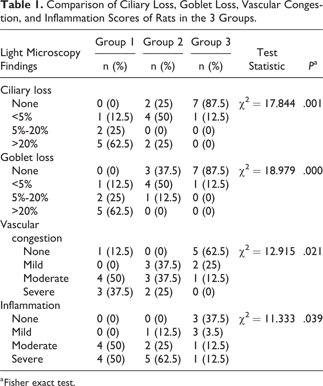
a Fisher exact test.
Comparison of BM Thickness of the 3 Groups.
Abbreviations: BM, basement membrane; SD, standard deviation.
a Kruskal-Wallis variance analysis.
Expression Profiles of GSTA1, CYP1A1, and CYP2E1
No statistically significant difference was determined between groups 1, 2, and 3 with respect to the GSTA1, CYP1A1, and CYP2E1 scores (P > .05; Table 3).
Comparison of GSTA1, CYP2E1, and CYP1A1 Scores of Rats in the 3 Groups.
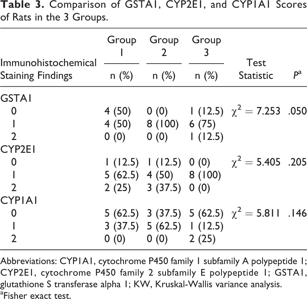
Abbreviations: CYP1A1, cytochrome P450 family 1 subfamily A polypeptide 1; CYP2E1, cytochrome P450 family 2 subfamily E polypeptide 1; GSTA1, glutathione S transferase alpha 1; KW, Kruskal-Wallis variance analysis.
aFisher exact test.
Transmission Electron Microscopy
In group 1, vacuoles were observed inside the cytoplasm of nasal mucosa cells. The inflammatory secretions of the goblet cells were distributed in large areas between the nasal mucosal cells. Swelling of the mitochondria was a prominent finding in this group. The other organelles and cell nuclei were observed to be normal ultrastructurally.
In group 2, vacuoles inside the cytoplasm of nasal mucosa cells were observed to be smaller in size than in group 1. Pathological changes in the mitochondria were also less prominent than in group 1. Inflammatory secretions of the goblet cells were observed at a small amount in a very few areas. The other organelles and cell nuclei were normal ultrastructurally (Figure 2).
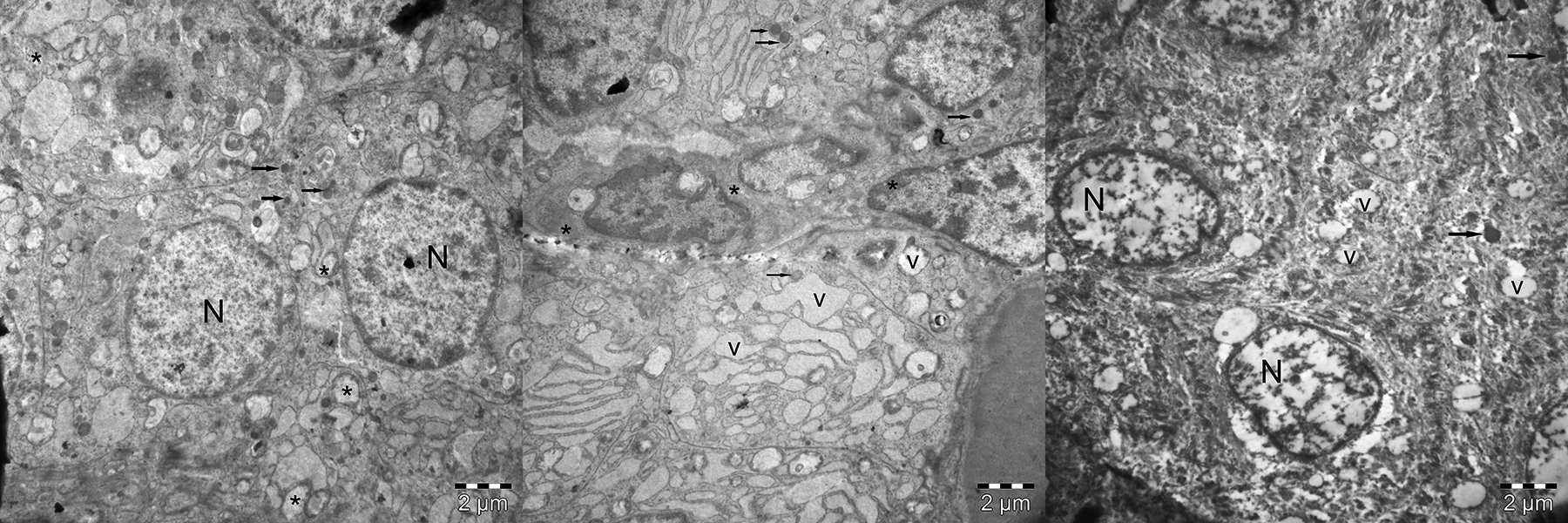
Different electron microscopic appearance of nasal mucosa in groups. Left (group 3): euchromatin nuclei (N) of the nasal mucosal cells and their organelle-rich cytoplasm were seen. The mitochondria (arrows) which were the first organelles effected in stress conditions were also seen normal. The cells contained many secretory granules (*). Scale bar: 2 µm. Middle (group 1): Vacuoles (v) were observed inside the cytoplasm of nasal mucosa cells. The inflammatory secretions of the goblet cells (*) distributed to huge areas in between the nasal mucosal cells. In this group, swelling of mitochondria (arrows) is one of the prominent findings. Scale bar: 2 µm. Right (group 2): Vacuoles (v) were observed inside the cytoplasm of nasal mucosa cells were smaller than the group 1. However, swollen mitochondria (arrows) were same with the group 1. Scale bar: 2 µm.
In group 3, the ultrastructure of the nasal mucosa was observed to be normal. The nuclei of the mucosal cells contained euchromatin more dominantly. The nuclear membranes were normal ultrastructurally. The cytoplasm of the mucosal cells was observed to be rich in organelles, which was a normal finding of the cell. The goblet cells were also normal ultrastructurally. The mitochondria, which are the first organelles affected in stress conditions, were also normal ultrastructurally. The nasal mucosal cells contained many secretory granules.
Discussion
Two important results emerged from this study: (1) the negative effects of smoking were evident on the nasal mucosa and (2) regular aerobic exercise diminishes some of the negative effects of smoking on the nasal mucosa. This study is one of only a few studies showing that smoking triggers all inflammatory and carcinogenetic events on the nasal mucosa and disrupts the mucociliary functions of the nasal mucosa. The nasal respiratory epithelium is basically formed of ciliary cells, goblet cells, striated cells, and basal cells. Irritants such as cigarette smoke cause an impairment in nasal physiology by creating hyperactivity in ciliary cells and causing the loss of goblet cells. In a previous study conducted on rats, it was reported that mucous hypersecretion could be determined within a short period, such as 4 weeks. 13 Increased secretion in the seromucinous glands is known to have a protective purpose by washing the nasal mucosa. However, this increase in secretion also creates a predisposition for inflammatory events as it forms mucous stasis at the same time. 14,15 The fact that mucus hypersecretion, which has a protective role in the early period after smoking exposure, actually triggers inflammatory events in the long term suggests that mucus hypersecretion may be an indirect indicator of smoking. In this study, objective evaluation criteria such as goblet and ciliary cell loss, vascular congestion, severity of inflammation, and basal membrane thickness were determined directly by both light and electron microscopy methods. Thus, the study can be considered of great importance in that it directly demonstrates the harmful effects of smoking on the nasal mucosa methodologically.
Nasal septum and sinus mucosa have different histopathological features. Therefore, they may exhibit different histopathological behavior in different pathological conditions. The nasal mucosa is thicker than the sinus mucosa and contains more goblet cells. 16 In this study, nasal septum mucosa was obtained in all patients in order to provide standardization between the groups due to this feature of the sinus mucosa and easy sampling.
Cilia are organelles found in almost all cells of the human body, especially in the respiratory tract. With the effect of mucosal secretions and cilia, nasal mucociliary clearance is a protective property that is effective in the elimination of dust particles and foreign bodies in the nasal mucosa from the nasal cavity toward the nasopharynx. By disrupting this protective property and increasing inflammation, cigarette smoke has an important role in the development of inflammatory diseases such as asthma and chronic rhinosinusitis. The results of the current study support the studies of cigarette smoke by Paul et al and Trombitas et al, showing a significant level of loss of ciliary cells, which are known to have a protective effect against infectious diseases. 11,14,17
The results of the study demonstrated that regular aerobic exercise partially improved the negative effects of smoking in rats by reducing goblet cell loss and basal membrane thickness. Regular aerobic exercise has been proven to have positive effects on the brain and cardiac and respiratory systems. 6 In addition, there are known to be changes during exercise such as vasoconstriction of nasal mucosa, increasing ala nasi muscle activity, increased nasal air flow, and hyperventilation. One of the systems in the body showing the greatest change during exercise is the respiratory system. Important changes are seen in the nose, which has the greatest resistance in the respiratory tract during exercise. 18,19 In a previous study, it was reported that exercise increased mucociliary clearance in smokers and thus reduced the development of infection. However, no histopathological effect was reported in that study. 20 Although there are many studies showing that the effects of cigarette smoke on lung tissue can be prevented by regular aerobic exercise, there is no study evaluating the effect on the nasal mucosa, which is in the entry to the airways. 21 -23 To the best of our knowledge, this is the first experimental animal study in the literature to demonstrate that regular aerobic exercise can prevent the harmful effects of smoking on the nasal mucosa. The light and electron microscopy findings of the current study show that cigarette smoke increased activity in the secretion cells present in the nasal mucosa, with subsequent widespread mucous pooling formed as a result of the excessive secretion. It has been proven that regular aerobic exercise reduces secretion hyperactivity and has a protective effect against infections. There are many studies in the literature investigating the effects of exercise on rhinitis and reporting different results. 24 -27
Cigarette smoke is also known to cause malignancies with intracellular organ damage. 2 Prevention of exposure to cigarette smoke is the most effective way to prevent both infectious and malignant diseases. However, since this is not always possible, it is important to find other methods to reduce the harmful effects of smoking. Previous studies examining this subject have reported N-acetyl-L-cysteine, vitamin C, β-carotene, and lycopene to have a protective effect against cigarette smoke. 7,8 Similarly, in the current study, regular aerobic exercise has a protective effect against the harmful effects of smoking on the nasal mucosa.
In previous histopathological studies, cigarette smoke has been reported to cause histopathological findings such as basal membrane thickening, proliferation, and irregularities around tumor cells. 28 Basal membrane thickening is known to be a pathological condition caused by carcinogenic agents in addition to irritants such as smoke. 29 In the literature, there are studies showing that smoking can cause nasal cancer as well as lung cancer. 30 -33 In the cigarette smoking group of the current study, there were some significant reactive changes secondary to inflammation, and in some areas, there were multiple layers of squamous epithelium in place of the respiratory epithelium and this had caused mitosis in the epithelium in addition to a significant degree of basal membrane thickening. However, with regular aerobic exercise, the basal membrane thickening was found to have significantly decreased. Therefore, the results of this study demonstrate that regular aerobic exercise may not only reduce infection but also reduce the risk of malignancy by preventing the histopathological changes created by cigarettes.
Greiser et al reported that cigarettes are a significant risk factor for nasal carcinomas. It was stated that the reason that stopping smoking significantly reduced the risk of malignancy developing was associated with an increase in physical activity that occurred together with stopping smoking. 34 Similarly, in the current study, there were seen to be positive effects of regular aerobic exercise on the prevention of infection and the development of carcinoma, despite the continuation of smoking.
GSTs protect cells against toxic agents and form a defense mechanism against carcinogenesis created by chemicals. Cigarette smoke has been shown to induce cytochrome P450 activity in the liver and other tissues. Expression of CYP and changes in CYP activity cause an increase in oxidative stress and are risk factors for cancer development. It has also been reported that cigarette smoke causes a reduction in antioxidant enzymes such as glutathione peroxidase and superoxide dismutase. It was reported in a previous study that CYP2E1 is expressed in nasal mucosa in addition to the liver, lungs, kidneys, and other organs. 35 Another study stated that cigarette smoke increased expression of these enzymes in the liver and lungs, but there was not the same effect in nasal mucosa.
Chronic exposure to cigarette smoke is known to cause an increase in nasal cavity cancers. 36 The results of the current study did not show that CYP1A1, CYP2E1, and GSTA1 enzymes were expressed in nasal mucosa with cigarette smoke in a 3-month period. On the premise of the information that chronic cigarette smoke exposure leads to malignancies, it can be considered that a longer period of exposure is needed for these enzymes to be expressed in the nose. The most important limitation of the study is the low number of subjects, as it was an experimental study with a rat model. In addition, the absence of an exercise control group is another limitation of the study in terms of the inability to evaluate the positive effects of exercise on the normal mucosa. Therefore, further studies are needed to support or disprove these results.
Conclusion
The results of this study demonstrated that aerobic exercise had a protective effect against the histopathological changes induced by cigarettes on the nasal mucosa of rats.
Footnotes
Acknowledgments
The authors declared no conflict of interest and received no financial support for the study.
Declaration of Conflicting Interests
The author(s) declared no potential conflicts of interest with respect to the research, authorship, and/or publication of this article.
Funding
The author(s) received no financial support for the research, authorship, and/or publication of this article.