
Abstract
Radiotherapy is a method of treatment used on malignant head and neck tumors; however, it may lead to adverse effects by influencing other tissues because its effects are not specific to tumor tissues. These adverse effects limit the effectiveness of the treatment and sometimes lead to termination of the treatment. This study aims to histopathologically and biochemically investigate the protective effect of whortleberry against the cellular degeneration and oxidative stress that take place in salivary glands due to radiotherapy. The rats were divided into 6 groups. One group was given radiotherapy only, one group was given radiotherapy and 100 mg/kg of whortleberry, and one group was given radiotherapy and 200 mg/kg of whortleberry. The remaining 3 groups were designated as whortleberry, sham, and control groups. At the end of the study, samples collected were histopathologically and biochemically analyzed. In the group given radiotherapy only, acinar areas were reduced histopathologically, whereas ductal areas increased (P < .01). Oxidative stress increased only in the group given radiotherapy, whereas the oxidative stress levels in the other groups were close to those in the control groups. In conclusion, whortleberry reduces cellular degeneration and oxidative stress that take place in salivary glands due to radiotherapy.
Introduction
Radiotherapy is a method of treatment used on malignant head and neck tumors; however, it may lead to adverse effects by influencing other tissues because its effects are not specific to tumor tissues. These adverse effects limit the effectiveness of the treatment and sometimes lead to termination of the treatment, most commonly including xerostomia, dysphagia, oral mucositis, taste problems, and increases in oral infections. 1,2 Although salivary glands are highly radiosensitive, unlike other radiosensitive tissues, they have slow proliferation and consist of cells that show a high degree of differentiation. According to the data obtained in animal experiments analyzing the effects of radiotherapy on salivary glands, the most significant finding is reduction in saliva flow and gland weight. 2,3 The 2 most important causes of radiotherapy-related adverse effects are direct DNA damage to healthy tissue created by radiation and the damage induced by oxygen radicals formed indirectly by radiation. 4 Various protective drugs have been used in attempts to prevent damage to salivary glands. These drugs usually demonstrate antioxidant characteristics against the damage caused by radiotherapy. 5,6 Additionally, the effects of drugs such as amifostine have been clinically demonstrated. 7
Whortleberry (Vaccinium myrtillus) is a member of the genus Vaccinium, which is a genus of bilberry that contains over 200 species. It is known to have high contents of anthocyanin, flavonoids, and phenolic acid. It is known that these anthocyanins, flavonoids, and other phenolic acid compounds have several biological effects, including antioxidant, antimutagenic, antineoplastic, anti-inflammatory, antihypertensive, antihyperlipidemic, antiproliferative, and antimicrobial effects. Because of all these biological effects, the interest in whortleberry is increasing daily, and studies are being conducted for its usage in various areas. 8,9
The purpose of this study is to histopathologically examine the protective effects of whortleberry on radiotherapy-related damage to salivary glands and biochemically investigate oxidative stress parameters as mechanisms of damage occurring locally in the salivary glands and systemically in the blood.
Materials and Methods
Permission was received from the Experimental Animals Ethics Board of Recep Tayyip Erdogan University to conduct this study (decision no: 2015/18). The rats used for the study were obtained from Recep Tayyip Erdogan University Basic Medical Sciences Experimental Animal Practices Unit, and the entire study was conducted in this laboratory. This work was supported by Recep Tayyip Erdogan University Scientific Research Project Unit under the project number of 282.
Study Protocol
The study included 48 adult male Wistar albino rats weighing 250 to 280 g. The rats were randomly divided into 6 groups of 8 rats. The groups and treatments are shown in Table 1. All invasive procedures were carried out under anesthesia. For anesthesia, 50 mg/kg of intraperitoneal (IP) ketamine hydrochloride (Ketalar; Eczacıbaşı Parke-Davis, Istanbul, Turkey) and 10 mg/kg IP xylazine HCl (Alfazyne; Alfasan International B.V., Woerden, the Netherlands) were used. The rats’ rectal temperatures were regularly measured under anesthesia, and a warm blanket was laid on them to keep their body temperatures at approximately 35°C. The rats were fed and maintained in a 12/12-hour light/dark cycle, 22°C (3°C) room temperature, and a humidity rate of 55% to 60%.
Groups and Implemented Treatments.
Abbreviation: IP, intraperitoneal.
On the tenth day, after 12 hours following the treatment of the groups, intracardiac blood samples were collected from the rats under anesthesia for biochemical analyses, and the rats were killed. Submandibular salivary glands were dissected and removed for histopathological and biochemical analysis.
Preparation of Whortleberry Extract
The whortleberry extract, which was prepared as a dietary supplement to include 330 mg of whortleberry extract in 1 mL with a half-and-half mixture of distilled water and ethanol (Bilberry [V myrtillus] Herbal Liquids; Health Aid, Harrow, United Kingdom), was again diluted in a half-and-half mixture of ethanol and distilled water under sterile conditions to obtain 50 mg of extract in 1 mL.
Radiotherapy Treatment
Planning tomography scans were performed on the rats before radiotherapy treatment, and their conformal planning was carried out in the planning system (CMS Xio, Ver. 5.0; Elekta, Stockholm, Sweden). With a linear accelerator (Elekta Synergy), a total of 8 Gy of external radiotherapy was applied in one fraction using 6 MV of energy on the neck area of the rats at skin source distance (SSD) 100 cm from the anterior area and using a 0.5 bolus under anesthesia.
Histopathological Analysis
In all groups, the submandibular gland tissues of the rats were given code numbers and put into bottles containing 10% neutral formaldehyde. After 72 hours, the following tissue process was applied for these tissues: dehydration in an increased alcohol series, clearing through a xylene series, immersion in liquid paraffin, and embedding in paraffin blocks. From the paraffin blocks of each rat, four 5-μm serial sections with intervals of 50 μm were taken using a microtome (Leica RM2125RT, Nussloch, Germany).
The obtained sections were brought from deparaffinization to the water and stained with hematoxylin and eosin (H&E). Later, the cover-slipped sections were photographed with a camera attached to a light microscope (Nikon Eclipse E600, Tokyo, Japan). Management of the same light settings was performed for photographing, especially in the histochemical analysis, in order to permit unbiased evaluation.
In this study, the mean granular duct and seromucous acinus areas were calculated using the nucleator method, one of the stereological methods with an unbiased counting frame. The Stereo Investigator (MicroBrightField 9.0; Colchester, Vermont, California) software system was used. This system consists of a camera attached to a light microscope, a motorized system that carries a microscope tray, and a computer with a software system. The H&E-stained sections were put on the microscope tray, and their sectional boundaries were determined using this program. After determining the area, frames separated from each other were determined by systematic random sampling of the sections, according to the rules of space fragmentation with the step interval of the x- and y-axes. Then, in 20 different selected areas, the mean areas of the seromucous acini and granular ducts of all groups were measured following the method described by Gundersen et al. 10
Biochemical Analysis
Intracardiac blood samples were collected from all groups under anesthesia at the end of the study. The blood samples taken for biochemical analyses were centrifuged at 3500 rpm for 5 minutes and then serum samples at the top were collected. Submandibular gland tissues were washed, then homogenized by keeping them in a serum buffered by phosphate (pH 7.4) for 1 minute. The tissues were then centrifuged at 4500 rpm for 20 minutes, and samples were taken for analysis. The total antioxidant statuses (TAS) and total oxidant statuses (TOS) of the collected blood samples were measured at the Faculty of Medicine’s Biochemistry Laboratory at Recep Tayyip Erdogan University using an autoanalyzer (Abbott C16000; Abbott Diagnostics, Abbott Park, Illinois) and Rel Assay TAS and TOS test kits (Mega Tıp, Gaziantep, Turkey).
Total antioxidant status measurement is based on the principle of loss of color in the colorful 2,2′-azino-bis(3-ethylbenzothiazoline-6-sulfonic acid) cationic radical in proportion to the ratio of all antioxidant molecules in a sample, as a result of the radical being reduced by the antioxidant molecules. The results are expressed in units of mmol Trolox-equivalent/L. 11
Measurement of the total oxidant levels was made using the colorimetric method based on the principle of ferrous ion being cumulatively oxidized into ferric ion by oxidant molecules in the sample. The results are expressed in units of μmol H2O2 equivalent/L. 12 The ratio of the TOS value to the TAS value was determined as the oxidative stress index (OSI).
Statistical Analysis
The data were analyzed using SPSS version 15.0 for Windows (SPSS Inc, Chicago, Illinois). The results are presented as mean (standard deviation). Comparisons among the groups were made using 1-way analysis of variance and post hoc Bonferroni tests. P < .05 was accepted as the statistically significant level.
Results
Histopathological Analysis Results
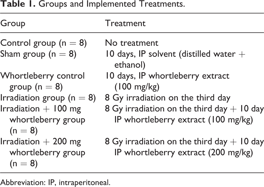
In the submandibular gland tissues of the control group, the structure of the seromucous acini, ducts, and connective tissue was observed to be normal (Figure 1A and B). The submandibular gland histology in the sham (Figure 1 and D) and whortleberry control groups (Figure 1E and F) was normal in structure; the seromucous acini, ducts, and connective tissue were normal. In the irradiation group, intense acinar atrophy and the presence of intense substation of the parenchyma by fibrous tissue were detected. Moreover, excessive vacuolization of glandular cells was observed. In light microscopic sections obtained from the submandibular gland tissue of individuals belonging to irradiation groups, the submandibular tissues contained eosinophilic secretor granular material and glandular fibroses. Remarkably, intense acinar atrophy and glandular fibrosis were detected (Figure 1G and H). In the irradiation + 100 mg whortleberry (Figure 1I and J) and irradiation + 200 mg whortleberry (Figure 1K and L) groups, regular submandibular gland histology was observed.
Photograph of the submandibular gland in the lower and higher magnification for all groups. Hematoxylin and eosin (H&E) staining. Mucous acinus (arrow). Serous acinus (arrowhead). Interlobular duct (IC). A and B, Control group: normal lobule, mucous (arrow), and serous acinar (arrowhead) structure. C and D, Sham group. E and F, Whortleberry control group. G and H, Irradiated group: intense mucous (blue arrow) and serous (blue arrowhead) acinar atrophy, the presence of intense substitution of the parenchyma by fibrous tissue. Fibrosis (tailed arrow). Vacuoles (arrowhead). Congestion (*). I and J, Irradiation + 100 mg whortleberry group: typical lobule, mucous (arrow), and serous acinar (arrowhead) structure. K, L, Irradiation + 200 mg whortleberry group: regular lobule, mucous (arrow), and serous acinar (arrowhead) structure. H&E indicates hematoxylin and eosin.
The mean acinar areas were reduced in the irradiation group in comparison to the control group (P < .01). No significant differences were observed between the control group and the other groups (P > .05; Table 2).
Assessments of Mean Seromucous Acinus Area and Mean Granular Duct Area of All Groups.a
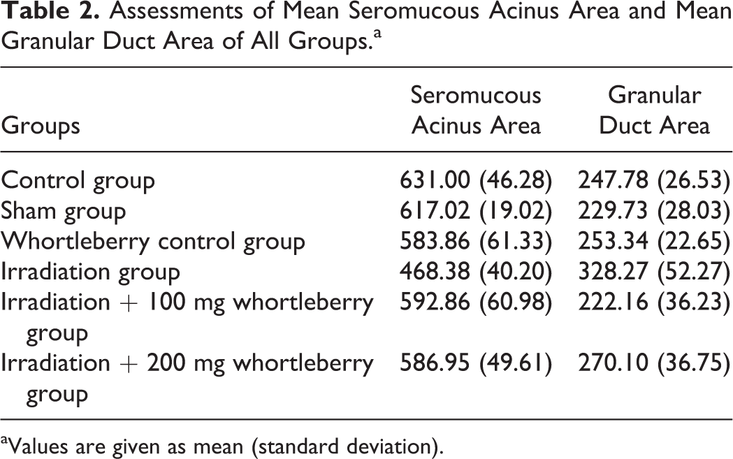
aValues are given as mean (standard deviation).
The mean ductal areas were larger in the irradiation group than in the control group (P < .01). No significant differences were observed between the control group and the other groups (P > .05; Table 2).
Biochemical Analysis Results
Table 3 shows the blood oxidative stress parameters in the groups. According to these results, the TOS values in the irradiation group were significantly higher than those in the control group (P = .001). Additionally, a significant increase was also observed in the irradiation + 100 mg whortleberry group in terms of TOS values in comparison to the control group (P = .031). No significant differences were observed between the control group and the other groups in terms of TOS values (P > .05).
Blood Oxidative Stress Parameters in the Groups.a
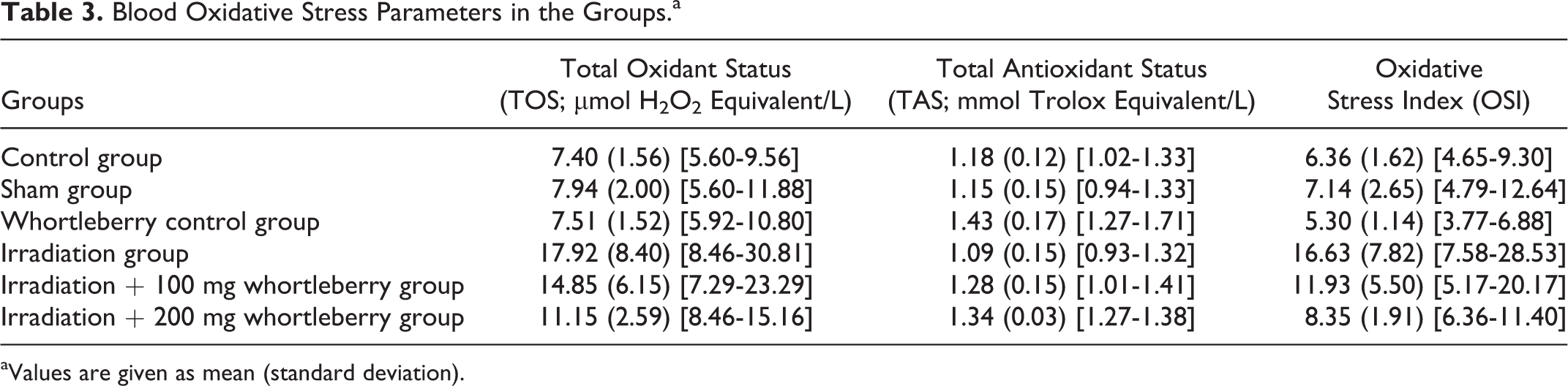
aValues are given as mean (standard deviation).
The highest TAS values were obtained in the whortleberry control group, and these values were significantly higher than those in the control group (P = .009). No significant differences were observed between the control group and the other groups in terms of TAS values (P > .05). In parallel to these findings, whereas the OSI in the irradiation group was significantly higher than that in the control group (P < .001), there were no significant differences between the control group and the other groups. The OSI was significantly lower in the irradiation + 200 mg whortleberry group than that in the irradiation group (P = .005), whereas the difference was not significant in the comparison between the irradiation + 100 mg whortleberry and irradiation groups (P = .463). This result demonstrates that the high dosage of whortleberry extract was more effective than the low dosage in reducing radiotherapy-induced oxidative stress in systemic circulation.
Table 4 summarizes the oxidative stress parameters of the submandibular gland tissues in the groups. Accordingly, the TOS values in the irradiation group were found to be significantly higher than those in the control group (P = .001). No significant differences were observed between the control group and the other groups in terms of TOS values (P > .05). In the comparison of the control group and other groups in terms of TAS values, a significant increase was found in the whortleberry control group (P = .004), whereas the other groups did not differ significantly.
Tissue Oxidative Stress Parameters in the Groups.a
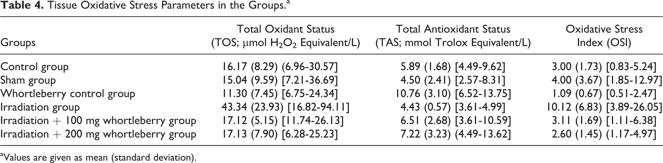
aValues are given as mean (standard deviation).
In the comparison of the OSI values obtained in the submandibular gland tissues in the groups, it was seen that the value was significantly higher in the irradiation group than in the control group (P = .002). No significant differences were observed between the control group and the other groups (P > .05). Additionally, the OSI value of the irradiation group was significantly higher than those of both the irradiation + 200 mg whortleberry group (P = .001) and the irradiation + 100 mg whortleberry group (P = .002). These results suggest that although high dosages of whortleberry extract are needed to reduce radiotherapy-related oxidative stress in the blood, low dosages of the extract may be sufficient to reduce the oxidative stress in tissues.
Discussion
The histopathological findings of this study show that whortleberry reduces salivary gland damage induced by radiotherapy. Moreover, the biochemical data analyzed show that whortleberry has antioxidant effects locally in the tissue and systemically in the blood against oxidative stress, which leads to an indirect mitigation of the damages caused by radiotherapy.
Radiotherapy is a treatment method used to eradicate or control malignant illnesses. However, the adverse effects that may occur during or after the treatment limit the area of usage for radiotherapy. One of the most frequently encountered adverse effects, especially in treatment of head and neck cancers, is xerostomia, which occurs because of salivary gland damage. Studies have shown that the main factor in the occurrence of xerostomia is the reduction in acinar areas in the salivary glands. 1 -3 Similar to the findings of other studies, in the radiotherapy model, we established the 8 Gy external radiotherapy applied on the head and neck areas of the rats led to decreases in acinar areas and increases in ductal areas. We did not observe any adverse effect that would lead to a necessity of exclusion of any rats.
Studies have been conducted on the effectiveness of the protective effect of various substances against radiotherapy-induced histopathological degeneration in salivary glands. The findings of these studies have shown that antioxidant substances decrease radiation-related cellular damage in salivary glands, prevent reduction of acinar areas, and reduce signs of degeneration, such as vacuolization. 2,5,6,13 In our study, only the irradiation group showed histopathological signs of degeneration, such as acinar atrophy, vacuolization, and fibrosis, whereas this degeneration did not occur in the groups given whortleberry extract in addition to radiotherapy. Additionally, although decreases in acinar areas and increases in ductal areas were seen in the group given radiotherapy only, no significant difference was observed in the groups given whortleberry in addition to radiotherapy. These findings show that whortleberry reduces radiotherapy-related histopathological degeneration in both low and high dosages.
In addition to direct DNA damage in tissues, radiotherapy also leads to tissue damage through the oxidative stress it indirectly induces. The findings of other studies have shown that some substances that are demonstrated to be effective against the adverse effects of radiotherapy also lead to reductions in oxidative stress parameters. 4,14 In comparison to the control group, the TOS and OSI values measured in our study in both the submandibular gland tissue and the blood were found to be significantly higher only in the group given radiotherapy without additional treatment. This finding in the radiotherapy model we established shows that radiotherapy also leads to increases in oxidative stress in addition to histopathological degeneration. Considering the tissue oxidative stress parameters, both low and high dosages of whortleberry given in addition to radiotherapy reduced oxidative stress, whereas the TOS values in the blood in the irradiation + 100 mg whortleberry group were found to be higher than that in the control group. Moreover, the OSI in the irradiation group was significantly higher than the OSI in the irradiation + 200 mg whortleberry group, whereas no significant difference was found between the irradiation and irradiation + 100 mg whortleberry groups in terms of the OSI. These results suggest that whereas high dosages of whortleberry extract are needed to reduce radiotherapy-related oxidative stress in the blood, low dosages of the extract may be sufficient to reduce oxidative stress in the tissue.
In conclusion, it is known that radiotherapy leads to damage in the salivary glands through cellular degeneration and increased oxidative stress. According to the findings of our study, whortleberry, which has high antioxidant contents, reduces histopathological degeneration and local oxidative stress induced in salivary glands by radiotherapy. Additionally, high dosages of whortleberry also lessen oxidative stress that radiotherapy induces systemically in the blood.
Footnotes
Authors’ Note
The study was performed in accordance with the 2011 Guide for the Care and Use of Laboratory Animals.
Declaration of Conflicting Interests
The author(s) declared no potential conflicts of interest with respect to the research, authorship, and/or publication of this article.
Funding
The author(s) disclosed receipt of the following financial support for the research, authorship, and/or publication of this article: This work was supported by Recep Tayyip Erdogan University Scientific Research Project Unit (RTEUBAP) under the project number of 282.