
Abstract
To address the challenges of low target contrast and severe scattering noise in turbid media imaging, this paper proposes an underwater active imaging method that integrates range-gated and polarization-difference joint modulation. By establishing a physical model of spatio-polarimetric joint modulation, we systematically analyze the differences in both depolarization characteristics and time-delay properties between target-reflected light and backscattered light. A dual mechanism combining time gating for primary scattering suppression and polarization difference for residual noise elimination is utilized to achieve high-precision separation and enhancement of the target signal. Through a combination of MATLAB simulations and experimental studies, using fat emulsion solutions to emulate turbid water and a 450 nm laser as the light source, key parameters including the medium absorption coefficient and scattering intensity were adjusted to identify system-sensitive parameters and optimization strategies. Results demonstrate that the proposed polarization-difference range-gated imaging method outperforms conventional imaging approaches in both image signal-to-noise ratio and target contrast, confirming its effectiveness and advancement for target detection in turbid media. This work provides a new perspective for high-resolution optical imaging in complex underwater environments.
Introduction
With the rapid development of marine resource exploration, ecological monitoring, and underwater archaeology, there is an urgent need for high-resolution underwater optical imaging technologies. However, in turbid water, light scattering by suspended particles significantly enhances backscattering noise, severely reducing the signal-to-noise ratio (SNR) and contrast of images, which constrains the improvement of imaging quality. 1 Traditional underwater imaging techniques primarily rely on active light source illumination but are limited by water absorption and scattering effects, making it difficult to achieve long-distance, high-definition imaging in turbid environments. 2 To address this, researchers have proposed various noise suppression methods. Among them, range-gated imaging technology utilizes the temporal relationship between laser pulses and camera gating to filter backscattering noise between the target and the camera through spatial slicing. 3 For example, Hongsheng Lin et al. developed a gated imaging system based on laser pulses, which improved the suppression efficiency of forward-scattered light and backscattering noise using Jaffe-McGlamery and Fourier optical models. 4 Polarization imaging technology further suppresses residual noise by exploiting the polarization differences between target-reflected light and backscattered light. 5 For instance, Pengfeng Liu et al. extracted clear object information by decomposing the matrix into sparse and low-rank components. 6
However, the slice depth of field in range-gated imaging is constrained by laser pulse width and gate width parameters, making it difficult to balance noise suppression with large-depth target observation. Polarization imaging is susceptible to optical energy attenuation and depolarization effects, leading to significant performance degradation in long-distance or high-attenuation water bodies. In recent years, hybrid techniques combining range-gated and polarization imaging have become a research focus. Shuaibao Chen et al. integrated polarization imaging with range-gated laser imaging, expanding the practicality of underwater laser imaging scenarios and enhancing captured images. 7 Feng Huang et al. proposed an imaging method based on range-gated detection combined with active polarization dehazing and denoising optimization, improving contrast and SNR in dense fog scenarios. 8
Although current studies have demonstrated the capabilities of polarization-gated imaging, there remains a lack of systematic integration of physical models addressing the coupled effects of target reflection, backscattering, and device noise. This study proposes a turbid medium target detection and enhancement method based on range-gated polarization difference imaging. By establishing a physical model for polarization-gated imaging, we quantitatively analyze target reflected light and medium backscattered light. Through experimental and simulation comparisons, optimized selection of sensitive parameters is achieved.
Physical model of polarization-difference range-gated imaging (p-RGI)
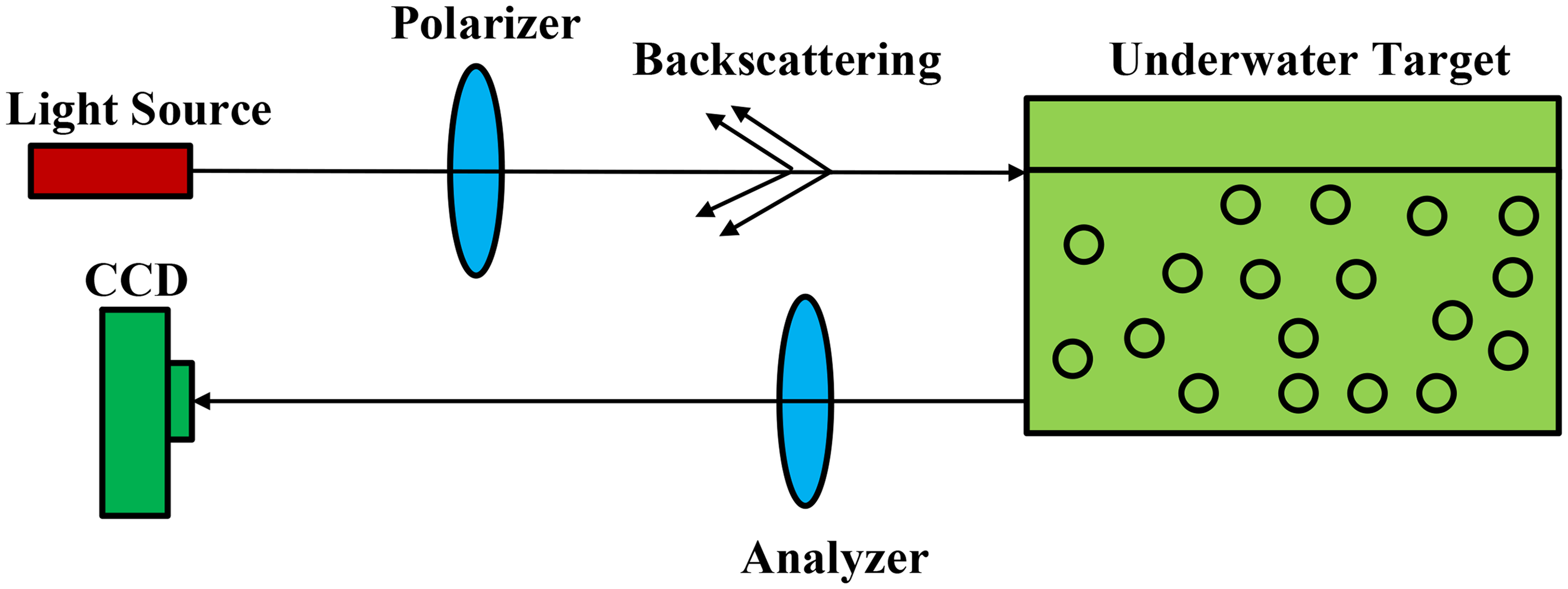
Figure 1 shows the schematic diagram of underwater range-gated imaging. Range-gated imaging is an active optical imaging technology based on time gating, with its core principle being the separation of target reflected light and water scattered light through precise temporal control. The system coordinates the timing between the laser and camera via a synchronization controller. The laser emits high-intensity short pulses (pulse width Tp) to instantaneously illuminate the underwater target area. The camera delays opening the gating gate (exposure window) by time T₁ after laser emission, with an exposure duration of Ts. The pulsed laser propagates through water to the target and reflects back, with a round-trip time of T₀=2*d/c (where d is the target distance and c is the speed of light in water). During the laser round-trip (0 < t < T0), the camera gate remains closed, blocking backscattered light generated by suspended particles in water. When the reflected light reaches the camera (t = T₀), the gating gate synchronously opens (T1 = T0) and receives the target reflected signal during Ts. Setting T₁=T₀ ensures that the gating gate opens only when the target reflected light arrives. Setting Tp = Ts allows the exposure time to fully cover the pulse echo, thus maximizing target signal reception while minimizing the introduction of stray light. By employing delayed exposure, only the target reflected light (time window: T0 ≤ t ≤ T0 + Ts) is captured, while excluding early-arriving forward-scattered light and delayed multiple-scattered light. This process achieves temporal control (synchronous triggering) → optical transmission isolation (gating to block scattered light) → signal extraction (capturing target reflected light) → SNR enhancement. Through dual time-energy domain filtering, scattering interference in water is effectively suppressed, making it particularly suitable for high-contrast imaging in turbid waters.9,10

Schematic diagram of underwater range-gated imaging.
The polarization characteristic of light waves is one of their fundamental physical properties, which, together with amplitude, wavelength, and phase, constitutes a complete description system of electromagnetic waves. Polarization manifests as the periodic directional variation of the electric field vibration vector during light propagation. Based on differences in polarization states, light in nature can be classified into three major categories: natural light, partially polarized light, and completely polarized light. Completely polarized light can be further subdivided into linearly polarized light, elliptically polarized light, and circularly polarized light based on the spatial trajectory of the electric vector11,12 (as shown in Figure 2). In the formation mechanism of completely polarized light, the superposition relationship and phase difference between two orthogonal electric vibration components play a decisive role: when the two orthogonal components maintain a constant phase relationship with a phase difference of 0° or 180°, the planar projection of their resultant vector exhibits a linear trajectory, forming linearly polarized light; if the phase difference between the two components falls within the range of 0° to 90°, the endpoint trajectory of the resultant vector exhibits elliptical polarization in the propagation plane, with its handedness (left-handed or right-handed) determined by the sign of the phase difference; when the phase difference is exactly 90° and the amplitudes are equal, the elliptical trajectory degenerates into a perfect circle, resulting in circularly polarized light. This series of polarization formation laws profoundly reveals the essential characteristics of electromagnetic wave vector superposition.
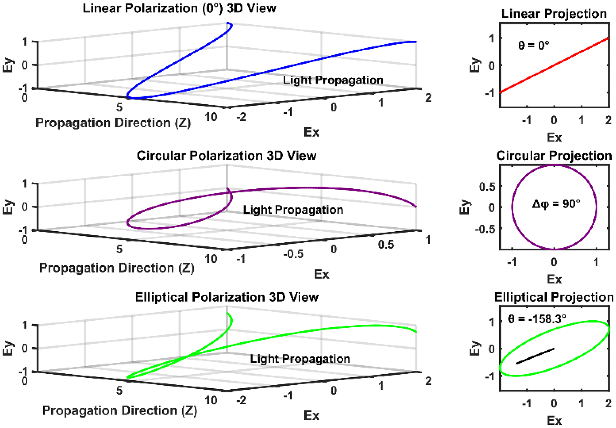
Classification of light polarization states.
The mechanism of underwater polarization difference imaging technology is illustrated in Figure 3. It achieves clear reconstruction of degraded underwater images by extracting intensity information of different polarization states from the target scene. This technique involves capturing intensity images of the target object at four typical polarization directions (0°, 45°, 90°, and 135°) under identical environmental conditions. Based on the Stokes vector, the degree and angle of polarization of the background light are calculated, thereby inverting the intensity distribution of the medium-scattered light. By establishing a separation model for the polarization characteristics of target-reflected light and background-scattered light, it effectively suppresses the interference of backscattering noise on imaging quality.13–15
Schematic diagram of underwater polarization difference imaging.
From the perspective of polarization transmission characteristics, underwater backscattered light exhibits significant polarization preservation, maintaining high consistency with the incident laser's polarization state. In contrast, when the laser is reflected by the target surface, depolarization occurs due to medium inhomogeneity and multiple scattering, leading to a notable attenuation in the degree of polarization (DoP) of the reflected light. Polarization imaging uses a filter to receive only light polarized in a specific direction (e.g. horizontal or vertical). Adjusting the filter's orientation can suppress polarization components consistent with the scattered light from the turbid medium, thereby enhancing the contrast of the target area. 16
Range-gated imaging technology is one of the key physical methods for target detection in turbid media. By emitting laser pulses and synchronously triggering a gated camera, it utilizes a time-gating mechanism to capture only the time-window signal of the target-reflected light, thereby filtering out most of the backscattered light and aquatic background noise. However, due to limitations in time-gating accuracy and medium scattering characteristics, residual backscattering noise remains within the gated slice, which restricts further improvement in imaging SNR.
To address this, optical polarization imaging is integrated with range-gating technology, proposing a range-gated polarization difference imaging method: first, the primary scattering noise is suppressed via time gating; then, the gated images are subjected to polarization splitting. Based on the Stokes vector, polarization difference images are calculated to further eliminate residual backscattered light within the target area; finally, high SNR image reconstruction is achieved by fusing multiple gated images with polarization parameters.17,18
In active imaging systems within turbid media, where laser serves as the active illumination source, the detected signal consists of three components (as shown in Figure 4): (1) medium backscattered light, which is the noise component resulting from direct backscattering of incident light by scattering particles in the medium toward the detector; (2) target ballistic light, which is the effective signal formed by incident light reflected directly from the target surface into the detector; and (3) target forward-scattered light, which represents the interference component generated by photons carrying target information undergoing forward scattering during propagation. Among these, the spatial overlap of backscattered light and target ballistic light causes image blurring, while target forward-scattered light reduces image contrast through wavefront distortion. In existing theoretical models, the effect of forward-scattered light is often incorporated into the category of backscattering noise. Thus, the signal received by the detector can be simplified as a linear superposition of backscattered light and target reflected light. Therefore, the core of image restoration lies in accurately separating these two types of intensity components.

Signals during active detection processes.
Range-gated imaging achieves preliminary noise suppression by precisely matching the temporal sequence between the laser pulse and the detector's time gate, leveraging the difference in time delay between backscattered light and target reflected light. On this basis, the introduction of polarization difference imaging technology further exploits the difference in polarization characteristics between the two types of optical signals: backscattered light maintains high consistency with the polarization state of the incident laser due to its polarization-maintaining properties, while target reflected light exhibits attenuated DoP caused by depolarization effects. By constructing a set of polarization modulation equations to quantitatively analyze the difference in their polarization parameters, a noise removal model coupled with multiple physical fields can be established, ultimately forming a range-gated polarization difference imaging system that combines the advantages of both temporal and polarization resolution. This technology significantly improves the detail resolution and scene adaptability of target imaging in turbid media through innovation in physical mechanisms.
Temporal gating serves as a coarse-scale geometric filter, exploiting path-length differences to block the majority of backscattered photons arriving outside the target signal's time window. This initial stage removes bulk noise, prevents detector saturation, and provides a foundational SNR improvement. Polarization differencing subsequently acts as a fine-scale vectorial filter, processing the light that passes the temporal gate. This residual signal contains the depolarized target reflection alongside polarized noise photons that coincidentally arrived within the time gate due to multiple scattering. The polarization filter selectively suppresses the residual noise photons based on their preserved polarization state—a property undetectable by temporal filtering alone.
The core of the synergy lies in mutual compensation: temporal gating addresses the limitation of polarization imaging by drastically reducing the total light volume, thereby preserving the polarization contrast between the depolarized target signal and the still-polarized residual noise. Conversely, polarization differencing purifies the temporal slice by removing noise photons that the time gate could not discriminate based on arrival time alone. This collaboration creates an emergent capability to resolve targets in regimes where either method alone would fail. For instance, in high-concentration media where polarization contrast diminishes, the initial SNR gain from time gating enables effective polarization-based discrimination. Similarly, at very close ranges where time-of-flight differences are minimal, polarization differencing becomes the primary mechanism for target-background separation.
Simulation verification
Backscattered light characteristics
Figure 5 illustrates the relationship between light intensity, polarization properties, and concentration in the scattering medium. The upper subplot displays the normalized intensity distribution curves under different concentrations, showing a Gaussian-like profile with slight shifts in the peak position. As concentration increases, the intensity curves exhibit significant peak reduction and distribution broadening. At low concentrations, the intensity is concentrated with a high peak value, while at high concentrations, the distribution becomes dispersed with a markedly reduced peak. The decrease in peak intensity is attributed to the reduced photon mean free path caused by higher scattering concentrations, leading to intensified attenuation. The broadening mainly results from enhanced multiple scattering, which randomizes photon paths and spatially expands the intensity distribution.

Relationships between light intensity/polarization characteristics and concentration in scattering media.
The lower subplot shows the DoP distribution curves across concentrations. The polarization profile exhibits a declining trend, with overall DoP decreasing and curve slope diminishing as concentration increases. This indicates strengthened depolarization effects due to increased multiple scattering at higher concentrations, which randomizes the polarization state of photons. The reduction in slope further reflects decreased polarization preservation capability and reduced disparity in DoP across different spatial locations.
Figure 6 shows the variation in intensity and polarization distributions of backscattered light with increasing concentration. As the concentration rises, the central peak of the intensity distribution gradually decreases, the distribution broadens, and inhomogeneity becomes more pronounced. At a concentration of 0.7%, the intensity profile becomes highly diffuse, with significantly reduced contrast between the center and peripheral regions. This is primarily due to the increased number density of scattering particles, which enhances multiple scattering effects.

Intensity and polarization distribution of backscattered light versus concentration.
The DoP distribution also decreases with increasing concentration, though the extent of change is much smaller compared to that of the intensity distribution. Even at the high concentration of 0.7%, the DoP maintains relatively good spatial uniformity, owing to the strong retention capability of polarization information.
Performance evaluation of polarization-enhanced imaging
Figure 7 demonstrates the imaging results of conventional range-gated imaging (RGI) and polarization-based range-gated imaging under varying concentrations. At a low concentration (0.1%), both methods produce relatively clear images with well-defined central bright regions and smooth edge transitions. As the concentration increases, the image quality of conventional RGI deteriorates significantly. At a concentration of 0.4%, the central bright region becomes noticeably diffused, with increased edge blurring and substantial noise enhancement. Under high concentration conditions (0.7%), the conventional RGI image is severely degraded, exhibiting a marked reduction in contrast between the center and the periphery.
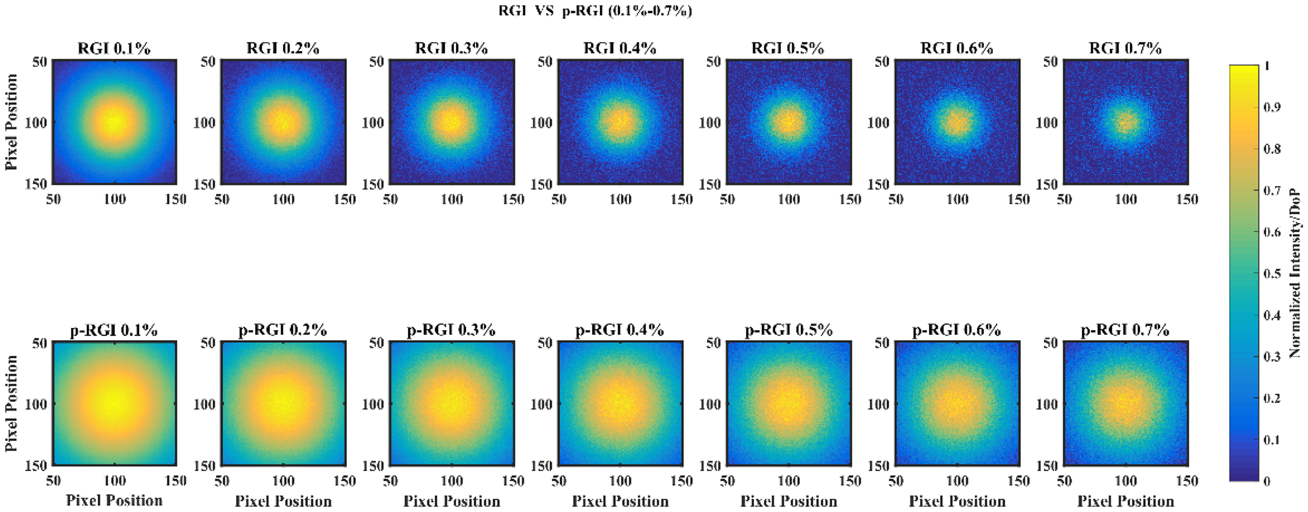
Comparison between underwater range-gated imaging and polarization-based range-gated imaging.
The primary factor contributing to the pronounced performance decline of conventional RGI with increasing concentration is the enhanced scattering effect. A higher concentration of fat emulsion solution leads to an increased number density of scattering particles, causing photons to undergo more scattering events and follow more randomized paths. This results in greater spatial inhomogeneity in the light intensity distribution and significantly aggravated image degradation.
In contrast, p-RGI maintains superior imaging quality across different concentrations. Even at a high concentration (0.7%), the images retain high uniformity with clearly outlined central bright regions. The stronger anti-interference capability of p-RGI stems from the stability of polarization information. Although polarized light also undergoes depolarization during scattering, changes in its polarization state occur more gradually compared to variations in light intensity. Polarization characteristics depend mainly on the number of scattering events and the angular distribution of scattering, demonstrating lower sensitivity to absolute scattering coefficients. Therefore, even at elevated concentrations, polarization information remains relatively stable, thereby providing higher-quality images.
Quantitative analysis of background uniformity
To quantify background uniformity, this study employs the standard deviation parameter (σ) to characterize intensity dispersion:
Figure 8 illustrates the variation trends of the standard deviation for both RGI and p-RGI under different fat emulsion concentrations. Both curves exhibit a declining trend, indicating that the uniformity of both imaging methods improves as concentration increases. The RGI curve (blue squares) consistently remains above the p-RGI curve (red circles), demonstrating that the standard deviation of conventional RGI is higher than that of p-RGI across all tested concentrations. The standard deviation of RGI decreases from 0.28 at 0.1% concentration to 0.08 at 0.7% concentration, a reduction of 71.4%; meanwhile, the standard deviation of p-RGI decreases from 0.15 at 0.1% concentration to 0.07 at 0.7% concentration, a reduction of 53.3%. At all concentration points, the standard deviation of p-RGI is significantly lower than that of RGI.

Standard deviation of underwater images for two imaging methods.
This phenomenon is related to changes in the scattering mechanism. Under low concentration conditions, scattering events are relatively few, but the impact of individual scattering events is more pronounced, resulting in poorer image uniformity. As concentration increases, the number of scattering events rises; however, as photons undergo more scattering events, their spatial distribution becomes more uniform, thereby improving image homogeneity.
For RGI, increased concentration leads to more scattering events. Although this amplifies the disparity in photon path lengths, it also promotes spatial homogenization of light intensity. For p-RGI, increased concentration similarly contributes to a more uniform spatial distribution of polarization information. However, due to the inherent insensitivity of polarization information to scattering, the improvement in uniformity is more pronounced, resulting in consistently lower standard deviation values for p-RGI compared to RGI.
Target feature recognition and contrast analysis
Figure 9 presents the one-dimensional distribution curves of RGI and p-RGI under different concentrations. The RGI curve (dashed line) exhibits a single-peaked Gaussian distribution with its peak at pixel position 60, and the distribution width gradually broadens with increasing concentration. At low concentration (0.1%), the RGI curve appears relatively sharp with a high peak intensity; as concentration increases, the curve gradually flattens with reduced peak intensity.
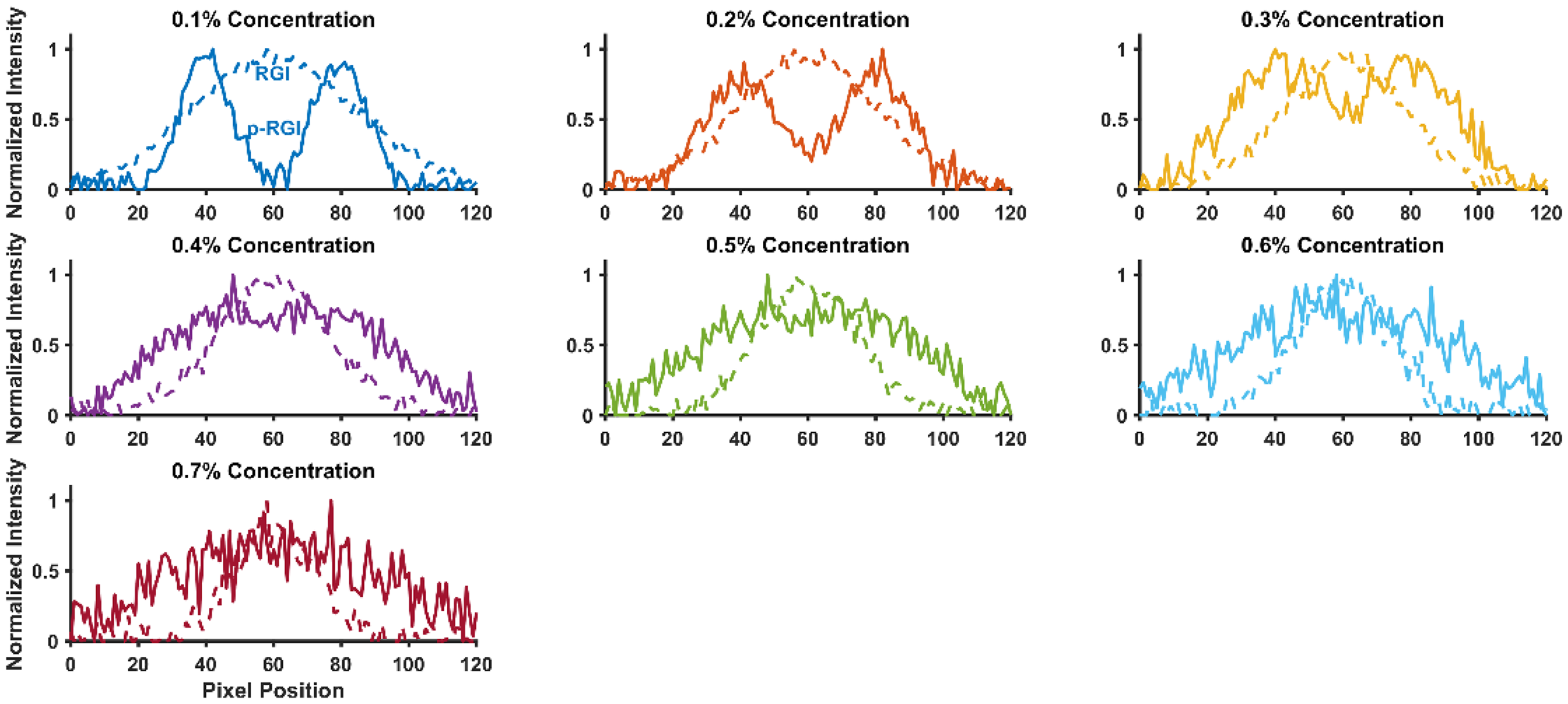
Numerical curve comparison between range-gated and polarization-based range-gated imaging under different scattering conditions.
Experimental data reveal two distinct characteristic peaks in the p-RGI curve, whose spatial positions precisely match the geometric structure of the target. In contrast, the target characteristic peaks are almost indistinguishable in the RGI curve. This indicates that when target and background have similar reflectivity, relying solely on intensity information makes effective target recognition difficult, whereas polarization-difference techniques can overcome this limitation by extracting differences in polarization properties of scattered light.
Analysis of the curve evolution shows that as fat emulsion concentration increases, the intensity distribution curves of both imaging methods exhibit an overall amplitude attenuation trend. The intensity difference between characteristic peaks and background valleys in the p-RGI curve gradually decreases with enhanced scattering. This phenomenon stems from the dual role of scattering characteristics in turbid media: on one hand, increased scattering events cause target-reflected light to undergo more depolarization, significantly reducing its DoP; on the other hand, when the polarization-difference imaging algorithm filters out backscattered light with high polarization preservation, partially depolarized target-reflected light may be misclassified as noise and suppressed. This coupling effect of target signal attenuation and residual noise leads to continuous deterioration of target-background intensity contrast with increasing scattering.
Depolarization characteristics and imaging distance
Figure 10(a) quantitatively reveals the differences in depolarization characteristics between the target and background in turbid media and their response to scattering intensity, with the DoP employed as the quantitative indicator of depolarization effects. 19 The target DoP (solid red line with circles) decreases monotonically from 0.79 at 0.10% concentration to 0.28 at 0.70% concentration, representing a reduction of 64.6%. The background DoP (dashed blue line with squares) decreases from 0.42 to 0.26, a reduction of 38.1%. Both curves show declining trends, but the target DoP decreases at a significantly faster rate than the background DoP.

Depolarization characteristics of objects versus backgrounds and object-background contrast in polarization-enhanced range-gated imaging.
This differential depolarization behavior originates from distinct scattering mechanisms in target and background regions. The target area typically exhibits specific surface characteristics and material composition, where depolarization is influenced by both surface scattering and volume scattering. As concentration increases, the higher number density of scattering particles in the fat emulsion solution enhances photon-particle interactions, leading to faster loss of polarization information. The slower depolarization in the background region is primarily because background scattering is dominated by volume scattering, resulting in a more uniform and gradual depolarization process. The target region, with its interface reflections and more complex scattering processes, shows greater sensitivity to concentration changes, thus exhibiting faster depolarization rates.
The subplot shows the target-background contrast curve (solid black line with triangles) and its quadratic fitting curve (dashed line) as functions of concentration. The contrast decreases continuously from 0.71 at 0.10% concentration to 0.19 at 0.70% concentration, exhibiting an overall non-linear decreasing trend. The fitted curve indicates a significant quadratic relationship between contrast and concentration, with the rate of decrease gradually slowing as concentration increases. The primary reason for contrast reduction is the diminishing difference between target and background depolarization. Under low concentration conditions, the substantial difference in DoP between target and background results in higher contrast. As concentration increases, the rapid decrease in target DoP combined with the relatively slower decrease in background DoP reduces their difference, consequently lowering contrast. The quadratic fitting relationship demonstrates that contrast reduction is not a simple linear process but tends to saturate at higher concentration regions. This reflects the complex characteristics of polarized light propagation in turbid media: beyond a certain concentration threshold, further increases in concentration have diminishing effects on polarization properties.
Figure 10(b) displays the variation curves of depolarization rates for both target and background with increasing concentration. The target depolarization rate increases from 0.21 to 0.72, while the background depolarization rate rises from 0.58 to 0.74. The rapid growth of the target depolarization rate reflects the sensitivity and variability of the target surface to polarization information.
Figure 10(c) presents a three-dimensional surface illustrating the complex relationship between contrast, concentration, and DoP. The surface demonstrates that contrast reaches its maximum values in regions of low concentration and high DoP, while decreasing to minimum values in areas of high concentration and low DoP. The surface morphology reflects the fact that contrast is simultaneously influenced by both concentration and DoP. This three-dimensional relationship reveals the coupling effects of contrast influencing factors. Concentration primarily affects polarization information by increasing the number of scattering events, while DoP directly reflects the preservation DoP information. The combined effect of these two factors determines the final contrast performance.
Figure 11 quantitatively compares the image contrast performance of three techniques: intensity imaging, polarization-difference imaging, and p-RGI. The contrast values of the three techniques consistently follow the same descending order across all conditions: p-RGI demonstrates the highest contrast, followed by polarization-difference imaging, with intensity imaging showing the lowest values. This indicates the absolute advantage of p-RGI technology in maintaining high contrast.
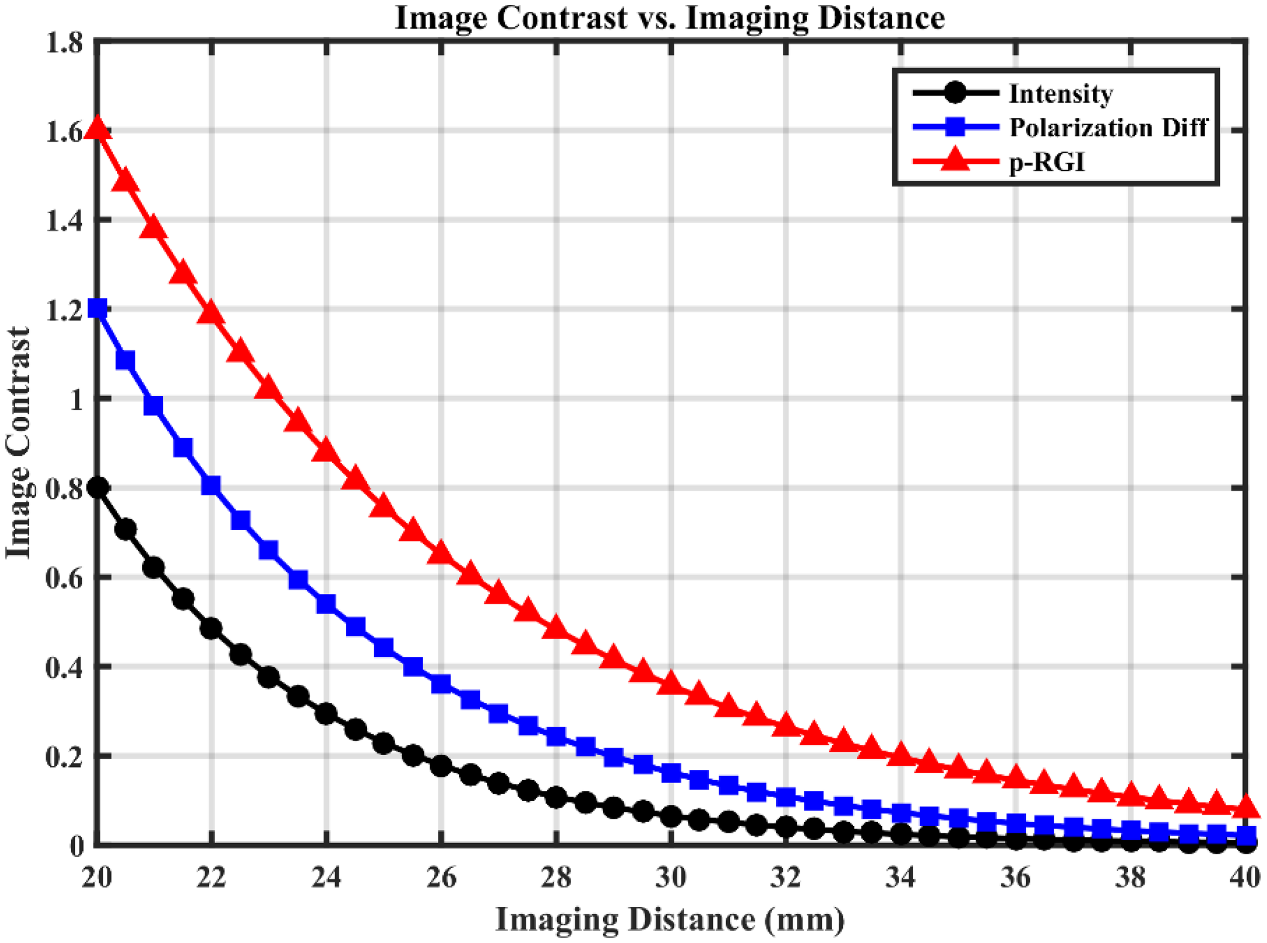
Image contrast versus imaging distance for three imaging modalities.
Regarding anti-attenuation capability, the three curves exhibit significantly different decay rates. The contrast of intensity imaging decreases most rapidly with increasing distance, while the p-RGI curve shows the most gradual decline. This demonstrates that p-RGI technology not only provides higher initial contrast but also possesses superior ability to resist scattering attenuation and maintain effective imaging distance.
In intensity imaging, signals are susceptible to interference from multiply scattered light. As imaging distance (penetration depth) increases, scattering effects intensify dramatically, causing rapid attenuation of target signals and significant enhancement of background noise, thereby resulting in the fastest contrast degradation.
Polarization-difference imaging utilizes the differential modulation capability of targets and scattering media on polarized light. By calculating the difference between orthogonally polarized images, it effectively suppresses most non-modulated (multiply scattered) background light. Consequently, it retains more effective signals and exhibits slower contrast decay than intensity imaging.
p-RGI combines the advantages of both polarization-difference and range-gating techniques. Range-gating technology selects photons based on their time of flight, enabling precise separation of earliest-arriving, unscattered ballistic photons and a few forward-scattered photons, while virtually eliminating late-arriving, noise-dominated multiply scattered light. When combined with polarization-difference imaging, it forms a dual-filtering mechanism that maximizes effective signal extraction and suppresses background noise. Therefore, p-RGI achieves the highest contrast with the slowest distance-dependent attenuation, demonstrating optimal potential for deep-layer imaging.
Experimental validation
Figure 12 shows the experimental setup of the range-gated polarization difference imaging system. The system employs a 450 nm Diode-Pumped Solid-State Laser as the light source L. The beam is shaped into a 15 mm diameter collimated beam through a beam expansion and collimation system E, and then modulated into linearly polarized light by a linear polarizer P1 before incident into the water tank W. The target is a standard reflector (CD disk) suspended in the water tank, whose reflected light together with the medium backscattered light constitutes the return signal. At the receiving end, a polarizer P2 serves as an analyzer, combined with a 450 nm bandpass filter to achieve polarization selection and spectral filtering. The filter F is placed between the CCD and the analyzer P2 to select light with a central wavelength of 450 nm. By rotating the azimuth angle of P2, intensity images of parallel (I∥) and perpendicular (I⊥) polarization components can be acquired respectively. Their difference operation (ΔI = I∥−I⊥) forms the basis of polarization difference imaging.

Experimental setup for range-gated polarization difference imaging.
The experimental procedure employs a dynamic medium modulation method to achieve range-gating functionality: first, orthogonal polarization images of the original solution (without absorbing medium) are recorded; then, fat emulsion solution is progressively injected to increase the medium absorption coefficient. When the target reflected light is completely absorbed due to excessive optical path length, background images containing only short-range scattered light are recorded. By calculating differential images of corresponding polarization components before and after ink addition (I∥Δ=I∥original−I∥background, I⊥Δ=I⊥original−I⊥background), the target polarization characteristics after range gating can be extracted. Finally, a secondary difference of the gated orthogonal components (ΔIΔ=I∥Δ−I⊥Δ) is performed to achieve spatiotemporal-polarization jointly modulated noise suppression.
This setup establishes a physical separation model between scattering noise and target signals through precise control of medium absorption characteristics and imaging geometric parameters. During the experiment, by adjusting the concentration of fat emulsion solution and target position, the coupling relationships among scattering intensity, imaging distance, and polarization characteristics were quantitatively characterized.
Figure 13 presents the parameter sensitivity analysis results of p-RGI technology in turbid media. Using a 26 mm imaging distance as the baseline (fat emulsion absorption coefficient = 1 m−1, noise level = 0.05, beam diameter = 5 mm, wavelength = 450 nm, R-para = 0.3, R-perp = 0.1), the study systematically reveals the influence mechanisms of various physical parameters on imaging quality by controlling fat emulsion absorption coefficient, noise intensity, beam diameter, incident wavelength, and target reflectivity:
Medium absorption characteristics: The absorption coefficient of fat emulsion solution governs the light transmission attenuation rate through the Beer-Lambert law. When the absorption coefficient increases from 1 to 10 m−1, the penetration depth of target reflected light significantly decreases, leading to reduced separation between target and background signals. Consequently, the contrast of differential images exhibits nonlinear attenuation characteristics due to effective signal annihilation. Scattering noise characteristics: Noise intensity is positively correlated with the medium scattering coefficient. Its enhancement increases the proportion of background scattered light, reducing the image SNR. While Gaussian filtering can suppress high-frequency noise, its sensitivity to low-frequency noise decreases compared to direct signal attenuation. Optical system parameters: The beam diameter determines the spatial distribution characteristics of scattering noise. Larger diameters result in broader scattered light distribution, reduced spatial resolution, and blurred target edges. Variations in beam diameter affect the filtering scale of the Gaussian kernel, thereby altering the local correlation of noise, but have relatively limited impact on the overall contrast of differential signals. Spectral modulation effects: The incident wavelength regulates noise intensity through the Rayleigh scattering coefficient. Shorter wavelengths (e.g. 450 nm) produce stronger scattering noise, making target signals more susceptible to be obscured. While wavelength variation significantly modulates noise, its overall sensitivity is moderate due to suppression by filtering and absorption coefficients. Target polarization characteristics: The difference in orthogonal polarization reflectivity (ΔR = R∥−R⊥) is the core source of differential signals. Larger reflectivity differences yield stronger differential signals and higher target-background contrast.
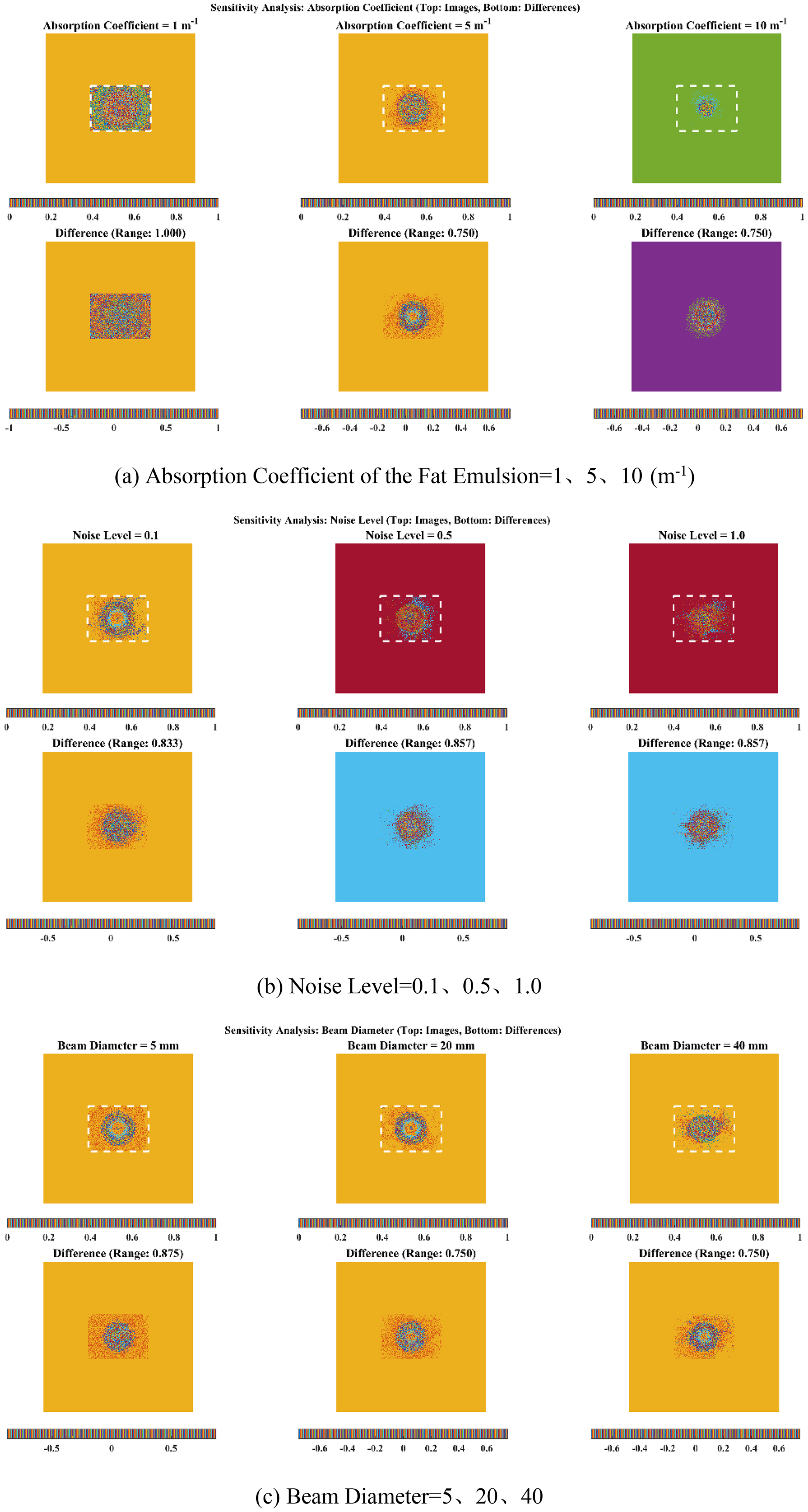
Parameter sensitivity analysis of range-gated polarization difference imaging performance.
These patterns demonstrate that the performance of range-gated polarization difference imaging is jointly constrained by medium absorption characteristics, scattering noise properties, optical system parameters, spectral modulation effects, and target polarization characteristics.
Based on the parameter sensitivity analysis results (shown in Figure 13), the absorption coefficient of the fat emulsion solution is identified as the core control parameter in the range-gated polarization difference imaging system. Its mechanism of action follows the Beer-Lambert attenuation law. When the absorption coefficient of the fat emulsion solution increases from 1 to 10 m−1, the effective penetration depth of the target reflected light decreases by approximately a specific value, resulting in an exponential degradation of the signal-to-noise separation. Experimental data indicate that even minor fluctuations in the absorption coefficient can cause significant changes in the SNR of the differential image, which directly determines the feasibility boundary of the imaging system.
In contrast, other parameters such as the target reflectivity difference (ΔR) and incident wavelength (λ), while important for imaging quality, are constrained by material intrinsic properties or physical laws. For example, ΔR is determined by the microscopic structure of the target surface and is difficult to actively optimize through external means; wavelength selection must balance the trade-off between Rayleigh scattering and detector quantum efficiency. Therefore, in practical system design, priority should be given to establishing an accurate calibration model for the absorption characteristics of the turbid medium, and maintaining reliable extraction of the target signal by dynamically adjusting laser power or optimizing the gating time window.
Conclusion
This study systematically investigates the application efficacy and methodological mechanisms of polarization-based range-gated imaging for target detection and image enhancement in turbid media through theoretical modeling, simulation analysis, and experimental validation. The main conclusions are as follows:
A spatiotemporal-polarization dual-dimensional collaborative imaging method is proposed, integrating range-gating and polarization-difference physical mechanisms to effectively suppress backscattering noise and target signal ambiguity. Experimental results demonstrate that this method reduces image intensity fluctuations and achieves significant improvement in SNR compared to conventional intensity imaging, while maintaining high target discernibility particularly under moderate turbidity conditions (0.4% fat emulsion concentration). The differential depolarization mechanisms between targets and backgrounds are revealed, validating the enhancement effect of polarization-difference on target recognition capability. Under conditions where target and background reflectivity are similar, contrast improvement can be achieved based on differences in DoP, indicating that polarization-derived features exhibit better robustness and separability in complex optical environments. Parameter sensitivity analysis identifies the absorption coefficient of fat emulsion solution as the primary influencing factor for system performance, whose variation directly determines signal attenuation degree and system imaging limits. Beam diameter and incident wavelength require optimization trade-offs between spatial resolution and scattering noise suppression according to practical application scenarios.
The established physical model and experimental system demonstrate excellent extensibility and adaptability, providing theoretical foundation and technical support for applications such as underwater high-definition imaging, biological tissue detection, and target observation in adverse weather conditions.
Footnotes
Funding
The authors received no financial support for the research, authorship, and/or publication of this article.
Declaration of conflicting interests
The authors declared no potential conflicts of interest with respect to the research, authorship, and/or publication of this article.