
Abstract
We present a female in her mid-70s with COVID-19 infection who developed worsening renal function along with systemic symptoms over months. The patient was initially monitored for long COVID and was referred to the nephrologist after she was found to have further worsening of her renal function. She underwent a renal biopsy and findings on light microscopy, immunofluorescence, and electron microscopy were consistent with crescentic glomerulonephritis, pauci-immune type with chronicity, and acute interstitial nephritis. These findings and a positive anti-neutrophilic cytoplasmic antibody immunofluorescence assay (ANCA IFA) and myeloperoxidase antibody confirmed myeloperoxidase-ANCA-positive pauci-immune glomerulonephritis. The patient was started on treatment with corticosteroids and Rituximab, resulting in improvement in her symptoms and renal function.
Introduction
SARS-COV-2 infection has caused more than one million deaths in the US and significant economic and life expectancy losses. 1 10–20% of patients infected with SARS-COV-2 develop persistent or new symptoms after acute infection, known as post-COVID syndrome. Endothelial dysfunction secondary to vascular inflammation, autoimmunity, residual infection, and persistent inflammation from dysregulated cytokine signature are thought to play a major pathophysiologic role in the development of the syndrome. 2 COVID-19 is known to cause both anti-neutrophilic cytoplasmic antibody (ANCA)-vasculitis and renal disease (tubular and glomerular disease) early during the course of disease and later after the resolution of early infection. 3
ANCA-associated vasculitis (AAV) is defined as a necrotizing vasculitis with few or no immune deposits and predominantly affecting small vessels and associated with ANCA positivity for myeloperoxidase (MPO-ANCA) or proteinase 3 (PR3-ANCA). 4 Pauci-immune glomerulonephritis is the most common pattern of injury seen with AAV, and more than 85% of pauci-immune glomerulonephritis cases have ANCA positivity.5,6 Few case reports have shown COVID-19 as a potential trigger for AAV, although its exact role in the occurrence of the vasculitis is not completely understood.7,8 Here, we report a unique case of MPO-ANCA-positive pauci-immune glomerulonephritis in a relatively healthy female post-COVID-19 infection. This article was previously posted to Authorea preprint server on 14 September 2024. 9 The reporting of this study conforms to CARE guidelines. 10
Case presentation
A relatively healthy woman in her mid-70s, working as a yoga teacher with past medical history of Covid-19 infection presented to our center in August with worsening renal function. The patient was tested positive for COVID 19 in January, when she had milder symptoms and did not require any treatment or hospitalization. Initially, she felt better, but after a month, she began to develop intermittent episodes of sore throat and nasal congestion. She also developed malaise, myalgia, and arthralgia, which limited her daily yoga activity. Additionally, she reported poor appetite, a sweet metallic taste, and a weight loss of more than 12 pounds over 4–6 months. Furthermore, she reported intermittent palpitations and pleuritic chest pain that worsened with deep breathing and waxed and waned without specific exacerbating or relieving factors. The physical exam was unremarkable except for thin built lady with weight 57 kg (BMI 20).
The patient reported having seen her primary care physician and being referred to ENT, cardiology, and pulmonology over the period of 6 months, where she underwent various tests, including echocardiogram, CT chest, and CT sinuses, with no identifiable cause. Her symptoms were initially attributed to salivary gland swelling. Later, she was suspected of having possible long COVID and was closely monitored. She denied any fever, chills, cough, hemoptysis, dysuria, hematuria, skin rash, nasal polyps or crusting, hoarseness, wheezing, paresthesia, numbness, focal weakness, hearing impairment, or additional rashes or swelling. The patient reported that she was never vaccinated for COVID-19. She also mentioned that the only medication she was taking was nattokinase, which she started after conducting her own research and purchasing it over-the-counter for long COVID. She denied any family history of autoimmune conditions. She also denied any recreational drug use. The patient reported that, after 3–4 months of her COVID-19 infection, her renal function worsened, leading to her referral to the nephrologist. She was finally able to get an appointment at the end of July and was subsequently referred for renal biopsy and further workup to our center.
Her chest x-ray (Figure 1) and CT scan of lungs showed no infiltrates. EKG showed normal sinus rhythm. Urinalysis and other important labs are presented in Tables 1 and 2. Renal ultrasound showed normal cortical echogenicity (right kidney 8.13 cm, left kidney 10.02 cm) with no evidence of hydronephrosis (Figure 2). A graph depicting the trends in serum creatinine, P-ANCA titer, and MPO antibody index over time is shown in Figure 3.
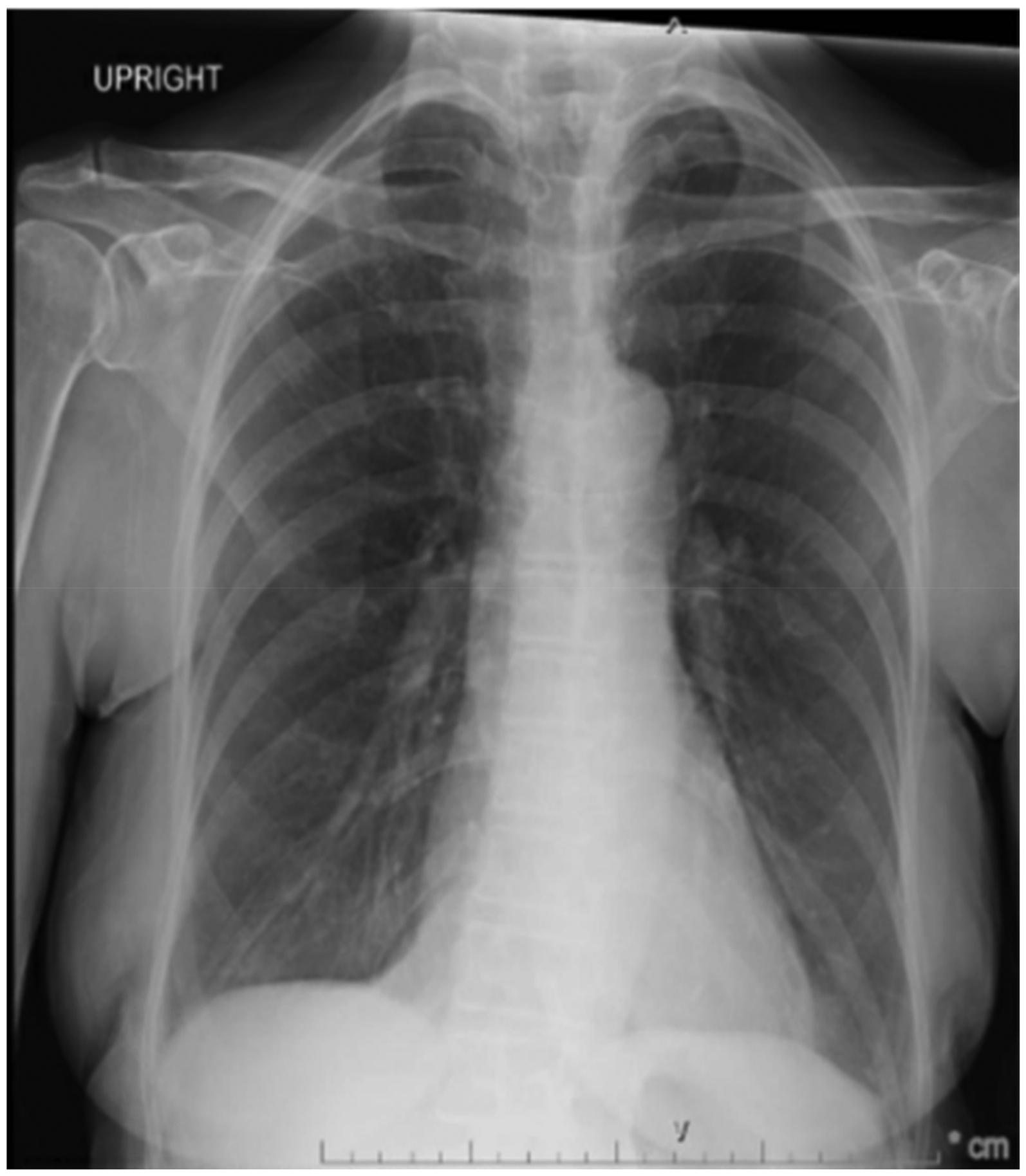
CXR obtained during hospital admission with no acute process.

Renal USG showing kidney size and echogenicity. Bilateral renal ultrasound images. The kidneys are normal in size and demonstrate preserved corticomedullary differentiation with normal echogenicity. No evidence of hydronephrosis is seen.

Trends in creatinine, P-ANCA, and MPO over time. Graph showing serum creatinine levels (blue line with circles), P-ANCA titers (orange dashed line with squares), and MPO antibody index (red dotted line with triangles) across different months. Only month labels are displayed to maintain patient confidentiality. Creatinine levels are reported as mg/dl, P-ANCA titers as dilution factors, and MPO levels are expressed as antibody indices (AI). P-ANCA: perinuclear anti-neutrophil cytoplasmic antibody; MPO: myeloperoxidase.
Important lab findings on review from her PCP's office and hospital lab after admission.

Notes: The values in parentheses indicate the normal range.
PCP: primary care physician; BUN: blood urea nitrogen.
Additional laboratory findings on review from her PCP's office and hospital lab after admission.

PCP: primary care physician; WBC: white blood cell count; CRP: c-reactive protein; TSH: thyroid-stimulating hormone; p-ANCA: perinuclear anti-neutrophil cytoplasmic antibody; MPO-Ab: anti-myeloperoxidase antibody; CCP: cyclic citrullinated peptide; HPF: high-power field; ANA: antinuclear antibody.
Three cores of renal cortex were submitted for histopathological evaluation. One core was submitted for light microscopy examination, showing 26 to 38 glomeruli per level section, of which 14–22 per level section were non-viable. Non-viable glomeruli showed extensive collagen deposition, suggestive of chronicity with a prominent surrounding inflammatory reaction (Figure 4(a) and (b)). Trichrome special stain highlighted the fibrosis in the non-viable (Figure 4(c)). The interstitium showed robust activity with mixed inflammatory infiltrates.

Renal biopsy findings. (a) Light microscopy showing non-viable glomeruli (black arrow) with collagen deposition (*) and associated prominent inflammatory reaction (red arrow), in comparison with viable glomeruli (white arrow) that are histologically unremarkable. (PAS stain x200). (b) Closer section of non-viable glomeruli, replace it with fibrosis and associated prominent mixed inflammatory reaction, including eosinophils (black arrow) and mononuclear cells (*) [H&E stain x400]. (c) Section from the renal biopsy shows a non-viable glomerulus with collagen deposition, consistent with chronicity (black arrow) highlighted by trichrome (Trichome x400). (d) Electron microscopy section, with thin and delicate capillary loops (CL), no increase mesangial matrix, swollen endothelial cells (EC), and extensive effacement of podocytes process (PP), and reactive podocyte cell (P). No immune deposits were identified. [Electron microscopy: XR160VwrkGDIRH77, exposure(ms): 800, Gain: 1, Bin: 1. Direct Magnification: 2500x].
One core was submitted to immunofluorescence and stained for antisera to IgA, IgG, IgM, C3, C1q, fibrinogen, albumin, Kappa, and Lambda light chains. Examination revealed 39 glomeruli, of which all were non-viable. No specific Ig deposition was identified (not shown). The last core was submitted to electron microscopy, showing eight glomeruli, of which two were viable. Upon ultrastructural organization, the viable glomeruli show glomerular basement membranes with average thickness with the typical architectural organization. Mesangial areas appear normal. The epithelial cells’ foot processes are extensively effaced, consistent with the history of proteinuria. Rare intramembranous electron density is present, with no other evidence of immune deposits (Figure 4(d)).
All findings are consistent with crescentic glomerulonephritis, a pauci-immune type with chronicity, and acute interstitial nephritis, likely secondary to glomerulonephritis.
The patient was diagnosed with MPO-ANCA-positive Pauci-immune glomerulonephritis. Her Cocci panel, QuantiFERON TB, HIV, Hepatitis panel were negative. Signed consent was obtained to treatment from the patient. She was started on induction treatment with rituximab and a reduced corticosteroid taper dose per PEXIVAS trial. Methylprednisolone 500 mg on day 1, 250 mg on day 2, 125 mg on day 3. Rituximab 1 gm on day 4. The patient was started on PEXIVAS reduced dose – Prednisone 60 mg (week 1), 30 mg (week 2), 25 mg (week 3–4), 20 mg (week 5–6), 15 mg (week 7–8), 12.5 mg (week 9–10), 10 mg (week 11–12), 7.5 mg (week 13–14), 2.5 mg daily and maintenance Rituximab 2 weeks apart. She experienced significant improvement in her symptoms, including sore throat, sinus issues, weakness, and tiredness, after the initial treatment course. She was also given prophylaxis for infections (Bactrim, fluconazole) and osteoporosis (vitamin D and calcium). During her hospital stay, the patient had one episode of atrial fibrillation but continued to remain in sinus rhythm since then. Her CHA2DS2-VASc score was 3 (age and gender). Patient was counseled on the risk and benefit of anticoagulation. Patient declined to be started on blood thinner and wanted to discuss further with her cardiology outpatient. She was discharged on M cot monitor.
The patient followed-up with cardiology where she continued to be in sinus rhythm. Anticoagulation for her paroxysmal atrial fibrillation was discussed again, but she declined and chose to undergo continuous e-patch monitoring. She also followed-up with Nephrology and continued maintenance rituximab therapy, showing improvement in her symptoms. However, her renal function showed only slow improvement (creatinine trended down to 1.57 on follow-up), and her CRP was normal (<3). Her urinalysis was negative for hematuria or proteinuria, and her microalbumin-to-creatinine ratio was normal. The patient is currently being followed-up by nephrologist and is under maintenance rituximab. She is responding well to treatment, with improvement in symptoms and objective markers, including a decline in serum creatinine, reduction in P-ANCA titers, and a significant decrease in MPO antibody index.
Written informed consent was obtained from the patient for publication of this case report and accompanying images.
Discussion
Most cases of pauci-immune glomerulonephritis are secondary to systemic small-vessel vasculitis, including granulomatosis with polyangiitis (GPA), microscopic polyangiitis (MPA), or Churg-Strauss syndrome, with fewer cases being renal-limited vasculitis. Pauci-immune glomerulonephritis is a type 3 rapidly progressive glomerulonephritis (RPGN) characterized by scant or absent immune deposits on pathology. ANCA antibodies are present in 80–90% of patients with pauci-immune glomerulonephritis (hence referred to as AAV) and play a crucial role in its pathogenesis. Exposure to certain triggers (such as infections, drugs, environmental factors, or injury) is believed to activate ANCA autoantigens, which are released from the cytoplasmic granules to the surface of neutrophils and into the microenvironment in genetically susceptible individuals.5–8,11 ANCA antibodies then bind to these antigens, leading to the activation of neutrophils and monocytes. This results in the release of pro-inflammatory cytokines and the recruitment of more immune cells to the site of inflammation within the glomeruli, via activation of the alternative complement pathway and the release of the chemoattractant C5a.11,12
ANCA antibodies activate neutrophils, with subsequent leukocyte adhesion to the endothelial cells in the capillary loops followed by damage and eventual disruption of the capillary wall and leakage of leukocytes and fibrinoid material to the urinal space, stimulating the parietal epithelial cells (earliest lesion). Due to the constant stimulation, the parietal epithelial cells proliferate, forming cellular crescents that can mature with fibroblasts and collagen deposition, evolving into fibroepithelial and fibrinous crescents, representing the transition from active lesions to more chronic ones.5–8,11,12
Cytomegalovirus, Epstein-Barr, and dengue viruses are known to trigger ANCA vasculitis, but little is known about COVID-19, and only a few case reports of ANCA vasculitis from COVID-19 have been reported. 12 COVID-19 has been associated with various glomerular diseases, with collapsing focal segmental sclerosis being the most common, along with podocytopathy, membranous nephropathy, pauci-immune crescentic glomerulonephritis, and thrombotic microangiopathy.13,14 Covid-19 and AAV share some common pathophysiological mechanisms of disease. Neutrophil extracellular traps and activation of alternative complement pathway C5a-C5aR1 receptor axis is predominant in both Covid and AAV. 15 Molecular mimicry and ANCA autoantibody production might play a role in the development of pauci-immune glomerulonephritis in susceptible Covid-19 patients.
The findings of a rapid decline in GFR by more than 50% over a period of at least 2–3 months, associated with microscopic hematuria, non-nephrotic range proteinuria, and normal blood pressure, suggest RPGN in our case. 16 Based on the histopathological findings, our case fits Type 3 pauci-immune RPGN/ pauci-immune glomerulonephritis. P-ANCA/MPO antibodies are often associated with chronic lesions compared to C-ANCA, as seen in our renal biopsy findings. This is believed to be due to either delayed presentation or a different renal pathological process. 16 Extra-renal findings typically help differentiate various AAV types, although our patient did not exhibit clear symptoms or findings to differentiate the types of AAV or post-COVID syndrome.
Our patient had symptoms of malaise, myalgia, arthralgia, poor appetite, weight loss, sore throat, nasal congestion, metallic taste, intermittent chest pain, dyspnea, which are non-specific and can be present in both vasculitis and post-COVID syndrome. 2 Her intermittent palpitation was likely due to her atrial fibrillation. Although she had nasal congestion and a sore throat, which she attributed to sinus issues, she had no asthma, lung infiltrates or eosinophilia making Churg-Strauss less likely.
Based on the 2022 ACR/EULAR classification criteria, she scored higher in the MPA class (MPO positivity +6, Pauci-immune glomerulonephritis +3, −3 for possible nasal passage involvement although she did not have any imaging evidence of sinusitis/mastoiditis with total score 6) and equivocal score of 5 for GPA (nasal congestion +3, Pauci-immune glomerulonephritis +1, MPO positivity −1, granulomatous inflammation +2 which was seen on renal biopsy in our case) making both a possible classification type. 8 MPO-ANCA-positive GPA is thought to be a unique subset of AAV, which could also be possible, and the above classification criteria might be overestimating the score of MPO positivity in this case and underestimating the score of necrotizing granulomatous inflammation.17,18
However, the treatment modality remains the same for MPA and GPA classes of AAV, with Rituximab being the preferred induction regimen along with steroids. 19 Our patient did not qualify for Plex therapy due to creatinine cutoff.
In evaluating the cause of renal dysfunction, age-related decline, hypertensive or diabetic nephropathy, and drug-induced injury were considered. However, the subacute onset of symptoms, absence of nephrotoxic medications or diabetes, and negative findings for other systemic autoimmune conditions made these less likely. Notably, the patient tested positive for MPO-ANCA, which is strongly associated with MPA. 20 Renal biopsy revealed pauci-immune crescentic glomerulonephritis, the histopathologic hallmark of ANCA-AAV, which helps differentiate it from other glomerular diseases such as diabetic or hypertensive nephropathy. 21 These findings, in combination with the clinical context, support AAV as the most plausible etiology. Our case is unique in that our patient developed symptoms a month after diagnosis and presented with clinical features overlapping with post-COVID syndrome, which raised a diagnostic challenge and a delay in appropriate diagnosis. Other case reports have shown early symptom presentation, with the majority of cases presenting within one week of COVID-19 infection. MPO positivity was found in 43.7% of cases with a mean age of 62.5 years, and the majority were white females like our case. 12
It is important to note that although patient's symptoms of vasculitis were more pronounced following COVID 19 infection, the possibility of undiagnosed ANCA-associated glomerulonephritis prior to COVID 19 infection cannot be completely ruled out.
Conclusion
COVID-19 could potentially trigger AAV or lead to a delay in diagnosis due to similar symptomatology with post-COVID syndrome. AAV should be considered in patients presenting with renal dysfunction following COVID-19 infection.
Footnotes
Acknowledgments
None
Ethical considerations
This case was approved as not human research by Institutional Review Board.
Consent
Written informed consent was obtained from the patient for publication of this case report and accompanying images. A copy of the written consent is available for review by the editor-in-chief of this journal on request.
Authors’ contributions
Funding
The authors received no financial support for the research, authorship, and/or publication of this article.
Declaration of conflicting interests
The authors declared no potential conflicts of interest with respect to the research, authorship, and/or publication of this article.