
Abstract
Objective
The current study aimed to investigate the role of the myocardial infarction-associated transcript (MIAT)/microRNA-326 (miR-326) axis in regulating the migration of vascular smooth muscle cells (VSMCs) during the progression of atherosclerosis (AS).
Methods
Bioinformatic analysis of MIAT and miR-326 in two AS-related GEO datasets was performed via the online web tool GEO2R. MIAT and miR-326 expression in 46 paired plasma samples and in oxidized low-density lipoprotein (ox-LDL)-treated VSMCs was analysed via RT–qPCR. Western blot analysis was used to determine the expression of monocyte chemotactic protein 1 (MCP-1) after diverse ox-LDL treatments. The correlation between MIAT and miR-326 was analysed by Spearman correlation analysis. Transwell assays were performed to determine the changes in migration after different MIAT or miR-326 interventions. RNA-fluorescence in situ hybridization (FISH) assays were performed to determine the subcellular localization of MIAT and miR-326. The targeted binding effect between MIAT and miR-326 was confirmed via a luciferase assay.
Results
MIAT was upregulated and miR-326 was downregulated in 46 plasma samples from patients with AS compared with those from patients without AS (non-AS). A negative correlation was found between MIAT and miR-326 (r = − 0.6591, P < 0.0001). The expression of MIAT in plaque samples from advanced AS patients was markedly greater than that in plaque samples from early AS patients according to the GEO dataset GSE28829 (P < 0.0001). The expression of miR-326 in platelet samples from patients with first acute myocardial infarction (FAMI) was significantly lower than that in healthy controls (P = 0.0034). MCP-1 was upregulated in ox-LDL-treated VSMCs. MIAT knockdown by specific MIAT small interfering RNAs (siRNAs) suppressed VSMC migration. Upregulation of miR-326 by transfection of miR-326 mimics also inhibited VSMC migration. Dual luciferase assays indicated that miR-326 targets MIAT. The upregulation of MIAT increased the migration of VSMCs. However, this effect was attenuated by a miR-326 mimic.
Conclusions
MIAT was upregulated and miR-326 was downregulated in AS plasma and in ox-LDL-treated VSMCs. MIAT binds to miR-326 via theoretical miRNA response elements. MIAT promoted migration by sponging miR-326 in ox-LDL-induced VMSCs. The MIAT/miR-326 axis may represent a novel therapeutic target for the treatment of AS, offering potential insights into AS progression and its clinical management.
Introduction
As a leading cause of cardiovascular-related death and morbidity worldwide, AS contributes largely to the development of coronary atherosclerotic heart disease (CHD), ischaemic stroke, and peripheral arterial disease.1,2 As an important switch in AS, the dedifferentiation, migration, and transdifferentiation of VSMCs contribute greatly to the pathogenesis of AS. 3 Under certain pathophysiologic conditions, such as hypertension-related mechanical stress and reactive oxygen species (ROS), VSMCs present increased proliferative and migratory capacities and facilitate the progression of atherosclerotic lesions. Changes in the migratory characteristics of VSMCs also control vessel smoothness and contractility in diverse vascular diseases. 4 Consequently, identifying a key migration-related molecule for VSMCs and verifying its underlying mechanism in CHD are urgently needed.
Long noncoding RNAs (lncRNAs), a large type of transcript with a length of more than 200 nt, play vital roles in various biological processes via transcriptional, post-transcriptional and epigenetic regulation. 5 MIAT, located on chromosome 22q12.1, was first identified in 2006, and MIAT has been revealed as a risk locus in myocardial infarction (MI). 6 MIAT participates in regulating heart failure and CHD by mediating advanced atherosclerotic lesion formation, atrial fibrillation and inflammation.7,8 MIAT was reported to be correlated with the formation of advanced atherosclerotic lesions via regulation of the early growth response 1 (EGR1)/ETS transcription factor (ELK1)/extracellular signal-regulated kinase (ERK) pathway. 9 MIAT was found to be involved in cell motility changes in diverse cancerous diseases.10–13 However, the functional role of MIAT in VSMC-mediated AS progression remains largely unknown.
LncRNAs function via diverse mechanisms, such as binding to DNA, RNA and protein to act as regulators in various pathological and physiological processes, in which lncRNAs regulate downstream gene expression either as protective factors or inhibitory elements. 14 Among the different mechanisms of lncRNAs, the competing endogenous RNA (ceRNA) theory, first proposed by Leonardo Salmena, has gained widespread and consistent recognition worldwide. 15 MIAT has been reported to be an important regulator in certain inflammatory diseases, such as allergic rhinitis (AR), diabetic nephropathy (DN), pneumonia and diabetic cardiomyopathy, via the sponging of miRNAs.16–19 In the present study, MIAT was upregulated in patients with AS and in LPS-induced AS cell models. Additionally, the association between MIAT and miR-326 and the potential effect of MIAT on the miR-326-mediated migration of VSMCs were further explored.
Materials and methods
Patients and tissue samples
This study recruited 46 patients with AS and 46 paired non-AS patients from hospitalized individuals at the Central Hospital Affiliated to Shenyang Medical College (Shenyang, China) between January 2016 and December 2019. Written informed consent was obtained from these patients. AS patients were enrolled on the basis of the following predefined inclusion criteria: (a) all patients were diagnosed with AS, which was confirmed by the assessment of various clinical indicators; (b) no patients received any treatment; and (c) all patients had complete clinical data available. 20 Patients with chronic infections, vasculitis, inflammatory diseases, neoplastic diseases, immunosuppressive therapy, or chemotherapy were excluded. For comparability between the two groups, the control group consisted of healthy individuals admitted during the same period and without AS. The control group was matched with the AS group in terms of age, sex, and other factors, and no clinical symptoms or diagnostic markers were found to be related to AS. Rigorous screening ensured the homogeneity of the control group, minimizing the potential impact of confounding factors on the study results. Plasma samples were collected from both groups for analysis. Baseline parameters, including age and sex, were not significantly different between the 46 AS patients and their paired non-AS counterparts. The clinical baseline of the AS patients and controls are shown in Table 1. This study was approved by the Ethics Committee of the Central Hospital Affiliated to Shenyang Medical College (Shenyang, NO.2016SEP12-12, 2016) and adhered to the ethical principles outlined in the Declaration of Helsinki (1975, revised in 2024).
Clinical baseline data of the study subjects.
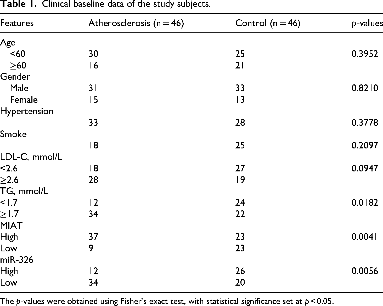
The p-values were obtained using Fisher's exact test, with statistical significance set at p < 0.05.
GEO database analysis and prediction of miRNAs
The differentially expressed genes (DEGs) in the AS-related GEO dataset GSE28829 were downloaded from the GEO database (https://www.ncbi.nlm.nih.gov/geo/query/acc.cgi?acc=GSE28829) and analysed via the online software GEO2R (https://www.ncbi.nlm.nih.gov/geo/geo2r/) with a filtration criterion of adjusted P [P (adj)] < 0.05. The differentially expressed miRNAs in the AS-related GEO dataset GSE24548 were also downloaded from the GEO database (https://www.ncbi.nlm.nih.gov/geo/query/acc.cgi?acc=GSE24548) and were analysed by GEO2R with a filtration criterion of P (adj) < 0.05. The potential miRNAs that might interact with MIAT were predicted via the online databases DIANA-LncBase 21 and miRDB 22 on the basis of the instructions of the websites. The identified miRNAs from GSE24548 and the predicted miRNAs from the abovementioned two online databases were subjected to the online Venn diagram tool jvenn 23 to screen potential miRNAs that might interact with MIAT during AS.
Cell culture and ox-LDL treatment
VSMCs were obtained from American Type Culture Collection (ATCC; Manassas, VA, USA; RRID: CVCL_4009) and maintained in DMEM (Gibco, El Paso, TX, USA) as previously described. 24 All media were supplemented with 10% foetal bovine serum (FBS, Gibco), 100 IU/ml penicillin (Solarbio, Beijing, China), 100 mg/ml streptomycin (Solarbio), and 250 ng/ml amphoteracin B (Solarbio). The VSMCs were cultured in a humidified atmosphere containing 5% CO2 at 37 °C.
For generation of low-AS VSMC and high-AS VSMC models, 10 μg/mL ox-LDL and 50 μg/mL ox-LDL were added to the culture medium for 24 h as previously reported, 25 and the expression of MCP-1, an inflammatory marker of VSMCs, was measured by a western blot analysis. 26 The treated VSMCs were used for further functional assays.
Western blot analysis
The procedure was performed as previously described. 27 The VSMC clumps were collected and then lysed with radioimmunoprecipitation assay (RIPA) buffer. The cell pellet was discarded after centrifugation, and the supernatant was collected. A bicinchoninic acid (BCA) protein quantitative analysis was used to determine the protein concentration. After protein quantification, loading buffer was added to the protein mixture, and the protein mixture was denatured by heating at 100 °C for 5 min. An 8% sodium dodecyl sulphate (SDS) polyacrylamide gel was used for protein electrophoresis, and the protein was transferred to a polyvinylidene fluoride (PVDF) membrane with a constant current of 400 mA, incubated with high-efficiency blocking solution at room temperature for only 10 min, incubated with a primary antibody targeting MCP-1 (Abcam, Cambridge, MA, UK) at room temperature for 1 h and incubated overnight at 4 °C. After being washed with TBST 3 times, the membrane was incubated with secondary antibody at room temperature for 60 min. A gel imaging system was used to scan the sample, and the results are expressed as the grayscale value/internal reference grayscale value.
RNA extraction and reverse transcription–quantitative (Rt–qPCR)
This procedure was executed as described previously. 28 In brief, total RNA from plasma samples or from VSMC models was extracted with TRIzol according to the manufacturer's instructions (Invitrogen, Carlsbad, CA, USA). The isolated RNA was reverse transcribed into cDNA using PrimeScript RT Master Mix (TaKaRa Bio, Inc., Dalian, China), and qPCR was carried out with a SYBR Green PCR kit (TaKaRa) according to the manufacturers' instructions. The endogenous controls for MIAT and miR-326 were GAPDH and U6, respectively. The relative expression of MIAT and miR-326 was determined using the 2−ΔΔCt method. The primer sequences are listed in Table 2.
Primer and oligonucleotide sequences were used in the present research.

Plasmid and oligonucleotide transfection
Specific small interfering RNAs targeting MIAT (siMIAT-1 and siMIAT-2), scrambled small interfering RNAs (siSCR) and an overexpression plasmid loaded with MIAT (oeMIAT) were designed and chemically synthesized by RiboBio Co., Ltd (RiboBio, Guangzhou, China). The miR-326 mimics and corresponding negative control mimics (NC mimics) were also synthesized by RiboBio Co., Ltd VSMCs were seeded in 6-well plates at a concentration of 2 × 10⁵ cells/well. The cell transfection procedure was performed by using Lipofectamine 3000 (Invitrogen, Carlsbad, CA, USA) following the manufacturer's instructions. The sequences of the siRNAs, plasmids and oligonucleotides used are listed in Table 2. For establishment of stable MIAT-overexpressing cell models, VMSCs were treated with ampicillin (8 μg/ml) for one week. Different expression levels of MIAT in selected cell clones were quantified via RT–qPCR, and the selected cell clones with stably upregulated MIAT were used for further functional assays. For determination of the effect of miR-326 on MIAT-mediated migration, miR-326 mimics and NC mimics were transfected into MIAT stable overexpression (oeMIAT) VMSCs as described above. Changes in the migration of VSMCs were determined by a Transwell assay.
Transwell assay
Transwell assays were performed to evaluate changes in the VSMC migratory ability as described previously. 29 In brief, after diverse treatments, VSMCs were seeded on the upper layer of a Transwell chamber (Corning, New York, USA) containing a polycarbonate membrane filter (pore size: 8 μm). Culture medium without serum was added to the upper wells, and medium containing 10% FBS was added to the lower wells. After 24 h of incubation at 37 °C with 5% CO2, nonmigrated cells were removed. After that, all the wells were fixed in 4% paraformaldehyde and stained with crystal violet. The cells were washed with PBS 3 times, and the results were observed per chamber with a 100 × inverted microscope (Olympus, Tokyo, Japan).
Fish
The procedure was carried out as described previously. 30 Specific probes targeting MIAT or miR-326 were synthesized by RiboBio Co., Ltd Briefly, VSMCs were seeded onto glass coverslips (0.8 × 0.8 cm) and grown to 80–95% confluence. The coverslips were washed with PBS twice and then fixed with 4% paraformaldehyde for 10 min, followed by blocking with 5% BSA (Sigma, St Louis, MO, USA) for 1 h. After permeabilization with 0.3% Triton X-100 (Sigma) for 15 min, the coverslips were incubated in hybridization solution containing 1 μM RNA probes supplemented with 1% bovine serum albumin (BSA) in a humid chamber at 37 °C overnight. The next day, the coverslips were gently washed with 2 × SSC (with 50% formamide for 5 min), 1 × SSC (for 5 min) or 0.5 × SSC (for 10 min) at 37 °C. The coverslips were subsequently incubated with 4′,6-diamidino-2-phenylindole (DAPI) for 8 min in the dark to stain the nucleus. Finally, the coverslips were washed with PBS, dried at room temperature, and photographed with a Leica SP5 confocal microscope (Leica Microsystems, Mannheim, Germany).
Dual-luciferase reporter assay
The experimental procedure was implemented as previously described. Wild-type or mutant MIAT luciferase reporter plasmids (MIAT-wt and MIAT-mut) containing miR-326 response elements or substitutions of target sites were synthesized by RiboBio Co., Ltd For verification of the targeted binding effect between MIAT and miR-326, the synthesized reporter plasmids were cotransfected with miR-326 mimics or NC mimics into VSMC models according to the instructions of Lipofectamine 3000 (Invitrogen). Forty-eight hours later, the luciferase activity was measured with a Dual-Luciferase Reporter Assay System (Promega, Madison, WI, USA) according to the manufacturer's instructions.
Statistical analysis
Statistical data were analysed with GraphPad Prism 8.0 (GraphPad Software, San Diego, California, USA). For comparisons between two groups, statistical analyses were performed by unpaired two-tailed Student's t test. Comparisons of multiple groups were made by one-way ANOVA, and statistical significance among multiple groups was determined by Tukey's post hoc test (for pairwise comparisons of means). All the results are presented as the means ± SDs from at least three independent experiments. Multivariate logistic regression analysis was used to analyse the influencing factors of AS (Table 3). *P < 0.05, **P < 0.01 and ***P < 0.001 were used to indicate significant differences.
Multivariate logistic regression analysis.
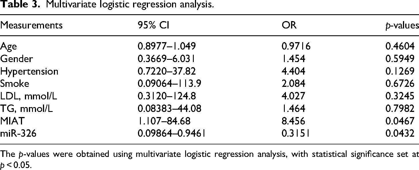
The p-values were obtained using multivariate logistic regression analysis, with statistical significance set at p < 0.05.
Results
MIAT was upregulated in patients with as and in ox-LDL-treated VSMCs
By using the online software GEO2R, we analysed the DEGs in the AS-related GEO dataset GSE28829. As shown in Figure 1(a), according to the filtration criteria of P (adj) < 0.05, a total of 3709 DEGs were differentially expressed in 13 early AS plaques from human carotid samples and in 16 advanced AS plaques from human carotid samples. MIAT (probe ID of 228658_at) was significantly upregulated in advanced AS plaques compared with early AS plaques (Figure 1(b) and (c)). Additionally, the expression of MIAT in 46 plasma samples from patients with AS and in paired plasma samples from patients without AS was determined by RT–qPCR. As shown in Figure 1(d) and (e), the expression of MIAT was markedly elevated in most (41/46, 89.13%) plasma samples from patients with AS. At the cellular level, low-AS (10 μg/mL ox-LDL) and high-AS (50 μg/mL ox-LDL) VSMC models were first constructed as previously reported. 31 The expression of MCP-1, a well-known biomarker of VSMC injury during the progression of AS in the above cell models, was detected by western blot analysis. As shown in the representative image in Figure 1(f), compared with that in the control group, the protein expression of MCP-1 was significantly increased in the low-AS and high-AS groups, indicating that the atherosclerotic VSMC model was constructed successfully. Finally, the expression of MIAT in the constructed cell models was measured via RT–qPCR. As shown in Figure 1(g), the expression of MIAT was upregulated in the low-AS and high-AS VSMC models.

MIAT was upregulated in patients with AS and ox-LDL-treated VSMCs. (a) The DEGs associated with advanced AS plaques and early AS plaques in the AS-related GEO dataset GSE28829 were analysed using GEO2R. (b) The expression of MIAT in 16 advanced AS plaque samples and in 13 early AS plaque samples. Probe ID 228658_at represents MIAT. (c) The expression of MIAT in 16 advanced AS plague samples was significantly greater than that in 13 early AS plague samples. ****P < 0.0001. (d) MIAT expression was upregulated in most (41/46, 89.13%) plasma samples from patients with AS, as detected via RT–qPCR. ****P < 0.0001. (e) MIAT expression in 46 plasma samples from patients with AS and in 46 paired plasma samples from patients without AS, as determined via RT–qPCR. ****P < 0.0001. (f) MCP-1 expression in ox-LDL-treated VSMC models. ****P < 0.0001. Con, treated with double distilled water; low-AS, treated with 10 μg/mL ox-LDL; high-AS, treated with 50 μg/mL ox-LDL. (g) The expression of MIAT in ox-LDL-treated VSMC models was measured via RT–qPCR. ****P < 0.0001.
MIAT knockdown suppressed the migration of ox-LDL-treated VSMCs
As a main reason for AS, the anomalous migration of VSMCs from the media to the intima contributes to the thickening of the arterial intima.32,33 In this section, the functional role of MIAT in VSMC migration was further explored. The expression of MIAT in the constructed low-AS and high-AS VSMC models was first knocked down via the transfection of specific siRNAs targeting MIAT (Figure 2(a)). Changes in the migration of low-AS- and high-AS-expressing VSMCs were subsequently evaluated by a Transwell assay. As shown in the representative images presented in Figure 2(b), MIAT knockdown decreased the migration of low-AS and high-AS VSMCs.

MIAT knockdown suppressed the migration of ox-LDL-treated VSMCs. (a) The expression of MIAT in ox-LDL-treated VSMC models after transfection with scramble small interfering RNAs (scramble siRNA, siSCR) or specific siRNAs targeting MIAT (siMIAT-1 and siMIAT-2) was confirmed via RT–qPCR. **P < 0.01. (b) Cell migration changes after the transfection of ox-LDL-treated VSMC models with siRNAs targeting MIAT. **P < 0.01. Magnification:×400; scale bar: 200 μm.
miR-326 is downregulated in as and ox-LDL-treated VSMCs
The most prevalent working mechanism of lncRNAs is that they serve as ceRNAs via the absorption of certain miRNAs. To explore the potential miRNAs that might interact with MIAT, we initially analysed GSE24548, a miRNA-related GEO dataset concerning AS, in the present research. With the filtration criterion of P (adj) < 0.05, a total of 68 miRNAs (4 upregulated and 64 downregulated miRNAs) were screened out (Figure 3(a)). Two online software programs, DIANA-LncBase 21 and miRDB, 22 were subsequently used to predict potential miRNAs that might interact with MIAT during AS. As shown in Figure 3(b), miR-326 was the only miRNA that overlapped in all three abovementioned datasets or databases. Interestingly, miR-326 was downregulated in four platelet samples from patients with FAMI compared with three platelet samples from healthy people (control) (Figure 3(c) and (d)). Additionally, miR-326 was downregulated in 46 plasma samples from patients with AS and in paired plasma samples from patients without AS according to a series of RT–qPCR analyses (Figure 3(e)). Moreover, the correlation between MIAT and miR-326 was further analysed. Encouragingly, as shown in Figure 3(f), MIAT was negatively correlated with miR-326 (r = − 0.6591, P < 0.0001). Additionally, the results of an RT–qPCR assay indicated that miR-326 was downregulated in low-AS and high-AS VSMCs (Figure 3(g)). For determination of the functional role of miR-326 in VSMC migration, miR-326 mimics were first transfected into low-AS and high-AS VSMC models. As shown by the RT–qPCR results presented in Figure 3(h), the expression of miR-326 in the low-AS and high-AS VSMC models was significantly upregulated in the miR-326 mimic group. Furthermore, a Transwell assay was performed to detect changes in migration after various types of miR-326 intervention. As shown in the representative images in Figure 3(i), upregulation of miR-326 weakened the migration of VSMCs.

miR-326 was downregulated in AS- and ox-LDL-treated VSMCs. (a) The differentially expressed miRNAs in 17 platelet samples from patients with FAMI and in 17 matched platelet samples from control patients were analysed in GSE24548 via GEO2R with the filtration criterion of adjusted P(adj) < 0.05. (b) The predicted miRNAs that might interact with MIAT in the LncBase and miRDB databases overlapped with the identified miRNAs in GSE24548. miR-326 was selected. (c-d) The expression of miR-326 in platelet samples of FAMI and in platelet samples of the control is presented. **P < 0.01. (e) The differential expression of miR-326 in 46 plasma samples from patients with AS and in 46 paired plasma samples from patients without AS was quantified via RT–qPCR. ****P < 0.0001. (f) The relationship between MIAT and miR-326 in 46 plasma samples from patients with AS was analysed via Spearman correlation analysis. r = − 0.6591, P < 0.0001. (g) The expression of miR-326 in ox-LDL-treated VSMC models was determined via RT–qPCR. ****P < 0.0001, *P < 0.05. (h) The expression of miR-326 after transfection of miR-326 mimics and corresponding negative control mimics (NC mimics) in ox-LDL-treated VSMC models was determined via RT–qPCR. ****P < 0.0001. (i) Cell migration changes after diverse miR-326 interventions in ox-LDL-treated VSMC models were evaluated by a Transwell assay. Magnification:×400; scale bar: 200 μm. **P < 0.01.
MIAT sponged mir-326 to facilitate the migration of ox-LDL-treated VSMCs
In this study, MIAT and miR-326 were first shown to colocalize in the cytoplasm of VSMCs, indicating that MIAT and miR-326 have a biological basis of interaction (Figure 4(a)). By using DIANA-LncBase, MIAT was predicted to supply theoretical binding sites for miR-326 (Figure 4(b)). A luciferase assay was subsequently performed to verify the targeted binding effect between MIAT and miR-326. As shown in Figure 4(c) and (d), cotransfection of miR-326 mimics and wild-type reporter plasmids (wt-MIAT) containing wild-type miR-326 response elements (MRE-326) resulted in a significant decrease in luciferase activity. When the predicted MRE-326 was mutated (cotransfection of miR-326 mimics and mutant reporter plasmids, mut-MIAT), the luciferase activity was increased. These findings strongly suggest that miR-326 directly targets MIAT. Finally, through a Transwell assay, upregulation of MIAT promoted low-AS and high-AS VSMC migration, whereas this effect was attenuated by further upregulation of miR-326 (Figure 4(e)). Taken together, these findings indicate that MIAT facilitates the migration of VMSCs via miR-326 sponging.

MIAT sponged miR-326 to facilitate the migration of ox-LDL-treated VSMCs. (a) MIAT and miR-326 were colocalized in the cytoplasm of VSMCs, as determined by a FISH assay. (b) MIAT supplies theoretical miR-326 response elements (MRE-326) as predicted by DIANA-lncBase. (c) Diagram of the wild-type and mutated luciferase reporter plasmids. (d) The targeted binding effect between MIAT and miR-326 was verified by a luciferase assay. **P < 0.01, n.sP > 0.05. (e) Cell migration changes after diverse MIAT and miR-326 interventions in ox-LDL-treated VSMC models were evaluated via a Transwell assay. Magnification:×400; scale bar: 200 μm. ****P < 0.0001, n.sP > 0.05.
Discussion
Currently, lncRNAs play important roles in the regulation and genetic programming of various cardiovascular diseases.34,35 The lncRNA-mediated switching of VSMCs from a contractile phenotype to a synthetic phenotype contributes greatly to the progression of AS.3,36 As a basic pathological condition, AS, which involves the migration and proliferation of VSMCs, is now closely associated with epigenetic regulation powered by genetic materials. 37 MIAT, located on chromosome 22q12.1, is reported to be a key regulator in diverse diseases. 38 In the present study, via online bioinformatic analysis, MIAT was significantly upregulated in advanced AS plaques from human carotid samples, indicating its potential function in AS; therefore, MIAT was selected for further study. In the present study, ox-LDL was used to simulate VSMC injury during AS progression as previously reported. The expression of MCP-1, a well-known biomarker of VSMC injury during the progression of AS, was quantified via a western blot analysis to evaluate the success of the AS cell model. Francesca and colleagues reported that knockdown of MIAT weakened the proliferation and migration of human carotid artery smooth muscle cells (SMCs) via regulation of the early growth response 1 (EGR1)-ETS transcription factor ELK1 (ELK1)-extracellular signal-regulated kinase (ERK) pathway. 9 Similar to previous studies, further RT–qPCR verified that MIAT was upregulated in AS both at the tissue level and at the cellular level. Functionally, in the present study, the results of the transfection of specific siRNAs targeting MIAT revealed that the downregulation of MIAT suppressed VSMC migration.
Previous studies have verified that there are different kinds of mechanisms by which lncRNAs mediate cell programs, such as scaffold, decoy and transcriptional effects. 39 In addition, lncRNAs are located in the cytoplasm and function as miRNA sponges that inhibit the effects of miRNAs. 40 The ceRNA theory, first proposed by Leonardo Salmena in 2011, has been broadly applied in noncoding RNA research. In the present study, MIAT was found to be located mainly in the cytoplasm of VMSCs, indicating that MIAT might function via miRNA sponging. Through bioinformatic analysis, miR-326 was predicted to bind to MIAT. The function of miR-326 in various systemic diseases, such as those of the digestive, urinary and endocrine systems,41–43 has been investigated, and recent research has revealed the regulatory role of miR-326 in cardiac hypertrophy. 44 The current research focused on the interaction between MIAT and miR-326. To investigate whether miR-326 also participates in the progression of AS, we investigated the expression and cellular function of miR-326 in VSMCs. Notably, miR-326 was downregulated in AS samples and cell models, and miR-326 was negatively correlated with MIAT. Furthermore, we investigated whether miR-326 could interact with MIAT. A FISH assay revealed that miR-326 was colocalized with MIAT in the cytoplasm of VSMCs, indicating that miR-326 and MIAT share a biological basis of interaction. Cytoplasmic miRNAs often directly bind to lncRNAs. Therefore, a luciferase assay was designed to confirm the targeted binding effect between MIAT and miR-326. Convincingly, the findings of the luciferase assay confirmed that wt-MIAT but not mut-MIAT bound to miR-326. Further overexpression assays revealed that MIAT-mediated migration of VSMCs could be reversed by overexpression of miR-326. Thus, MIAT promoted the migration of VSMCs by acting as a ceRNA by decoying miR-326 in AS.
However, this study validated only the role of the MIAT/miR-326 axis in an in vitro VSMC model. To gain a more comprehensive understanding of the role of this axis in physiological processes, future studies should include animal models. Another limitation is the lack of formal sample size analysis, although the sample size in this study is sufficient to address the core research questions. As part of the complex ceRNA network, the downstream targets of miR-326 are likely intricate. Further exploration is needed to verify the interactions between MIAT, miR-326, and specific mRNAs.
Conclusion
The migration of VSMCs plays important roles in AS pathogenesis and is facilitated by numerous molecules and pathways. Our present study revealed that MIAT serves as a promoter for the migration of VSMCs and that MIAT functions by sponging miR-326, which plays a negative role in the regulation of AS. As a regulator of AS, MIAT has good potential as a diagnostic marker and therapeutic target in AS. Therefore, the MIAT/miR-326 axis may provide a novel target for the treatment of AS at the molecular level.
Supplemental Material
sj-zip-1-sci-10.1177_00368504251335854 - Supplemental material for Long noncoding RNA MIAT regulates VSMC migration by sponging miR-326
Supplemental material, sj-zip-1-sci-10.1177_00368504251335854 for Long noncoding RNA MIAT regulates VSMC migration by sponging miR-326 by Yuxin Bao, Yinzhou Luo, Hanjie Zhai, Jie Lu, Man Zhang and Ningning Wang in Science Progress
Footnotes
Ethical Considerations
All atherosclerosis tissues and matched normal tissues were processed in accordance with the ethical guidelines of Central Hospital Affiliated to Shenyang Medical College (Approval No.: 2016SEP12-12, September 12, 2016). Written informed consent was obtained from every patient before the study. All patients whose data were approved could be used for studies.
Author contributions/CRediT
WN conceived and designed the study and revised the manuscript. BY, LY and ZH made efforts to acquire the data and design Tables. Y, LY and LJ drafted the article or revised it critically for important intellectual content. WN and HM and HX and ZM wrote the paper. All the authors read and approved the final manuscript.
Funding
The present study was supported by grants from the Scientific Study Project for Institutes of Higher Learning, Ministry of Education, Liaoning Province (grant no. LJKZ1155) and the Technological Innovation Fund of Shenyang Technology Division (grant no. 21-173-9-20).
Conflicting interests
The authors declared no potential conflicts of interest with respect to the research, authorship, and/or publication of this article.
Data availability
The authors declare that the data supporting this study's findings are available within the paper. Should any raw data files be needed in another format, they are available from the corresponding author upon reasonable request.
Supplemental material
Supplemental material for this article is available online.
References
Supplementary Material
Please find the following supplemental material available below.
For Open Access articles published under a Creative Commons License, all supplemental material carries the same license as the article it is associated with.
For non-Open Access articles published, all supplemental material carries a non-exclusive license, and permission requests for re-use of supplemental material or any part of supplemental material shall be sent directly to the copyright owner as specified in the copyright notice associated with the article.