
Abstract
The ceramic-polymer composite materials are widely known for their exceptional mechanical and biological properties. Polycaprolactone (PCL) is a biodegradable polymer material extensively used in various biomedical applications. At the same time, barium titanate (BT), a ceramic material, exhibits piezoelectric properties similar to bone, which is essential for osseointegration. Furthermore, a composite material that combines the benefits of PCL and BT results in an innovative composite material with enhanced properties for biomedical applications. Thus, this review is organised into three sections. Firstly, it aims to provide an overview of the current research on evaluating biological properties, including antibacterial activity, cytotoxicity and osseointegration, of PCL polymeric matrices in its pure form and reinforced structures with ceramics, polymers and natural extracts. The second section investigates the biological properties of BT, both in its pure form and in combination with other supporting materials. Finally, the third section provides a summary of the biological properties of the PCLBT composite material. Furthermore, the existing challenges of PCL, BT and their composites, along with future research directions, have been presented. Therefore, this review will provide a state-of-the-art understanding of the biological properties of PCL and BT composites as potential futuristic materials in biomedical applications.
Introduction
Bone is a remarkable tissue with a great potential for regeneration following injury. Throughout history, numerous traditional medicinal practices across various cultures have been centered on bone regeneration. Despite significant advancements in biomaterials and medicine over the past few decades in managing bone defects, large bone defects still necessitate the insertion of bone grafts. Most of the time, cadaveric, autologous and xenogeneic tissue is used to create bone grafts; this carries a risk of transplant rejection and unfavourable inflammatory and immunological reactions. New treatments and medical equipment are being developed to enhance patient quality of life by preventing or minimising this type of response from the body. Tissue engineering aims to restore or regenerate damaged tissues and organs. The scaffolds have been mixed with bioactive agents, such as growth factors, to facilitate tissue growth and mimic the extracellular matrix.1,2
Natural or synthetic polymers, ceramics and their composite materials have been widely used as biomaterials, most frequently employed for building scaffolds and implant coating materials that support the formation of bone tissue.3–6
Synthetic biodegradable polymers find numerous applications in the field of biomedicine, while biopolymers are desirable materials for biomedical implants due to their high bioactivity, non-toxicity and resorbability properties. Furthermore, polymers can be considered ‘smart’ materials that have gained popularity as polymer coatings in various applications, such as corrosion resistance, surface functionalisation, wear resistance, enhancement of bioactivity and even switchable smart materials. 7 A crucial component of regenerative medicine is the creation of porous scaffolds from synthetic polymers that are biocompatible and biodegradable. 8
Ceramic-polymer bioactive composites have been developed to integrate the inherent qualities of each component and optimise the physicochemical and biological properties required for complex tissue engineering. Such composite systems offer a degree of flexibility, favourable mechanical properties, enhanced biological activity and improved osteoconductivity when bioactive ceramics are combined with biodegradable polymers such as polycaprolactone (PCL), polyglycolic acid, polylactic acid (PLA) and their copolymers. 9
While bioactive ceramics are biologically active and osteoconductive, they are also quite brittle and lack flexibility and moldability. As a result, their use has been limited to non-load-bearing sections. The ceramics must first undergo a high-temperature sintering to incorporate them into a bulk body. However, their brittleness makes the sintered part unsuitable for load-bearing locations. The density of bioceramics is also affected by sintering. Generally, densification eliminates pores acting as defects in the microstructure. Although the high-temperature treatment improves densification and reduces porosity, the bulk material may crack. High porosity promotes corrosion and releases material ions induced by the body's environment. Reduction of porosity by high temperature promotes cracks that might result from the difference in thermal coefficients of materials. This affects the expansion and shrinkage differences between the composite materials at heating and cooling during the sintering process, thus resulting in thermal stress. 10 Combining bioactive ceramics with flexible and moldable polymers is, therefore, one of the most effective ways to create integrated ceramics that are easy to handle. 11 A concern when using polymers in the in vivo condition is the acidic environment created during their degradation, which can lead to inflammatory issues. Therefore, ceramic buffering fillers have been introduced to maintain a neutral pH level. 12
The qualities of ceramics significantly impact the degradation rate and the mechanical and biological properties of ceramic/polymer composites. 13 This paper aims to investigate the antibacterial, cytotoxicity and osseointegration biological properties of ceramic material (barium titanate [BT]), polymer (PCL) and their composites.
Polycaprolactone
Polycaprolactone belongs to the family of aliphatic polyesters and is a hexanoate repeat polymer. Its molecular weight and degree of crystallinity influence its thermal, mechanical and physical properties, affecting its ability to degrade under physiological conditions via hydrolysis of its ester bonds. Polycaprolactone is highly semicrystalline, hydrophobic, easily processable due to its low melting temperature, highly soluble at room temperature and exhibits excellent mix compatibility. As the molecular weight of PCL increases, its crystallinity tends to decrease.14,15 The melting point of PCL is between 59°C and 64°C, and its glass transition temperature is −60°C. Polycaprolactone is soluble in solvents such as acetic acid, formic acid, benzene, carbon tetrachloride, chloroform, dichloromethane, toluene, 2-nitropropane and cyclohexanone at room temperature. Polycaprolactone is insoluble in diethyl ether and petroleum ether with low solubility in acetonitrile, acetone, ethyl acetate, 2-butanone and dimethylformamide (DMF). Because of its superior solubility properties, chloroform was the main solvent employed for PCL. The particles created with this solvent typically have small pores because of the quick evaporation of chloroform. However, the biomedical field has avoided employing this solvent as much due to concerns regarding its toxicity and carcinogenicity. Acetic acid exhibits favourable solubility characteristics for PCL. Because DMF has partial solubility parameters for PCL, using it as a co-solvent can change the chain entanglement, particle shape and chain–solvent interactions. Dimethylformamide can potentially be hepatotoxic, even though its toxicity varies depending on the patient's liver health, dosage and duration of exposure. Although they have been added to chloroform, formic and acetic acid solutions in several investigations to increase electrospinnability, methanol and ethanol are not PCL solvents.16–19 The PCL degradation rate can be controlled by molecular weight, crystallinity or modifying the structure using hydrophilic polyethylene glycol (PEG), ceramics or the creation of copolymers with other polymers.14,20
Polycaprolactone has been approved by the FDA (Food and Drug Administration) and (Conformité Européenne [CE] marking) for use in a wide range of drug delivery and medical devices, but only a few have been marketed or translated into clinical research. In physiological conditions, PCL is degraded via hydrolysis of its ester bonds (such as in the human body). Polycaprolactone degrades in two stages: first, non-enzymatic hydrolytic cleavage of ester groups, and second, when the polymer is more highly crystalline and has a low molecular weight (less than 3000), the polymer is shown to undergo intracellular degradation, as evidenced by experiments of PCL fragments uptake in macrophage and giant cell phagosomes, as well as within fibroblasts.14,21
A promising material for biomedical applications can be generated by functionalising the fibrous structures of PCL using nanoparticles to give them new physical and chemical properties (improving the strength and endurance or enhancing antimicrobial and anti-inflammatory capabilities). It would also be necessary to add growth factors to obtain suitable properties for adequate tissue regeneration. 22
Degradation studies of non-woven materials consisting of PCL nanofibers in vitro and in vivo revealed that electrospun PCL materials disintegrated considerably faster in vivo than in vitro, owing to enzymatic and hydrolytic degradation of PCL. 23 Polycaprolactone/β-Tri-calcium phosphate (TCP) composite scaffolds break down more quickly in vivo than PCL homopolymer scaffolds after six months. 24
Antibacterial properties of PCL and its combinations
Polycaprolactone alone does not possess antibacterial properties. This specific characteristic is crucial. Polycaprolactone is a form of polymer that may be broken down by a wide variety of bacteria. Polycaprolactone's qualities may be changed and improved by adding micro and nano-sized chemicals. 25 In addition to increasing susceptibility to infection, tissue damage caused by surgery or ingesting foreign materials also activates the host's defenses and encourages the generation of inflammatory mediators, which are boosted by bacterial activity and toxins. 26 Implants can be specifically employed in laryngology, as the sinus and nasal surgical fields are not entirely sterile. Numerous studies indicate the need for PCL changes to improve its bactericidal potency. Polycaprolactone itself has the potential to carry antimicrobial agents, and its alterations typically involve adding chemicals with well-known bactericidal properties, such as graphene, bioglass, copper, silver, zinc oxide or silver.27,28 Low MBC (minimal bactericidal concentration; the minimum concentration needed to kill at least 99.9% of the bacteria cells) and minimal inhibitory concentration; the minimum concentration required to resist bacterial growth) values were indicators of the copolymers’ high antibacterial activity. 26 Table 1 summarises the antibacterial effects of PCL combined with different materials in the form of composites.
The antibacterial effect of PCL and composite of PCL and other components.

Cytotoxicity studies of PCL and its associated compounds
One of the methods for assessing and screening biological properties is the cytotoxicity test, which examines the effects of medical devices on cell growth, reproduction and morphology using tissue cells grown in vitro. 54 The ISO 10993-5 standard specifies three types of cytotoxicity tests: extract, direct and indirect contact (including agar overlay assay and filter diffusion). The extracted test is typically consistent with the results of animal toxicity testing and is useful for determining the toxicity of soluble components of medical devices. The direct contact assay is the most sensitive test for detecting even minimal cytotoxicity caused by medical devices. Molecular filtration and bulk filtering methods are both appropriate for assessing the biocompatibility of the hazardous components of small molecular-weight medical devices. The agar overlay assay is suitable for evaluating highly toxic medical devices.55,56 Polycaprolactone's hydrophobic nature presents a challenge for tissue engineering, as it hinders cell adhesion. Therefore, various surface treatments have been explored to create a more conducive environment for cell growth and proliferation, such as treating PCL with different solutions to enhance its hydrophilicity and promote cell attachment to the polymer. 57 Prior attempts at using pure PCL could not provide the best possible mechanical strength and biocompatibility. Many studies have attempted to modify the PCL. Polycaprolactone scaffolds with a double protein coating have been shown to have better early cell adhesion, proliferation and colonisation. 58 When tested on isolated human tonsil-derived mesenchyme stem cells, Lee et al. demonstrated that a 3D PCL scaffold has osteonduction. The cells rapidly underwent differentiation to become osteoblast-like cells with osteo-prompting properties. 59 Lin et al. designed a PCL composite membrane that contains 20 wt% of cobalt-substituted hydroxyapatite powder, significantly increased cell proliferation (over 90% after seven days of culture) was observed. 34 Ho et al. investigated the effects of Biodentine/PCL composite on human dental pulp cells. The bioactivity, proliferation and odontogenic differentiation of human dental pulp cells cultured on the composite exhibited a good apatite-forming ability and capability to support proliferation and differentiation. 60 The PCL / PEG hydrogel composite inhibits mineralisation and osteoblastic differentiation of cultured calvarial cells. 61 The PCL-Biosilicate composite showed adequate mechanical properties without increasing toxicity. 62 In vitro test of gelatin collagen (GC) and PCL with two different ratios (GC: PCL; 1:8 and 1:20) showed better cell proliferation on the composite with a lower collagen content when tested on adipose-derived stem cells. 63 Table 2 summarises the cytotoxicity effects of PCL and its associated composites.
Summary of cytotoxicity effect of PCL and PCL blended compound.

Osseointegration of PCL composites
Osseointegration is calculated using a sample of the implant and peri-implant bone that has been stained to determine the amount of peri-implant bone and bone-implant contact. Although accurate measurement is beneficial, long-term research cannot use it because of the invasive and damaging process. It is utilised in non-clinical research and testing. It is evaluated before, during and after surgery. 67 Due to PCL's hydrophobicity and lack of functional groups that encourage cell growth and proliferation, cellular interactions are restricted. Modifications are required to improve the PCL's cellular compatibility and capacity for bone repair. Protein adsorption, necessary for cell identification and attachment, decreases at very low contact angles due to an increased water intake. However, low surface energy and very high contact angle result in poor cell conductivity and protein denaturation. Arginine-glycine-aspartic acid-PCL composite demonstrated smoother surfaces and early bone deposition onto composite surfaces.68,69 In vivo blending of 1% Sr2+ with PCL greatly accelerated bone tissue regeneration without causing localised irritation. 70 Combining calcium lactate and cellulose acetate solutions with a PCL nanofibrous scaffold significantly improves the composite fibre's bio-physio-chemical characteristics. 71 When PCL is functionalised with polydopamine and rhBMP2, the surface wettability, cell proliferation and osteogenic potential are improved. 72 Compared to pure PCL, hydroxyl-functionalised PCL exhibited 70% porosity, improved cell adhesion, higher metabolic activity and osteogenic potential. 73 According to Spalthoff, 74 PCL-TCP composite indicated that a significant portion of the composite material had deteriorated and been replaced by vital bone.
Compared to the pure PCL, cell growth is noticeably greater in composites, including those made of recombinant PCL/SF (Silk Fibroin) composite, spider silk protein/PCL/gelatin, PCL/chitosan composite and PCL/biphasic calcium phosphate hybrid composite.75–77
Increased cell proliferation is made possible by adding HA to the PCL/SF composite scaffold. At the same time, collagen-I synthesis is lowered, and osteoinductivity is highly favoured by adding bone morphogenetic protein 4-expressing bone marrow stromal cells.78,79 Table 3 lists the effects of pure and blended PCL when implanted inside a live animal.
Summary of osseointegration effect of pure PCL and blended PCL.

Barium titanate
Barium titanate (BaTiO3) is a polycrystalline ceramic material with ferroelectricity properties with perovskite structure (ABO3) (Figure 1). The unique features and widespread use in a variety of devices, ferroelectrics are gaining popularity.87,88 The bulk physical characteristics and crystal structure of BaTiO3 have been extensively studied since Wul and Goldman discovered BaTiO3 ferroelectricity in 1945. 89

Crystalline structure of BT material. 67
The BaTiO3 exhibits four crystal phases in bulk: cubic, tetragonal, orthorhombic and rhombohedral. At high temperatures (T > 120°C), it has the conventional perovskite structure (i.e. cubic phase), in which Ba2+ and O2− ions form the FCC lattice (Ba2+ at the corners and O2− in the center of each face of the lattice). At the same time, the smaller Ti4+ cation occupies the octahedral interstitial spaces, forming [TiO6] in the FCC array.90,91
The cubic phase structure of BaTiO3 is very symmetric, with cation and anion centers coinciding, resulting in no polarisation in the crystal structure and, therefore, no ferroelectricity (i.e. paraelectric). Upon reducing the temperature to less than 120°C, the cubic symmetric structure becomes non-symmetric due to a deviation of the cation center from the anion center, leading to net spontaneous polarisation in the crystal structure and transition from paraelectric to ferroelectric phase, exhibiting a tetragonal structure. Further cooling between −5°C and −90°C leads to the conversion to the orthorhombic phase, which can be considered the elongation of the cubic unit cell in the face diagonal direction. Finally, at T < −90°C, the rhombohedral phase occurs, which represents the elongation of the cubic unit cell along with the body diagonal direction. Non-symmetry in the crystal structure results from distortions along with these directions, with the deviation of the cation center from the anion center, resulting in ferroelectric phases with net spontaneous polarisation (Figure 2).89,91

A schematic representation of the four different crystalline structures of BaTiO3. 89
Barium titanate (BaTiO3) piezoelectric ceramic is often used to stimulate bone regeneration by mimicking natural bone stress-generated potentials, which produce micro-electric currents and enhance calcium salt deposition at the bone defect site. The piezoelectric polarisation stimuli generated by BaTiO3 can modify bone development, shaping and reconstructing the tissue as native bone undergoes deformation. 92
The domain in piezoelectric ceramic materials refers to many unit structures having the same polarisation orientation. Each part has its polarisation direction, randomly orientated to keep the material's net polarisation at zero without an external electric field (EF). 93 A strong external voltage applied at a high temperature aligns the domains parallel to the direction of the applied EF. 94
The piezoelectric action of BaTiO3 was discovered to enhance osteoblast development. It should be highlighted that most prior research used bulk BT materials. Bone tissue regeneration occurs mainly at the implant-tissue interface during the first four months of the healing period for hard tissue replacement materials. As a result, tiny micro-scale BT coatings coated on artificial metal (Ti or Zr alloys) implants will be more favourable. The latter could provide protective coatings for implants while also achieving electroactive performance. The solid-state reaction, sol-gel technique and physical vapor deposition are now used to make BT coatings or films. 95
Barium titanate demonstrates good biocompatibility and the ability to develop a strong interfacial connection with the surrounding bone. The biocompatibility of BT in its nanoparticle form has been confirmed through various animal studies, which revealed no signs of a foreign body or inflammatory reactions at the implant-tissue interface. These results indicate that BT has excellent systemic non-toxicity, paving the way for developing new biomedical applications based on BaTiO3-based materials. 88
On CpTi, BaTiO3/TiO2 double layers’ electrolytic deposition has been successfully conducted to examine its properties. Nanocrystalline BaTiO3/TiO2 specimens have a smaller grain size and a denser structure, which improves corrosion resistance and bioactivity. Furthermore, it was demonstrated that the piezoelectric ceramic (BaTiO3/TiO2) double layers exhibit higher biocompatibility and strong substrate adhesion. 96
Coatings of HA/BT/CS nanocomposites with various HA/BT ratios were placed on 316L stainless steel substrates. The hardness diagram and impedance spectra data demonstrated that adding BT to the implant coating increased the coating's corrosion resistance. The samples’ SEM and AFM data also revealed apatite forms on the implants’ surface after 28 days of immersion in the SBF solution, suggesting the coating's bioactivity. 97
Antibacterial properties of BT
The majority of bioimplant treatments fails and requires revision surgery due to bacterial infections. 98 A biofilm is initially produced during the development and spread of bacterial contamination. Gram-positive (GP) and Gram-negative (GN) bacteria can cause infections. GP bacteria comprise Staphylococcus aureus and Streptococcus species, while Pseudomonas aeruginosa, Enterococcus species and Enterobacter aerogens are examples of GN bacteria. 99 The treatment of bone infections has made considerable use of antibiotic applications. Nevertheless, an appropriate antimicrobial stewardship program is needed for complete infection control. According to this viewpoint, an antibacterial agent would treat a chronic infection for up to four weeks. 100
Numerous strategies have been used to completely eradicate infections, including altering the composition of implants, altering the level of roughness of the coating and applying external factors such as electric and magnetic stimulation. Based on the substance used, the seriousness of the infection, and the patient's reaction to therapy, antibiotics may be given to a patient for a short or long period of time. 101 Using a polarised EF has a huge advantage since it works as a microbiological inhibitor due to the harmful effects of using antibacterial drugs in biomaterials simultaneously on the human body. Bacterial negatively charged cells typically develop that charge on their surface due to the ionisation of functional groups such as phosphate, amino, hydroxyl and carboxyl groups. Exogenous agents, including electric and magnetic fields, have recently been applied to suppress bacterial populations.
In contrast, the human body's electrical charge maintains the control of cell action. To manage bacterial activity without endangering human health, it is important to thoroughly explore the stimuli created by an intrinsic electrical charge in treating bacterial infections.102,103 Both internal and external factors influence any material's biological reaction. Their size and shape significantly influence the internalisation of nanoparticles. Different forms of endocytosis, such as phagocytosis, pinocytosis, receptor-mediated cytosis, etc., are possible. Additionally, the surface charge of the nanoparticles, their structural composition and the position of ions in the lattice structure all affect how cells behave, which significantly impacts how biologically responsive the nanomaterials are under investigation.104,105
Reactive oxygen species (ROS), produced when high-intensity laser beams come into contact with metal-based nanoparticles, might be used to generate new antibacterial substances. Shah et al. assessed the antibacterial activity of BaTiO3 nanoparticles (approximately 100 nm) against human pathogenic bacteria. Through this work, BaTiO3 NPs have shown a dose-dependent substantial reduction in gram-positive and gram-negative bacterial growth and strongly affected biofilm formation against clinical isolates of P. aeruginosa and S. aureus. 106
Sasikumar et al. demonstrated the antimicrobial activity of hydrothermally synthesised cubic-shaped BaTiO3 NPs. At very low doses, these NPs have shown antimicrobial activity against C. albicans and various multidrug-resistant pathogens. These antimicrobial activities result from decreased ergosterol biosynthesis in C. albicans. Incorporating BaTiO3 NPs in the PLA fibres shows both bactericidal and bacteriostatic properties. They showed a dose-dependent response to the growth of Staphylococcus epidermidis.107,108 Table 4 lists the antibacterial application of BT and its composite materials.
Summary of antibacterial application of barium titanate and its blended materials.

Cytotoxicity studies of BT
The breakdown of BaTiO3 biomaterial resulted in the release of Ba2+, a distinctive element in the human body, but is present in trace amounts in natural bone minerals (2.54 ± 0.16 ppb). This element may be cytotoxic.112,113 Although investigation into the underlying processes governing the toxicity of BaTiO3 is ongoing, oxidative damage might be due to the overproduction of ROS that have been connected to these mechanisms.114,115
In one investigation, human lung cancer cells (A549) with a dosage rate of 50 g/mL of BaTiO3 showed substantial oxidative stress. This concentration range of BaTiO3 was emphasised to exhibit a decrease in cell viability. 114 Similar findings were also reported by Staedler et al. (50 g/mL) of BaTiO3 was tested for its time-dependent cytotoxicity on A549 cells, with cell survival rates of almost 91%, 84% and 81% following exposures of 24 h, 48 h and 74 h, respectively. 116
The type of cell lines and the duration of exposure significantly impact the dose to be provided and the cell survival rate. In this work, the cytotoxicity of BaTiO3 NPs (50 g/mL) was assessed on adenosquamos carcinoma cell line (HTB-178), non-tumoural BEAS-2B cells and lung squamous carcinoma cell line (HTB-182). Interestingly, HTB-178 exhibited the lowest percentage of cell survival at about 74% after a 72-h exposure. 117
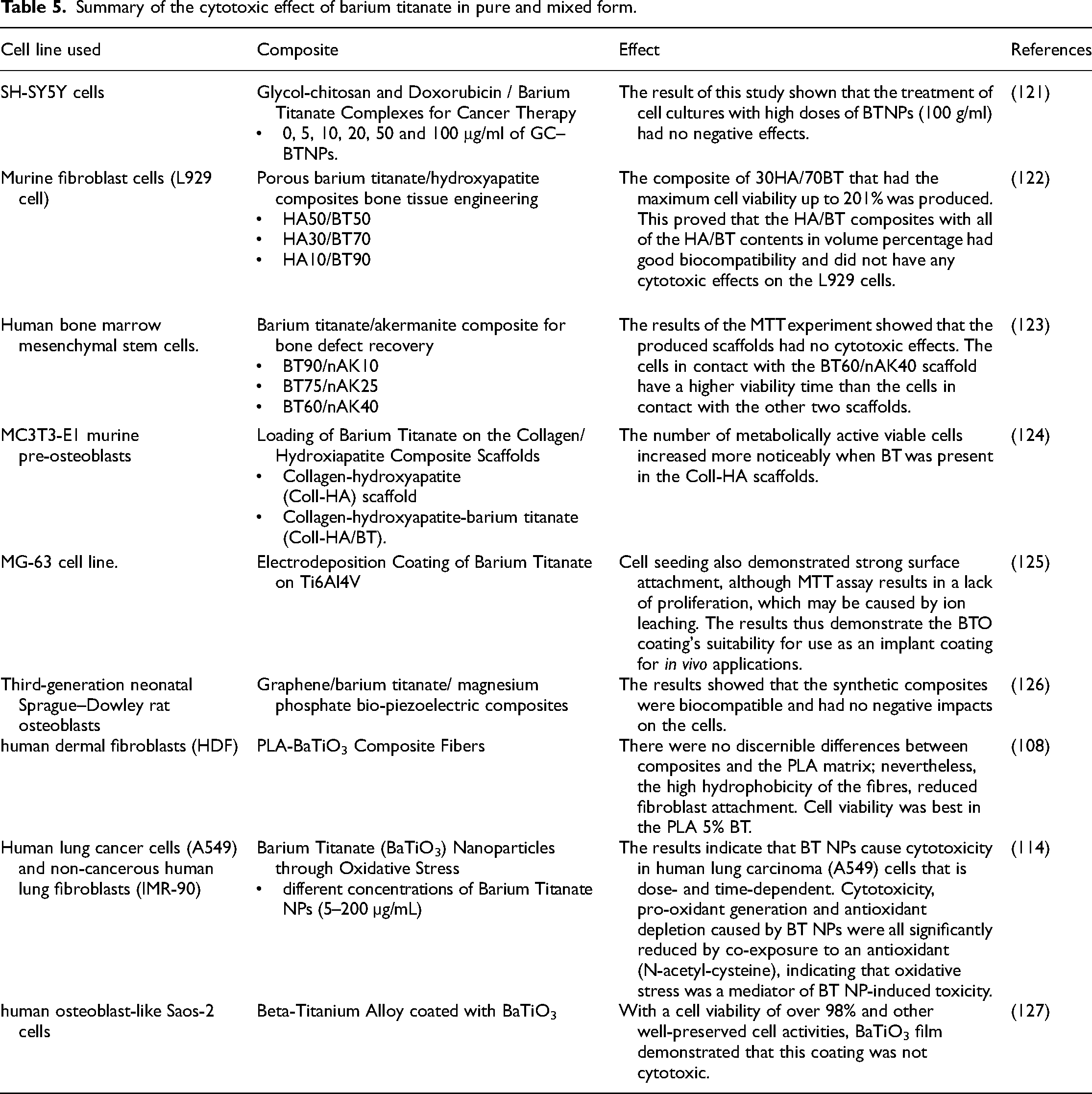
Similar results were observed by Bonacino et al., where a range of harmonic nanoparticles, including BaTiO3, were investigated for cytotoxicity effect on A549, BEAS-2B, HTB-178 and HTB-182 cells for an exposure time of (5 and 24) h. 118 Most cell lines showed a 20–30% reduction in cell viability at a 50 g/mL dosage rate of BaTiO3. Genchi et al. reported a slight influence on the percentage of viable cells when studying the response of human neuroblastoma cells (SH-SY5Y) towards P(VDF-TrFE)/ BaTiO3 NPs films. 119 There is still much to discover about the precise processes behind cell responses to piezoelectric stimulation. The size of the BaTiO3, the mechanism of functionalisation and the kind of cells under examination are only a few variables that may impact their internalisation, followed route and associated cellular expressions. 120 Table 5 summarises BT's cytotoxic effect in pure and mixed form.
Summary of the cytotoxic effect of barium titanate in pure and mixed form.
Osseointegration evaluation of BT
The analysis of the BT implants through histology provided some valuable insights regarding the tissues grown in the pores, its high compatibility with hard tissues, and the frequent occurrence of direct bone apposition to the implant surface. Short-term studies indicated an increase in actively depositing osteoblasts near the implant. In contrast, long-term studies showed a slight reduction in the number of osteoblasts and the transformation of trabecular bone into compact bone, a typical response of healthy bone to a biocompatible implant. A comparison between a PTFE membrane and a composite of poly (vinylidene fluoride trifluoroethylene), P(VDF-TrFE) and BT (BaTiO3) (P(VDF-TrFE)/BT) revealed through histomorphometric and gene expression analyses that the latter promotes new bone formation when implanted in rat calvarial bone defects. Therefore, this composite could replace the current biomaterials used in GBR treatments. 128 Fan et al. 92 reported that electroactive BT-coated titanium scaffold conditions significantly accelerated osteogenesis and osseointegration six and twelve weeks after implantation in extensive segmental bone defects in the rabbits’ radius, according to histomorphology and the peak pull-out load.
The BT/PLA (barium titanate/poly lactic acid) composite film, created using the solution casting process, has strong biocompatibility and piezoelectric capabilities. It had a good osteogenic effect in an in vivo study of rat cranial defects with the ability to direct bone tissue and support bone tissue regeneration. 129
Barium titanate-PCL (BT-PCL) composite
BaTiO3 has well-known piezoelectric and biocompatible characteristics. Piezoelectricity has been demonstrated to be effective in biological processes, particularly those strongly related to bone activity. The bones’ ability to convert functional stress into the electrical stimulation required for regrowth and repair has been well demonstrated, and it's essential to mention that the drawbacks of high brittleness and strong resistance to deterioration accompany the high piezoelectric coefficient of piezoelectric ceramic. The biopolymers are biodegradable but have no piezoelectric or mechanical strength, limiting their usefulness. With the help of the piezoelectric property, the mechanical strength of the piezoelectric composite of biopolymer and piezoelectric ceramic can be increased, and the signal of the cells may be recognised. PCL is a biodegradable polymer widely employed in implantable biomaterials.130,131 The PCL-BT composites’ d33 piezoelectric coefficient increased as the amount of BaTiO3 increased. Significantly, as compared to the unmodified PCL specimen, the BaTiO3 inclusion up to 25 vol.% steadily improved the piezoelectric response to 1.2 pC/N; more specifically, the PCL-45BT and PCL-65BT specimens showed an improvement in the piezoelectric response of 2.4 and 2.6 pC/N, respectively. Similar findings about BaTiO3 were also noted in some previous works.122,132 For example, Liu et al. 82 found that when the BaTiO3 incorporation extent rose over 35 vol.% in the PCL matrix, there was a significant increase in the d33 values, up to 3.9 pC/N. The dense dispersion of BaTiO3 particles in the PCL-45BT and PCL-65BT specimens is the cause of the sharp increase in the d33 coefficient. A stronger electroactive response is produced due to the network of interacting BaTiO3 particles creating an EF through tight spacing. The PCL-25BT specimens, on the other hand, had less electroactive reactions due to the BaTiO3 particles’ sparse distribution.
Zhang et al.'s investigation of porous HA-BaTiO3 composites revealed d33 values ranging from 0.3 to 2.8 pC/N. 122 The polarised HA-BaTiO3 piezoelectric ceramics with a BaTiO3 concentration ranging from 80% to 100% have d33 values that Tang et al. 132 observed in the 1.3–6.8 pC/N range. High-volume BaTiO3 contents are required in the HA-BaTiO3 composites since some with less than 80 vol.% BaTiO3 did not show any piezoelectric action. 133
Conversely, due to the tight packing density of the piezoelectric BaTiO3 particles in a sintered sample, specific bulk-sintered HA-BaTiO3 composites showed exceptionally high d33 values (>50 pC/N). 134 BaTiO3 particles are not closely packed in a polymer matrix, which prevents polymer–ceramic composites like PCL-BT from ever displaying such a strong piezoelectric response. A strong scaffold-mediated piezoelectric response for bone regeneration is unnecessary because the bone has a piezoelectric response between 0.7 and 2.3 pC/N. Compared to HA-BaTiO3 composites made using various traditional methods, the d33 values of the 3D-printed PCL-BT composites were comparable to the piezoelectric response of bone. Therefore, the PCL-BaTiO3 composites’ noted piezoelectric response emphasises its enormous potential for osteogenesis and bone remodelling, accelerating orthopedic and dental applications.131,135
Liu et al. prepare the PCL/BT composite using a solution blending technique. Particles of PCL and BT were dissolved in tetrahydrofuran at different ratios (PCL with 5%, 10%, 15%, 20%, 25% BT). The composite flocs were precipitated by adding the PCL and BT liquid mixture to the ethanol. No cytotoxicity was observed when the composite was tested against the osteoblast line MG63 in DMEM. 131
Composite scaffolds for skin regeneration composed of PCL 3D-printed scaffolds containing 500 mg/ml of BT piezoelectric nanoparticles were prepared. With a distance of 11 cm, a flow rate of 1.5 ml/h and a voltage of 10 kV, BT and PCL solutions were electrospun. Cell proliferation capacity and cellular responsiveness to the piezoelectric substrates were studied in vitro. The proliferation of NIH 3T3 fibroblasts was analysed while cultivated on PCL and PCL/BTNP scaffolds. When electrospinning is used with 3D-Bioplotting, a scaffold with multi-scale porosity is produced. In vitro cell viability analyses on this piezoelectric composite scaffold have shown that it provides a healthy environment for 3T3 and SaOS-2 cell adhesion, growth and proliferation. Compared to nanofibrous mats and 3D-printed scaffolds without fibres, the number of viable cells on composites is large. 136 Piezoelectric BT nanoparticles have been incorporated into the framework to mimic the responsiveness of the wound dressing. Recently, some research teams have started looking into how an EF affects the healing process of wounds. These investigations show that an electric stimulus, which mimics the physiologic endogenous EFs that arise with an injury, can efficiently stimulate skin regeneration. 88
Successfully created PCL-BT 3D-printable filaments that can be used for maxillofacial, cranial and dental applications in addition to orthopedic ones. Activating calcium-sensing receptors or boosting Ca2+ influx into osteoblast cells are two ways Ca2+ regulates osteoblast development. As a result, the electrical stimulation (caused by the piezoelectric effect) from the PCL-BT scaffolds activated the calcium-sensing receptors, started the electrically sensitive Ca2+ signal transduction and increased the Ca2+ influx into MC3T3-E1 cells.14,130 Ibrahim et al. 137 explained that adding 36% of BT to 18% of PCL improved the wettability performance of the composite coating on CpTi and Ti3Nb13Zr alloys. Improving the wettability property increases the performance of the composite in osseointegration.
This review has several limitations: 1. The methods of fabricating scaffolds or coatings or the possibility of these methods affecting biological properties were not discussed. 2. biodegradability, bioresportion, biomineralisation and the factors affecting them were not evaluated. 3. No specific criteria were specified when collecting information from different references. 4. The effect of piezoelectricity on biological properties has not been discussed. These limitations will be the main topics of investigation in future research work.
Challenges and future perspective of PCL, BT and their composites
BT-PCL composites exhibit significant potential for utilisation in various biomedical applications. PCL-BT composites offer numerous benefits, including cost-effectiveness and the convenience and adjustability of manufacturing processes. The PCL-BT composites exhibited significant cellular adhesion and excellent biocompatibility in the in vitro studies.
The most effective approach for producing this composite has yet to be ascertained, including the parameters for a particular procedure or the fabrication techniques for producing appropriate structures. Only a few articles establish the optimal concentration of BT to be used in the composite. Each approach specifies a distinct quantity of ceramic material that differs from the other method. The investigation of the degradability of BT-PCL has not been explored yet. The existing methodologies, which include the utilisation of BT-PCL composites, typically require careful adjustment to optimise the desired structure for a particular application. There is a need for comprehensive investigations regarding the utilisation of this composite in the in vivo investigations. Additionally, further research is warranted to examine the impact of this composite on bacterial organisms. Furthermore, it is imperative to explore the mechanical characteristics of the composite, evaluate its potential as a material for dental implants, assess its viability as a coating material and investigate the influence of BT and PCL concentrations on the compound's adhesion to the implant.
Conclusion
The PCL, BT and their associated composites exhibit excellent activity against bacteria with minimum cytotoxicity effect. PCL-BT composites combine good mechanical, piezoelectric and biological properties to accelerate the osseointegration and the overall healing process. PCL and BT show promising properties for biomedical applications, but further investigation is needed in the future.
Footnotes
Acknowledgements
The authors would like to thank Baghdad University (www.uobaghdad.edu.iq) and Mustansiriyah University (![]() ) Baghdad- Iraq, for their support in the present work.
) Baghdad- Iraq, for their support in the present work.
Declaration of conflicting interests
The author(s) declared no potential conflicts of interest with respect to the research, authorship, and/or publication of this article.
Funding
The author(s) received no financial support for the research, authorship, and/or publication of this article.
Author biographies
Sabreen Waleed Ibrahim is Lecturer in Prosthodontic, her area of research in advanced and composite materials,coatings deposition and characterization, implants surface treatment and esthetics.
Thekra Ismael Hamad is Prof in Prosthodontic, her area of research in materials and implants.
Julfikar Haider is Senior Lecturer in Engineering, his area of research in composite materials, surface engineering, Tribology, metal casting, metal cutting machining, advanced welding, manufacturing systems, artificial intelligence, and finite element modelling.