
Abstract
Forensic science is currently fast-growing for the development detection of the latent fingerprint. Currently, chemical dust quickly enters the body through touch or inhalation and will be affected by the user. In this research, a study on the comparison of natural powder from four species of medicinal plants (Zingiber montanum, Solanum Indicum L., Rhinacanthus nasutus, and Euphorbia tirucall) for the detection of latent fingerprints is carried out that has fewer adverse effects on the user's body by using such natural substances instead. In addition, the fluorescence properties of the dust have been found in some natural powder for sample detection and appear on multi-colored surfaces to show that the latent fingerprints are more pronounced than ordinary dust. In this study, medicinal plants have also been applied to detect cyanide, as it has been known that it is hazardous for humans and can be used as a poisonous compound to kill someone. The characteristics of each powder have also been analyzed using naked-eye detection under UV light, Fluorescence spectrophotometer, FIB-SEM, and FTIR. All the powder obtained can then be used for high potential detection of latent fingerprints on the non-porous surface with their specific characteristics and trace amounts of cyanide using turn-on-off fluorescent sensing method.
Keywords
Introduction
Cyanide (CN-) is a highly toxic ion to human health because cyanide can inhibit critical biological processes with oxidative metabolism and cellular respiration. Cyanide poisoning usually occurs due to being found in raw or processed foods that contain cyanogenic compounds. Cyanide compounds are also being used intentionally to kill someone because of their toxic properties.1–3 Detection of cyanide in water and foods currently has been developing with many methods such as mass spectrometry, 4 ion chromatograph, 5 flow injection, 6 Raman spectrometry, 7 colorimetry, 8 and others.
Fingerprints are said to be a golden standard for use to identify people, especially in forensic science, and it is also used for biometrics and scientific evidence in the police field. Papillary protrusions on the tips of the fingers, which contain rows of pores connected to the sweat glands, make each person's fingerprints unique. The only exception would be a person who had been in an accident so serious that the papillary tissue on the fingers, palms, soles of the feet, and feet were damaged because fingerprints are unique to each person, cannot be changed, are easy to verify, and leave marks on every object a person touches, they are routinely used as evidence in court and by the police.9–28 To detect latent fingerprints, investigators need some physical or chemical processes (water, amino acids, oils, and some other substances) to enhance the fingerprint residue because latent fingerprints cannot be seen by the naked eye.29–33 Detection of latent fingerprints currently using traditional methods based on chemicals such as ninhydrin, 34 ferric oxide, 35 cyanoacrylate, 36 and DFO. 37 However, various research studies involving the synthesis of fluorescent dust remain limited in many areas, such as the complexity of synthesis. The toxicity of raw materials and dust powder with a high price, for that reason, hinders its practical use. 38 Therefore, the research team is interested in a highly effective method of preparing fluorescent dust powder used in examining latent fingerprints from medicinal plants that have the potential ability to glow under UV light and can detect trace amounts of cyanide using the medicinal plants by using fluorescence turn on-off method, other than having a property that can glow under UV light. The medicinal dust powder can be prepared inexpensively and is environmentally friendly.42,43 Most of the chemical compounds of each medicinal plant are shown in Table 1. It is shown that almost powder compound is antibacterial and anti-inflammatory, meaning the powder is not toxic to the human body. The factors affecting latent fingerprints were also studied, such as different material surfaces, temperature, storage time, and the identification of latent fingerprints from overlapping fingerprints.
Major chemical compounds and their applications in selected four medicinal plants.

Experimental
Chemicals and reagents
All chemicals used in this research are analytical grade. Potassium cyanide (KCN) was obtained from Spectrum Chemical (USA). Four common species of Thai medicinal plants were selected and used in this study including Zingiber montanum, Euphorbia tirucall, Rhinachantus nasutus, and Solanum indicum collected during a short winter time (November 2021–January 2022) from our flower park of Science Building, Khon Kaen University, Thailand.
Instruments
The pretreatment of the medicinal plant powder was carried out in a heating oven (BINDER ED 115UL-120V), grind by using a high-speed multi-function crusher 2500A, and filtered by using a laboratory test sieve 100 mesh. The dried and ground powder was examined by an optical property using a fluorescence spectrophotometer (RF-5301 PC, Shimadzu), and their characteristics were identified by FTIR (Fourier transform infrared spectrometer, Bruker TENSOR 27), FIB-SEM (Helios NanoLab G3 CX). For the detection application of latent fingerprint, the material surface will be checked using a UV chamber (Trans-illuminator Model TM-26 (302 nm)) and TLC UV Cabinet (365 nm).
Preparation of the medicinal plant powder
The 500-g rhizome of the plant Zingiber montanum and 500 g leaf of Solanum indicum L., Euphorbia tirucall and Rhinacanthus nasutus, each was cut into small sizes and dry in the oven for 48 h at 75°C, after that the plant will be ground in high-speed multi-function crusher 220 V for 5 min, then filtered using laboratory test sieve 100 mesh. The final fine powder was kept in a capped plastic bottle at room temperature for further use.
Method detection of a latent fingerprint
To detect the latent fingerprint using the common brushing method, a volunteer rubbed his forehead with a finger and printed his fingerprint on the different material surfaces for 5 s and check its trace immediately under a UV chamber.
Method detection of cyanide
Cyanide has been prepared by using KCN, and the KCN was diluted using DI water until varying concentrations 5, 6, 7, 8, 9, and 10 µM. After finishing the preparation, all the reagents were stirred using a magnetic stirrer for 5 min to ensure that KCN was completely dissolved. One milliliter from each solution adding to the vial with 0.25 medicinal solution and 9 mL DI water. The analysis will be using a fluorescence spectrophotometer (RF-5301 PC, Shimadzu) with a maximum wavelength of 380 nm to excite the ground-state electron of the CDs, and then the excited electron undergoes an energy transfer, which is visible as a fluorescence spectrum.44,45
Results and discussion
Characteristics and optical properties of the medicinal plant powder
Similar characteristic patterns of four selected medicinal plant powders have been analyzed by using FTIR as shown in Figure 1(a). There are OH/N-H groups on 3284 cm−1, 2929 cm−1 for C-H bonding, 1632 cm−1 for C=C, and C=N, C=C, and C-OH bands were found on 1451 cm−1, 1004 cm−1 was representative for C-H bending peak, and CO2 vibration was found in Zingiber montanum. The FTIR of Solanum indicum L. can be seen in Figure 1(b) having OH/N-H group on 3278 cm−1, 2850 cm−1 for C-H bonding, 1617 cm−1 for hexion vibration N-H, C-H band was found on 1372 cm−1, 1031 was representative on C-O bending, and C-H bonding on 781 cm−1. Figure 1(c) shows the FTIR of Rhinacanthus nasutus OH/N-H group was found on 3275 cm−1, 2918 cm−1 for C-H bounding, 1605 and 1408 cm−1 for C-C band, C-O bonding was found on 1020 cm−1, and C-H bonding on 896 cm−1. The FTIR of Euphorbia tirucall was also shown in Figure 1(d) having a C-H group on 2850 cm−1, 1720 cm−1 for C=O bonding, 1389 cm−1 for C=H bond, and C-O band was found on 1026 cm−1, and C-H bonding was found on 664 cm−1.46–55

Fourier transform infrared spectra of (a) Zingiber montanum, (b) Solanum Indicum L., (c) Rhinacanthus nasutus, and (d) Euphorbia tirucall.
The medicinal powder has been observed for their morphology images with FIB-SEM as shown in Figure 2. It can be seen that the particle of the powder is different from each other depending on the type of plants used. Figure 2(a) shows the characteristic powder from Euphorbia tirucall, it is shown that the powder has a non-smooth surface, and an irregular shape, then by using SEM the average sized of the powder is more than 200 μm. Figure 2(b) gives the characteristic powder from Zingiber montanum, it is shown that the powder has a non-smooth surface and has an oval-shape, and the average size of the powder is less than 100 μm by using SEM. Figure 2(c) shows the characteristic powder from Solanum indicum L., it is evident that the powder has a smooth surface with an elongated part. Figure 2(d) shows the characteristic powder from Rhinacanthus nasutus, indicating the powder has a non-smooth surface with an irregular shape, the average size of the powder is around 200 μm by using SEM.
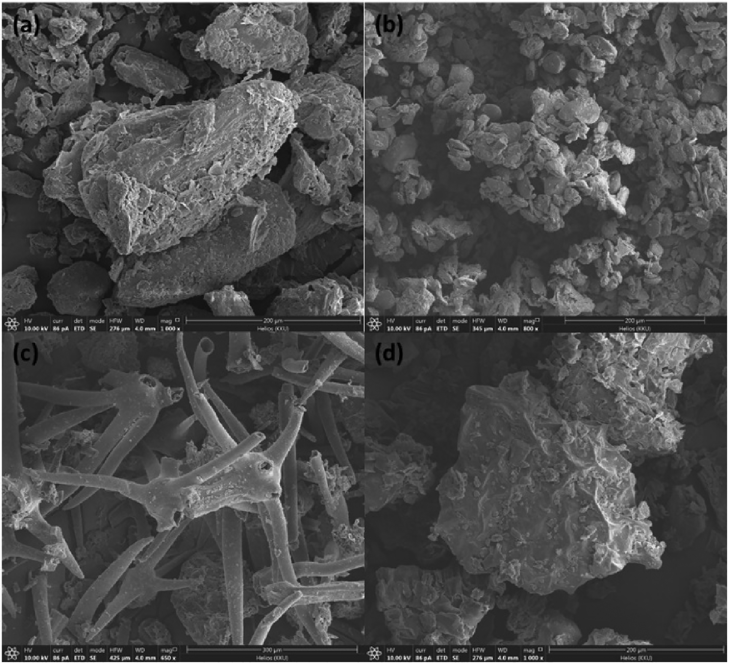
Focused ion beam scanning electron microscope of (a) Euphorbia tirucall, (b) Zingiber montanum, (c) Solanum Indicum L., and (d) Rhinacanthus nasutus.
The optical property of the plant powder has also been characterized using a fluorescence spectrophotometer, UV–Vis spectrophotometer, and UV light chamber. The 0.05-g from each powder has been diluted with 10 mL DI water. 1 and was analyzed by fluorescence spectrophotometer. Figure 3 shows that the fluorescence spectra with excitation 360 nm from Zingiber montanum powder is higher compared with the others, while that of Rhinacanthus nasutus didn’t show the intensity of the fluorescence spectrum.

Fluorescence spectra of the powder samples from medicinal plants.
Comparison of the medicinal plant powder vs black powder for latent fingerprints detection on a non-porous surface under UV light and daylight
The plant powder was applied to detect latent fingerprints on the non-porous surface, as shown in Figure 4

Comparison of the medicinal plant powder vs black powder for latent fingerprints detection on non-porous surface under UV light and day light (The pictures were taken using UV Transilluminator Model TM-26) (a) Zingiber montanum, (b) Rhinacanthus nasutus, (c) Solanum Indicum L., (d) Euphorbia tirucall, and (e) Black powder (commercial powder).
Detection of the latent fingerprints on the non-porous surface has been tried in various surface materials. The plant powder was applied on the surface after the volunteer pressed the fingerprint in the material. The surface was checked under a UV lamp to compare the contrast of fluorescence of latent fingerprints.
Various surface materials have also tried to detect latent fingerprints on the non-porous surface. After the volunteer pressed the fingerprint print on the material, the plant powder was applied on the surface. The surface was checked under a UV lamp to compare the contrast of the fluorophore's emission from the latent fingerprints tracer. However, data shown in Table 2 demonstrated that all the natural plant powder could be used to detect latent fingerprints on many types of surfaces such as plastic, iron, ceramic, and paper. Even the extent of the fade detection could not be differentiated from the visualized details, which can be seen in SI1. The resistance and durability also have been tested in this study. We made comparing on the 1st day after the volunteer pressed the fingerprint with the day 30th. The non-porous material surface was kept in the forensic science laboratory Khon Kaen University, at room temperature (about 27°C). The volunteer pressed the fingerprint on the iron material. In this test, iron material from a knife was applied, and the result is shown in Figure 5. The results showed that all the natural powder could be used to detect the latent fingerprints on the non-porous surfaces, particularly in this case, only after one month the fingertips were pressed on the material surfaces.

Comparison of the plant powder on 1st day and 30th day after pressing the fingertips on non-porous surface. (The pictures were taken under TLC UV light cabinet.)
Application of four medicinal plants powder on various surface materials.

Detection of cyanide by using the powder sample of the medicinal plants
Medicinal plant fluorescence turn-off was investigated at different KCN concentrations in the range of 5–10 µM as shown in Figure 6. The quenching effect of the fluorescence intensity may occur due to different reasons such as energy transfer, surface absorption, or surface characteristic. 0.05 g of each medicinal powder was diluted with 10 mL of DI water. For detection using FL spectrophotometer, 0.25 µL of each medicinal liquid, adding 1 mL of KCN and 9 mL DI water with the setting condition of maximum excitation wavelength of 360 nm, after measurement the spectral data were obtained as shown in Figure 6(d). The cyanide detection by using Euphorbia tirucall, powder shows that the powder can be used to detect trace cyanide ion in various concentrations from 5–10 µM with the limitation of detection (LOD) of 1.2 µM as shown in Figure 6(a). Various concentrations of cyanide also can be detected by using Solanum Indicum L. (Figure 6(e)) with 2.8 µM LOD. Figure 6(f) shows that Zingiber montanum can be used to detect cyanide using turn on-off method under the suitable conditions of various concentration titration of cyanide with 1.04 µM LOD. The LOD was calculated by using the following formulation:

Detection LOD of (a) Euphorbia tirucall (b) Solanum Indicum L. (c) Zingiber montanum Emission spectra detection KCN of (d) Euphorbia tirucall (e) Solanum Indicum L. (f) Zingiber montanum, Turn-Off methods sensitivity test of (g) Euphorbia tirucall (h) Solanum Indicum L. (i) Zingiber montanum, Wavelength Ex = 360 nm slit width 5/5.
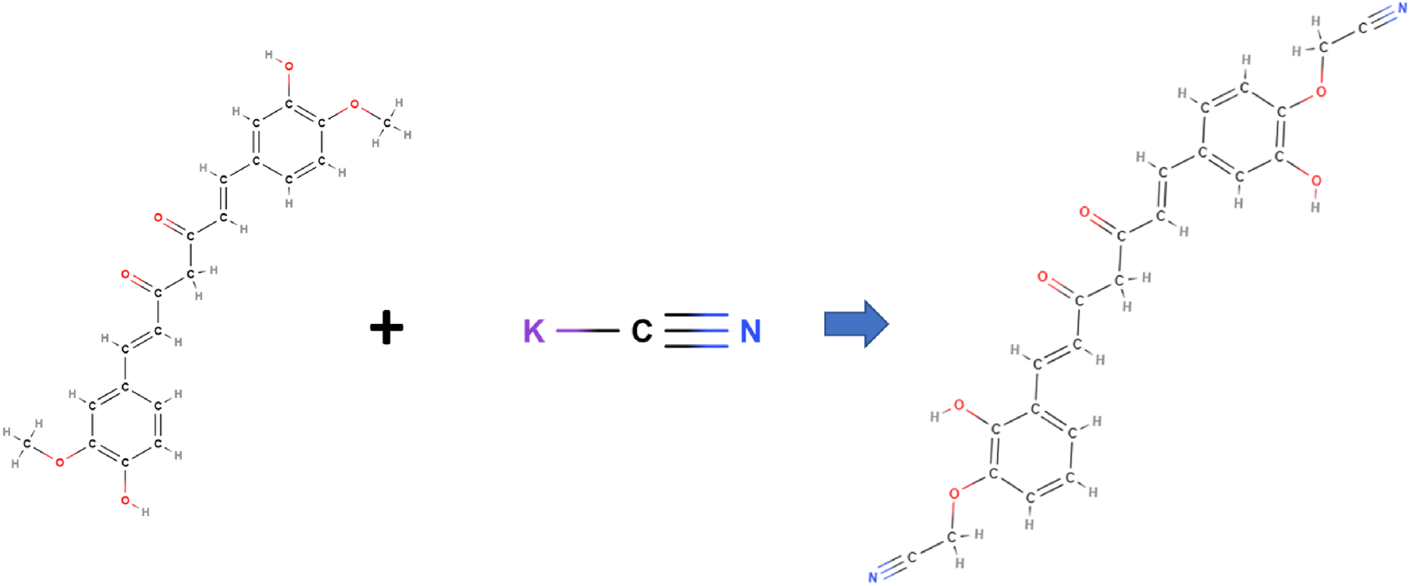
Prediction Mechanism reaction detection KCN Zingiber Montanum (curcumin).
Conclusion
The natural characteristics of medicinal plants can be simply used for the pronounced detection of latent fingerprints on non-porous surfaces. Some plants exhibit distinct fluorescence under UV excitation, which has quickly been evident under UV light chamber and fluorescence spectrophotometry in the routine analysis. The characteristic powder and its optical properties were observed using fluorescence spectrum, FIB-SEM, and FTIR, demonstrating that the medicinal powder has a different characteristic under FIB-SEM and UV light visualization. Zingiber montanum has the highest intensity fluorescence compared to other powders. The application for latent fingerprint detection was carried out with experiments on various non-porous materials (iron, plastic, etc.). The experimental optimization was also done with the effect of temperature and storage time of the materials used. The effect of storage time of the plant powder could be worked satisfactorily as shelf-life for 30 days. The powder samples of the medicinal plants also have been analyzed for the detection of cyanide. It was found that the Rhinacanthus nasutus can't be used to detect trace cyanide concentrations.
Supplemental Material
sj-docx-1-sci-10.1177_00368504231156217 - Supplemental material for Fluorophores- rich natural powder from selected medicinal plants for detection latent fingerprints and cyanide
Supplemental material, sj-docx-1-sci-10.1177_00368504231156217 for Fluorophores
Footnotes
Acknowledgements
The authors thank the research financial support to: (1) Research Center for Environmental and Hazardous Substance Management (EHSM), Khon Kaen University; (2) Materials Chemistry Research Center, Department of Chemistry and Center of Excellence for Innovation in Chemistry (PERCH-CIC), Faculty of Science, Khon Kaen University, Thailand.
Declaration of conflicting interests
The author(s) declared no potential conflicts of interest with respect to the research, authorship, and/or publication of this article.
Funding
The author(s) received no financial support for the research, authorship, and/or publication of this article.
Supplemental material
Supplemental material for this article is available online.
References
Supplementary Material
Please find the following supplemental material available below.
For Open Access articles published under a Creative Commons License, all supplemental material carries the same license as the article it is associated with.
For non-Open Access articles published, all supplemental material carries a non-exclusive license, and permission requests for re-use of supplemental material or any part of supplemental material shall be sent directly to the copyright owner as specified in the copyright notice associated with the article.