
Abstract
Leptospirosis is a neglected disease in Vietnam. Only a few studies have evaluated the status of Leptospira infection in both humans and animals. To our knowledge, this is the first serological survey of Leptospira in both domestic and wild animals, which may act as reservoirs of this agent. This study aimed to evaluate the seroprevalence of Leptospira in animals that are in close contact with humans in different geographical areas in Vietnam. Sera were collected from 1205 individual animals of six species, including buffaloes, cattle, cats, dogs, swine, and rats. The microscopic agglutination test (MAT) against 25 serovars of Leptospira spp. has been employed to detect serovars of Leptospira among the studied population. Overall, 44.2% of buffaloes, 24.9% of cattle, 10.2% of swine, 32.9% of dogs, 12.2% of cats, and 16% of rats were seropositive. A total of 17 different serovars were detected, of which serovars Hebdomadis and Canicola circulated in all the studied animal species. Variability of the predominant serovars circulating in animal species and in different geographical areas of Vietnam has been noted. We conclude that this study showed a high prevalence of Leptospira circulating in animals that are in close contact with humans, raising an alert of the important sources of pathogenic leptospires transmission to humans in Vietnam. These findings prove an imperative need for effective measures for disease prevention.
Introduction
Leptospirosis is caused by bacteria of the genus Leptospira, which affects humans and animals. The WHO estimated that 0.1–1 in 100,000 people living in temperate climates and 10 or more in 100,000 people living in tropical climates are affected each year by this pathogen. These figures are assumed to be underestimated because leptospirosis is commonly misdiagnosed as other febrile illnesses such as dengue, scrub typhus, and chikungunya. 1 The Health Statistics Yearbook from the Vietnam Ministry of Health annually reported <20 cases nationwide, while individual studies showed that Leptospira seroprevalence in humans ranged from more than 10%–80%, indicating high circulation of the pathogen in this country.2−6
Humans can be infected by direct contact with urine of infected animals or indirect contact with contaminated soil and water, where Leptospira spp. can survive for a long period. 7 A wide range of domestic and wild animals in close contact with humans are reservoir hosts of Leptospira, such as cattle, swine, dogs, and rats.8−11 In Vietnam, 65% of the population living depends on agriculture, of which approximately 7 million and 32,349 households produce swine and cattle in 2016, respectively. 12 According to the Department of Animal Health, approximately 3.8 million households kept about 7.7 million dogs have been roughly estimated in 2017 throughout the country. 13 Several studies in Vietnam revealed that the overall seroprevalence of Leptospira in swine ranged from 8.17% to 21.05% depending on the study location and more than 20% seropositivity among dogs tested.8,9,14,15 A similar proportion of rats were infected with Leptospira. 16 To our knowledge, only a few studies in Vietnam have revealed the status of Leptospira infection in humans and animals. Accordingly, this study aimed to evaluate the seroprevalence of Leptospira in animals that are in close contact with humans.
Methodology
Study site
A cross-sectional study was designed to determine the seroprevalence of antibodies against various Leptospira serovars in animals that are in close contact with humans from three provinces, Thai Binh, Ha Tinh, and Can Tho, which represent different geographical and climatic zones in Vietnam. Thai Binh is located in the center of the Red River Delta, North of Vietnam, and has a subtropical humid climate, while Ha Tinh lies in the Central Coast region, where it is very drought in summer, but suffers from many typhoons. Can Tho, is located in the south of Vietnam and has tropical dry and wet seasons and interlacing canals (Figure 1).
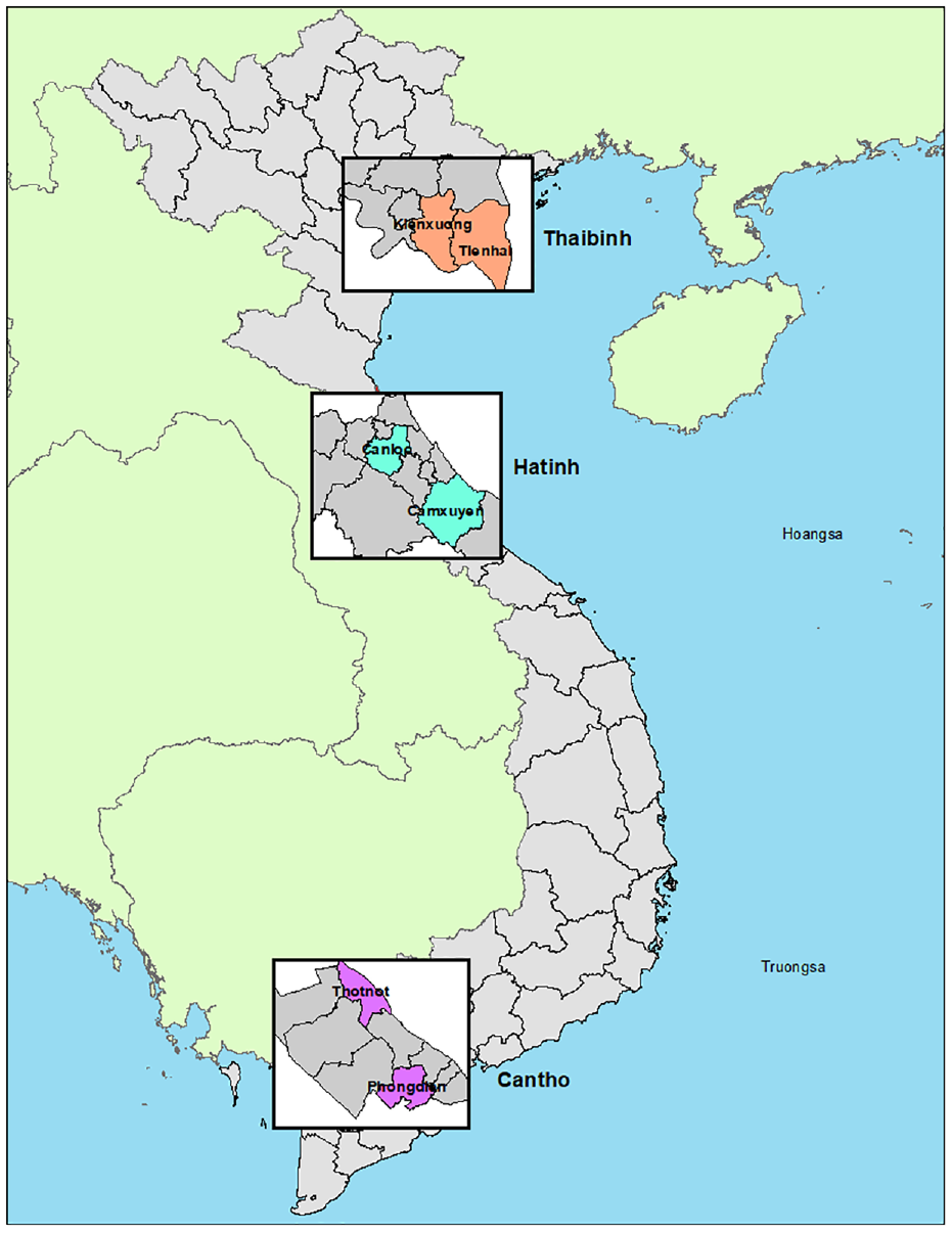
Map of sampling location.
Sampling
Six animal species, namely, dog, cat, cattle, buffalo, swine, and rat that are in close contact with humans, were subjectively targeted to samples from the three above-mentioned provinces. All sampled animals must be raised in the selected households for at least 2 months or trapped (for rats) in the respective provinces, but not imported from other provinces. Blood samples were collected from the cephalic vein in dogs and cats, from the jugular vein in swine, and from the tail vein in cattle and buffaloes. For rats, blood was collected via heart puncture from live rats almost on the same day of trapping. Prior to sampling, the owners were asked if the animals (except rats) manifested any common symptoms of sickness, such as fever, fatigue, diarrhea, constipation, or jaundice, within 1 month ago. All samples were centrifuged for serum extraction on the same day and stored at −20°C until transportation in a cool box to the National Center for Veterinary Diagnosis (NCVD) in Hanoi for laboratory analysis. A total of 1205 serum samples were analyzed, of which 52 were from buffaloes (buffalo samples were not taken in Can Tho because buffaloes are not commonly raised in this province), 233 from cattle, 164 from cats, 219 from dogs, 381 from swine, and 156 from rats. The sample collection was conducted from October to December 2019.
Laboratory analysis
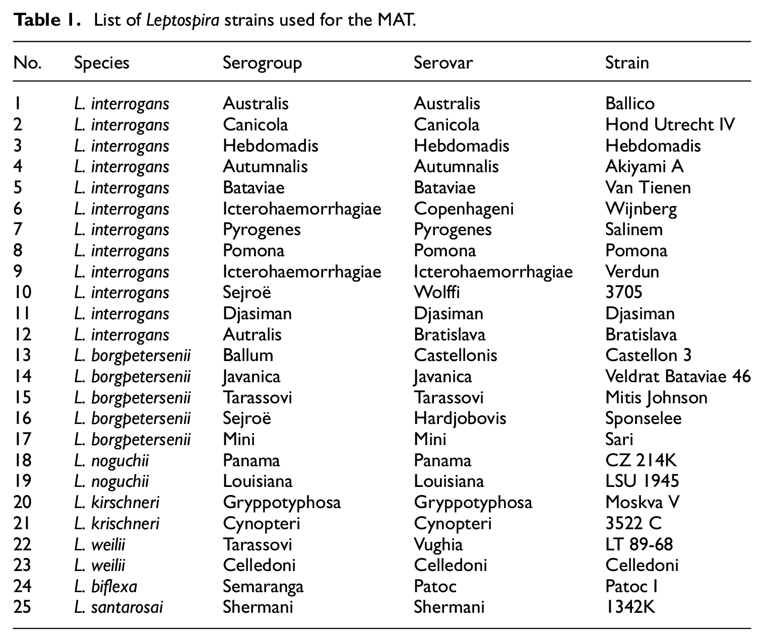
All sera samples were subjected to the microscopic agglutination test (MAT) to identify Leptospira serovars. In this study, we used a set of 25 serovars, which were serovars from The Institut Pasteur de Nouvelle-Calédonie and NCVD (Table 1). A sample was considered positive if the MAT was 1:100 for at least one of these serovars. A two-fold serial dilution was used starting at 1:100 dilution up to 1:800, considering the highest positive dilution to be the titer of serum.
List of Leptospira strains used for the MAT.
Statistical analysis
All data were recorded in Microsoft Excel 2010 and statistically analyzed using IBM SPSS version 23.0. The seroprevalence and 95% confidence intervals (CIs) were estimated. The Fisher’s exact test was performed to compare the prevalence of infection among animal species. A value of p < 0.05 was considered the critical level of significance. The ArcGIS 9.3 was used to generate the map.
Findings
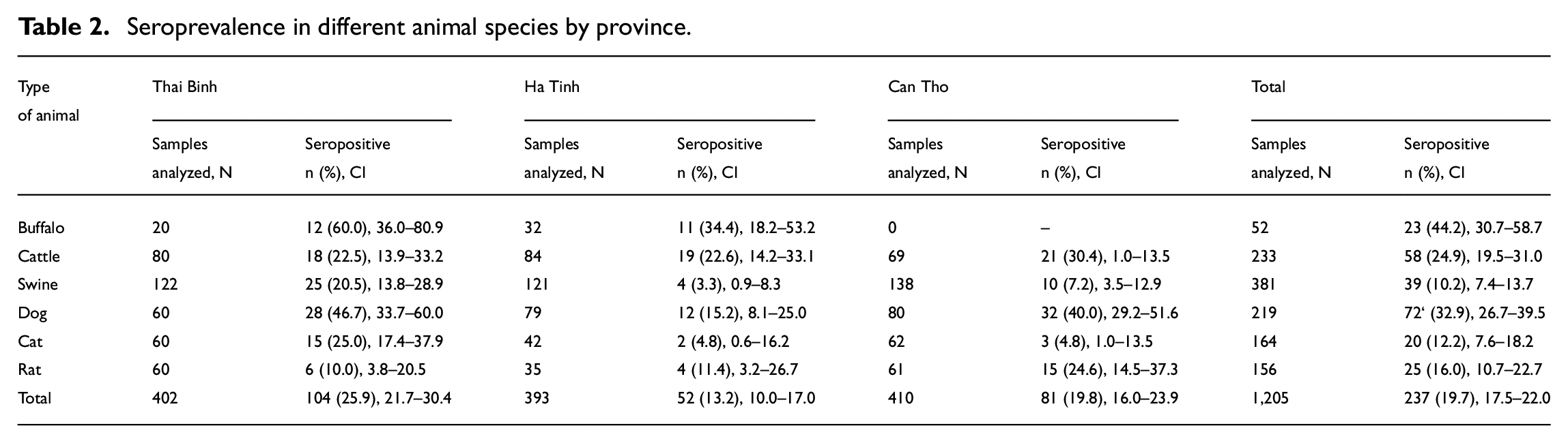
Overall, 19.7% of the 1205 samples tested were seropositive for at least one Leptospira serovar, with titers ranging from 1:100 to 1:200, of which 67 positive samples were multiple with two to seven serovars. Among the sera of six animal species analyzed, seroprevalence in buffaloes reached the highest proportion (44.2%), followed by dogs (32.9%). The data also showed 24.9% seropositivity in cattle, 16% in rats, 12.2% in cats, and only 10.2% in swine sera. By province, Thai Binh showed the highest seroprevalence (25.9%), while it was lower in Ha Tinh and Can Tho (13.3% and 19.8%, respectively) (Table 2).
Seroprevalence in different animal species by province.
Using a low cut-off titer (1:100), 17 different serovars were detected in 237 positive sera, of which the most frequently detected serovar was Hebdomadis (2.3%), followed by Patoc (1.6%), Castellonis (1.5%), and Javanica (1.4%). Moreover, serovars Panama, Tarassovi, Australis, and Autumnalis occurred in all three provinces, while serovar Bratislava was found only in Can Tho province. Additionally, all 17 Leptospira serovars were detected in Can Tho, while 12 and eight serovars were detected in Thai Binh and Ha Tinh, respectively. With a cut-off titer of 1:200, nine serovars were found, of which Javanica was the most predominant (0.6%) and the highest in Thai Binh (1.7%) (Figures 2 and 3).

Distribution of Leptospira serovars by province using cutoff titer ≥ 1:100.

Distribution of Leptospira serovars by province using cutoff titer ≥ 1:200.
Among the six studied species, the cattle sera showed the highest diversity with 15 different serovars, followed by dogs and rats. Only six different serovars were detected in cat sera. The most frequent Leptospira spp. serovars were Hebdomadis (18.9%) and Canicola (15.5%) in cattle; Hardjobovis (15.3%) and Patoc (12.5%) in dogs; Louisiana (24%) in rats; Castellonis (20.5%) and Patoc (20.5%) in swine; Hebdomadis (17.4%) in buffaloes; and Javanica (25%), Louisiana (15%), Hebdomadis (10%), and Castellonis (10%) in cats. It was noted that a certain number of multiserovars were the most predominant in buffaloes (43.6%) and cats (30%). Serovars Hebdomadis and Louisiana were circulating in all six species, and except swine, serovar Javanica also occurred in five species (Figure 4).
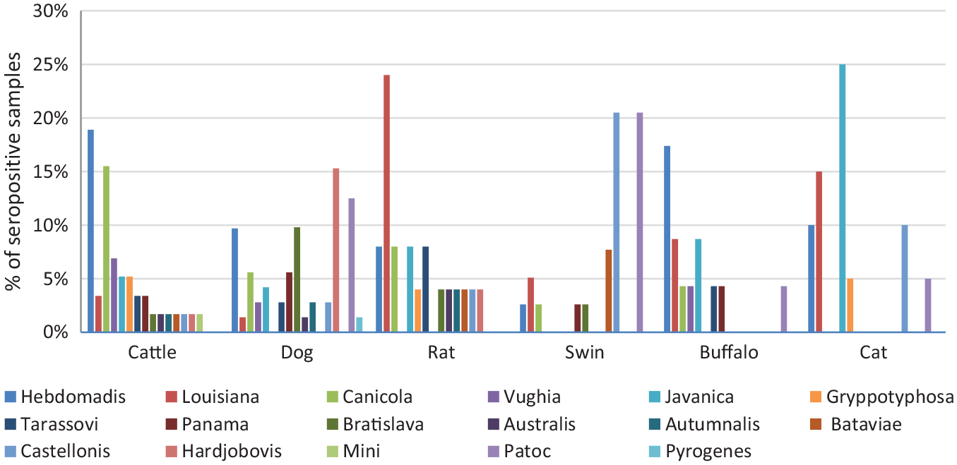
Distribution of Leptospira serovars in six animal species.
Discussion
A high seropositive proportion with 17 different serovars was detected in all studied animals, which indicates the diversity of Leptospira in Vietnam. Among the three provinces, Thai Binh appeared to have the highest seroprevalence of all species compared to the other two provinces. The results were similar to those of a previous study in Vietnam, where low land is at a higher risk of Leptospira circulation than highland and mountainous areas. 17 Nevertheless, we did not select buffaloes in Can Tho province for analysis because this species is rarely raised in households for food or plow purposes; buffalo species were most frequently infected with Leptospira, particularly in Thai Binh (60%). Our data were similar to those of a recent study that showed MAT positive prevalence in 63.6% of buffalo serum samples in Amazon and 58.73% in Iran.18,19 Leptospirosis in buffaloes has been investigated in several countries such as the Philippines, Thailand, India, and Sri Lanka, indicating different prevalence and even reported as a public health concern in some countries. The most prevalent serovars found in buffaloes in our study were Hebdomadis, Louisiana, and Javanica, and other serovars have been detected in other countries, indicating serological diversity of the spirochetes in different geographical areas, such as serovar Canicola in Iran; serovars Shermani, Bratislava, and Panama in Thailand; and serovars Sejroe, Autumnalis, and Pomona in the Brazilian Amazon.20−22
In the ruminant species, cattle also showed high seropositivity (24.9%), similar to a study infection that investigated 22.61% of dairy cows with Leptospira in Can Tho Province of Vietnam. 23 However, these figures were much higher than those reported in previous studies in Brazil (6.44%) and Tanzania (7.08%).10,24 Two predominant serovars in cattle were found, namely Hebdomadis and Canicola, in which Hebdomadis was also the most infected buffalo, and Canicola was one of the primary hosts in cattle. 25 In our study, 15 different serovars were detected in cattle, indicating the most diversified among animal species and also compared to other studies, such as Australis, Grippotyphosa, and Sejroe in France and Ranarum and Shermani in Thailand.21,26
The dog population in our study was the second most prevalent serovar in Leptospira, with 13 serovars detected, of which serovars Hardjobovis and Patoc were the most prevalent. Worldwide, predominant serovars in dog populations differ from country to country. For example, Canicola and Icterohaemorrhagiae have been the most infected serovars in dogs in Brazil, while Hebdomadis are the major serovars in Japan.27,28 Overall, Leptospira infection among dog populations in our study was also higher than that of other studies conducted in 2011 and 2016 in similar areas of Vietnam and a study in Brazil.15,23,29,30
In at least 160 mammalian species affected by Leptospira, cats are considered to be resistant to leptospirosis but an important source of transmission to humans. 31 In our study, seroprevalence stood out the most in Thai Binh (25%), where the cat is regarded as a special food that increases the risk of transmission. The overall seroprevalence in cats was higher than that reported in similar studies in Massachusetts, Malaysia, and Lisbon. Even though the fewest serovars were detected in cats among the studied animal species in our study, it showed more serovar variety in cats in Vietnam than in other nations.32−35
Swine have been studied the most among mammalian species affected by Leptospira, as it is a common disease in swine worldwide. Seropositive serum was detected in approximately 10.2% of swine serum, which is much lower than that of a previous study in 10 provinces of Vietnam in 2017 (21.05%), but similar to that of a study in five provinces of Vietnam in 2016 with 8.17%.8,9 In 2019, Vietnam livestock had suffered from an African swine fever outbreak that led to the death and culling of approximately 5.9 million pigs, accounting for approximately 22% of the total swine population.36,37 Consequently, new swine herds have been raised after the outbreak, which may have affected our findings. Nevertheless, serovars Castellonis and Patoc were the most frequently detected in our swine serum, unlike previous studies in Vietnam, suggesting a diversity of Leptospira serovars circulating in this species.8,9,38,39
Rats are the most well-known primary reservoir of Leptospira worldwide, and their infection varies widely from country to country and region to region. The number of rats to be sampled in our study was limited because we used the catch and release traps for trapping rats and requested that those with live rats returned on the same day for blood collection. The seroprevalence of rat serum in our study was much lower than that of studies worldwide, but 12 different serovars showed more diversity than other studies. It has been noted that Leptospira was more frequent in the South than in other provinces, where rats can be considered as a special food, with a possible risk of infection. The seropositivity in this recent study was similar to that reported in previous studies in the Mekong Delta region.16,40,41
Overall, a predominant variety of a wide range of serovars (17 different serovars) was found across all animal species. Serovars Patoc and Castellonis are the most prevalent in Thai Binh, while serovars Hebdomadis and Canicolla commonly circulate in Ha Tinh and Can Tho. Two serovars, Hebdomadis and Louisiana, were detected in all species, suggesting cross-transmission among the studied animals. 42
In short, this study showed a high prevalence of Leptospira circulating in both domestic and wild animals, increasing the risk of pathogenic leptospires transmission to humans in Vietnam. These findings prove an imperative need for effective measures for disease prevention. Additionally, the diversity of Leptospira serovars detected by MAT suggests that development of a multiserovar vaccine for animals should be an effective measure to reduce disease transmission in domestic animals, and consequently, environmental contamination and human transmission.
Footnotes
Acknowledgements
We thank the Institut Pasteur de Nouvelle-Calédonie and Dr. Cyrille Goarant for providing technical support. We also thank the Institut Pasteur de Paris and Mr. Yves Froehlich for coordinating the work of the project, including this study. We would like to thank Editage (![]() ) for English language editing.
) for English language editing.
Declaration of conflicting interests
The author(s) declared no potential conflicts of interest with respect to the research, authorship, and/or publication of this article.
Funding
The author(s) disclosed receipt of the following financial support for the research, authorship, and/or publication of this article: This work was financed by the Agence Française de Développement under the ECOMORE project. We gratefully acknowledge the staff of the Center of Disease Control in Thai Binh, Ha Tinh, and Can Tho provinces for coordinating the fieldwork.
Ethical issue
The project was approved by the Institutional Review Board (IRB) of NIHE.