
Abstract
Moringa oleifera (M. oleifera) Lam belongs to the family Moringaceae. It is an important multipurpose tree that is largely distributed globally and has been used almost in every aspect of traditional medicine for the treatment of various illnesses including cancers, diabetes mellitus, asthma, arthritis, etc. This study investigated the effects of oral acute and sub-acute administration of M. oleifera hydroethanolic leaf extract (MOHE) in ICR-mice. Its major phenolic compounds were also determined. Ten (10) female, 8-week old mice were grouped into control and treatment groups for acute toxicity study. A dose of 2000 mg/kg MOHE was given once to the treatment group via oral gavage. However, for the sub-acute toxicity study, 25 mice were grouped into groups A (control), B (125 mg/kg), C (250 mg/kg), D (500 mg/kg) and E (1000 mg/kg). MOHE was given via oral gavage to groups B, C, D and E daily for 28 days. Group A received only distilled water. The mice were sacrificed at the end of the experiments and samples were collected for evaluation. The results of the chemical profiling of MOHE revealed the presence of glucomoringin, niaziminine, quercetin and kaempferol as the major compounds. The treated mice in the acute toxicity study were slightly anaemic and showed evidence of stress leukogram. Moreover, a slight increase in creatinine, significant increases in AST and CK, hepatic degeneration and necrosis, none-obstructive sinusoidal dilatation, renal tubular necrosis, interstitial nephritis and renal interstitial oedema were observed. It is concluded that the LD50 of MOHE is higher than 2000 mg/kg. However, oral administration of MOHE causes acute mild anaemia and moderate hepato-nephrotoxicity in ICR-mice. Its major phenolic compounds are glucomoringin, niaziminine, quercetin and kaempferol.
Introduction
Plants usually contain chemical compounds known as secondary metabolites, which have wide range of biochemical and pharmacological activities ranging from antioxidation, antiinflammation, antiallergic to antithrombotic effects. 1 These metabolites may include saponins, tannins, oxalates, phytates, phenolic compounds, flavonoids and glycosides. Some of these metabolites may be used directly as medicinal agents. 2 Every medicinal plant species, in addition to having pharmacologically active compounds, possesses other nutritional phytochemicals that are essential for the physiological functions of human and animal body, thereby providing the energy requirements for metabolic processes. These nutrients include carbohydrates, fats, proteins, vitamins, antioxidants, 3 minerals, fatty acids and fibre. 4 Phytochemical analyses of edible plants and vegetables play an important role in assessing their nutritional and medicinal potentials.5,6
Moringa oleifera Lam an important multipurpose plant, widely distributed worldwide and largely used in virtually every aspect of traditional Medicine. 7 It belongs to a mono-generic family, the Moringaceae, which has about 14 species of deciduous tree and is natural to the sub-Himalayan tracts of India and Pakistan. Moreover, the plant is nowadays inhabitant in humid and sub-humid regions 8 ; extensively cultivated in African countries, America, India, Malaysia, Mexico, Philippines and Sri-Lanka. The plant is also commonly used as a wholesome herb, as food (leaf and pods), in industries, and almost every part of the plant (seeds, leaves, oil, sap, bark, roots and flowers) possesses valuable pharmacological activities. 9 Moringa oleifera is known in English as Horseradish tree, Drumstick tree and Ben oil tree. It is known as Pokok Kelor in Malay. The plant is used as food supplement and plant-based medicine for the treatment of several diseases, malnourishments/starvation as well as health conditions including antimicrobial, antihyperlipidemic, anticancer, hepatoprotective, arthritis, rheumatism, prostrate problems, among others. The inclusive range of pharmacological activities associated with M. oleifera was credited to the different active phytochemicals reported in different parts of the plant; namely the vitamins, carotenoids, polyphenol, flavonoids and phenolic compounds. 10
So far, there is little or no report on the phytochemicals, nutritional elements and toxicity studies of M. oleifera cultivated in Selangor-Malaysia. Some groups of phytocompounds (flavonoids, glycosides, Saponins, steroids, etc.) in M. oleifera cultivated elsewhere were reported previously. 5 Nevertheless, the exact phytochemical compounds in M. oleifera cultivated in Selangor have not been sufficiently studied.11,12 Furthermore, the few studies that have studied the active phytochemicals of the plant elsewhere are not consistent in their reports, perhaps as a result of variations in agro-climatic settings of the plants, genetic influences, changes in cultivation techniques, drying and extraction methods.10,13 Moreover, Förster et al. 14 suggested that bioactive compounds of medicinal plants should be verified in each field trials in different cultivation areas. This is because of the susceptibility of the secondary metabolites of the plants to various environmental conditions. 14 Similarly, it is important to evaluate the toxicity effects of MOHE because the existing literature generally reported the toxicity effects of the aqueous leaf extracts of the plants.15–17 Factors including type of solvent, solvent concentration, time, temperature, pH, liquid to solid ratio and particle size of the plant material have significant effects on the efficacy of solvent extraction. 18 Organic solvents (including acetone, ethanol and methanol) and combination of organic and aqueous solvents (e.g. acetone and water; ethanol and water) were reported to be the best solvents for the extraction of bioactive compounds in most of the medicinal plants.9,19–21 The combination of aqueous and organic solvents would results in the extraction of more bioactive or phytocompounds, compared to either of the organic or aqueous solvents alone, this is because the combination would extract both polar and non-polar compounds from the plants.9,22
In this study, the phytochemical compounds of the dried leaf powder of M. oleifera cultivated and collected from Selangor-Malaysia were chemically profiled and identified using liquid chromatography-mass spectrometry (LC-MS) and MS. Furthermore, the effects of single oral administration of high dose and repeated lower doses of MOHE on ICR-mice were also investigated. This study would be useful in the evaluation of the safety of the plant as well as the role of the natural plant products in health and medicine.
Materials and methods
Plant materials
Fresh leaves of M. oleifera were collected locally from Institute of Bioscience (IBS), Universiti Putra Malaysia (UPM), between April to May 2017. The collected leaves were washed thoroughly to take away any dust and soil deposits. It was then air dried under shade at room temperature for 5 days to get rid of the moisture content. Thereafter, the dried leaves were ground to a fine powder (1.0 mm) using an electric Laboratory grinder. The dry matter content (DMC) and the percentage yield (biomass) of the plant’s leaves were calculated as follows:

Botanical identification
The plant was botanically identified at the IBS, UPM, by Dr. Mohd Firdaus Ismail, where the kingdom, family and species of M. oleifera was confirmed. The voucher specimens were deposited in herbarium IBS, UPM (SK3168/17).
Extraction procedure
The extraction method described by9,23,24 was employed in this study with modifications. The dried powdered leaves were extracted using 70% ethanol (hydroethanolic solvent) at the ratio of 1:10, that is, 100 g of M. oleifera (powdered leaves) was dissolved in 1000 mL of 70% ethanol in a clean glass flask. The flask was covered with aluminium foil and sonicated at 25°C for 30 min using ultrasonic bath kudos (SK8210HP) sonicator. Following sonication process, Whatman number one filter paper in Buchner funnel was used to filter the extract using suction pump and concentrated using rotary evaporator (Heildolph Rotavac vario tec, Germany). The procedure was repeated two times to extract the phytochemicals as much as possible and the concentrated extract was combined. It was then freeze-dried using Laboratory freeze dryer (Scanvac CoolSafe 95-15 PRO, Denmark) and stored at −20°C until needed for further experiments.
Liquid chromatography-electrospray ion mass spectrometry (LC-ESI-MS/MS)
Preparation of samples
Samples were prepared according to the method described by Aliyu et al. 25 About 1000 mg of powdered MOHE was dissolved into 1 mL of LC-MS-grade methanol and sonicated for 5 min at 25°C by Bransonic Ultra sonic cleaner (2510E-DTH, USA). The samples were later filtered using N Nylon (NY) 0.45 µm filter paper (Sartorius, Germany) into LCMS vials for further analyses.
Materials and equipment
Standard compounds used for this experiment were LCMS grade acetonitrile (ACN), formic acid, methanol (MeOH) and LCMS grade water. The compounds were purchased from Fisher Scientific (Fair Lawn, USA).
High performance liquid chromatography (HPLC) separation was performed using a Thermo Scientific™ Dionex Ultimate 3000 LC system (Thermo Fisher Scientific, USA) with a Thermo Hypersil Gold aQ (1.9 µm, 100 mm × 2.1 diameter). A Thermo Scientific Q Exactive Focus (Thermo Fisher Scientific, USA) equipped with a pump: HGP-3200RS, Autosmpler: WPS3000TRS, column compartment: TCC3000RS a degasser: SRD3400, DAD (Diode Array Detector), Orbitrap mass analyser with a heated-electrospray ionization (H-ESI II), and software of Xcalibur and Chromaleon was used for LC–MS and LC–MS/MS detection.
LC-ESI-MS/MS for compounds identification
The bioactive compounds in crude extract of MOHE were determined according to the method described by Coppin et al. 26 with modifications. Mobile phase containing solvents B and C in linear gradient was used for the liquid chromatography (LC) separation. Solvent B was 0.1% formic acid (v/v) in acetonitrile, while C was 0.1% formic acid (v/v) in water. The gradient was set as follows: 10–30% B in 20 min and 30% B in 20–30 min at a flow rate of 0.2 mL/min. The injection volume was 10 µL and the UV detector was set at 254, 280 and 370 nm. The eluent was observed using a Thermo Scientific ion max API source (H-ESI II), under positive and negative ion modes and scanned from m/z 190 to 800. Needle voltages of 4.2 and 3.5 kV were respectively used for the conduction of (ESI) for positive and negative modes under optimum collision energy level of 30. Dry gas (99.999% high-purity nitrogen) was used at a capillary temperature of 320°C and a flow rate of 12 L/min. Nitrogen was used as nebulizer at 40 psi.
Ethical approval
Ethical approval for these studies (acute and sub-acute toxicity studies) was obtained from the Animal Ethic Committee (AEC) of Malaysian Agricultural Research and Development Institute (MARDI) (approval reference number: 20170717/R/MAEC00023).
Animal welfare
The present study followed institutional guides for humane animal treatment and complied with the relevant legislation, according to the guide for the care and use of laboratory animals of MARDI.
Acute and sub-acute toxicity studies of moringa oleifera hydroethanolic leaf extract
The acute and sub-acute toxicity studies were conducted according to the principles of Organisation for Economic Co-operation and Development (OECD) guidelines (OECD 425 and 407 respectively) with slight modifications. The studies were conducted at the Animal Metabolism, Toxicology and Reproductive Centre (AMTREC), MARDI, Serdang. Each mouse was placed in a polycarbonate plastic cage and acclimatised to the optimum housing conditions with temperature ranges between 22°C and 25°C, humidity within the range of 40%–70% and 12 h light/12 h dark cycle for 1 week. The cages were cleaned regularly, bedding and water were changed accordingly.
Ten female mice, at 8-week old were randomly divided into 2 groups A and B using randomised complete block design (RCBD), with 5 mice each. Group A served as the control group and was given distilled water, while mice in group B were fasted for about 3 h and administered with a single dose of 2000 mg/kg MOHE via oral gavage in the morning. 16 The weight of each mouse was monitored on day 0 (prior to the extract’s administration) and then repeated weekly for 14 days. Changes in the body weight of each mouse in the treatment group were measured and compared with that of the control group. The extract was freshly dissolved in 5% dimethyl sulfoxide (DMSO) before being administered orally to the mice. Subsequently, mice in groups A and B had free access to water and commercial chow ad libitum and were observed for any signs of toxicity, behavioural changes (agitation, dullness, restlessness) and/or mortality for 14 days.
The sub-acute toxicity study was carried out according to OECD guidelines 407. Twenty five female 8-week-old mice were randomly divided using RCBD into five groups (A to E) of five mice each; group A was administered with distilled water and served as the control group, while groups B, C, D and E received 125, 250, 500 and 1000 mg/kg MOHE (dissolved in 5% DMSO) respectively. The extract was administered once daily in the morning for 28 days at a volume of 1 mL/100 g body weight. The mice were observed daily for any signs of toxicity and were sacrificed using CO2 chamber on the 29th day of the experiment. In both study, administration of the herb was through oral gavage (using stainless steel needle) to symbolise the normal consumption of the herb in animals or human.
Weekly body weight gain
The weekly body weight of the mice in each group was determined throughout the study period using electric weighing scale and recorded as described by Aliyu et al. 25 and Asyura et al. 27 The body weight gain was calculated weekly by deducting the average weight gain of each mouse in the preceding week from that of the present week. 25
Collection of samples
The mice from both acute and sub-acute toxicity studies were respectively sacrificed humanely on days 15 and 29 of the experiments, using CO2 chamber (Labquip Sdn. Bhd, Malaysia). Blood samples were collected using 26-gauge needle and 1 mL syringe, from the heart into clean bottles containing anticoagulant [ethylenediamine tetra acetic acid (EDTA)]. The blood samples were used for haematological and biochemical analysis.16,28
Necropsy was conducted on each mouse and tissue samples were collected from liver, kidneys, heart, brain, spleen, lungs and uterus. The weight of these organs was determined and recorded, afterwards the organs weight ratios were calculated as described by Aliyu et al. 25
Haematological analyses
The blood samples collected in anticoagulated (EDTA) tubes were transported in ice packs to the Haematology and Clinical Biochemistry Laboratory, Faculty of Veterinary Medicine, Universiti Putra Malaysia and analysed for complete blood count using an automated haematology analyser (ABC Vet®, ABX Diagnostics, France). The parameters investigated included total red blood cells, total white blood cell, platelet count, haemoglobin concentration, mean corpuscular volume (MCV) and mean corpuscular haemoglobin concentration (MCHC).29,30 Blood smears were prepared and stained with Wright stain and examined under a light microscope. Differential WBC count, the absolute values of each WBC type, packed cell volume (PCV) and plasma protein concentration were determined as described previously.25,27,29,31
Plasma biochemical analysis
After the ETDA blood was processed for analysis of hemogram, the blood was centrifuged using a benchtop centrifuge (Centrifuge S417R, Eppendorf, CA, USA) for 15 min at 3000 rpm to obtain plasma. The plasma was further analysed by a completely automated clinical chemistry analyser (BioLis 24i Chemistry Analyzer, Japan) for urea, creatinine, alanine aminotransferase (ALT), aspartate aminotransferase (AST), creatinine kinase (CK), total protein (TP), albumin (ALB) and globulins.25,29,32
Histopathological analysis
The tissue samples collected from the liver and kidneys from each mouse at the end of the experiment were processed at the Histopathology Laboratory, Faculty of Veterinary Medicine, Universiti Putra Malaysia according to the methods described previously.25,31 The tissues processed on the slides were stained with haematoxylin and eosin (H&E) and examined under a light microscope at different magnifications of 40, 100, 200 and 400. 25
Lesion scoring
Lesions including acidophilic hepatocytes, pyknotic, karyolitic and karyorrhectic nuclei, activated Kupffer cells, none-obstructive sinusoidal dilatation, degenerative (cytoplasmic vacuolation), regenerative and inflammatory changes were scored for each stained section of liver from both groups of mice.25,27,31 In the kidney, acidophilic epithelial cells, pyknotic nuclei, degenerative change (hydropic swelling), hyaline and epithelial casts formation were scored for each mouse. Methods of scoring and determination of severity of lesions in liver and kidney is showed on Table 1.
Assessment for the severity of lesions seen in the liver and kidneys of female ICR-mice in acute toxicity study of Moringa oleifera hydroethanolic leaf extract.

Values in the same row with different superscripts differ significantly (p < 0.05). Adopted from Aliyu et al. 25
Statistical analysis
The results were expressed as mean ± standard error of the mean (SEM) and were accordingly subjected to students’‘t’ test statistical tool (between two treatment groups) as well as one-way analysis of variance (ANOVA) statistical tool using *IBM *SPSS statistics 23 (IBM Corp., NY, USA). 29 Differences between groups were determined using tukey post hoc test 29 (between three or more treatment groups). However, the results from the histopathological lesion scoring were subjected to non-parametric Mann Whitney U test tool (between two treatment groups) and non-parametric Kruskal Wallis H test 29 (between three or more treatment groups). Statistical significant was set at p values < 0.05.
Results
Dry matter and percentage yield
The total moisture content of M. oleifera cultivated in Selangor-Malaysia was found to be 74.64%. Hence 25.4% was recovered as the dry matter from the experiment. Moreover, the percentage yield (biomass) of the plant leaves at the end of the extraction was found to be 10% of the dried powdered leaves.
Liquid chromatography-mass spectrometry
The total ion chromatograms (TIC) of the bioactive compounds detected by positive and negative ion modes in of M. oleifera hydroethanolic leaf extract are presented in Figures 1 and 2. Table 2 shows the characteristics of the four compounds identified, their retention time, protonated molecular ions and the characteristic fragment ions (Table 2). The compounds identified include glucomoringin, niaziminine, quercetin and kaempferol (Table 2). The identification was based on the LCMS and MS/MS data evaluation. The structure of each of the four compounds detected are shown on Figures 3 to 6.

Total ion chromatograms (TIC) of the compounds in MOHE (positive ion mode).

Total ion chromatograms (TIC) of the compounds in MOHE (negative ion mode).
Phytochemical compounds identified in Moringa oleifera Hydroethanolic (MOHE) leaf extract.


The MS spectra and structure of Glucomoringin.

The MS spectra and structure of Niaziminin A.
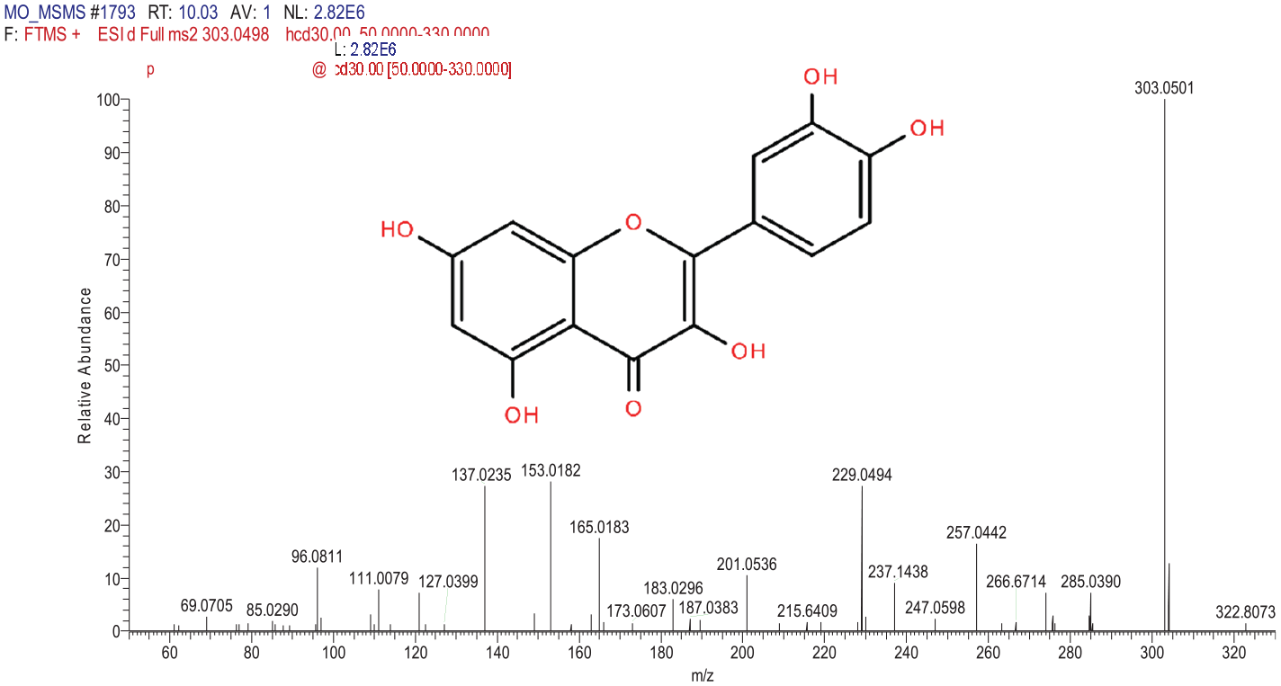
The MS spectra and structure of Quercetin.

The MS spectra and structure of Kaempferol.
Acute toxicity study of MOHE
Behavioural changes
There were neither behavioural changes nor mortality observed in the treated mice throughout the period of observation (Table 3).
Behavioural observations for female ICR-mice in acute toxicity studies of MOHE in both control and treated groups (2000 mg/kg body weight MOHE).
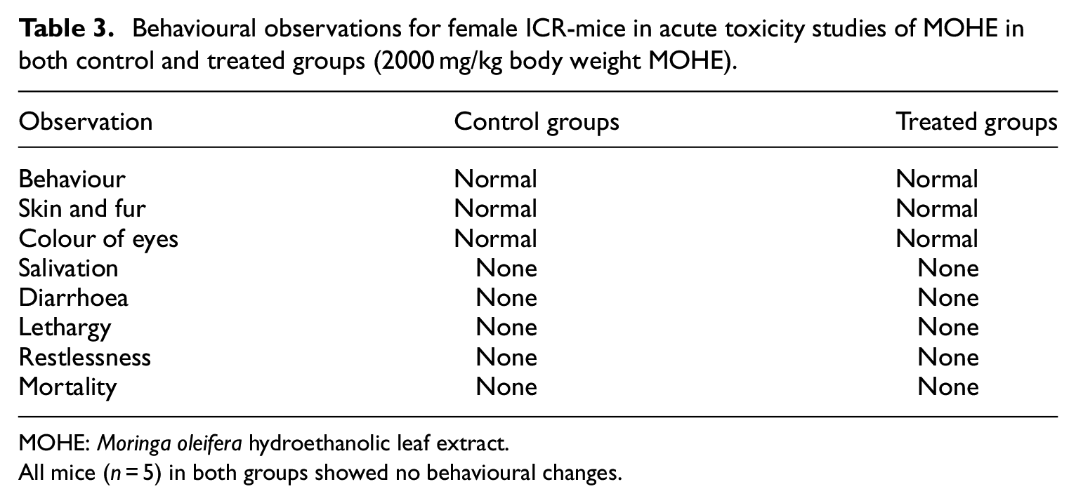
MOHE: Moringa oleifera hydroethanolic leaf extract.
All mice (n = 5) in both groups showed no behavioural changes.
Bodyweight
Figure 7 showed the effects of 2000 mg/kg MOHE on the average bodyweight gain of mice following single oral administration. There were no significant (p > 0.05) differences in the body weight gain of the mice in group B compared to A throughout the 14 days observation period. However, the mice in group B showed 187.24% and 512.25% reductions (p > 0.05) in the body weight gain at weeks 1 and 2 of the experiment respectively, compared to group A (Figure 7).

Average (mean ± SEM) weekly body weight gain (g) of female ICR-mice in acute toxicity study of Moringa oleifera hydroethanolic leaf extract; A = control, B = 2000 mg/kg MOHE, (p > 0.05).
Relative organs weight
The effects of oral administration of 2000 mg/kg MOHE on relative organs weight (%) of mice is presented in Table 4. There were no significant (p > 0.05) differences in the relative organs weight of liver, kidneys, spleen, heart, lungs, uterus and brain between the two groups of mice. (Table 4).
Relative organ weights (mean ± SEM) of female ICR-mice in acute toxicity study of Moringa oleifera hydroethanolic leaf extract.
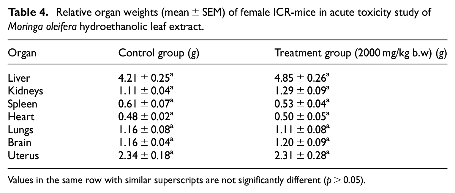
Values in the same row with similar superscripts are not significantly different (p > 0.05).
Haematological parameters
Table 5 illustrated the effects of 2000 mg/kg MOHE on haematological parameters of mice following single oral administration. The values did not show any significant (p > 0.05) differences between treatment and control groups. Importantly, haemoglobin and PCV values of mice in the treatment group were lower than the control, which indicate anaemia. Slight lymphopaenia and monocytosis indicating stress leukogram were also observed in the treated mice (Table 5).
Haemogram (mean ± SEM) of female ICR-mice in acute toxicity study of Moringa oleifera hydroethanolic leaf extract.

Values in the same row with similar superscripts are not significantly different (p > 0.05).
indicate the vales are lower than control and/or reference range.
indicates the value is higher than control and reference range.
Plasma biochemical parameters
The effects of oral administration of 2000 mg/kg MOHE on plasma biochemical parameters of ICR-mice are shown on Table 6. There was 3.1% decrease (p > 0.05) in urea level in group B (11.48 ± 0.50 mmol/L) compared to A (11.85 ± 1.35 mmol/L), as well as 22.4% increase (p > 0.05) in creatinine level in group B (40.40 ± 3.94 µmol/L) compared to A (33.00 ± 1.14 µmol/L). Moreover, the level of plasma ALT was 3.27% higher (p > 0.05) in group B (58.40 ± 2.98 U/L) compared to A (56.55 ± 4.42 U/L) (Table 6). Furthermore, there was 93% significant (p < 0.05) increase in the level of AST in group B (397.80 ± 17.64 U/L) compared to A (206.00 ± 11.34 U/L), as well as 52% significant (p < 0.5) increase in CK in group B (790.20 ± 103.43 U/L) compared to A (517.30 ± 51.56 U/L). (Table 6). Additionally, the albumin level was 4.3% lower (p > 0.05) in group B (34.44 ± 1.02 g/L) compared to A (36.00 ± 0.29 g/L), Moreover, the values for albumin in group B were 4.3% lower than the reference interval (36–43 g/L), while the plasma level of creatinine was 14.3% higher than the reference ranges in the treated group (Table 6).
Biochemical parameters (mean ± SEM) of female ICR-mice in acute toxicity study of Moringa oleifera hydroethanolic leaf extract.

Values in the same row with asterisk differ significantly (p < 0.05).
indicates the value is higher than the reference range.
indicates the value is lower than the reference range.
Histopathological evaluation of liver
Table 7 and Figure 8 showed the histopathological effects of single oral administration of 2000 mg/kg MOHE on the liver of ICR-mice. Mann-Whitney U test showed significant (p < 0.05) differences in the lesion scores between control and treatment groups. Pairwise comparisons test showed a significant (p < 0.05) mild to moderate necrosis indicated by acidophilic cytoplasm (acidophilic hepatocytes) and sinusoidal dilatation. Moreover, the results also showed a significant (p < 0.05) increase in the activity of Kupffer cells, and mild to moderate degenerative changes (cytoplasmic vacuolation) in the liver of treatment group (Table 7).
Lesion scores (mean ± SEM) for histopathological changes seen in the liver of female ICR-mice in acute toxicity study of Moringa oleifera hydroethanolic leaf extract.

indicate the values are significantly different (p < 0.05) between groups.

Lesions seen in the liver of female ICR-mice orally administered with Moringa oleifera hydroethanolic leaf extract (MOHE) at one dose level of 2000 mg/kg body weight (acute toxicity study): (a) and (b) photomicrographs of liver sections from mice in control group showing normal architecture of liver parenchyma (H&E stain, ×100), (c) photomicrograph of liver section from a treated mouse showing marked portal-portal bridging necrosis (H&E stain, ×100), (d) higher magnification of (c) showing acidophilic hepatocytes with pyknotic (top right), karyolitic and normal nuclei (bottom part) (H&E stain, ×200), (e) photomicrograph of liver section from a treated mouse showing marked acidophilic hepatocytes with pyknotic nuclei (bottom half and top right) at the periportal area (H&E stain, ×100), (f) higher magnification of (e) showing none-obstructive sinusoidal dilatation and activated Kupffer cells (centre left) (H&E stain, ×100), (g) photomicrograph of liver section from a treated mouse showing mild portal-portal bridging necrosis, degenerative change at the centrilobular area and prominent Kupffer cells activity (H&E stain, ×200) and (h) photomicrograph of liver section from a treated mouse showing mild focal inflammatory changes in liver parenchyma (H&E stain, ×400), scale bars represent 100 µ.
Histopathological evaluation of kidney
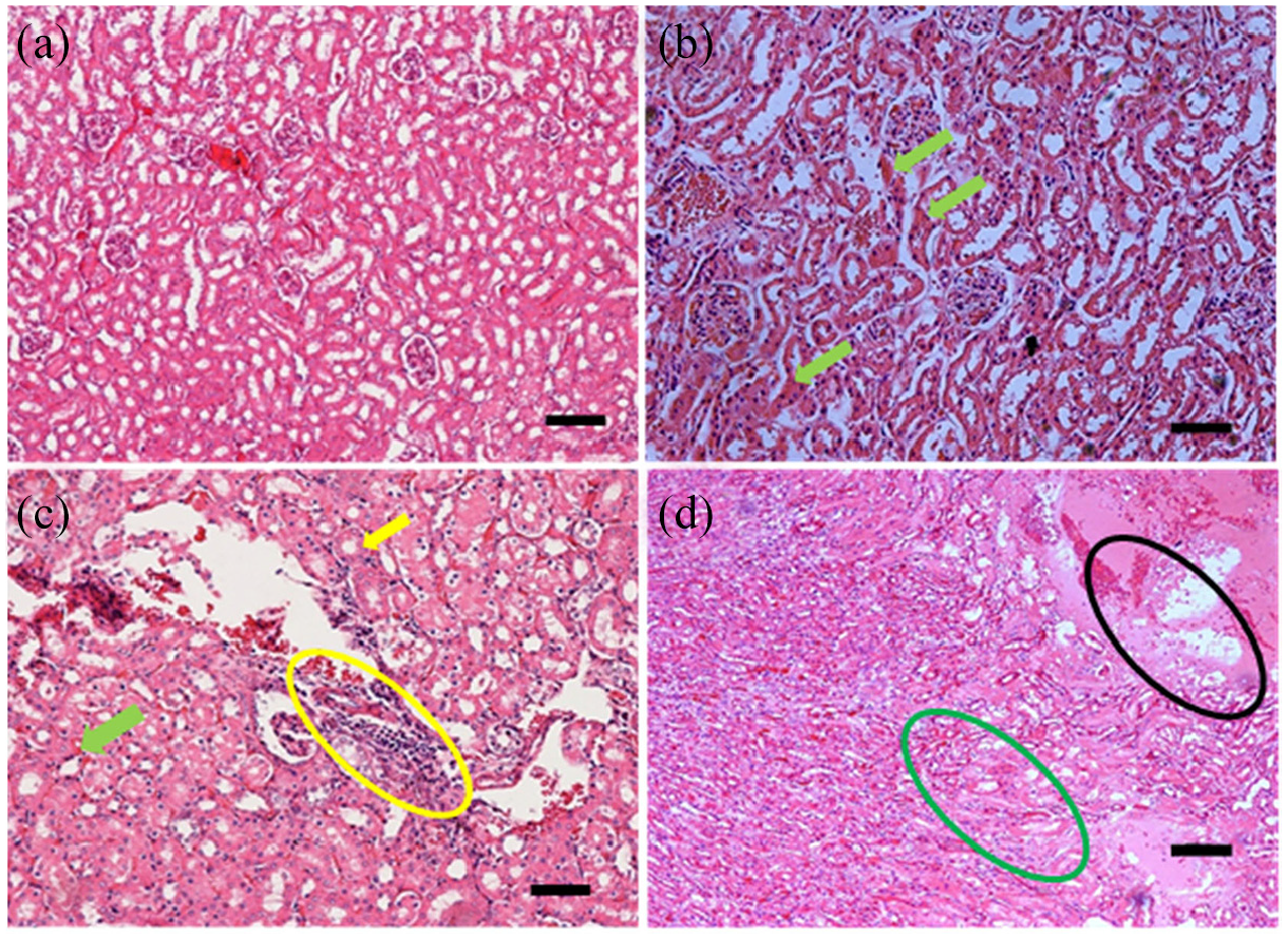
Significant (p < 0.05) histopathological changes were seen in the group of mice treated with 2000 mg/kg MOHE. These include renal tubular necrosis, interstitial nephritis and renal interstitial oedema (Table 8 and Figure 9). The other lesions (degenerative changes, pyknosis, hyaline and epithelial casts formation) observed in the treated mice were not significantly (p > 0.05) different from the control group.
Lesion scores (mean ± SEM) of the kidney of female ICR-mice in acute toxicity study of Moringa oleifera hydroethanolic leaf extract.
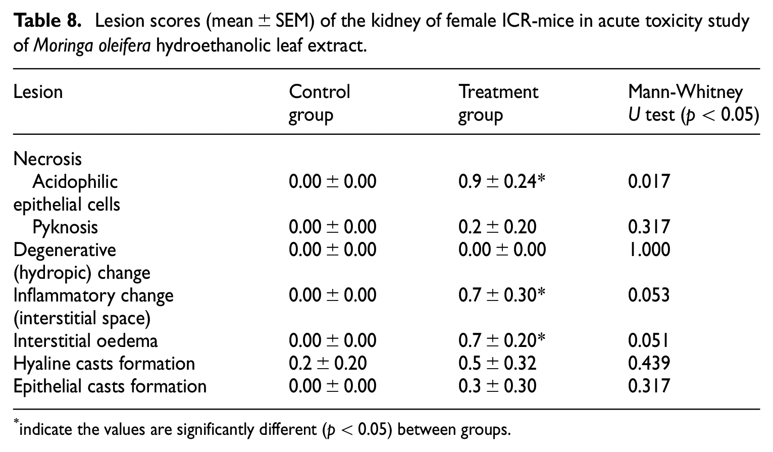
indicate the values are significantly different (p < 0.05) between groups.
Lesions seen in the kidney of female ICR-mice orally administered with Moringa oleifera hydroethanolic leaf extract (MOHE) at one dose level of 2000 mg/kg body weight (acute toxicity study): (a) photomicrograph of kidney section from a mouse in control group showing normal architecture of kidney (H&E stain, ×100), (b) photomicrograph of kidney section from a mouse in group B showing interstitial nephritis (H&E stain, ×100), (c) photomicrograph of kidney section from a mouse in group B showing interstitial oedema with infiltration of inflammatory cells and diapedesis of erythrocytes, (d) photomicrograph of kidney section from a mouse in group B showing interstitial oedema (H&E stain, ×100) and (e) and (f) photomicrographs of kidney sections from mice in group B showing inflammatory changes and necrosis of renal tubular epithelial cells characterised by acidophilic cells (E: generalise and F: bottom left) (H&E stain, ×100), scale bars represent 100 µ.
Sub-acute toxicity study
Body weight gain
The average body weight gain of the treated mice was significantly (p < 0.05) affected by the repeated administration of MOHE, as shown by repeated measures ANOVA and Bonferroni post hoc test (Figure 10). There were 569.83% and 150.29% significant (p < 0.05) increases in the body weight gain of the mice in group B at weeks 1 (1.55 ± 0.59 g) and 2 (0.88 ± 0.36 g) respectively, compared to group A at weeks 1 (0.23 ± 0.36 g) and 2 (0.35 ± 0.31 g). However, at weeks 3 (−0.12 ± 0.09 g) and 4 (0.91 ± 0.20 g), the mice in group B showed 107.48% (p < 0.05) and 32.99% (p > 0.05) reductions in the body weight gains respectively, compared to group A at weeks 3 (1.66 ± 0.24 g) and 4 (1.35 ± 0.54 g) (Figure 10). Furthermore, there were 170.69%, 92.57%, 53.19% and 50.15% decreases in the body weight gain of the mice in group C at weeks 1 (−0.16 ± 0.99 g; p > 0.05), 2 (0.03 ± 0.74 g; p > 0.05), 3 (0.78 ± 0.43 g; p < 0.05) and 4 (0.67 ± 0.60 g; p < 0.05) respectively, in relation to group A at weeks 1, 2, 3 and 4 (Figure 10). Similarly, there were 368.97%, 0.571% and 192.60% decreases in the body weight gain in group D at weeks 1 (−0.62 ± 0.67 g; p < 0.05), 2 (0.35 ± 0.90 g; p > 0.05) and 4 (−1.25 ± 1.58 g; p < 0.05) of the study respectively, in relation to group A (Figure 10). Correspondingly, the treated mice in group E presented 31.03%, 312.57%, 123.76% and 174.85% reductions in the mean body weight gains at weeks 1 (0.16 ± 0.64 g; p > 0.05), 2 (−0.74 ± 0.74 g; p < 0.05), 3 (−0.39 ± 0.51 g; p < 0.05) and 4 (−1.01 ± 0.18 g; p < 0.05) respectively, compared to group A (Figure 10).

Average (mean ± SEM) weekly body weight gain (g) of female ICR-mice in sub-acute toxicity study of MOHE; A = control, B = 125 mg/kg MOHE, C = 250 mg/kg MOHE, D = 500 mg/kg MOHE, E = 1000 mg/kg MOHE.
Relative organs weight
The relative organs weight of the treated mice was not significantly (p > 0.05) affected by 28 days repeated administration of MOHE between groups as shown by one-way ANOVA (Table 9). However, the relative weight of liver was 25.3%, 18.4%, 22% and 26.3% lower (p > 0.05) in groups B, C, D and E respectively, compared to A (Table 9). Similarly, the relative weights of right kidneys were 29.1% and 16.5% lower (p > 0.05) in groups D and E respectively, compared to A (Table 9).
Relative organ weights in % (mean ± SEM) of female ICR-mice in sub-acute toxicity study of MOHE.
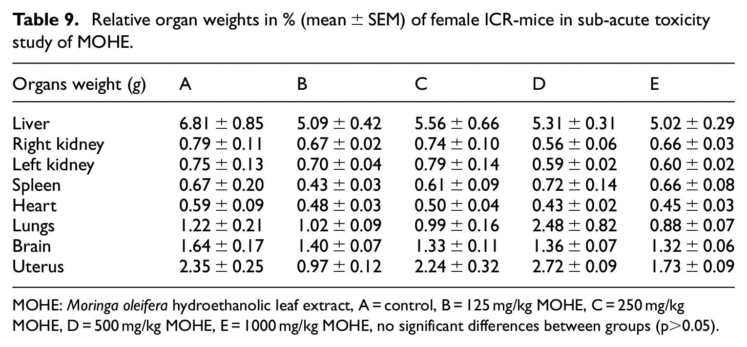
MOHE: Moringa oleifera hydroethanolic leaf extract, A = control, B = 125 mg/kg MOHE, C = 250 mg/kg MOHE, D = 500 mg/kg MOHE, E = 1000 mg/kg MOHE, no significant differences between groups (p>0.05).
Haematological parameters
Table 10 shows the effects of repeated daily administrations of MOHE for 4 weeks on the haematological parameters of female ICR-mice. There was significant (p < 0.05) difference in the haematological parameters between groups (Table 10) as determined by one-way ANOVA. Tukey post hoc test revealed that there was 11.7% decrease (p > 0.05) in the values of red blood cells in group E (9.85 ± 0.22 × 10¹²/L) compared to A (11.15 ± 0.65 × 10¹²/L). Correspondingly, the haemoglobin concentrations were 15% and 13.2% lower (p > 0.05) in groups D (150.00 ± 13.19 g/L) and E (153.04 ± 4.39 g/L) respectively, in relation to A (176.40 ± 8.35 g/L). Moreover, there was also 7.6% significant (p < 0.05) decrease in the values of MCV in group D (60.80 ± 0.86 fL) compared to A (65.80 ± 0.97 fL), as well as 12.9% significant (p < 0.05) decrease in the values of MCH in group D (13.50 ± 0.51 g/L) compared to A (15.86 ± 0.28 g/L). Moreover, the platelets were 36%, 1%, 32.4% and 59% higher (p > 0.05) in groups B (1355.00 ± 276.2 × 10⁹/L), C (1007.40 ± 158.2 × 10⁹/L), D (1310.80 ± 197.9 × 10⁹/L) and E (1573.80 ± 121.4 × 10⁹/L) respectively, compared to A (989.80 ± 197.4 × 10⁹/L) and the values in group E (1573.80 ± 121.4 × 10⁹/L) were 2% higher than the reference interval (926−1539 × 109/L). The plasma proteins were 13% higher (p > 0.05) in group D (82.00 ± 5.83 g/L) compared to A (72.00 ± 2.00 g/L) Table 10. Furthermore, the values for total white blood cells (TWBC) were 49.32%, 9.70% and 7.82% higher (p > 0.05) in groups B (11.08 ± 0.71 × 10⁹/L), D (8.14 ± 1.43 × 10⁹/L) and E (8.00 ± 1.24 × 10⁹/L) respectively, compared to group A (7.42 ± 0.95 × 10⁹/L). Correspondingly, the values for neutrophils were also 38.2%, 24.16%, 29.21% and 42.13% higher in groups B (2.46 ± 0.26 × 10⁹/L), C (2.21 ± 0.46 × 10⁹/L), D (2.30 ± 0.47 × 10⁹/L) and E (2.53 ± 0.77 × 10⁹/L) respectively, compared to group A (1.78 ± 0.35 × 10⁹/L). The lymphocytes were however, 4.47%, 1.36% and 8.37% lower (p > 0.05) in group C (4.91 ± 0.27 × 10⁹/L), D (5.07 ± 1.14 × 10⁹/L) and E (4.71 ± 0.53 × 10⁹/L) respectively, compared to group A (5.14 ± 0.76 × 10⁹/L) (Table 10). However, there were 51.06%, 23.4%, 21.28% and 21.28% decreases (p > 0.05) in the levels of monocytes in groups B (0.71 ± 0.03 × 10⁹/L), C (0.58 ± 0.12 × 10⁹/L), D (0.57 ± 0.16 × 10⁹/L) and E (0.57 ± 0.15 × 10⁹/L) respectively, compared to group A (0.47 ± 0.09 × 10⁹/L) (Table 10).
Haematological parameters (mean ± SEM) of female ICR-mice in sub-acute toxicity study of MOHE.

MOHE: Moringa oleifera hydroethanolic leaf extract, A = control, B = 125 mg/kg MOHE, C = 250 mg/kg MOHE, D = 500 mg/kg MOHE, E = 1000 mg/kg MOHE, values in the same row with asterisk differ significantly (p < 0.05), MCV: mean corpuscular volume; MCH: mean corpuscular haemoglobin; MCHC: mean corpuscular haemoglobin concentration.
Plasma biochemical parameters
The biochemical parameters analysed in the plasma differed significantly (p < 0.05) between the groups as demonstrated by one-way ANOVA (Table 11). Tukey post hoc test revealed 29.2% significant (p < 0.05) decrease in the plasma level of urea in group E (8.24 ± 0.79 mmol/L), in relation to A (11.64 ± 0.94 mmol/L), as well as 23% and 81.9% significant (p < 0.05) increases in creatinine levels in groups D (23.20 ± 3.65 µmol/L) and E (34.20 ± 1.66 µmol/L) respectively compared to A (Table 11). Furthermore, there were 160.9% and 68.2% significant (p < 0.05) increases in the level of ALT in groups D (337.20 ± 52.85 U/L) and E (217.40 ± 17.09 U/L) respectively compared to A (Table 11), as well as 30.2% significant (p < 0.05) decrease in AST level in group E (260.40 ± 23.16 U/L) compared to A (373.00 ± 8.93 U/L). The plasma levels of CK were 125.1%, 121.9% and 99.9% significantly (p < 0.05) higher in groups C (1411.80 ± 124.09 U/L), D (1391.60 ± 141.18 U/L) and E (1253.60 ± 95.83 U/L) respectively compared to A (627.20 ± 20.53 U/L) Table 11.
Biochemical parameters (mean ± SEM) of female ICR-mice in sub-acute toxicity study of MOHE.

MOHE: Moringa oleifera hydroethanolic leaf extract, A = control, B = 125 mg/kg MOHE, C = 250 mg/kg MOHE, D = 500 mg/kg MOHE, E = 1000 mg/kg MOHE, values in the same row with asterisk differ significantly (p < 0.05).
Histopathological evaluation of liver
There was significant (p < 0.05) differences in the histological alterations in the liver of the various groups of mice as determined by Kruskal Wallis H test (Table 12). Pairwise comparisons test showed a mild sinusoidal dilatation (Figure 11(d)) and acidophilic cytoplasm (Figure 11(b) as well as moderate pyknosis of the hepatocytes (Figure 11(b) and (c)). The moderate sinusoidal dilatation was significantly (p < 0.05) higher in groups D (2.30 ± 0.20) and E (1.90 ± 0.48) compared to A (0.00 ± 0.00). There was moderate (p < 0.05) hepatic eosinophilic cytoplasm in group D (2.30 ± 0.20) compared A (0.00 ± 0.00), as well as moderate hepatic karyolysis (p < 0.05) in group E (2.40 ± 0.10) compared to A (0.00 ± 0.00). These findings agreed with those of plasma biochemistry, where significant (p < 0.05) differences were observed in the liver parameters between the control and groups treated with 500 and 1000 mg/kg of the extract (Table 11).
Lesion scores (mean ± SEM) of the liver of female ICR-mice in sub-acute toxicity study of MOHE.

MOHE: Moringa oleifera hydroethanolic leaf extract, A = control, B = 125 mg/kg MOHE, C = 250 mg/kg MOHE, D = 500 mg/kg MOHE, E = 1000 mg/kg MOHE.
significantly different at p < 0.05.

Effects of repeated oral administration of MOHE for 28 days on the histology of liver of female ICR-mice: (a) photomicrograph of liver section from a mouse in a control showing normal architecture of liver (H&E stain, ×100), (b) photomicrograph of liver section from a mouse in group E showing sinusoidal dilatation and hepatic degeneration characterised by cytoplasmic vacuolation (yellow encircled), hepatic necrosis characterised by eosinophilic cytoplasm (black encircled) and pyknosis (yellow arrow) of the hepatocytes (H&E stain, ×100), (c) photomicrograph of liver section from a mouse in group E showing pyknosis (yellow arrow) and hepatic degeneration characterised by cytoplasmic vacuolation (yellow encircled) (H&E stain, ×200) and (d) photomicrograph of liver section from a mouse in group E showing sinusoidal dilatation (blue encircled) and cellular infiltration (green arrow) (H&E stain, ×100).
Histopathological evaluation of kidney
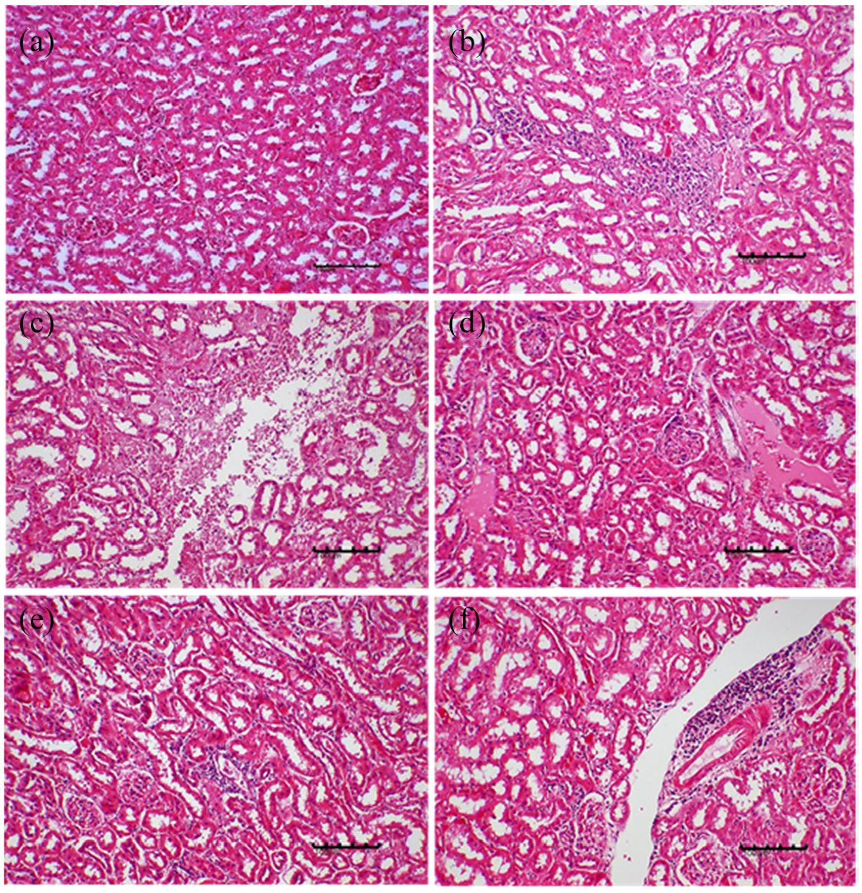
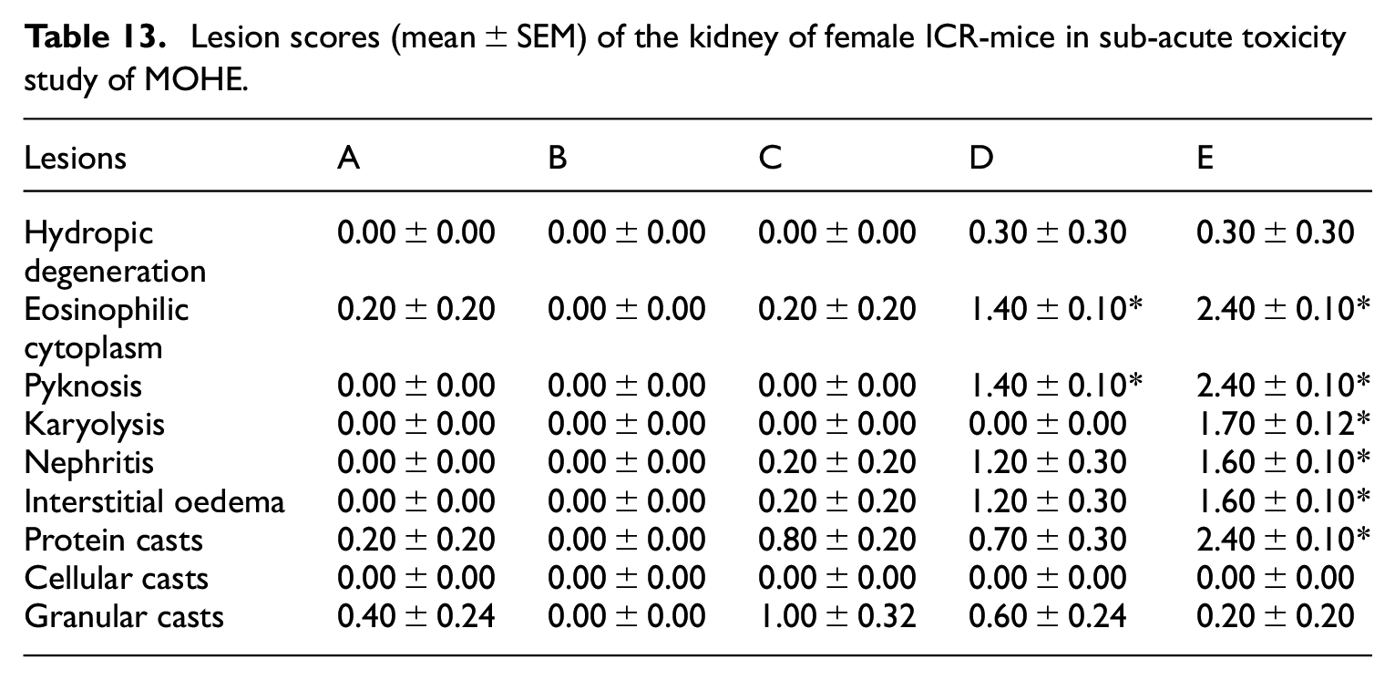
Significant (p < 0.05) histopathological changes were observed in the kidneys of different groups of mice treated with MOHE for 28 days, as presented by Kruskal Wallis H test (Table 13). Pairwise comparisons test showed a mild to moderate kidney necrosis characterised by significant mild to moderate eosinophilic cytoplasm (Figure 12(b) and (c)), pyknosis and karyolysis of the renal tubules (Figure 12(c)). The eosinophilic cytoplasm was significantly (p < 0.05) higher in group E (2.40 ± 0.10) compared to A (Table 13). Similarly, pyknosis was significantly (p < 0.05) higher in group E (2.40 ± 0.10) compared to control. Additionally, there was a significantly (p < 0.05) higher karyolytic tubular lesion in group E (1.70 ± 0.12) compared to A (Table 13). The renal tubular necrotic lesions were mild in group D and moderate in group E (Table 13). Besides, there was mild significant (p < 0.05) tubular and interstitial nephritis (Figure 12(c)) in groups D (1.20 ± 0.30) and E (1.60 ± 0.10) respectively compared to A, as well as significant (p < 0.05) moderate tubular protein casts and interstitial oedema (Figure 12(d)) in group E (2.40 ± 0.10) compared to A (Table 13).
Lesion scores (mean ± SEM) of the kidney of female ICR-mice in sub-acute toxicity study of MOHE.
Effects of repeated oral administration of MOHE for 28 days on the histology of kidney of female ICR-mice: (a) photomicrograph of a kidney section (H&E stain, ×100) from a mouse in control showing normal architecture of kidney, (b) photomicrograph of a kidney section (H&E stain, ×200) from a mouse in group E (1000 mg/kg MOHE) showing renal necrosis characterised by eosinophilic cytoplasm (green arrow), (c) photomicrograph of a kidney section (H&E stain ×200) from a mouse in group E showing glomerulonephritis characterised by cellular infiltration (yellow encircled) and (d) photomicrograph of a kidney section (H&E stain, ×100) from a mouse in group E showing interstitial oedema (mainly at the top right).
Discussion
Results for the chemical profiling analysis of M. oleifera hydroethanolic leaf extract in this study revealed four major compounds; namely glucomoringin, niaziminin A, quercetin and kaempferol. Leone et al. 12 and Coppin et al. 26 demonstrated that M. oleifera leaf cultivated in Sharawi camps, Chad and Haiti and Ghana, Senegal and Zambia have high level of phenolic compounds including the four compounds detected in the M. oleifera leaf cultivated in Selangor Malaysia. The first compound, glucomoringin (GMG), is a glucosinolate (GL) presents in a large quantity in M. oleifera seeds. 33 Giacoppo et al. 33 hydrolysed GMG from M. oleifera seeds to 4-(α-L-rhamnosyloxy) benzyl isothiocyanate (GMC-ITC; moringin) using myrosinase, and mixed the GMC-ITC compound with alpha-cyclodextrin (α-CD) to form a α-CD-MAC complex. The complex exhibited anti-inflammatory effects on lipopolysaccharide (LPS)-stimulated RAW 264.7 macrophages cells 34 and also inhibits the proliferation of malignant cell line (SH-SY5Y human neuroblastoma cell line) via inhibiting the survival of PI3K/Akt/mTOR and MAPKs pathways, which eventually resulting in cell death. 33 For the second compound, niaziminin or commonly known as niaziminin A, it belongs to the class of organic compounds known as phenolic glycosides, and contain phenolic structures which include lignans and flavonoids. 35 The compound is also found in fats and oils, herbs and spices and green vegetables. 35 Murakami et al. 36 discovered that niaziminin isolated form the leaves of M. oleifera, holds a strict structural requirement for inhibition of tumour-promoter-induced Epstein-Barr Virus activation. The third compound present in the extract is quercetin, which is a polyphenol flavonoid with potential chemo-preventive activity. It is present ubiquitously in fruits such as red grapes, raspberries, cranberries, citrus fruit, tomato, broccoli and other leafy green vegetables. 37 It inhibits proliferation of tumour cells via modulation of either estrogen growth factor receptor (EGFR) or estrogen receptor mediated signal transduction pathways. 38 Moreover, Goodla et al. 39 reported that ethanolic extract of Ammannia baccifera is a potent antioxidant and hepatoprotective agent because of the flavonoids and phenolic compounds it contains, including quercetin. 39 The fourth compound that was identified in the M. oleifera leaf extract is kaempferol. It is a natural flavonoid, which has been isolated from Delphinium, Witch-hazel, grapefruit and other plant sources. 40 It mainly acts as an antioxidant by reducing oxidative stress and could be used as a possible cancer treatment. 41
As all the compounds possess a potential anticancer activity, it should not be forgotten that the compounds could also cause negative effects to healthy cells as what other chemotherapeutic drugs do. A few studies have been conducted to evaluate the safety level of the plant extracts.15–17,27,31,42,43 by determination of its lethal dose (LD). Basically, the smaller is the LD value, the more toxic is the substance, and vice versa. According to Hodge and Sterner 44 toxicity scale, LD50 value (administered through oral route) of 1 mg/kg body weight or less, falls under toxicity rating level 1, is considered extremely toxic, while 15,000 mg/kg body weight and above, falls under toxicity level 6, is considered relatively harmless, and the doses are equivalent to a drop and 1 L, respectively, for probable lethal dose for human. It should be remembered that LD50 only indicates that the substance is safe to be taken at single dose, when or where it would not cause acute death, but consumption at that particular dose over longer periods of time should be carefully evaluated before it is conclusively considered safe to be consumed for supplementary and medicinal purposes. Taken all the consideration, herbal toxicity studies are considered very important before determination of its medicinal potentials.
Acute toxicity study conducted by Adedapo et al. 17 revealed 17% and 33% of rats orally gavaged with 1,600 and 2,000 mg/kg body weight of M. oleifera leaf aqueous extract, respectively, died within 48 h. The rats were slightly dull for the first 5 h after administration of the extract. Based on the percentage of mortality, the LD50 of the extract is concluded more than 2000 mg/kg body weight (48-h, oral LD50, rat). Okumu et al. 43 also reported 48-h oral LD50 of M. oleifera leaf aqueous extract in rats is higher than 2000 mg/kg body weight. Further assessment on the level of serum liver enzymes revealed significant increase in AST and insignificant increase in bilirubin (but both parameters were still within the normal limits), and liver histopathology revealed mild distortions in the architecture of hepatocytes. 43 Another study done by Awodele et al. 16 revealed that mice orally gavaged with 1600, 3200 and 6400 mg/kg body weight of M. oleifera leaf aqueous extract also showed no mortality in 24 h, although the mice were slightly dull within 2 h of administration. They concluded oral LD50 of the extract is higher than 6,400 mg/kg body weight (24-h, oral LD50, mouse). They also determined i.p. LD50 of the extract and the LD50 is estimated at 1,585 mg/kg body weight (24-h, i.p. LD50, mouse). Using ethanolic method of extraction, Osman et al. 42 reported higher i.p. LD50 for M. oleifera leaf extract in rats and rabbits, which are 6,616.67 and 26,043.67 mg/kg body weight, respectively (48-h, i.p. LD50, rats and rabbits). Histopathology of the heart revealed cardiac muscle degeneration and haemorrhage, while kidney and liver revealed haemorrhage, necrosis and degeneration of epithelial renal tubules and hepatocytes, respectively. 42 Using different part of the plant, Kasola et al. 45 conducted acute toxicity study on the root of M. oleifera. They demonstrated that LD50 of aqueous and ethanolic extracts of M. oleifera roots are 17,800 and 15,900 mg/kg body weight, respectively, in mice.
In this study, single oral administration of 2000 mg/kg M. oleifera hydroethanolic leaf extract induced neither symptoms of acute toxicity nor mortality in female ICR-mice. The extract did not cause any significant changes in relative organs weight, but insignificant decreased body weight gain was noted. The mice in group B had mild anaemia and showed evidence of stress indicated by stress leukogram. The extract also caused significant increases in AST and CK levels, which suggest liver and/or muscular injuries. Further evaluation on liver architecture of the mice, confirmed its hepatotoxicity. Normal ALT level in the mice could be related to its short half-life as compared to AST (mitochondrial AST). ALT is solely localised in the cytoplasm whereas AST localised in both cytoplasm (20%) and mitochondria (80%). The ALT half-life is 48 h as compared to 87 h for the mitochondria AST; cytoplasm AST is shorter than ALT, which is 17 h. Another reason that could explain increase in the AST is, released of this enzyme by other tissues including kidneys, skeletal muscle, erythrocytes, brain, lungs, pancreas and heart. As CK was also elevated (an enzyme specific for muscle injury), further investigation on heart muscle architecture is necessary (not examined in this study). Evaluation on kidney architecture of the treated mice revealed nephrotoxicity (which further explain increase in serum AST level), and it was consistent with the elevation of serum creatinine level in the mice. The presence of hyaline cast in the renal tubules, further supports kidney injury and it was also portrayed by the evidence of hypoalbuminaemia/hypoproteinaemia that could be the outcome of protein losing nephropathy (PLN). Hypoproteinaemia encountered by the mice decreases the active colloid osmotic/oncotic pressure gradient, and subsequently increases the flux of transcapillary fluid 46 depicted by oedema of the renal interstitium (interstitial oedema) in the treated mice. Besides PLN, reduction in serum albumin level could be due to reduce feed/protein intake and liver disease. The formation of oedema in the interstitial tissues of the kidneys was also associated with inflammation of the renal interstitium (interstitial nephritis). Inflammation would increase vascular permeability and lead to effusion of fluid into the renal interstitium. 46 Inflammation would also result in the release of inflammatory mediators causing vasodilation and induce infiltration of leukocytes. Vascular smooth muscle cells relaxation in the arterioles and precapillary sphincters cause reduction in the upstream resistance, which subsequently increases capillary pressure (hydrostatic pressure) and the number of open capillaries for flow of blood. These alterations, in addition to the increased effective pore radii in the microvascular barrier lead to the formation of filtrate that is rich in protein, which increases interstitial fluid volume, interstitial fluid pressure, and tissue colloid osmotic pressure. 46
The sub-acute toxicity study of MOHE in this study, showed significant reduction in the body weight gain of the mice treated with 1000 mg/kg MOHE daily for 28 days. This could be due to decreased feed intake or reduced fats deposition in the treated mice. 47 These reports were comparable to those reported by Kasolo et al., 15 where rats treated with aqueous and ethanol extracts of M. oleifera gained less weight compared to the control group.
The repeated oral administration of MOHE for 28 days in this research resulted in some plasma biochemical alterations. The substantial decrease in the plasma level of urea in the group of mice treated with 1000 mg/kg MOHE may suggest that the sub-acute administration of the extract at this dose might have some degree of liver injury to the mice. This is because urea has been reported to decrease due to liver failure, low protein diet, anabolic steroids, or diabetes insipidus. 48 On the other hand, the creatinine level in the groups treated with 500 and 1000 mg/kg doses of the extract daily for 28 days was significantly elevated. This may indicate that the extract might have caused some degree of renal injury in the treated mice.49,50 This is because creatinine has been routinely used as marker for evaluation of renal function. 51 There was also a significant increase in the levels of ALT in the group treated with 500 and 1000 mg/kg MOHE daily for 28 days. At the same time, the AST level was significantly elevated at 1000 mg/kg, so also the CK level was remarkably elevated in the groups treated with 250, 500 and 1000 mg/kg doses of the extract. The significant increase in ALT and AST could indicate that the extract at 500 and 1000 mg/kg induced some degree of liver injury in the mice.48,52 This is because ALT is known to increase in liver injury particularly hepatic necrosis. 48 Significant increase in the serum levels of ALT and AST was reported previously in rats treated with 400 and 1600 mg/kg M. oleifera aqueous leaves extract daily for 21 days. 17 Similarly, Awodele et al. 16 reported that oral administration of M. oleifera aqueous leaf extract at 250, 500 and 1500 mg/kg caused a reduction in hepatic enzyme marker (SGOT) in rats, suggesting possible hepatic injury in the treated mice. The results of the biochemical analyses of this study also agrees with the reports of Olayemi et al., 53 where a remarkable decrease in the liver enzymes, AST, ALP and ALT was reported in the rats treated with both seed and leaf methanolic extracts of M. oleifera at 100, 200, 400 and 1000 mg/kg daily for 28 days. Awodele et al. 16 reported an elevation in the serum levels of urea and creatinine in rats treated with 250, 500 and 1500 mg/kg M. oleifera aqueous leaf extracts daily for 60 days, probably due to renal lesions in the rats. 49 However, Olayemi et al. 53 described a remarkable reduction in the creatinine level in the group of rats treated with 1000 mg/kg M. oleifera methanolic leaf extract daily for 28 days. The decreased creatinine levels could be associated with muscular dystrophy or hepatic injury.48,52
The histological evaluation of liver and kidneys of the mice treated with different doses of MOHE daily for 28 days revealed some degrees of lesions. The moderate pyknosis and karyolysis as well as eosinophilic cytoplasm of the hepatocytes observed in the group treated with 500 and 1000 mg/kg of the extract daily for 28 days may suggest mild hepatic necrosis,27,31,54 and this corroborates the results of plasma biochemical analysis, especially that the mice administered with 1000 mg/kg showed significant hepatic karyolysis compared to the control and other treatment groups. This further suggested that the extract at 500 and 1000 mg/kg might have induced adverse effects on the liver of the treated mice.27,31,54 Similarly, the mild and moderate eosinophilic cytoplasm, pyknosis and karyolysis respectively observed in the renal tubules of the mice treated with 500 and 1000 mg/kg daily for 28 days suggest that the extract at these concentrations induced some degree of necrosis27,55,56 in the kidneys of the treated mice. The sub-acute adverse effects of the extract at these doses were further evidenced by the mild tubular and interstitial nephritis55,56 seen in group of mice treated with 500 and 1000 mg/kg MOHE, as well as the moderate tubular protein casts27,56 observed in group treated with 1000 mg/kg of the extract. Moreover, these findings supported the plasma biochemistry results that showed significant increase in the creatinine level at 1000 mg/kg dose.
The results of the histological evaluation of liver and kidney in this study have agreed with Olayemi et al. 53 who reported that Wistar rats treated with M. oleifera methanolic extract at 100, 200, 400 and 1000 mg/kg revealed visible changes in all the organs at all doses tested, even at 100 mg/kg. The histological changes observed by Olayemi et al. 53 included severe portal congestion, periportal cellular infiltration at 100 mg/kg, as well as severe diffuse vacuolar degeneration of the hepatocytes at 200 mg/kg. Moreover, there was generalised interstitial congestion of the renal parenchyma at 100 mg/kg and a moderate renal cortical congestion at 1000 mg/kg. 53 Remarkably, the results of Kwaghe et al. 57 were in agreement with the histological findings of this study. Administration of ethyl acetate soluble fraction of aqueous extract of M. oleifera leaves in cockerels orally at 100, 200 300 and 400 mg/kg daily for 28 days revealed histopathological lesions including renal multifocal haemorrhages, congestion, focal and diffused mononuclear cell infiltration of the interstitium, hyaline degeneration, tubular degeneration and necrosis, as well as renal tubular protein cast. 57 Furthermore, hepatic congestion of blood vessels, hyperplastic liver, bile duct hyperplasia, vacuolar degeneration particularly in the periportal area, hydropic changes, focal mononuclear infiltration as well as multifocal area of necrosis were observed in the liver of cockerels treated with different doses of ethyl acetate soluble fraction of aqueous extract of M. oleifera leaves. 57 Similarly, Akinlolu et al. 58 reported widening of renal capsular spaces in the kidneys of Wistar rats administered with varying doses (250, 500 and 750 mg/kg) of M. oleifera daily for 21 days, 58 suggesting possible adverse effects of the extract. Nevertheless, the histopathological findings of this research were contrary to some of the reports of previous researchers including Adedapo et al., 17 Kasolo et al. 45 and Awodele et al., 16 where the extract was reported not to be associated with any histological changes in the liver and kidney of the animals. This perhaps could be because those previous studies were carried out with aqueous extract of the plant, which has less ability to extract the phytochemical compounds present in the leaves as compared to the ethanolic extract of the plants9,19–21 used in this study.
Anaemia in mice treated with MOHE could be attributed to the presence of glucomoringin in the leaf extract. Glucomoringin, once hydrolysed, the compound is converted to isothiocyanate (ITC). 33 Two forms of ITC, which are allyl ITC (AITC) and benzyl ITC (BITC) are present in plants/vegetables. AITC is detected in cabbage, cauliflower, arugula, broccoli etc. A study conducted by Jenner et al. 59 indicated AITC oral LD50 is 340 mg/kg in rats with pronounce jaundice, while at 40 mg/kg via oral route, it induced acute toxicity on rats’ liver, thymus, kidney and blood. 60 Meanwhile, administration of AITC daily for 20 days at 50 mg/kg body weight in rats induced subacute stomach ulceration. For BITC, subacute daily administration at 200 mg/kg body weight in rats induced significant alterations in the haematological parameters. 61 In a study conducted by Adedapo et al. 17 on subacute toxicity evaluation of aqueous extract of M. oleifera leaves in rats, daily doses of the extract at 800 mg/kg and 1600 mg/kg body weight for 28 days induced anaemia. They also found a slight decrease in the mice body weight at the end of the experiment which strongly associated with decrease in feed intake, which lead to reduction in the deposition of fats in their body.
Toxicity results presented in this study are similar to our previous toxicity studies on other ethanolic extracts, which include Mariposa christia vespertilionis leaves 31 and Clinacanthus nutans leaves25,27 where significant histopathological changes in the liver and kidneys of rats and mice were observed using doses lower than 300 mg/kg body weight in subacute25,31 and sub chronic 27 repeated oral doses. However, alterations in the serum biochemical parameters related to the liver and kidney injury were not observed in the rats.27,31 In another toxicity study by our group 62 on nanoparticle-loaded phytic acid-chitosan-magnetic iron oxide (IP6-CS-MNPs) for its acute toxicity effects in mice, no alterations in liver and kidneys architecture were observed, but higher levels of urea and serum alkaline phosphatase (ALP) at 2000 mg/kg and 1000 mg/kg body weight of IP6-CS-MNPs, respectively, were observed in mice. Meanwhile, acute toxicity study of methanolic extract of Rhaphidophora decurvisa (Roxb.) Schott in rats using doses of 700, 2800 and 3500 mg/kg body weight showed no abnormalities in the body weight, blood parameters and also liver and kidneys histological structures. 63 The same findings were found when lower doses (70, 140 and 280 mg/kg body weight) of the extract were tested in rats for the subacute and subchronic toxicity studies. Using aqueous extract of Ficus deltoidea, Fazliana et al. 64 have shown the extract did not induce alterations in the haematological and biochemical parameters of rats subchronically administered with the extract at 100 and 300 mg/kg body weight. All the toxicity experiments conducted above indicate that level of toxicity is solely dependent on the type of herbs, extraction methods, exposure period and dosage of the herbs. Dose above 1000 mg/kg body weight could be safe for single administration or exposure, but dose lower than 1000 mg/kg might not be safe for repeated consumption or exposure. Therefore, assessment on the safety of herbal extracts for human consumption should be thoroughly evaluated.
Limitations of the study
The current study is limited to the toxicity effects of single oral administration of high dose (2000 mg/kg) and short-term duration (4 weeks) of repeated lower doses (125, 250, 500 and 1000 mg/kg) of the extract (MOHE) in mouse model. However, administration of herbal extracts in traditional medicine, requires longer repeated administration, especially in the treatment of chronic illnesses, including cancers. Consequently, it is recommended to further evaluate the sub-chronic and chronic toxicity effects of the extract in mouse model. Moreover, the quantification of the bioactive compounds identified in the current study has not been reported. It is therefore, recommended that the quantity of each of the compounds identified in M. oleifera leaves be evaluated. Furthermore, the sample size selection in the current study was limited to the OECD guidelines 425 and 407, which did not require power analysis. Hence future studies may consider power analysis for the selection of sample size.
Conclusions
It is concluded that MOHE cultivated in Selangor, Malaysia contains phenolic compounds and flavonoids. Its lethal dose (LD50) in female ICR-mice is higher than 2000 mg/kg. At that dose level the extract causes mild anaemia, stress leukogram and mild to moderate hepato-nephrotoxicity in female ICR-mice. Similarly, this study demonstrated that repeated daily oral administrations of MOHE for 28 days induced significant alterations in the parameters for injury markers in the liver, kidney and muscles at 500 and 1000 mg/kg in female ICR-mice. In addition, repeated oral administrations of MOHE daily for 28 days in female ICR mice induced moderate hepatic degeneration at 500 mg/kg and necrosis at 1000 mg/kg, as well as mild kidney necrosis at 1000 mg/kg. Therefore, the plant extract at 1000 mg/kg dose, is considered toxic and unsafe for repeated consumption as food supplementation and/or alternative medicine; however, lower doses and/or short time administration could be used safely for medicinal purposes.
Footnotes
Acknowledgements
The technical support from the staff of the Natural Product Laboratory, Institute of Bioscience, UPM (Mr Azizul Isa, Mrs. Zurina Zainal, Mr. Ahmad Fauzi Mokhtar), Histopathology Laboratory, Faculty of Veterinary Medicine, UPM (Mr. Mohd Jamil Samad, Mrs. Jamila Jahari, Mrs. Latifah Mohd Hanan, Ms. Zainatulaishah Abdul Manaf) and Haematology and Clinical Biochemistry Laboratory, Faculty of Veterinary Medicine, UPM (Mrs. Darulmuqamah Masud, Mrs. Noorain Azman, Mr. Abdullah Misron and Mr. Arman Addelan)) are highly acknowledged. The authors also wish to acknowledge the contributions of the Malaysian Agricultural Research Development Institute (MARDI).
Declaration of conflicting interests
The author(s) declared no potential conflicts of interest with respect to the research, authorship, and/or publication of this article.
Funding
The author(s) disclosed receipt of the following financial support for the research, authorship, and/or publication of this article: This work was supported by the IPS Research Grant (IPS/2018/9594600) Universiti Putra Malaysia, and the Tertiary education trust fund (TETFUND) Nigeria.