
Abstract
Sprague-Dawley rats are amongst the most widely used animals in biomedical research and malignant lymphoma has long been known to be a frequent neoplasm in these animals. A 9-month-old male control Sprague-Dawley rat from a toxicity study showed gelatinous material in the cranial cavity and dark, thickened cerebral meninges at necropsy. At microscopic evaluation of the temporal bone, neoplastic lymphocytes were seen invading several structures of the middle ear. The neoplastic cells appeared to extend from the marrow of the temporal bone, covered the dorsal part of the tympanic cavity wall, and surrounded and infiltrated the base of the tensor tympani muscle as well as the chorda tympani branch of the facial nerve. The lymphoma was generalized; neoplastic lymphocytes were also noted in numerous other tissues. Literature regarding neoplasms of the middle and inner ear in animals is scarce and, to our knowledge, this is the first report of a lymphoma involving the middle ear of a rat.
Introduction
Sprague-Dawley rats are amongst the most widely used animals in biomedical research and malignant lymphoma has long been known to be a frequent neoplasm in aged, and even middle-aged, individuals of this strain. The incidence of malignant lymphoma in aged Sprague-Dawley rats was reported to be close to 2% in males and up to slightly over 4% in females.1,2 In male Sprague-Dawley rats up to 50 weeks old, this neoplasm was identified as the most common tumor. 3 Malignant lymphomas in rats are well known to generalize to most organs but, to our knowledge, invasion of the rat middle ear by this neoplasm has never been reported.
Case presentation
A 9-month-old male Sprague-Dawley rat (Charles River Canada Inc., St-Constant, Quebec, Canada) from a 26-week oral gavage toxicity study was assigned to a group-housed control group and was administered a non-pharmacologically active, non-toxic reference item (citrate buffer in ultra pure water) daily. The animal was group-housed with two other control group males in a polycarbonate cage containing appropriate bedding, a hiding tube and chewing object, and equipped with an automatic watering valve. The room temperature was kept between 19 and 25℃, the humidity was between 30 and 70%, and the light cycle was 12 hours of light and 12 hours of darkness. The animal was fed PMI Nutrition International Certified Rodent Chow No. 5CR4 ad libitum. All animal procedures were conducted with guidance from the USA National Research Council and the Canadian Council on Animal Care, and under approval from the Institutional Animal Care and Use Committee of Charles River Laboratories.
The day before scheduled necropsy, the animal weighed 786 g compared to a group mean of 890.4 g and showed a 4% body weight loss during the previous week, after a constant weight gain during the rest of the study.
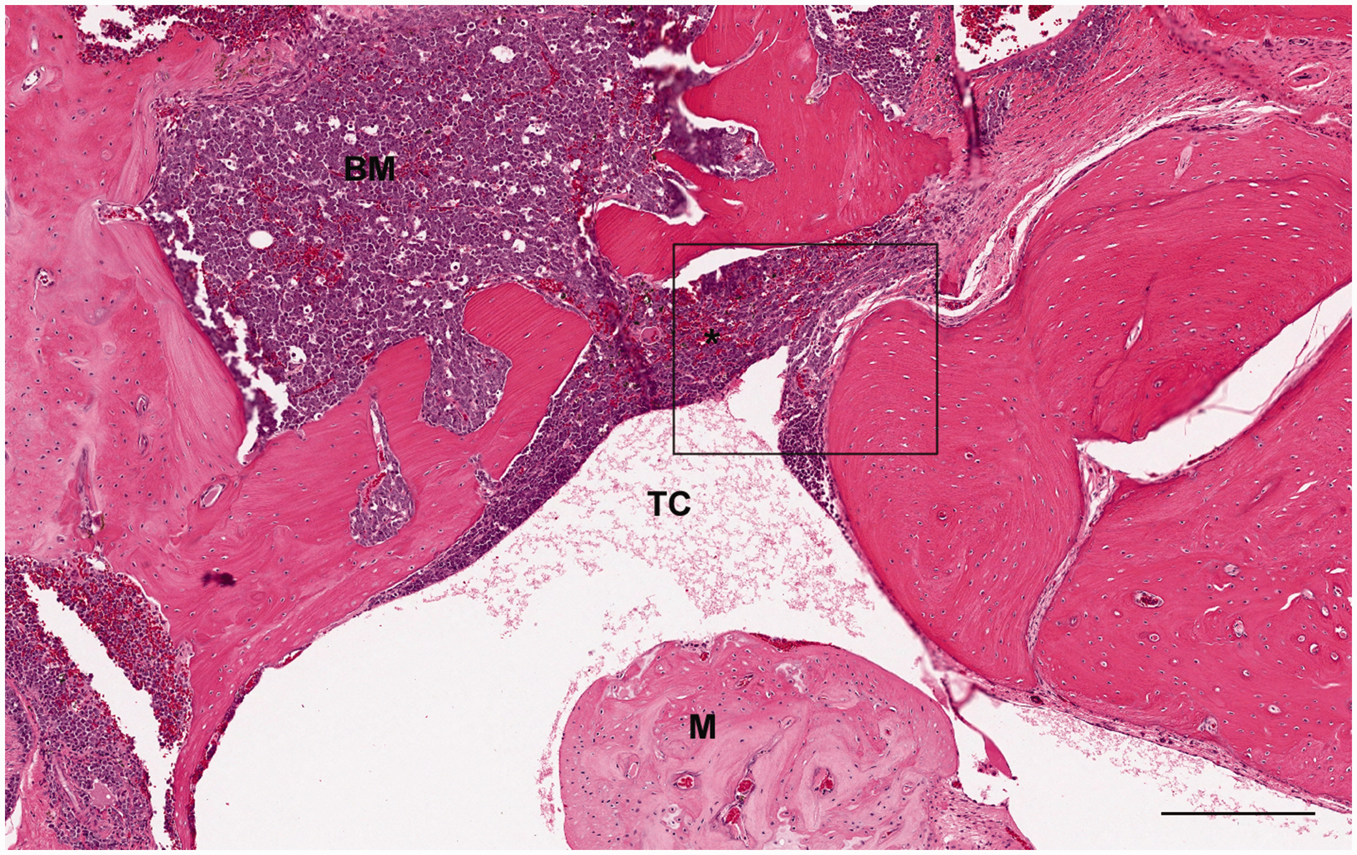
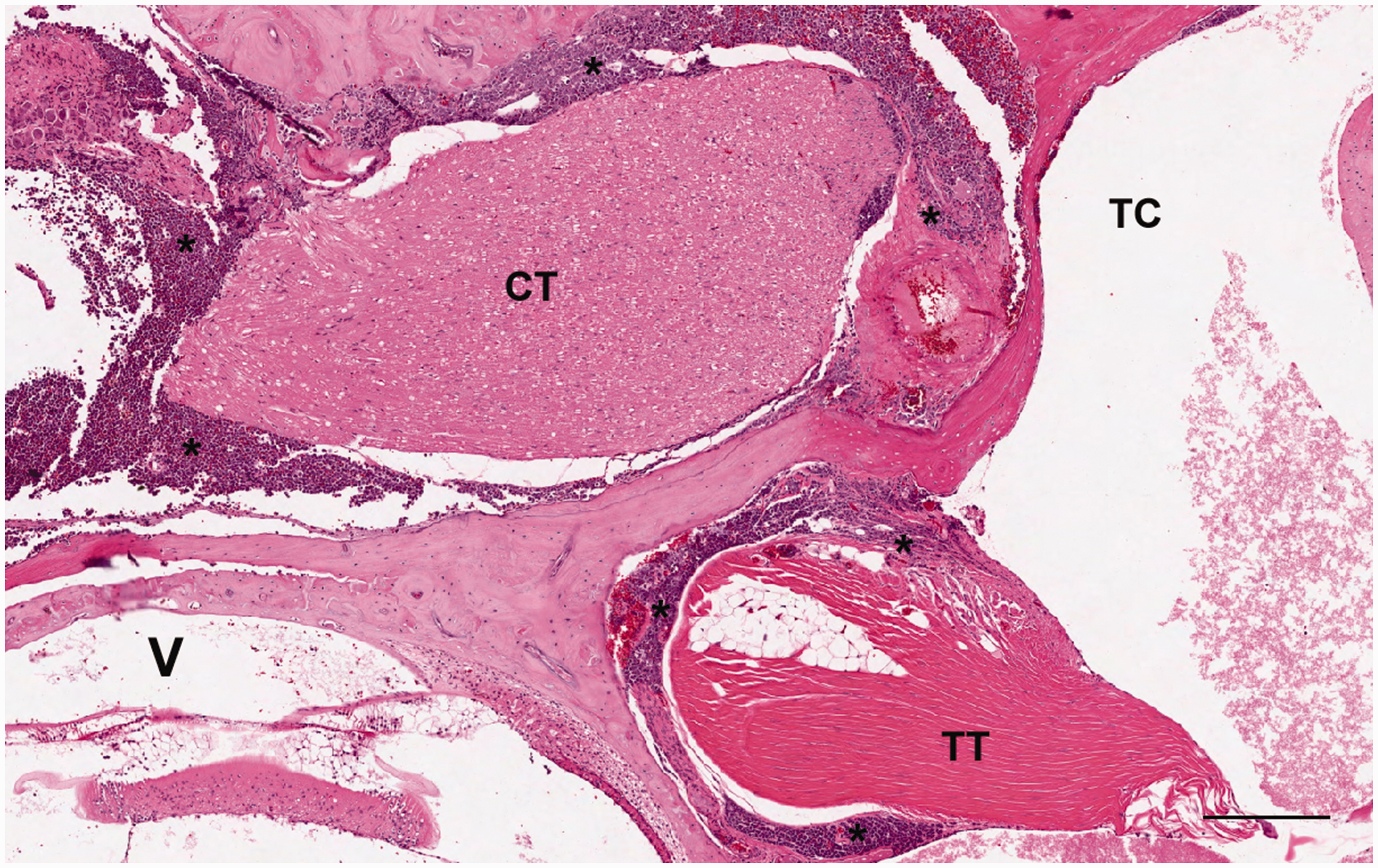
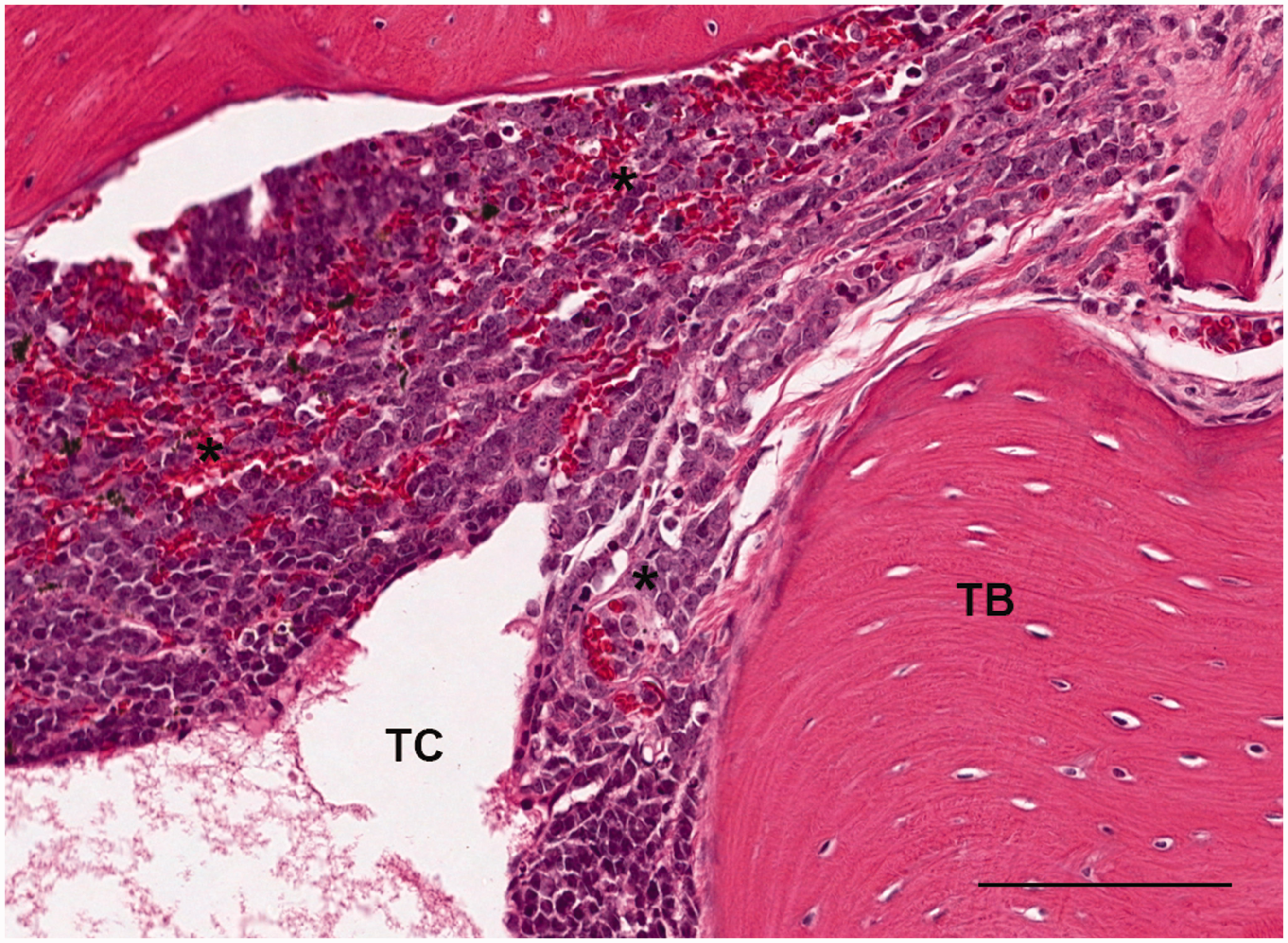
The animal was euthanized by exsanguination from the abdominal aorta after isoflurane anesthesia as planned at the end of the study, with no clinical signs other than oily fur consistent with decreased grooming. A complete necropsy examination was performed and included evaluation of the carcass and musculoskeletal system, all external surfaces and orifices, the cranial cavity and external surfaces of the brain and the thoracic, abdominal and pelvic cavities with their associated organs and tissues. Macroscopically, dark and gelatinous material was observed in the cranial cavity over the temporal bone and the cerebral meninges were thickened and dark. Additionally, the spleen was enlarged and there were dark foci on the mandibular lymph nodes. A standard and exhaustive list of tissues, along with the grossly abnormal meninges including the gelatinous material described above and underlying temporal bone, were sampled at necropsy, fixed in 10% buffered formalin, sectioned, and stained with hematoxylin and eosin for histopathological evaluation. In order to elucidate the origin of the gelatinous material, microscopic evaluation of the temporal bone was performed, and neoplastic lymphocytes were seen invading several structures of the middle ear (Figure 1). These neoplastic cells appeared to extend from the marrow of the temporal bone and formed a sheet several cells thick that covered the dorsal part of the tympanic cavity wall (Figure 2). Medially, neoplastic lymphocytes also surrounded and infiltrated the base of the tensor tympani muscle, as well as the chorda tympani branch of the facial nerve (Figure 3). The neoplastic lymphocytes were large, with scant eosinophilic to amphophilic cytoplasm and large nuclei, which often contained several conspicuous nucleoli (Figure 4). Mitotic figures and necrosis of individual neoplastic cells were frequently observed. The malleus was the only ossicle present in the sections examined and was not infiltrated by the neoplastic cells. The tympanic membrane and external ear canal appeared normal. The portions of the inner ear that were present in the sections examined, including part of the vestibule and cochlea, did not contain any neoplastic cells. The lymphoma was generalized and neoplastic lymphocytes were also noted in the adrenal glands, bone marrow, brain, femur, kidneys, larynx, liver, lymph nodes, meninges, nasal cavity, pituitary gland, prostate, spinal cord, spleen, sternum and thyroid glands. In the spleen and mandibular lymph nodes, the lymphoma correlated microscopically with the macroscopic lesions described above (i.e. enlarged spleen and dark foci in the lymph nodes).
Extension of neoplastic lymphocytes (*) from the bone marrow into the tympanic cavity lumen. Hematoxylin and eosin stain. Inset corresponds to Figure 4. Scale bar, 250 µm. Neoplastic lymphocytes (*) surrounding and infiltrating the base of the tensor tympani muscle and the chorda tympani branch of the facial nerve. Hematoxylin and eosin stain. Scale bar, 250 µm. Higher magnification showing large neoplastic lymphocytes (*) with scant cytoplasm and large nuclei infiltrating the tympanic cavity. Hematoxylin and eosin stain. Scale bar, 100 µm.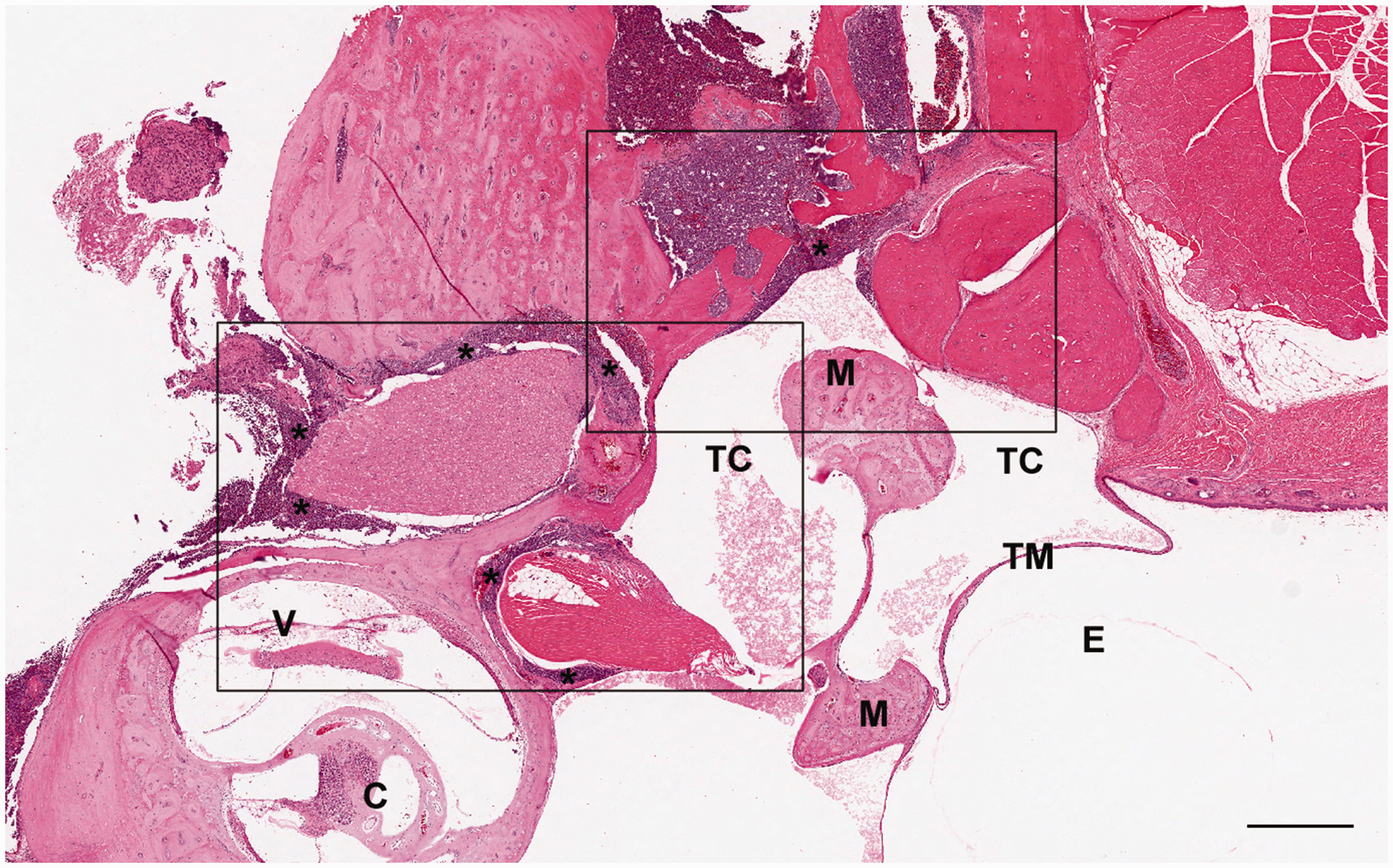
Discussion
The most common primary neoplasms of the middle and inner ear in humans are endolymphatic-sac tumors, squamous carcinomas, adenocarcinomas, rhabdomyosarcomas, multiple myelomas, plasmacytomas and lymphomas,3,4 and the middle ear is known to be a primary location of some carcinomas of the external auditory canal. 5 Middle and inner ear involvement in human lymphoma has been well described and the neoplastic lymphocytes are thought to reach these areas either following inner ear hemorrhage or by direct invasion of the solid structures of the middle ear, body labyrinth and inner ear.6–10
Clinically, rats with malignant lymphoma will show mainly non-specific signs such as depression, pallor, icterus and a palpably enlarged spleen. 11 At necropsy, typical findings include splenomegaly, hepatomegaly and lymphadenomegaly, 12 which correlate microscopically with infiltration by neoplastic lymphocytes.
Because of the complicated tridimensional anatomy of the middle and inner ear, and the difficult dissection, trimming and processing inherent to those anatomic features, these structures are often overlooked at necropsy and are seldom part of histopathological examination. Thus, literature regarding neoplasms of the middle and inner ear in animals is scarce and, to our knowledge, this is the first report of a naturally-occuring lymphoma involving the middle ear of a rat. However, in Wistar rats, an experimental model of infiltration of the inner ear scala tympani through the cochlear aqueduct following inoculation of neoplastic lymphocytes into the cisterna magna has been described. 13 Carcinomas of Zymbal’s gland, a small auditory sebaceous gland seen in mice as well, 14 is a common ear tumor in rats 15 but originates from the external ear and, to our knowledge, is not known to infiltrate the middle ear.
Most animal middle ear neoplasms reported appear to have been seen in dogs and were most frequently extensions of primary growths that originated in the external ear canal, including adnexal tumors and adenocarcinomas of the ceruminous glands. 16 Of note, a single case of lymphoma occupying the tympanic cavity was reported in a cat. 17
In conclusion, even though there was no such occurrence in the present case, lymphoma with inner ear involvement should be added to the list of possible diagnoses in middle-aged and older rats showing signs of vestibular disorder, along with the frequent pituitary gland neoplasms, in particular pars distalis adenomas.
Footnotes
Acknowledgement
The authors would like to thank Hamid Boubekeur for his invaluable help with the images.
Declaration of Conflicting Interests
The author(s) declared no potential conflicts of interest with respect to the research, authorship, and/or publication of this article.
Funding
The author(s) received no financial support for the research, authorship and/or publication of this article.