
Abstract
Alpha2-agonist anesthetic combinations are often used in rodent anesthesia but no information about their effects on cardiac function in chinchillas exists. The purpose of this study was to utilize echocardiography to evaluate the cardiovascular effects of dexmedetomidine–ketamine anesthesia in chinchillas. Echocardiographic examinations were performed in eight adult chinchillas under manual restraint and following dexmedetomidine (0.015 mg/kg) and ketamine (4 mg/kg) administration. Dexmedetomidine–ketamine anesthesia resulted in a significantly decreased heart rate, fractional shortening, cardiac output, and flow velocity across the aortic and pulmonic valves, and significantly increased left ventricular internal diameter in systole. The observed changes in echocardiographic parameters are similar to those previously reported in chinchillas anesthetized with isoflurane.
Keywords
Combinations of alpha2-agonists and ketamine have been advocated for injectable anesthesia in rodents, but only two reports exist of its use in chinchillas.1,2 No information is available concerning the effect of alpha2-agonists and ketamine protocols on echocardiographic variables in chinchillas.
Echocardiographic examination of chinchillas has been described with the animals either physically restrained or in animals under isoflurane anesthesia. 3 Isoflurane is a commonly used anesthetic in rodents but has been associated with negative changes in several cardiovascular parameters in chinchillas. 3
The objectives of this study were to evaluate the cardiovascular effects of anesthesia with dexmedetomidine–ketamine (D–K) in chinchillas. The study was approved by the University of Wisconsin–Madison, Institutional Animal Care and Use Committee. Animal use complied with the National Research Council Guide for the Care and Use of Laboratory Animals standard.
Eight healthy adult chinchillas (4 male, 4 female), ranging in age from two to five years and with a mean weight (±SD) of 0.71 ± 0.04 kg, were obtained from a commercial breeder (R and R Chinchillas, Jenera, OH, USA).
All the animals underwent cardiac auscultation by a board-certified veterinary cardiologist (RS), in order to rule out any cardiac arrhythmias or murmurs.
The animals underwent an echocardiographic examination twice within the same day between 08:00 h and 14:00 h. The initial echocardiographic exam was performed under manual restraint. Following the initial echocardiogram, the animals were anesthetized and the echocardiogram was repeated 15 min after administration of the anesthetic drugs. The animals were imaged in right and left lateral recumbency using a commercial ultrasound machine (Vivid-7; GE Healthcare, Waukesha, WI, USA) with a 10 MHz phased array probe (GE Healthcare). Echocardiographic measurements were obtained using standard views. Each echocardiographic measurement was the calculated average of three separate measurements. Cardiac output (CO) was calculated using velocity time integrals (VTI) derived from aortic Doppler spectral tracings and aortic diameter measured in two-dimensional images according to the following equation: aortic VTI * (π * aortic diameter/2) 2 * HR, with instantaneous heart rate (HR) derived from measurement of R–R intervals during Doppler interrogation. The same board-certified cardiologist (RS) performed all echocardiograms in this study.
Anesthesia was induced by intramuscular administration of dexmedetomidine (0.015 mg/kg, Dexdomitor; Pfizer Animal Health, New York, NY, USA) and ketamine (4 mg/kg, ketamine hydrochloride injection; Hospira Inc, Lake Forest, IL, USA). Following completion of the second echocardiogram, atipamezole (0.15 mg/kg, Antisedan; Pfizer Animal Health) was administered subcutaneously to reverse the effects of dexmedetomidine.
A two-tailed paired t-test was used to compare echocardiographic parameters between conscious and anesthetized animals. Data are reported as mean ± standard deviation (SD). A value of P < 0.05 was considered to be statistically significant.
Echocardiographic parameters measured in eight chinchillas before and during anesthesia with dexmedetomidine–ketamine (D–K).
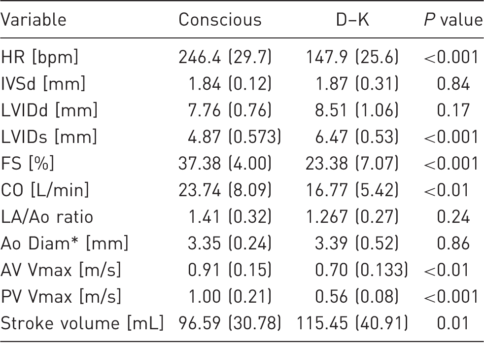
Values are reported as mean (standard deviation). HR: heart rate; IVSd: interventricular septum in diastole; LVIDd: left ventricular diastolic dimension; LVIDs: left ventricular systolic dimension; FS: fractional shortening; CO: cardiac output (calculated); LA/Ao: ratio of left atrial and aortic diameters; Ao Diam: aortic diameter (two-dimensional image); AV Vmax: maximal systolic velocity across the aortic valve; PV Vmax: maximal systolic velocity across the pulmonary valve. A value of P < 0.05 was considered to represent a statistically significant difference between groups. *Incomplete data-set (n = 5).
The administration of alpha2-agonists stimulates peripheral alpha2-receptors in arterioles, leading to vasoconstriction and increased peripheral vascular resistance.4,5 This resistance can result in an increase in afterload and a decrease in HR and CO. 4 Negative chronotropic and inotropic effects following administration of alpha2-agonist and dissociative agent combinations have been well-documented in mice. 6
Left ventricular systolic dimension was significantly larger following D–K anesthesia. While a direct myocardial depressant effect of this combination is possible, bradycardia secondary to use of dexmedetomidine in this drug combination may also explain an increase in LVIDs. 5 D–K was suspected as a cause of reduced systolic function in rats, demonstrated by an increase in left-ventricular end systolic dimensions. 5 Isoflurane has also been reported to increase LVIDs in chinchillas, with a mean increase (31%) which is similar to the results of this study (33%). 3
Fractional shortening was significantly reduced under D–K anesthesia. Decreases in FS have been demonstrated in several animal species following administration of an alpha2-agonist. In rats, medetomidine–ketamine anesthesia resulted in a lower FS percentage than two other anesthetic regimens. 5 FS is dependent upon left ventricular ejection time, which is affected by HR. 7
Isoflurane has also been demonstrated to result in a significantly decreased FS in chinchillas. 3 However, the decrease in FS with D–K was greater when compared to chinchillas administered isoflurane.
Cardiac output was also significantly decreased in this group of chinchillas. This noticeable reduction in CO can likely be attributed in part to the significant reduction in HR secondary to the effect of the dexmedetomidine, despite a compensatory increase in stroke volume. In rats, medetomidine–ketamine combinations resulted in overall lower CO values than other anesthetic drug regimens not containing an alpha2-agonist, although the difference between groups was not statistically significant. 5
Results of this study show that the administration of dexmedetomidine and ketamine has significant effects on echocardiographic parameters in chinchillas, which appear to be similar to those noted in chinchillas anesthetized with isoflurane. 3
Footnotes
Declaration of Conflicting Interests
The author(s) declared no potential conflicts of interest with respect to the research, authorship, and/or publication of this article.
Funding
The author(s) received no financial support for the research, authorship, and/or publication of this article.