
Abstract
In recent years our laboratory has developed an immunological hypothesis for the pathogenesis of atherosclerosis. We have shown that cellular and humoral immune reactions against heat shock proteins (Hsps) 60/65 expressed on the surface of stressed endothelial cells comprise the initial event in the pathogenesis of this disease. In the course of these studies, we also investigated normal, unaffected arteries for control purposes (carotid bifurcations from children aged 8 weeks to 10 years). This investigation led to the unexpected and previously unknown finding that mononuclear cells pre-exist in the intima at bifurcation sites. Our findings can be summarized as follows: Mononuclear cells are always found in the intima, primarily at sites subjected to major hemodynamic stress. Although the proportion of macrophages vs CD3+ T-cells differs, overall the latter clearly predominate. Most of the T-cells express the T-cell receptor (TCR)α/β, but TCRγ/δ cells are also present. We also identified dendritic cells and mast cells in the intima. Analogous to the mucosaassociated lymphoid tissue (MALT) we coined the designation “vascular-associated lymphoid tissue” (VALT) for these newly discovered cellular aggregates in the arterial intima.
O
Materials and Methods
Tissue Specimens
Carotid bifurcations were obtained from the Institute of Forensic Medicine (Innsbruck, Austria) from 12 children aged 8 weeks to 10 years (average age 3.9 years; two girls, 10 boys), victims of accidents or sudden infant death syndrome (SIDS; in accordance with the Helsinki Declaration of 1975). The arteries were snap-frozen and stored in liquid nitrogen for further immunohistochemical staining.
Immunohistochemistry
The entire procedure was performed at room temperature (RT). Cryostat sections (4 μm; cryostat CM 3000, Leica, Oberkochen, Germany) mounted on poly-
For detection of dendritic cells, immunoperoxidase single staining was performed in a three-step assay. Briefly, monoclonal antibodies against CD1a (a surface marker of dendritic cells susceptible to acetone fixation) were applied to 4-μm unfixed frozen sections, followed by a second-step incubation with peroxidase-labeled rabbit anti-mouse immunoglobulin (Dako) and a third-step horseradish peroxidaselabeled swine anti-rabbit immunoglobulin (Dako). Peroxidase activity was visualized with diaminobenzidine tetrahydrochloride with metal enhancer (Sigma).
Results
Macrophages and different subpopulations of T-lymphocytes were found in the intima of all investigated carotid bifurcations (Figure 1). Although the dominant cell types may differ, an overall assessment revealed a predominance of CD3+ T-lymphocytes (Figure 1A) over macrophages (Figure 1C). For example, in the carotid bifurcation of a 7-month-old infant, 67 CD3+ and only 43 CD68+ cells were found in the tunica intima, whereas in a bifurcation of another 8-year-old child, macrophages appeared to be the most abundant cell type (106 CD68+ vs 60 CD3+ cells; total number of cells counted per 5-mm2 section area). Both, CD4+ (Figure 1B) and CD8+ (not shown) T-cells were present in the tunica intima. In general, CD4+ predominated over CD8+ T-cells. Most of these T-cells carried the T-cell receptor TCRα/β, but an unexpectedly high number were also positive for the TCRγ/δ. This is noteworthy because TCRRγ/δ+ cells characteristically contribute to the local immune system and constitute important cellular elements of the MALT. Furthermore, earlier observations from our group had already shown an unexpectedly large proportion of TCRRγ/δ+ cells in early atherosclerotic lesions (Kleindienst et al. 1993).
The concept of the existence of a VALT was further corroborated by the finding of mast and dendritic cells at those sites. However, in contrast to Kaartinen et al. (1994), who described mast cells in the normal intima and adventitia, we rarely detected mast cells in the intima but found them consistently in the adventitia and the media, mainly in close proximity to the vasa vasorum (Figure 1E). Moreover, macrophages and T-cells were detected in the vicinity of these small vessels, which are important for the nourishment of arteries. Although Bobryshev and Lord (1995) and Bobryshev et al. (1997) described the presence of vascular dendritic cells in atherosclerotic plaques, we were now able to identify dendritic cells in the intima of carotid arteries of healthy children as well (Figure 1D). B-lymphocytes and NK/K cells were not found in the arterial intima in any of our experiments.
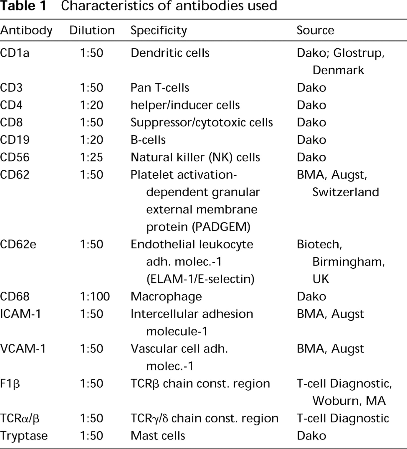
Characteristics of antibodies used
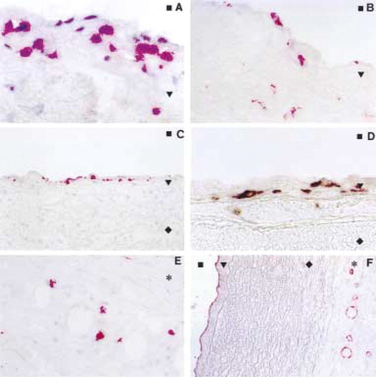
Immunohistochemical demonstration of cells in the carotid bifurcation of children (aged 8 weeks to 10 years) contributing to the formation of the vascular-associated lymphoid tissue (VALT). Positive cells are stained red (
As mentioned previously, the distribution of MNC accumulation was not homogenous but rather was concentrated at sites subjected to altered hemodynamic forces, primarily the lateral regions of bifurcations. A corollary of our studies was the observation that intimal MNC showed a preferential location at an area of the external carotid artery located opposite to the abutting external jugular vein. This may be due to the fact that the vein has a dampening effect on the neighboring arterial wall, thus reducing the stress by systolic pressure and inhibiting excessive pulse-dependent excursion of the arterial wall (Cordopatri 1993).
A prerequisite for the recruitment of MNC into the intima is the expression of appropriate adhesion molecules by endothelial cells (Springer 1994; Endres et al. 1997; Gimbrone et al. 1997). Significant expression of P-selectin (Figure 1F) and ICAM-1 (not shown) was found on endothelial cells of the tunica intima of all specimens studied. Interestingly, the vasa vasorum in the adventitia also showed constant expression of these two adhesion molecules, in contrast to the lack of expression of VCAM-1 and E-selectin.
Discussion
Thus far, we cannot provide any functional data indicating the origin of the immigrating MNC of the VALT, i.e. from the vascular lumen or the vasa vasorum via the adventitia and media. However, morphological evidence of MNC adhesion to and transgression through the endothelium of the lumen favors the former possibility. Our observations are in agreement with the hallmarks for the existence of a new site of the local immune system and support the concept of a vascular-associated lymphoid tissue (Wick et al. 1997). Of course, the VALT does not reach the extent of the highly organized MALT, e.g., the Peyer's patches (Croitoru and Bienenstock 1994; Heel et al. 1997) and related aggregates in other organs. If so, it would certainly have been discovered earlier. It can, however, be correlated with more diffusely distributed components of the MALT in the lamina propria, such as in the bronchus-associated lymphoid tissue (BALT) (Pabst and Tschernig 1995) or the inner ear (Glodeck and Arnold 1995). Although functional data on the VALT are still lacking, we propose that it serves a monitoring function similar to that known to operate in the MALT, except that it controls the internal surface of the vascular system for potentially harmful exogenous or autologous antigenic components and not an external surface in direct contact with the hostile environment. The fact that Hsps are known ligands for TCRγ/δ T-cells (Fu et al. 1994) further supports our assumption that Hsp 60 may be involved in the pathogenesis of atherosclerosis. Because protective cellular and humoral immunity against Hsp 60, a major antigen of bacteria and parasites and the envelope of many viruses (Kissling et al. 1991), is present in most humans, we may “pay” for this protection through the risk for crossreaction with highly conserved epitopes of our own Hsp 60, which is expressed on endothelial cells as well as on cells within the intima when we subject our vascular system to stressful conditions, such as classical risk factors for atherosclerosis (Wick et al. 1995; Xu and Wick 1996).
Footnotes
Acknowledgments
This work was supported by the Austrian Science Fund (grant no. 12213 to GW) and by the State of Tyrol.